Case Report

Adenoma-like Adenocarcinoma of the Colon: Case Report and Diagnostic Pitfalls of an Underrecognized Entity with Favorable Prognosis

by
Alfonso Agüera-Sánchez
1,2,*,
Emilio Peña-Ros
3,4,
Irene Martínez-Martínez
5 and
Francisco García-Molina
1,2
1
Department of Pathology, Hospital General Universitario Reina Sofía (HGURS), 30003 Murcia, Spain
2
Faculty of Medicine, Universidad de Murcia (UM), 30100 Murcia, Spain
3
Department of General and Digestive Surgery, Hospital General Universitario Reina Sofía (HGURS), 30003 Murcia, Spain
4
Faculty of Medicine, Universidad Católica San Antonio de Murcia (UCAM), 30107 Murcia, Spain
5
Centro Regional de Hemodonación, Department of Haematology and Medical Oncology, Hospital General Universitario Morales Meseguer, IMIB-Pascual Parrilla, Universidad de Murcia (UM), 30100 Murcia, Spain
*
Author to whom correspondence should be addressed.
Onco 2025, 5(3), 39; https://doi.org/10.3390/onco5030039 (registering DOI)
Submission received: 21 July 2025 / Revised: 21 August 2025 / Accepted: 22 August 2025 / Published: 23 August 2025

Simple Summary

Adenoma-like adenocarcinoma is an uncommon and recently defined subtype of colorectal cancer that closely mimics the appearance of tubulovellous adenomas, and it often poses a diagnostic challenge between malignant and benign entities. This case report describes an elderly patient whose lesion was initially interpreted as non-invasive, but was ultimately confirmed as invasive adenocarcinoma following surgical resection. Immunohistochemical analysis revealed loss of PMS2 expression, consistent with microsatellite instability. This case highlights the importance of recognizing the subtle histological and molecular features of this disease, which may have implications for treatment planning and genetic counseling. Given its typically indolent behavior and favorable prognosis, accurate diagnosis is essential to ensure appropriate clinical management and avoid overtreatment or misclassification.

Abstract

Adenoma-like adenocarcinoma (ALAC) of the colon is a recently recognized histological subtype of colorectal adenocarcinoma, characterized by a villous architecture, low-grade cytologic atypia, and deceptive bland morphology despite its invasive potential, which can mimic non-invasive adenomas, leading to underdiagnosis in limited biopsy samples. Herein, we report the case of an 81-year-old male presenting with right-upper-quadrant pain that was found to have a hepatic abscess and a 4 cm villous lesion in the ascending colon. Histopathological examination of the right hemicolectomy specimen revealed a villous adenocarcinoma with invasion of the muscularis propria, consistent with adenoma-like adenocarcinoma. Isolated loss of PMS2 indicated a mismatch repair deficiency. However, adjuvant therapy was not indicated. The patient remained recurrence-free for three years, until he died from unrelated causes in the context of progressive frailty and comorbidities, with no evidence of cancer progression. This case highlights the diagnostic challenges posed by ALAC and underscores the importance of recognizing its distinct morphological features. Awareness of this entity is essential to avoid misclassification and ensure adequate treatment, especially given its typically favorable prognosis with low metastatic potential.

1. Introduction

Colorectal cancer (CRC) is among the most prevalent malignancies globally, ranking as the third most commonly diagnosed cancer and the second leading cause of cancer-related mortality, with over 1.9 million new cases and approximately 930,000 deaths reported in 2020 [1]. Its incidence is rising in many regions, partly due to the recent implementation of colorectal cancer screening programs in many countries, making CRC a substantial public health burden with significant epidemiological impact, particularly in elderly populations [2].
According to the 5th edition of the World Health Organization (WHO) Classification of Tumors of the Digestive System (2019) [3], colorectal adenocarcinomas are categorized into multiple histological subtypes such as conventional adenocarcinoma, mucinous adenocarcinoma, serrated adenocarcinoma, medullary carcinoma, and micropapillary carcinoma. Adenoma-like adenocarcinoma (ALAC) of the colon is a newly recognized histological subtype of colorectal adenocarcinoma included in this classification (Table 1), characterized by a deceptively benign morphology that mimics conventional adenomas and poses multiple major pitfalls for pathologists in limited biopsies [3,4]. As a result, the true clinical impact of ALAC remains uncertain, and its incidence is likely underreported, with an estimated 3–9% of all colorectal adenocarcinomas [4,5,6].
From a histopathological standpoint, ALAC is characterized by villous or tubulovillous architecture with low-grade cytologic atypia, an expansive growth pattern, and minimal or absent expression of infiltrating invasive features, such as desmoplasia, budding, or cribriforming [4]. These findings can easily be misinterpreted as non-invasive adenomas on superficial biopsies, resulting in underdiagnosis and inappropriate clinical management. Deeper tissue levels often reveal subtle invasion with glandular dilatation in the muscularis mucosae, submucosa, or beyond, confirming that despite its deceptive bland features, this lesion represents true adenocarcinoma [3,4].
ALAC had previously been subsumed in the literature under ill-defined categories such as “villous adenocarcinoma” [5] and “papillary invasive adenocarcinoma” [7], reflecting the diagnostic uncertainty surrounding its classification for decades. However, adenoma-like adenocarcinoma is associated with significantly lower rates of lymph node involvement and disease-specific mortality than conventional colorectal adenocarcinomas [8,9], positioning it among the most prognostically favorable variants within the spectrum of invasive colonic neoplasms, as patients may benefit from less aggressive treatment approaches when complete surgical resection is achieved.
Recognizing ALAC is crucial to avoid misclassification and to ensure appropriate surgical management. In this report, we present a case of adenoma-like adenocarcinoma with detailed clinical and pathological correlation, highlighting the diagnostic challenges and histopathologic nuances of this underrecognized colonic adenocarcinoma subtype.

2. Methodology

This is a single-patient case report conducted according to the CARE guidelines. Clinical, imaging, endoscopic and pathology data were retrieved from our hospital’s institutional records.

2.1. Radiologic and Endoscopic Assessment

Our patient underwent abdominopelvic contrast-enhanced computed tomography (CT), using a multidetector scanner with arterial and portal venous phases and multiplanar reconstruction. Images were interpreted by board-certified radiologists, following standard institutional protocols for the evaluation of abdominal pain and fever. Subsequent colonoscopy was performed using a high-definition video endoscope with CO2 insufflation. Targeted biopsies and tattooing for lesion localization were performed according to routine practice by board-certified gastroenterologists.

2.2. Histopathological Processing

Colectomy specimen and prior endoscopic biopsies were fixed in 10% neutral buffered formalin, serially sampled, and embedded in paraffin. Four-micrometer-thick sections were stained with hematoxylin and eosin (H&E) using an automated protocol with Dako CoverStainer (Agilent Technologies, Glostrup, Denmark). Histological assessment was performed by two experienced gastrointestinal pathologists, who evaluated tumor architecture, cytologic atypia, depth of invasion, presence of lymphovascular and/or perineural invasion, intraepithelial tumor-infiltrating lymphocytes (iTILs) ratio, tumor budding, and margin status according to the WHO/AJCC criteria.

2.3. Ancillary Techniques

Immunohistochemical studies were performed on representative formalin-fixed paraffin-embedded (FFPE) tissue blocks using an automated Dako Omnis staining platform (Agilent Technologies, Glostrup, Denmark). The following antibodies against mismatch repair (MMR) proteins were used: MLH1 (mouse monoclonal, clone ES05, Ref. M3640, PD00439EN), MSH2 (mouse monoclonal, clone FE11, Ref. M3639, PD00440EN), MSH6 (rabbit monoclonal, clone EP49, Ref. IR086, PD00441EN), and PMS2 (rabbit monoclonal, clone EP51, Ref. IR087, PD00442EN), all in ready-to-use format. Complete loss of nuclear expression of any MMR protein in tumor cells, with retained expression in non-neoplastic epithelium and stromal cells as internal positive controls, was interpreted as mismatch repair deficiency (dMMR). Additional IHC markers (e.g., p53 and CDX2) were evaluated when clinically relevant.

3. Case Presentation

An 81-year-old male with a significant medical history of hypertension, chronic obstructive pulmonary disease (COPD), ischemic heart disease, and previous abdominal surgeries (sigmoid polypectomy and cholecystectomy) presented with a several-day history of abdominal pain, fever, and constitutional symptoms. Clinical examination revealed tenderness in the right upper quadrant and mild dehydration. Laboratory tests revealed elevated levels of inflammatory markers (CRP > 100 mg/L), leukocytosis (15 × 109/L) with neutrophilia, normocytic anemia (Hb 10.5 g/dL) and reactive thrombocytosis (405 × 109/L). CEA was not elevated (Table 2).
Abdominal contrast-enhanced computed tomography (CT) identified a large segment VIII hepatic abscess with surrounding phlegmon and an irregular, asymmetric mural thickening of the transverse colon near the hepatic flexure measuring approximately 4 cm. Colonoscopy confirmed the presence of a sessile, broad-based polypoid lesion with a villous surface and central depression occupying nearly half of the luminal circumference. The mass was friable and bled easily upon contact. The lesional area was tattooed, and multiple biopsies were obtained during the procedure and submitted for histological evaluation, which showed only focal high-grade dysplasia with lamina propria fibrosis.
Faced with inconclusive results from the initial endoscopic biopsies and a lack of alignment between the clinical and pathological findings, the case was presented to the institutional multidisciplinary tumor board. After evaluating the lesion’s dimensions and morphology, as well as the presence of concurrent hepatic abscess, the board advised proceeding with surgical management, and a right hemicolectomy was performed.
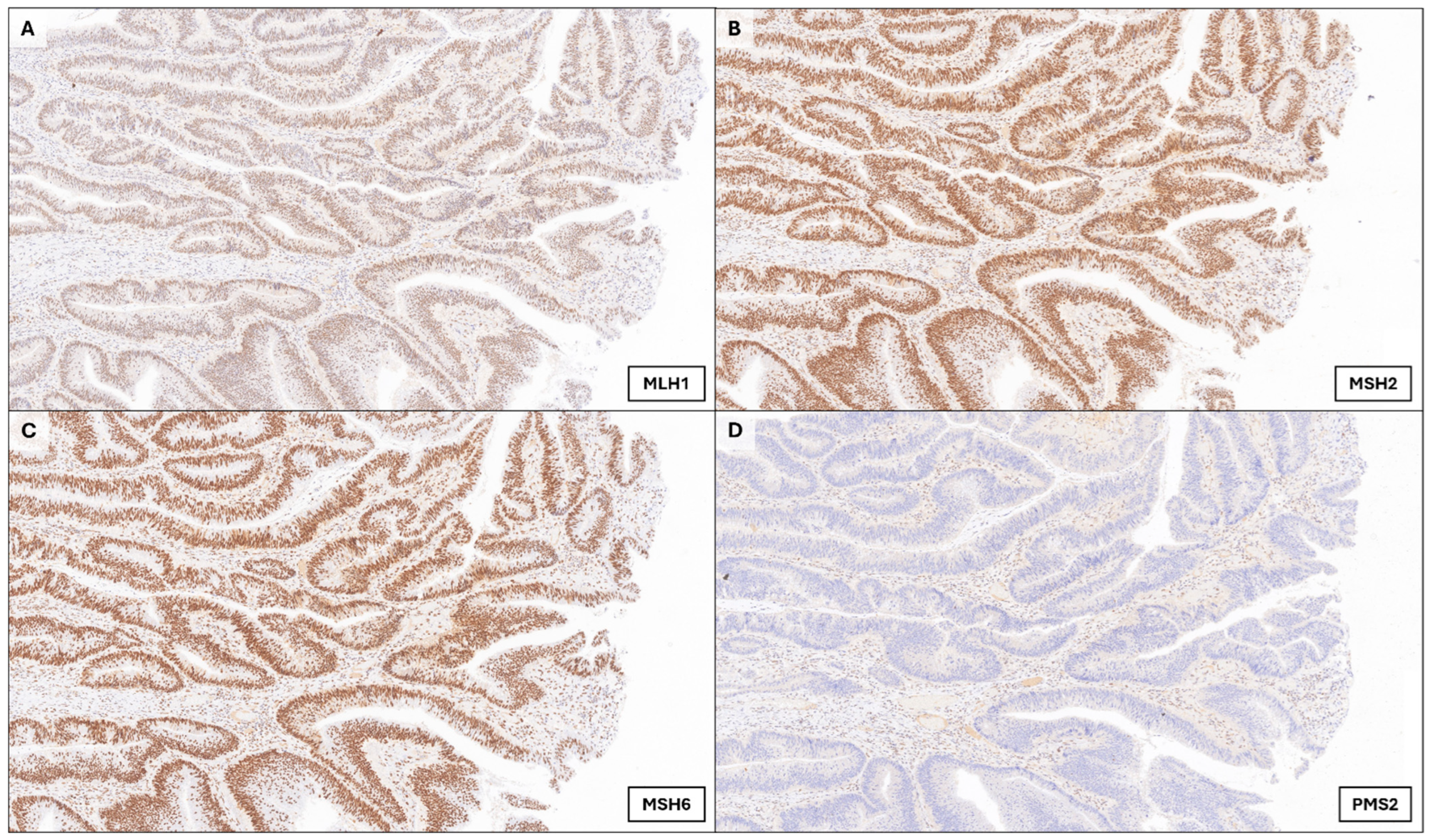
Histopathological evaluation of the right hemicolectomy specimen revealed a villous adenomatous lesion with focal infiltration of the muscularis propria (pT2) with a pushing front abutting pericolic adipose tissue (focal acellular mucin present, no unequivocal extramural invasion identified) (Figure 1). No lymphovascular or perineural invasion was identified, and surgical margins were clear (R0 resection). Twenty-one lymph nodes were examined, all of which were negative for metastasis (N0). A mild chronic inflammatory infiltrate was present in the vicinity of the tumor, with five intraepithelial tumor-infiltrating lymphocytes (iTILs) per high-power field. Immunohistochemical analysis showed retained expression of MLH1, MSH2, and MSH6, but isolated loss of PMS2, indicating mismatch repair deficiency (dMMR) (Figure 2). These findings, in conjunction with the bland cytology, villous architecture, and expansile invasive pattern of the lesion, were diagnostic of adenoma-like adenocarcinoma (ALAC) of the colon.
Given the early stage of the tumor, its dMMR status, and the patient’s advanced age and comorbidities, adjuvant chemotherapy was not administered. Genetic counseling was offered due to the isolated loss of PMS2, even though there was no personal or family history indicative of Lynch syndrome. No relevant alterations were identified.
During follow-up, the patient experienced progressive frailty, weight loss, and several hospital admissions due to chronic diarrhea, malnutrition, and cognitive decline. Approximately one year postoperatively, the patient was admitted with intestinal obstruction. Imaging studies excluded tumor recurrence or metastasis, and his condition was attributed to postoperative adhesions. Despite intensive supportive care including nutritional support and home palliative interventions, his illness progressively worsened. He developed end-stage organ dysfunction, including renal failure and respiratory compromise. The patient ultimately died under palliative care nearly three years after his initial diagnosis. Evidence of local recurrence or distant metastasis was ruled out; therefore, death was not attributable to tumor progression in any case.

4. Discussion

Adenoma-like adenocarcinoma (ALAC) exhibits unique biological behavior: although >2/3 are staged as pT2 or higher, only 20% have nodal metastases and 15% develop distant metastases [4]. When adjusted for age and stage, overall survival is significantly better than that of conventional well-differentiated colorectal adenocarcinoma, with a low disease-specific mortality rate of 12% [4]. Our case illustrates this pattern, with no subsequent recurrence after clear-margins segmental colectomy despite foregoing adjuvant therapy.
From a molecular perspective, ALAC seldom exhibits BRAF mutations, CpG island methylator phenotype (CIMP-high status), or MLH1 promoter hypermethylation, which are typical hallmarks of serrated adenocarcinomas [4,10]. Instead, ALAC often harbors key driver mutations in APC, KRAS, and TP53, with parallel chromosomal instability (CIN)-driven tumorigenesis typical of conventional colorectal cancers [11,12]. Surprisingly, KRAS mutations are expressed at rates comparable to or even higher than those of conventional adenocarcinomas (up to 58% in largest series), but with a lesser deleterious effect. In a dedicated study of colorectal cancers with residual adenoma components conducted by Lee et al. (2022), a high prevalence of KRAS mutations (~65%) was associated with well-differentiated polypoid tumors with favorable pathological features [13], as shown in ALAC. This supports the hypothesis that ALAC arises through the classic adenoma-to-carcinoma sequence rather than through the serrated pathway; however, further molecular investigation is required to elucidate why these tumors exhibit a more favorable prognosis despite frequent KRAS mutations. It is possible that the expression of other, yet unidentified, genetic or epigenetic alterations may mitigate the oncogenic impact of KRAS in this setting. For instance, there is growing opinion that cancer progression can also be understood as an ecological or microenvironmental process, where tumor cells interact with stromal, immune, and vascular compartments under spatial and resource constraints [14,15]. In ALAC, the combination of an expansile pushing border, scarce or absent tumor budding, minimal immature/myxoid desmoplasia, and the presence of intraepithelial lymphocytes, suggests the influence of a less aggressive tumor ecosystem, potentially limiting metastatic spread and reinforcing its indolent clinical course.
High-microsatellite status instability (MSI-H) is also particularly frequent in a subset of patients with ALAC [4]. While isolated PMS2 loss is often sporadic in elderly patients [16], current NCCN and European guidelines recommend universal germline testing for all dMMR colorectal cancers, irrespective of age or family history [17]. In real-world practice, this recommendation may present challenges when patients lack capacity, family support, or have limited life expectancy. Emerging tools, such as MLH1 methylation assays or targeted sequencing, may eventually help differentiate sporadic from hereditary causes, without relying solely on age or family history. Additional markers, such as abnormal p53 expression or loss of CDX2, can also be subtle indicators of malignancy rarely found in ALAC [18,19].

4.1. Tips and Clues to Avoid Adenoma-like Adenocarcinoma Misdiagnosis

One of the most pressing challenges surrounding ALAC is its under-recognition in routine diagnostic practice. Many cases continue to be classified simply as “well-differentiated adenocarcinomas” or misinterpreted as advanced adenomas, particularly when invasion is minimal or overlooked. To preclude this, we propose a structured approach that combines careful assessment of architectural and cytological features with attention to subtle invasion patterns, supported by ancillary studies when indicated.
The first key point is the limitation of biopsy sampling. Superficial mucosal biopsies, as in this patient, may capture only the adenomatous component. In the series conducted by Gonzalez et al. (2016), adenoma-like areas represented the dominant component in over 60% of the tumor volume in most cases, and preoperative biopsies frequently missed the invasive component [4]. Therefore, endoscopists and pathologists should remain vigilant when encountering a large broad-based often sessile mass with villous architecture that macroscopically exhibits a depressed (usually central) component or a “chicken-skin” appearance of the mucosa adjacent to the lesion, as thoroughly described in Fisac et al.’s paper (2024) [20]. Total resection (or at least extensive sampling of the neoplasia) is highly recommended in presence of the aforementioned changes.
Histologically, the main histological pitfall of ALAC lies in differentiating true invasion from architectural complexity within adenomatous tissue. Owing to the expansile rather than infiltrative growth pattern, staging may also become a thorough task. In the absence of desmoplasia, budding, or marked cytological atypia, pathologists must rely on subtle cues that require experience, such as glandular distortion, irregular contours, and misplaced crypts lacking lamina propria. In our case, the invasive front exhibited dilated and irregular glands embedded in an inflammatory stroma, with a vaguely expansile arrangement and focal total effacement of the muscularis propria. These features may closely resemble mucosal prolapse or pseudoinvasion, especially in lesions from the sigmoid colon or rectum [8]. A particularly informative feature in our case was the characteristic downward, finger-like pushing border at the tumor base protruding through the muscularis propria, a pattern reminiscent of low-grade infiltrative carcinomas elsewhere, but not widely described in colorectal pathology. Combined with architectural complexity and glandular spacing loss, this pattern strengthens the histological diagnosis of ALAC despite its low cytological grade. In borderline cases, immunohistochemistry can help delineate muscularis propria to demonstrate this pushing/expansile border, as shown in Figure 1C. Digital pathology tools and quantitative metrics may also support reproducibility in these cases, particularly when invasion is focal [21,22].
Beyond architectural clues, intraepithelial tumor-infiltrating lymphocytes (iTILs) can be assessed on routine biopsies and may aid in the recognition of ALAC. In the largest clinicopathological series of ALAC to date, this adenocarcinoma subtype showed a higher frequency of iTILs than conventional adenocarcinoma, together with absent tumor budding and lack of immature/myxoid desmoplasia, and overall presented with lower pT/pN stages and fewer nodal deposits [8]. Complementarily, a dedicated study in stages I–III CRC demonstrated that the presence of iTILs (≥5/HPF) is independently associated with better recurrence-free survival, and that iTILs inversely correlate with tumor budding and immature/myxoid desmoplastic reaction [23]. The finding of a moderate chronic inflammatory infiltrate with intraepithelial lymphocytes in our surgical specimen is concordant with these observations and supports the favorable-risk profile classically described for ALAC.

4.2. Limitations and Future Directions

The prognostic implications of dMMR in patients with ALAC remain unclear. In conventional colorectal cancer, MSI-high tumors are often associated with improved prognosis in early stages [24], but the low cellular turnover and subtle invasion patterns seen in ALAC may reduce the immune-stimulatory effect typical of MSI-high tumors, potentially modifying the prognostic impact of MMR deficiency in this context. Moreover, it has not yet been established whether ALAC exhibits the immune microenvironment characteristics of typical MSI-high cancers such as increased PD-1/PD-L1 expression [25,26].
This report describes a single patient and, by design, cannot support incidence estimates or robust correlations between the tumor immune microenvironment and outcomes. Consequently, additional prospective registries are required to assess the progression risk of this condition. Digital image analysis could help validate thresholds and define which patients might benefit from tailored surveillance or adjuvant strategies.

5. Conclusions

Adenoma-like adenocarcinoma (ALAC) constitutes a morphologically subtle yet clinically relevant subtype of colorectal carcinoma that lies at the biological intersection between benign and malignant colorectal lesions. Its architectural resemblance to conventional adenomas and absence of overt invasive features can lead to misdiagnosis, particularly in limited biopsy samples. Accurate identification of this entity is essential, as ALAC carries a favorable prognosis with a low rate of metastasis even without adjuvant therapy when complete surgical resection is achieved. Therefore, awareness of potential clinical and histopathological diagnostic pitfalls, and integration of ancillary techniques such as immunohistochemistry markers are key to ensuring correct classification and optimal patient care. Future research with a larger series should aim to clarify the molecular landscape and immune microenvironment of ALAC to better define its location within the spectrum of colorectal neoplasia, validate reproducible diagnostic criteria (potentially with the aid of digital pathology and artificial intelligence tools), and determine the optimal management of patients with this uncommon but clinically significant colorectal carcinoma subtype.

Author Contributions

Conceptualization and methodology: A.A.-S., I.M.-M. and F.G.-M.; formal analysis and investigation: A.A.-S., E.P.-R. and F.G.-M.; writing—original draft preparation: A.A.-S.; writing—review and editing: all authors; visualization: A.A.-S. and F.G.-M.; supervision: F.G.-M. All authors have read and agreed to the published version of the manuscript.

Funding

This research received no external funding.

Institutional Review Board Statement

Ethical review and approval were not required for this single-patient case report in accordance with local legislation and institutional requirements for case reports based on fully anonymized clinical information. The study was conducted in accordance with the principles of the Declaration of Helsinki.

Informed Consent Statement

Written informed consent was obtained from subject involved in the study.

Data Availability Statement

All data supporting the findings of this study are included in the article. Additional anonymized pictures are available from the corresponding author upon reasonable request.

Conflicts of Interest

The authors declare no conflicts of interest.

References

  1. Morgan, E.; Arnold, M.; Gini, A.; Lorenzoni, V.; Cabasag, C.J.; Laversanne, M.; Vignat, J.; Ferlay, J.; Murphy, N.; Bray, F. Global burden of colorectal cancer in 2020 and 2040: Incidence and mortality estimates from GLOBOCAN. Gut 2023, 72, 338–344. [Google Scholar] [CrossRef] [PubMed]
  2. Brenner, H.; Heisser, T.; Cardoso, R.; Hoffmeister, M. Reduction in colorectal cancer incidence by screening endoscopy. Nat. Rev. Gastroenterol. Hepatol. 2023, 21, 125–133. [Google Scholar] [CrossRef]
  3. WHO Classification of Tumours Editorial Board. Digestive System Tumours, 5th ed.; IARC Press: Lyon, France, 2019. [Google Scholar]
  4. Gonzalez, R.S.; Cates, J.M.M.; Washington, M.K.; Beauchamp, R.D.; Coffey, R.J.; Shi, C. Adenoma-like adenocarcinoma: A subtype of colorectal carcinoma with good prognosis, deceptive appearance on biopsy and frequent KRAS mutation. Histopathology 2015, 68, 183–190. [Google Scholar] [CrossRef]
  5. Loy, T.S.; Kaplan, P.A. Villous Adenocarcinoma of the Colon and Rectum. Am. J. Surg. Pathol. 2004, 28, 1460–1465. [Google Scholar] [CrossRef]
  6. Rubio, C.A.; Yanagisawa, A.; Kato, Y. Histologic phenotypes of colonic carcinoma in Sweden and in Japan. Anticancer Res. 1998, 18, 2649–2655. [Google Scholar]
  7. Palazzo, J.P.; Edmonston, T.B.; Chaille-Arnold, L.M.; Burkholder, S. Invasive papillary adenocarcinoma of the colon. Hum. Pathol. 2002, 33, 372–375. [Google Scholar] [CrossRef]
  8. González, I.A.; Bauer, P.S.; Liu, J.; Chatterjee, D. Adenoma-like adenocarcinoma: Clinicopathologic characterization of a newly recognized subtype of colorectal carcinoma. Hum. Pathol. 2020, 107, 9–19. [Google Scholar] [CrossRef]
  9. Remo, A.; Fassan, M.; Vanoli, A.; Bonetti, L.R.; Barresi, V.; Tatangelo, F.; Gafà, R.; Giordano, G.; Pancione, M.; Grillo, F.; et al. Morphology and Molecular Features of Rare Colorectal Carcinoma Histotypes. Cancers 2019, 11, 1036. [Google Scholar] [CrossRef] [PubMed]
  10. Conesa-Zamora, P.; García-Solano, J.; García-García, F.; Turpin, M.d.C.; Trujillo-Santos, J.; Torres-Moreno, D.; Oviedo-Ramírez, I.; Carbonell-Muñoz, R.; Muñoz-Delgado, E.; Rodriguez-Braun, E.; et al. Expression profiling shows differential molecular pathways and provides potential new diagnostic biomarkers for colorectal serrated adenocarcinoma. Int. J. Cancer 2012, 132, 297–307. [Google Scholar] [CrossRef]
  11. Fearon, E.R.; Vogelstein, B. A genetic model for colorectal tumorigenesis. Cell 1990, 61, 759–767. [Google Scholar] [CrossRef] [PubMed]
  12. Jungwirth, J.; Urbanova, M.; Boot, A.; Hosek, P.; Bendova, P.; Siskova, A.; Svec, J.; Kment, M.; Tumova, D.; Summerova, S.; et al. Mutational analysis of driver genes defines the colorectal adenoma: In situ carcinoma transition. Sci. Rep. 2022, 12, 1–10. [Google Scholar] [CrossRef] [PubMed]
  13. Lee, H.W.; Song, B.; Kim, K.; Yokoyama, S. Colorectal cancers with a residual adenoma component: Clinicopathologic features and KRAS mutation. PLoS ONE 2022, 17, e0273723. [Google Scholar] [CrossRef] [PubMed]
  14. Hanahan, D. Hallmarks of Cancer: New Dimensions. Cancer Discov. 2022, 12, 31–46. [Google Scholar] [CrossRef] [PubMed]
  15. Luo, W. Nasopharyngeal carcinoma ecology theory: Cancer as multidimensional spatiotemporal "unity of ecology and evolution" pathological ecosystem. Theranostics 2023, 13, 1607–1631. [Google Scholar] [CrossRef] [PubMed]
  16. Gill, S.; Lindor, N.M.; Burgart, L.J.; Smalley, R.; Leontovich, O.; French, A.J.; Goldberg, R.M.; Sargent, D.J.; Jass, J.R.; Hopper, J.L.; et al. Isolated Loss of PMS2 Expression in Colorectal Cancers: Frequency, Patient Age, and Familial Aggregation. Clin. Cancer Res. 2005, 11, 6466–6471. [Google Scholar] [CrossRef]
  17. NCCN Clinical Practice Guidelines in Oncology: Genetic/Familial High-Risk Assessment: Colorectal. Version 1. 2025. Available online: https://www.nccn.org/professionals/physician_gls/pdf/genetics_ceg.pdf (accessed on 21 July 2025).
  18. Yao, T.; Kajiwara, M.; Kouzuki, T.; Iwashita, A.; Tsuneyoshi, M. Villous tumor of the colon and rectum with special reference to roles of p53 and bcl-2 in adenoma–carcinoma sequence. Pathol. Int. 1999, 49, 374–382. [Google Scholar] [CrossRef]
  19. Nayak, J.; Mohanty, P.; Lenka, A.; Sahoo, N.; Agrawala, S.; Panigrahi, S.K. Histopathological and Immunohistochemical Evaluation of CDX2 and Ki67 in Colorectal Lesions with their Expression Pattern in Different Histologic Variants, Grade, and Stage of Colorectal Carcinomas. J. Microsc. Ultrastruct. 2021, 9, 183–189. [Google Scholar] [CrossRef]
  20. Fisac, J.; Burgos, A.; Méndez, M. Adenoma-like adenocarcinoma. Gastrointest. Endosc. 2024, 100. [Google Scholar] [CrossRef]
  21. Boyaval, F.; Fariña-Sarasqueta, A.; Boonstra, J.J.; Heijs, B.; Morreau, H. Recognition of pseudoinvasion in colorectal adenoma using spatial glycomics. Front. Med. 2024, 10, 1221553. [Google Scholar] [CrossRef]
  22. Wu, Y.; Li, Y.; Xiong, X.; Liu, X.; Lin, B.; Xu, B. Recent advances of pathomics in colorectal cancer diagnosis and prognosis. Front. Oncol. 2023, 13, 1094869. [Google Scholar] [CrossRef]
  23. González, I.A.; Bauer, P.S.; Liu, J.; Chatterjee, D. Intraepithelial tumour infiltrating lymphocytes are associated with absence of tumour budding and immature/myxoid desmoplastic reaction, and with better recurrence-free survival in stages I-III colorectal cancer. Histopathology 2021, 78, 252–264. [Google Scholar] [CrossRef] [PubMed]
  24. Saridaki, Z. Prognostic and predictive significance of MSI in stages II/III colon cancer. World J. Gastroenterol. 2014, 20, 6809–6814. [Google Scholar] [CrossRef]
  25. Llosa, N.J.; Cruise, M.; Tam, A.; Wicks, E.C.; Hechenbleikner, E.M.; Taube, J.M.; Blosser, R.L.; Fan, H.; Wang, H.; Luber, B.S.; et al. The Vigorous Immune Microenvironment of Microsatellite Instable Colon Cancer Is Balanced by Multiple Counter-Inhibitory Checkpoints. Cancer Discov. 2015, 5, 43–51. [Google Scholar] [CrossRef] [PubMed]
  26. Le, D.T.; Uram, J.N.; Wang, H.; Bartlett, B.R.; Kemberling, H.; Eyring, A.D.; Skora, A.D.; Luber, B.S.; Azad, N.S.; Laheru, D.; et al. PD-1 Blockade in Tumors with Mismatch-Repair Deficiency. N. Engl. J. Med. 2015, 372, 2509–2520. [Google Scholar] [CrossRef] [PubMed]
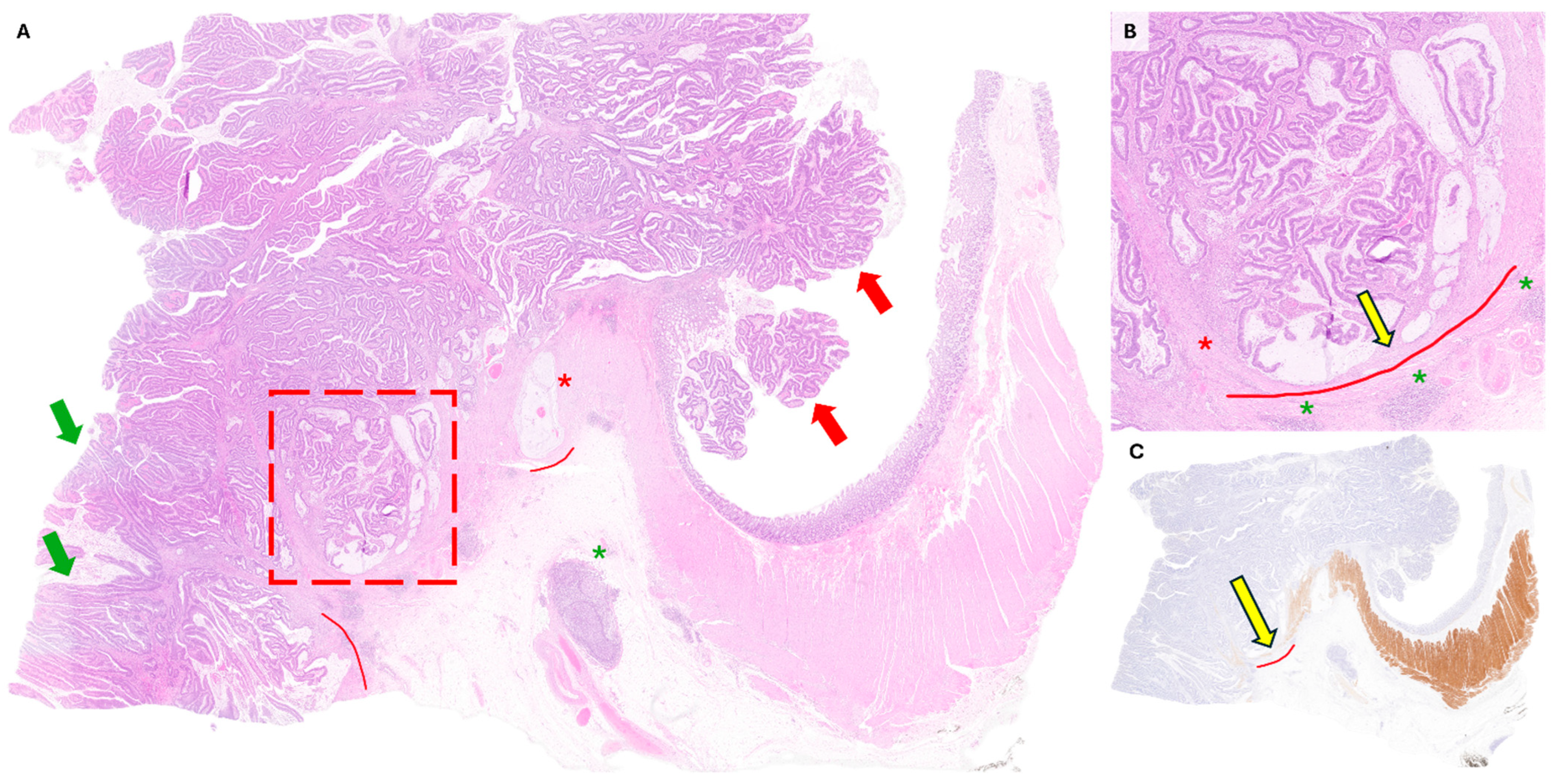
Figure 1. Histology and immunohistochemistry of ALAC. (A). Low-power panoramic view shows an invasive adenocarcinoma with admixed tubular (green arrows) and villous (red arrows) features. A striking expansive (“pushing”) growth is evident, with dilated glands (red dotted frame) and pools of acellular mucin protruding through the submucosa and muscularis propria (curved red lines). The invasive front lies immediately adjacent to pericolic adipose tissue, raising concern for incipient extension in some areas, occasionally represented by mucin pools devoid of epithelial cells (red asterisk). Close-by, an adjacent pericolic lymph node without metastatic involvement is also noted (green asterisk) (H&E, 20×). (B) At higher magnification, the invasive component is characterized by distorted, mucin-filled glands with papillary configuration that expand rather than infiltrate the surrounding stroma (yellow arrow). The typical surrounding desmoplastic reaction observed in conventional adenocarcinomas (red asterisk) is very subtle in this case, accompanied by a mild chronic lymphocytic infiltrate (green asterisks) (H&E, 100×). (C) Desmin immunohistochemistry highlights effacement of the muscularis propria by the expansile tumor front (yellow arrow), producing a characteristic downward, finger-like pushing border at the deepest portion of the tumor scored as pT2 (Desmin, 20×).
Onco 05 00039 g001
Figure 2. Mismatch repair deficiency (dMMR) status (colectomy specimen, 150×). (AC) Tumor cells show retained nuclear expression of MLH1, MSH2, and MSH6: the former demonstrated weak staining but was considered positive (+/+++), while MSH2 and MSH6 were strongly positive (+++/+++). (D) Tumor cells show isolated loss of PMS2 expression with positive internal control (−), indicating deficient mismatch repair.
Onco 05 00039 g002
Table 1. Comparative histopathological and molecular features of colorectal adenocarcinoma subtypes. Based on the World Health Organization Classification of Tumours, 5th Edition: Digestive System Tumours (2019) [3].
SubtypeKey Architecture &
Cytology
Invasion Front (Desmoplasia/Budding)Typical Molecular Profile (MMR/MSI; Drivers)Usual SitePrognosis/Notes
Adenoma-like adenocarcinoma (ALAC)Villiform/tubulovillous pattern; bland/low-grade cytology, deceptively “adenoma-like” Pushing/expansile border, mild desmoplasia, absent/low buddingNot infrequently dMMR; KRAS mutations relatively common, BRAF/CIMP uncommonAny (often right colon)Early stage, low nodal burden; favorable outcomes after R0 resection; superficial biopsies often underestimate invasion
Conventional adenocarcinoma (NOS, well-differentiated)Tubular ± villous; variable atypiaTypically infiltrative with desmoplasia; budding variableMixed: MSS or MSI; APC/KRAS/TP53 commonAnyPrognosis largely stage-driven
Serrated adenocarcinomaSerrated glands, eosinophilic cytoplasm; may mimic traditional serrated lesionsOften infiltrative; mucinous areas frequentMSI-H when MLH1 methylated; BRAF mutation and CIMP-high common Proximal
colon
Can show worse stage-adjusted survival vs. NOS in some series
Mucinous adenocarcinoma≥50% extracellular mucin pools with overt malignant epitheliumInfiltrative; tumor budding variableEnriched for dMMR/MSI-H; KRAS frequent, variable BRAFProximal
colon
Response to standard chemo may differ; outcomes vary with stage/MSI
Medullary carcinomaSolid sheets, vesicular nuclei; prominent lymphoid infiltrate; pushing borderPushing; budding typically lowAlmost always MSI-H/dMMR combined with BRAF mutation; frequent PD-L1 expressionProximal
colon
Favorable stage-adjusted outcomes; immune-rich microenvironment
Micropapillary adenocarcinomaTight morula-like clusters in retraction spacesMarked lymphovascular invasion; early nodal spreadUsually MSS;
drivers variable
Any (often left colon)Adverse prognosis
Table 2. Patient’s characteristics and baseline blood work values at presentation.
VariableDetails/Values at Presentation
Age/Sex81 yo/Male
ComorbiditiesFormer smoker (~60 pack-years; quit 3 years ago); Hypertension; COPD; ischemic heart disease; chronic kidney disease, prior abdominal surgery (segmental colectomy for polyp; cholecystectomy; umbilical/epigastric hernia repair; prostatectomy); cataracts; vertebrobasilar TIA 6 y ago; ECOG performance status 1
PharmacotherapyAspirin 100 mg; amlodipine 5 mg; bisoprolol 2.5 mg; furosemide 40 mg; omeprazole 40 mg; solifenacin 5 mg; fesoterodine 4 mg; pregabalin 25 mg; risperidone 2 mg; trazodone 100 mg
Leukocytes (WBC)15.0 × 10⁹/L (88% neutrophils)
RBC3.40 × 1012/L
Hemoglobin10.5 g/dL
MCV92 fL
Platelets405 × 109/L
Glucose115 mg/dL
Creatinine1.20 mg/dL
Sodium133 mmol/L
Potassium4.9 mmol/L
Bilirubin (total)1.3 mg/dL
Albumin3.2 g/dL
INR1.10
aPTT31 s
CRP110 mg/L
LDH280 U/L
CEA2.1 ng/mL
Medication doses are daily oral doses unless otherwise specified (pack-years = packs/day × years smoked). Abbreviations: COPD, chronic obstructive pulmonary disease; TIA, transient ischemic attack; ECOG, Eastern Cooperative Oncology Group; WBC, white blood cells; RBC, red blood cells; MCV, mean corpuscular volume; INR, international normalized ratio; aPTT, activated partial thromboplastin time; CRP, C-reactive protein; LDH, lactate dehydrogenase; CEA, carcinoembryonic antigen.
Disclaimer/Publisher’s Note: The statements, opinions and data contained in all publications are solely those of the individual author(s) and contributor(s) and not of MDPI and/or the editor(s). MDPI and/or the editor(s) disclaim responsibility for any injury to people or property resulting from any ideas, methods, instructions or products referred to in the content.