Review

Tumour Heterogeneity and Disease Infiltration as Paradigms of Glioblastoma Treatment Resistance

School of Medicine, Biodiscovery Institute, University of Nottingham, Nottingham NG7 2RD, UK
*
Author to whom correspondence should be addressed.
Onco 2024, 4(4), 349-358; https://doi.org/10.3390/onco4040024
Submission received: 9 August 2024 / Revised: 25 September 2024 / Accepted: 16 October 2024 / Published: 18 October 2024

Simple Summary

Glioblastoma is an aggressive and hard-to-treat brain cancer, with poor prognosis worldwide. The relationship between biological variation in different regions of each glioblastoma, including that of the residual infiltrative disease spared by standard treatment, is poorly understood. We summarize the current understanding of glioblastoma intra-tumour variation and consider antibiotic resistance as a helpful analogy for further insight.

Abstract

Isocitrate dehydrogenase wild-type glioblastoma, a Grade 4 malignant brain neoplasm, remains resistant to multimodal treatment, with a median survival of 16 months from diagnosis with no geographical bias. Despite increasing appreciation of intra-tumour genotypic variation and stem cell plasticity, such knowledge has yet to translate to efficacious molecular targeted therapies in this post-genomic era. Critically, the manifestation of molecular heterogeneity and stem cell biological process within clinically relevant infiltrative disease is little understood. Here, we review the interactions between neural plasticity, intra-tumour heterogeneity and residual infiltrative disease, and we draw upon antibiotic resistance as an insightful analogy to further explain tumour heterogeneity.

1. Introduction

Isocitrate dehydrogenase wild-type (IDH WT) glioblastoma (GBM) is one of the most lethal malignant brain cancers that arise de novo within the adult central nervous system (CNS). GBM accounts for 48.6% of malignant CNS tumours and occurs between 3.19 to 4.17 per 100,000 people annually [1,2,3,4]. GBM is a glioma, specifically classified as a Grade 4 astrocytoma, with a median survival time (upon multimodal standard treatment) of 12–15 months post-diagnosis [5]. Gliomas are an umbrella term for tumours formed from glial cells (non-neuronal cells that assist the function of neuronal brain parenchyma) which are known to be diffusely infiltrating. Gliomas are classified by which glial cell the tumour originates from—astrocytes form astrocytoma, oligodendrocytes form oligodendrogliomas, and ependymal cells form ependymomas—and are associated with the presentation of glioma-like clinical manifestations, aggressiveness, management strategy and extent of malignancy [6].
A key obstacle facing GBM treatment is the infiltrative margin that is left behind following maximal safe surgical resection, which, due to various oncogenic micro-environments, is the primary source of GBM recurrence [7]. The infiltrative margin is an invasive penetration of tumour cells into adjacent neural and glial tissue parenchyma, which is near impossible to remove safely by surgery. GBM cells invade the brain by following ‘roadmaps’ that are already present, for example, perivascular space, white-matter tracts, leptomeningeal space, and brain parenchyma [8]. Perivascular space and white-matter tracts are favoured by tumour cells as these are the primary structural components of the brain. The perivascular space hosts an abundance of blood vessels, which use endothelial chemo-attractants to attract tumour cells that have a high demand for oxygen and nutrients (due to the low supply of these as a result of the hypoxic nature of the primary mass), allowing the tumour cells to flourish [9,10]. White-matter tracts are an alternative preferred route for invasion. These tracts are composed of myelinated axons and provide the invading tumour cells with the ability to use the corpus callosum or anterior commissure (exemplar white matter tracts) to cross into the contralateral hemisphere, guided by proteins from the slit, netrin, and semaphoring families [11,12].
The standard-of-care application of T1-weighted contrast magnetic resonance imaging (MRI) cannot establish a parameter for the invading tumour, as these invasive tumour cells penetrate beyond an inch of the primary tumour mass, amid an overwhelming signal of healthy brain parenchyma [13]. An undefined tumour border poses a challenge to surgeons, as resecting any functional brain around the primary tumour mass in each direction (as a preventative measure for recurrence) would severely risk neurological damage. The highly diffuse behaviour of GBM infiltrative cells is not amenable for radiotherapy as an option to prevent recurrence [14]. The extent of infiltration is closely correlated with prognosis, whereby the larger the infiltrative margin, the worse the prognosis for the patient. This is primarily due to the fact that tumour cells in the infiltrative margin are more likely to have escaped detection and treatment, and are therefore more likely to remain after safe surgical resection, giving rise to recurrent GBM tumours. Tumour cells at the infiltrative margin are more likely to be resistant to chemo-radiotherapy treatments, as they are located in regions of the brain that are difficult to access and may have a different microenvironment compared to the primary tumour mass. It follows that the infiltrative margin of GBM is thus a critical factor in the management of these tumours, treatment response, and functional outcome.
Here, we aim to direct focus to the infiltrative margin by converging two concurrent and contemporary streams of GBM research—intra-tumour heterogeneity and tumour stem cell biology, which currently represents a scientific knowledge gap.

2. Image-Guided Surgery to Reveal the GBM Infiltrative Margin

A landmark clinical trial in 2006 demonstrated that the intraoperative use of the fluorescent marker 5-Aminolevulinic acid (5-ALA), compared to operating only under white light, leads to greater tumour resection with better survival outcomes and several months of progression free survival [15]. Biologically, the porphyrin biosynthesis intracellular pathway produces 5-ALA as an intermediate metabolite. Despite being absorbed by both tumour and healthy brain cells, 5-ALA causes the fluorescent protoporphyrin IX molecule (PpIX) to exclusively accumulate inside tumour cells. As a result of PpIX accumulation, it is feasible to perform radical surgeries and safely sample diffuse tumour regions, due to an improved fluorescence-guided visualisation of the infiltrative margin extent [16]. However, there remains a gap in the scientific knowledge as to the genetic profile and function of residual disease stem and progenitor cells.
In an attempt to determine the true magnitude of GBM infiltration, a pilot project used the concept of probabilistic tractography, an advanced version of MRI that uses diffusion scans. Tractography works by reconstructing the direction of osmosis in the brain. Probabilistic tractography is an algorithm that factors in the uncertainty of water diffusion, which presents a limitation in standard tractography [17]. The problem to overcome was that the infiltrative region does not appear on standard MRI images [18]. There were several advantages and disadvantages of the method chosen, but the study highlighted that probabilistic tractography provides useful information for patients regarding the pattern of progression and when the recurrent tumour is local to the site of resection, which is the case in 90–95% of patients [19].

2.1. Glioma Stemness at the Infiltrative Margin

GBM surgery induces substantial alterations to the tumour microenvironment at the infiltrative margin and is indeed considered a selection pressure of distinct resident cellular subpopulations. It is established that surgery promotes the recruitment and activation of fibroblasts, inflammatory cells, and mesenchymal stem cells at the invasive edge of the primary tumour. A recent study, which explored the impact of resective tumour surgery in orthotopic xenografts, identified an induction of glioblastoma stem cells (GSCs) and tumour-associated macrophages at the infiltrative margin. Pleiotropin secreted by both residual disease cells and macrophages may drive the proliferation of GSCs, promoting therapy resistance and GBM recurrence [20,21]. Consistent with an association between degree of stemness and patient outcome, pleiotropin mRNA levels retrieved from The Cancer Genome Atlas reveal that high pleiotropin expression is associated with poor overall survival and is an independent prognostic factor [20].

2.2. Infiltrative Margin: Photodynamic Therapy and Molecular Targets

Photodynamic therapy (PDT) can be utilised when exposing the tumour cells to 635 nm light, which results in PpIX becoming a photosensitiser [22,23]. Although it is established that GBM tumour cells are sensitive to PDT-induced phototoxicity [24], Schimanski et al. demonstrated the first in vitro testing in GSCs (attributed to be responsible for disease recurrence), which showed phototoxicity induced by 5-ALA/PDT as a result of PpIX accumulation [25]. GSCs were induced into apoptotic cell death as the 635 nm light could excite the accumulated PpIX, generating highly reactive oxygen species from a photochemical reaction [26]. As the extent of GBM resection is increased by using 5-ALA fluorescence, there is an opportunity for preclinical and clinical research efforts to investigate whether PDT with 5-ALA can induce tumour cell death (including that of GSCs) precisely at the infiltrative tumour margin.
As a corollary, we have shown that the isolation of a pure population(s) of invasive GBM cells using 5-ALA metabolism-based fluorescence-activated cell separation (i.e., 5-ALA-positive tumour cells and 5-ALA-negative non-tumour cells), followed by RNA-seq, reveals a unique molecular signature present in GBM invasive cells [27], including a non-canonical pro-invasion candidate, SERPINE1 [28]. We further demonstrated the proof of concept of therapeutic interventions focused on the GBM infiltrative margin by siRNA-mediated silencing and pharmacological inhibition of SERPINE1, whereby both strategies conferred a significant reduction of GBM invasion in vitro [28].

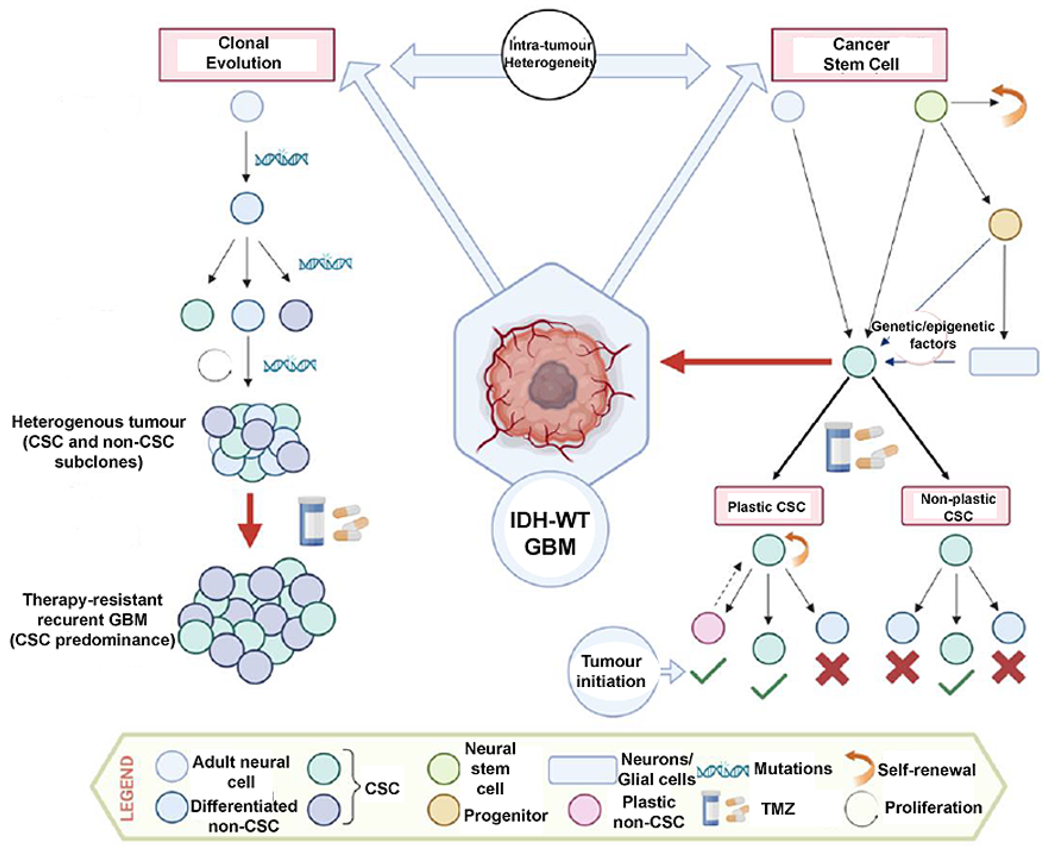
3. Intra-Tumour Heterogeneity and Cancer Stem Cells

Intra-tumour heterogeneity (ITH) has been studied in depth over the recent decade as a possible route to explain GBM resistance to treatment. Tumours are dynamic and over time, and malignant cancers tend to sub-clonally vary under a gradient of microenvironmental pressures. Due to this heterogeneity, the core tumour mass may contain a plethora of genetically varied cells with unique molecular fingerprints and varying levels of responsiveness to therapy. ITH may manifest in a non-uniform dispersal of genetically unique tumour cell subtypes throughout and within disease foci (spatial heterogeneity), or dynamic sub-clonal evolution over time (temporal heterogeneity) [29]. As implied by the ‘multiforme’ of the pre-2016 World Health Organisation classification, there has been a known association between the striking molecular heterogeneity and the dismal prognosis of GBM, establishing the clinical cornerstones of GBM: aggressive tumour growth and inevitable recurrence [30].
Cancer stem cells (CSCs) are a subpopulation of proliferating cancer cells that support tumour growth, whereas the cell of origin refers to healthy stem or progenitor cells in which oncogenic mutations initially appear and accumulate to cause tumour formation [31]. Although it is widely accepted that cancer cells with stem cell properties exist in solid tumours, the clinical significance remains unknown. It was demonstrated that targeting a small subgroup of CSC biomarkers is a futile attempt, as cells expressing CSC-associated markers in GBM are highly plastic, receiving cues from the tumour microenvironment. A dynamic process of reversible state transitions forms the non-hierarchical organisation of CSCs in GBM (i.e., GSCs), suggesting that the direction of research should focus on the dynamic state, rather than GSCs as a singular discreet entity. Four out of twelve GSC cell membrane markers have been shown to exhibit intra-tumour heterogeneity: CD133, CD44, CD15, and A2B5 [32].
Furthermore, there is an overlap between ITH and CSC in GBM. In 1976, the idea of clonal evolution was expounded to help explain the effect of cancer cellular heterogeneity. Clonal evolution suggests that somatic Darwinian evolution plays a role in how tumours acquire (epi)genetically and phenotypically distinctive cell subpopulations. Following this theory, (epi)genetic mutations may arise stochastically, with novel observable traits immediately exposed to natural selection pressures, which allow the most adapted cells (‘survival of the fittest’) to survive and flourish. When cancer cells are confronted with extrinsic or intrinsic selection pressures, such as those caused by therapeutic intervention or pH, oxygen, or nutrient gradients, respectively, an emergent resistant clone/phenotype enables a subtype of cells to survive, expand, and primarily recolonise the neoplasm [33]. With technological advancement and access to transcriptomic profiling that were not accessible in the 20th century, the CSC model is more widely accepted today. Heterogeneity in the CSC model is the premise of a spectrum of differentiation between CSC (tumorigenic) and non-CSC (non-tumorigenic) [34]. Although these models are several decades apart, they are not mutually exclusive, and both concepts can explain the origin of ITH. CSCs in neoplasms will encounter clonal evolution during the progression of the tumour [35]. The interactions of these concepts are outlined in Figure 1.

3.1. Transformation of Neural Stem/Progenitor Cells

In order to better understand tumour growth and develop treatment strategies for GBM, it is helpful to further elucidate the cellular origin of GBM, which is regarded as a neural stem or progenitor cell. There are two prominent theories regarding the origin of GSCs. The first theory posits that an accumulation of mutations in oncogenes or tumour suppressor genes in any somatic cell can transform it into a tumorigenic cell; the second theory is more focused on the self-renewing capacity of stem or progenitor cells, and it states that GSCs arise via oncogenic mutation(s) in these subpopulations. The former theory is less widely accepted based on the improbability that gain- or loss-of-function mutations would emerge in a mature, non-dividing cell with a finite lifespan. Critics of this theory endorse the latter explanation, known as the “cancer stem cell theory,” which is more widely recognised [6].
It is crucial to describe the non-diseased neural tissue hierarchy in order to comprehend the cell of origin. The three primary cell types of the CNS (oligodendrocyte, astrocyte, and neuron) are generated through the differentiation of intermediate progenitor cells from neural stem cells (NSCs) [36]. NSCs are present throughout the CNS and have the ability to generate new cell lineages that develop into differentiated neurons and glial cells.
In the highly proliferative subventricular zone (SVZ), located alongside the lateral ventricle, neural precursor cells are generated in a substantial amount during embryonic development. These precursor cells leave the niche of the SVZ, diffusing radially and differentiating into progenitor cells, which gain a restriction on self-renewal and multipotency [37].
In the context of gene expression profiles, unbiased quantitative analysis of 217 patients with GBM from the Cancer Imaging Archive indicated that tumour proximity to the SVZ is associated with an increased expression of stem-like gene signatures, which is reflective of an enrichment of CSC status. Proximity to the SVZ demonstrates enhanced stem-like mRNA expression profiles (e.g., CD133) and up-regulation of DNA repair enzymes (e.g., MGMT), contributing to a worse progression-free survival and prognosis [38].
Immature glial cells at the edge of the SVZ and GBM cells exhibit transcriptional similarities, suggesting that these glial cells could be precursors to neoplastic GBM cells [39]. Furthermore, GSCs also exhibit mutated genes expressed in NSCs, such as TP53, TERT, PTEN, PDGFα, and EGFR [40]. Combined with the location (proximity of tumours to the SVZ), matching variations in gene expression profiles, and dedifferentiation characteristics, there is a compelling case for NSCs to be the cell-of-origin lineage of GSCs [41].

3.2. GSC Plasticity

Brain tumours are known to be composed of cancer cells that exhibit stem cell surface antigens. These cells can be up to 25 times more common in high-grade tumours (for example, GBMs) than low-grade tumours [42,43]. The most widely recognised theories, which attempt to explain how tumours harbour CSCs, currently hypothesise that the astrocyte dedifferentiation theory, and/or the GBM stem cell theory, are responsible [44].
Healthy brain tissue possesses NSCs that unidirectionally differentiate into glial or neural progenitor cells, which then terminally differentiate into neurons, oligodendrocytes, astrocytes, and glial cells. This hierarchy loses plasticity significantly at each differentiation level. This is in contrast with GSCs that may dynamically differentiate into diverse tumour cell types (such as proneural, mesenchymal, classical, neural/oligodendrocyte progenitor cell-like, astrocyte-like, mesenchymal-like) due a retention of plasticity and cues from the tumour microenvironment, therapeutic interventions, and underlying genetics [45].
The astrocyte dedifferentiation theory outlines the principle of genetic mutations resulting in a backward differentiation of the astrocyte—reverting it back to a glial progenitor cell (GPC)—a type of NSC. The associated genetic mutations are responsible for a combination of suppression of tumour suppressor genes and the promotion of oncogenes, as evidenced by experiments in mice that showed induced tumorigenesis from manipulated astrocytes, depending on whether this manipulation acted on GPCs, which would only need oncogene alteration, or NSCs, which need both alterations [46]. The astrocyte dedifferentiation theory is linked to plasticity as it demonstrates that non-stem cells can be tumour progenitors.
Furthermore, astrocyte-like NSCs in the subventricular zone have intricately been shown, via genome-edited mouse models and corroborated primary patient brain tissue, to be the likely cell of origin which harbours GBM driver mutations. Indeed, single-cell sequencing confirmed that such astrocyte-like NSCs migrate from the subventricular zone and give rise to malignant glioma at distal anatomical sites [47].

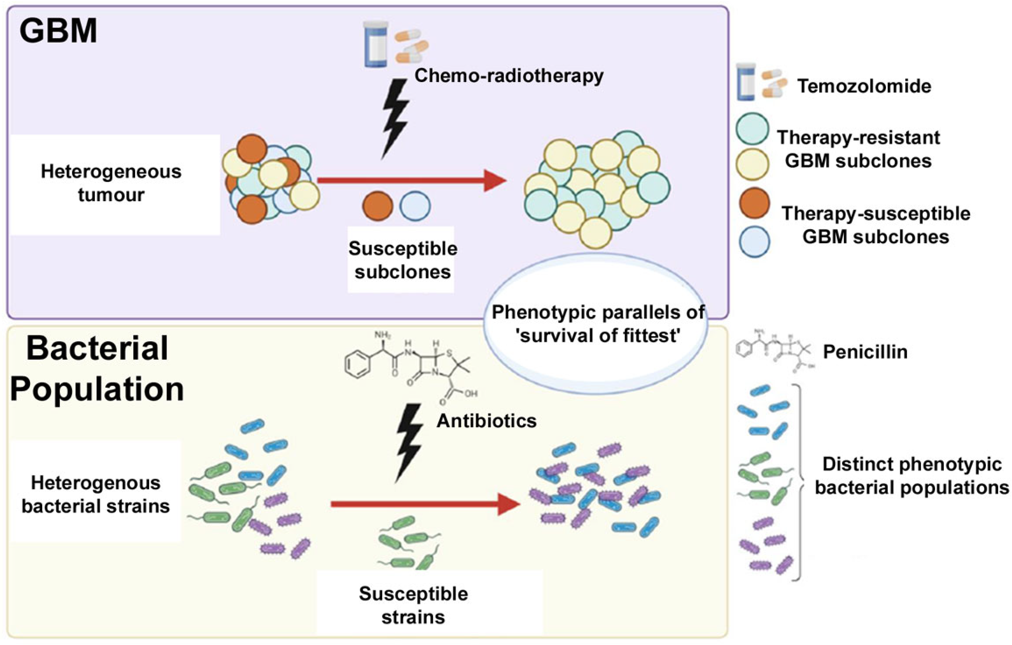
4. Comparison between CSCs and Antibiotic Resistance

Antibiotic resistance and GSCs are both phenomena that present challenges in the management of infections and cancer, respectively. Antibiotic resistance arises in bacteria and is driven by the selective pressure of exposure to antibiotics, while GSCs drive the genetic alterations that promote cancer clonal evolution [48]. While there are some similarities in the mechanisms by which they develop, such as through mutation and the acquisition of resistance genes through horizontal gene transfer, there are also important differences to consider. For example, strategies for preventing or reducing the development of antibiotic resistance, such as the judicious use of antibiotics and infection control measures, are not applicable to the prevention of GSC propagation in GBM.
GSCs are the cells most likely to survive standard-of-care treatment and have been shown to be the most tumorigenic. It is probable that the present standard of care for GBM is merely eliminating the vulnerable subpopulations, whilst the more resistant subpopulations survive and repopulate the tumour, given the inherent genetic and phenotypic heterogeneity of GBM. The cells that survive therapy have already been chosen as the most capable of forming tumours, surviving in a hostile environment, and retaining the most malignant attributes [49].
There is thus a similarity between antibiotic resistance and GSCs that can be drawn, akin to Darwinian selection. Both scenarios reflect a population of biological entities, in which each individual cell or organism is governed genotypically to determine how they respond when a selective pressure is applied (such as drug treatment). The individual cell or organism may be susceptible to the selective pressure and die, or it may survive. The survival of some cells means that a given subpopulation expands, and with time, can mutate further, leading to the recurrence of tumours or the proliferation of a resistant strain, which is refractory to further treatment. This unifying concept between antibiotic resistance and GSCs is analogous as both contain a series of random mutations that can lead to the development of non-random therapeutic adaptation and resistance (Figure 2). Human interventions induce this resistive mechanism and speed it up—as demonstrated by the sudden need for antibiotic stewardship.
The fact that the GSC mechanism of resistance is more complex than antibiotic resistance is crucial to note. Since most medications cannot penetrate healthy brain tissue, infiltrative GBM tumour cells that have spread throughout the brain are inaccessible to systemically administered drugs such as temozolomide (TMZ) and radiotherapy, as evidenced by GBM growth, which causes a heterogenous disruption of the blood–brain barrier, hampering evenly distributed drug delivery [50]. However, given that GBM is a heterogeneous tumour, it could be posited that the mutations causing the tumour’s propensity to penetrate brain parenchyma are themselves pro-survival characteristics. Furthermore, it is now well established that GBM cells generate tumour microtubes (membrane projections), forming functional multicellular network structures that contribute to neo-vasculature and confer resistance to TMZ [51].

5. Future Therapeutic Outlook

We contend that next-generation therapeutic approaches should prioritise GBM infiltrative disease, both as a means to circumvent intra-tumour heterogeneity and to ensure that the most clinically relevant tumour subpopulation is targeted. Recent attractive multidisciplinary therapeutic approaches have demonstrated potential in a preclinical setting. These include systemically administered iron oxide nanoparticles conjugated with a p32 cell surface antigen, conferring efficacy against an infiltratively-disseminating GBM orthotopic model [52]; inhibition of PAK4, normalising the tumour vascular microenvironment to sensitise orthotopic GBM xenografts to chimeric antigen receptor-T cell immunotherapy [53]; and combinatorial targeting using IL15-expressing oncolytic virus and EGFR chimeric antigen receptor natural killer cells eliciting a survival benefit in GBM-bearing mice [54].

6. Conclusions

Identifying the degree of stemness across heterogenous infiltrative cells remains a scientific challenge for a therapeutically relevant understanding of GBM biology. What is the genetic profile of residual disease stem cells? Are residual disease stem cells functionally associated with promoting infiltration and recurrence? Previous research has focused on single therapies directed at the primary GBM mass; however, future efforts will require combination therapies that target the primary mass, as well as the heterogenous infiltrating cell populations at the site of residual disease.

Author Contributions

Conceptualization, R.R.; writing—original draft preparation, P.M.; writing—review and editing, R.R.; supervision, R.R. All authors have read and agreed to the published version of the manuscript.

Funding

This research received no external funding.

Conflicts of Interest

The authors declare no conflicts of interest.

References

  1. Grochans, S.; Cybulska, A.M.; Simińska, D.; Korbecki, J.; Kojder, K.; Chlubek, D.; Baranowska-Bosiacka, I. Epidemiology of Glioblastoma Multiforme–Literature Review. Cancers 2022, 14, 2412. [Google Scholar] [CrossRef] [PubMed]
  2. Batash, R.; Asna, N.; Schaffer, P.; Francis, N.; Schaffer, M. Glioblastoma Multiforme, Diagnosis and Treatment; Recent Literature Review. Curr. Med. Chem. 2017, 24, 3002–3009. [Google Scholar] [CrossRef]
  3. Razavi, S.M.; Lee, K.E.; Jin, B.E.; Aujla, P.S.; Gholamin, S.; Li, G. Immune Evasion Strategies of Glioblastoma. Front. Surg. 2016, 3, 11. [Google Scholar] [CrossRef]
  4. Fabbro-Peray, P.; Zouaoui, S.; Darlix, A.; Fabbro, M.; Pallud, J.; Rigau, V.; Mathieu-Daude, H.; Bessaoud, F.; Bauchet, F.; Riondel, A.; et al. Association of Patterns of Care, Prognostic Factors, and Use of Radiotherapy–Temozolomide Therapy with Survival in Patients with Newly Diagnosed Glioblastoma: A French National Population-Based Study. J. Neurooncol. 2019, 142, 91–101. [Google Scholar] [CrossRef]
  5. Wen, P.Y.; Kesari, S. Malignant Gliomas in Adults. New Engl. J. Med. 2008, 359, 492–507. [Google Scholar] [CrossRef]
  6. Altmann, C.; Keller, S.; Schmidt, M.H.H. The Role of Svz Stem Cells in Glioblastoma. Cancers 2019, 11, 448. [Google Scholar] [CrossRef] [PubMed]
  7. Ross, J.L.; Cooper, L.A.D.; Kong, J.; Gutman, D.; Williams, M.; Tucker-Burden, C.; McCrary, M.R.; Bouras, A.; Kaluzova, M.; Dunn, W.D.; et al. 5-Aminolevulinic Acid Guided Sampling of Glioblastoma Microenvironments Identifies Pro-Survival Signaling at Infiltrative Margins. Sci. Rep. 2017, 7, 15593. [Google Scholar] [CrossRef] [PubMed]
  8. Seker-Polat, F.; Degirmenci, N.P.; Solaroglu, I.; Bagci-Onder, T. Tumor Cell Infiltration into the Brain in Glioblastoma: From Mechanisms to Clinical Perspectives. Cancers 2022, 14, 443. [Google Scholar] [CrossRef]
  9. Montana, V.; Sontheimer, H. Bradykinin Promotes the Chemotactic Invasion of Primary Brain Tumors. J. Neurosci. 2011, 31, 4858–4867. [Google Scholar] [CrossRef]
  10. Diksin, M.; Smith, S.J.; Rahman, R. The Molecular and Phenotypic Basis of the Glioma Invasive Perivascular Niche. Int. J. Mol. Sci. 2017, 18, 2342. [Google Scholar] [CrossRef]
  11. Chédotal, A.; Kerjan, G.; Moreau-Fauvarque, C. The Brain within the Tumor: New Roles for Axon Guidance Molecules in Cancers. Cell Death Differ. 2005, 12, 1044–1056. [Google Scholar] [CrossRef] [PubMed]
  12. de Gooijer, M.C.; Guillén Navarro, M.; Bernards, R.; Wurdinger, T.; van Tellingen, O. An Experimenter’s Guide to Glioblastoma Invasion Pathways. Trends Mol. Med. 2018, 24, 763–780. [Google Scholar] [CrossRef] [PubMed]
  13. Pinel, S.; Thomas, N.; Boura, C.; Barberi-Heyob, M. Approaches to Physical Stimulation of Metallic Nanoparticles for Glioblastoma Treatment. Adv. Drug Deliv. Rev. 2019, 138, 344–357. [Google Scholar] [CrossRef]
  14. Laperriere, N.; Zuraw, L.; Cairncross, G. Radiotherapy for Newly Diagnosed Malignant Glioma in Adults: A Systematic Review. Radiother. Oncol. 2002, 64, 259–273. [Google Scholar] [CrossRef] [PubMed]
  15. Stummer, W.; Pichlmeier, U.; Meinel, T.; Wiestler, O.D.; Zanella, F.; Reulen, H.J. Fluorescence-Guided Surgery with 5-Aminolevulinic Acid for Resection of Malignant Glioma: A Randomised Controlled Multicentre Phase III Trial. Lancet Oncol. 2006, 7, 392–401. [Google Scholar] [CrossRef]
  16. Smith, S.J.; Diksin, M.; Chhaya, S.; Sairam, S.; Estevez-Cebrero, M.A.; Rahman, R. The Invasive Region of Glioblastoma Defined by 5ALA Guided Surgery Has an Altered Cancer Stem Cell Marker Profile Compared to Central Tumour. Int. J. Mol. Sci. 2017, 18, 2452. [Google Scholar] [CrossRef]
  17. Kis, D.; Szivos, L.; Rekecki, M.; Shukir, B.S.; Mate, A.; Hideghety, K.; Barzo, P. Predicting the True Extent of Glioblastoma Based on Probabilistic Tractography. Front. Neurosci. 2022, 16, 886465. [Google Scholar] [CrossRef]
  18. Lasocki, A.; Gaillard, F. Non-Contrast-Enhancing Tumor: A New Frontier in Glioblastoma Research. Am. J. Neuroradiol. 2019, 40, 758–765. [Google Scholar] [CrossRef]
  19. de Bonis, P.; Anile, C.; Pompucci, A.; Fiorentino, A.; Balducci, M.; Chiesa, S.; Lauriola, L.; Maira, G.; Mangiola, A. The Influence of Surgery on Recurrence Pattern of Glioblastoma. Clin. Neurol. Neurosurg. 2013, 115, 37–43. [Google Scholar] [CrossRef]
  20. Knudsen, A.M.; Halle, B.; Cédile, O.; Burton, M.; Baun, C.; Thisgaard, H.; Anand, A.; Hubert, C.; Thomassen, M.; Michaelsen, S.R.; et al. Surgical Resection of Glioblastomas Induces Pleiotrophin-Mediated Self-Renewal of Glioblastoma Stem Cells in Recurrent Tumors. Neuro Oncol. 2022, 24, 1074–1087. [Google Scholar] [CrossRef]
  21. Gupta, K.; Burns, T.C. Radiation-Induced Alterations in the Recurrent Glioblastoma Microenvironment: Therapeutic Implications. Front. Oncol. 2018, 8, 503. [Google Scholar] [CrossRef] [PubMed]
  22. Olzowy, B.; Hundt, C.S.; Stocker, S.; Bise, K.; Reulen, H.J.; Stummer, W. Photoirradiation Therapy of Experimental Malignant Glioma with 5-Aminolevulinic Acid. J. Neurosurg. 2002, 97, 970–976. [Google Scholar] [CrossRef] [PubMed]
  23. Krammer, B.; Plaetzer, K. ALA and Its Clinical Impact, from Bench to Bedside. Photochem. Photobiol. Sci. 2008, 7, 283–289. [Google Scholar] [CrossRef]
  24. Etminan, N.; Peters, C.A.; Ficnar, J.; Anlasik, S.; Bünemann, E.; Slotty, P.J.; Hänggi, D.; Steiger, H.J.; Sorg, R.v.; Stummer, W. Modulation of Migratory Activity and Invasiveness of Human Glioma Spheroids Following 5-Aminolevulinic Acid-Based Photodynamic Treatment. J. Neurosurg. 2011, 115, 281–288. [Google Scholar] [CrossRef]
  25. Schimanski, A.; Ebbert, L.; Sabel, M.C.; Finocchiaro, G.; Lamszus, K.; Ewelt, C.; Etminan, N.; Fischer, J.C.; Sorg, R.V. Human Glioblastoma Stem-like Cells Accumulate Protoporphyrin IX When Subjected to Exogenous 5-Aminolaevulinic Acid, Rendering Them Sensitive to Photodynamic Treatment. J. Photochem. Photobiol. B 2016, 163, 203–210. [Google Scholar] [CrossRef]
  26. Castano, A.P.; Demidova, T.N.; Hamblin, M.R. Mechanisms in Photodynamic Therapy: Part Two—Cellular Signaling, Cell Metabolism and Modes of Cell Death. Photodiagn. Photodyn. Ther. 2005, 2, 1–23. [Google Scholar] [CrossRef] [PubMed]
  27. Andrieux, G.; Das, T.; Griffin, M.; Straehle, J.; Paine, S.M.L.; Beck, J.; Boerries, M.; Heiland, D.H.; Smith, S.J.; Rahman, R.; et al. Spatially Resolved Transcriptomic Profiles Reveal Unique Defining Molecular Features of Infiltrative 5ALA-Metabolizing Cells Associated with Glioblastoma Recurrence. Genome Med. 2023, 15, 48. [Google Scholar] [CrossRef]
  28. Smith, S.J.; Rowlinson, J.; Estevez-Cebrero, M.; Onion, D.; Ritchie, A.; Clarke, P.; Wood, K.; Diksin, M.; Lourdusamy, A.; Grundy, R.G.; et al. Metabolism-Based Isolation of Invasive Glioblastoma Cells with Specific Gene Signatures and Tumorigenic Potential. Neurooncol. Adv. 2020, 2, vdaa087. [Google Scholar] [CrossRef]
  29. Dagogo-Jack, I.; Shaw, A.T. Tumour Heterogeneity and Resistance to Cancer Therapies. Nat. Rev. Clin. Oncol. 2018, 15, 81–94. [Google Scholar] [CrossRef]
  30. Aubry, M.; de Tayrac, M.; Etcheverry, A.; Clavreul, A.; Saikali, S.; Menei, P.; Mosser, J. From the Core to beyond the Margin: A Genomic Picture of Glioblastoma Intratumor Heterogeneity. Oncotarget 2015, 6, 12094–12109. [Google Scholar] [CrossRef]
  31. Visvader, J.E. Cells of Origin in Cancer. Nature 2011, 469, 314–322. [Google Scholar] [CrossRef] [PubMed]
  32. Dirkse, A.; Golebiewska, A.; Buder, T.; Nazarov, P.V.; Muller, A.; Poovathingal, S.; Brons, N.H.C.; Leite, S.; Sauvageot, N.; Sarkisjan, D.; et al. Stem Cell-Associated Heterogeneity in Glioblastoma Results from Intrinsic Tumor Plasticity Shaped by the Microenvironment. Nat. Commun. 2019, 10, 1787. [Google Scholar] [CrossRef] [PubMed]
  33. Nowell, P.C. The Clonal Evolution of Tumor Cell Populations. Science 1976, 194, 23–28. [Google Scholar] [CrossRef] [PubMed]
  34. Shackleton, M.; Quintana, E.; Fearon, E.R.; Morrison, S.J. Heterogeneity in Cancer: Cancer Stem Cells versus Clonal Evolution. Cell 2009, 138, 822–829. [Google Scholar] [CrossRef]
  35. Kreso, A.; Dick, J.E. Evolution of the Cancer Stem Cell Model. Cell Stem Cell 2014, 14, 275–291. [Google Scholar] [CrossRef]
  36. Kim, H.J.; Park, J.W.; Lee, J.H. Genetic Architectures and Cell-of-Origin in Glioblastoma. Front. Oncol. 2021, 10, 615400. [Google Scholar] [CrossRef]
  37. Ming, G.; Song, H. Adult Neurogenesis in the Mammalian Brain: Significant Answers and Significant Questions. Neuron 2011, 70, 687–702. [Google Scholar] [CrossRef]
  38. Steed, T.C.; Treiber, J.M.; Taha, B.; Engin, H.B.; Carter, H.; Patel, K.S.; Dale, A.M.; Carter, B.S.; Chen, C.C. Glioblastomas Located in Proximity to the Subventricular Zone (SVZ) Exhibited Enrichment of Gene Expression Profiles Associated with the Cancer Stem Cell State. J. Neurooncol. 2020, 148, 455–462. [Google Scholar] [CrossRef]
  39. Pollen, A.A.; Nowakowski, T.J.; Chen, J.; Retallack, H.; Sandoval-Espinosa, C.; Nicholas, C.R.; Shuga, J.; Liu, S.J.; Oldham, M.C.; Diaz, A.; et al. Molecular Identity of Human Outer Radial Glia during Cortical Development. Cell 2015, 163, 55–67. [Google Scholar] [CrossRef]
  40. Matarredona, E.R.; Zarco, N.; Castro, C.; Guerrero-Cazares, H. Editorial: Neural Stem Cells of the Subventricular Zone: From Neurogenesis to Glioblastoma Origin. Front. Oncol. 2021, 11, 750116. [Google Scholar] [CrossRef]
  41. Verdugo, E.; Puerto, I.; Medina, M.Á. An Update on the Molecular Biology of Glioblastoma, with Clinical Implications and Progress in Its Treatment. Cancer Commun. 2022, 42, 1083–1111. [Google Scholar] [CrossRef] [PubMed]
  42. Bardella, C.; Al-Shammari, A.R.; Soares, L.; Tomlinson, I.; O’Neill, E.; Szele, F.G. The Role of Inflammation in Subventricular Zone Cancer. Prog. Neurobiol. 2018, 170, 37–52. [Google Scholar] [CrossRef] [PubMed]
  43. Singh, S.K.; Clarke, I.D.; Terasaki, M.; Bonn, V.E.; Hawkins, C.; Squire, J.; Dirks, P.B. Identification of a Cancer Stem Cell in Human Brain Tumors. Cancer Res. 2003, 63, 5821–5828. [Google Scholar]
  44. Beiriger, J.; Habib, A.; Jovanovich, N.; Kodavali, C.V.; Edwards, L.; Amankulor, N.; Zinn, P.O. The Subventricular Zone in Glioblastoma: Genesis, Maintenance, and Modeling. Front. Oncol. 2022, 12, 790976. [Google Scholar] [CrossRef] [PubMed]
  45. Yabo, Y.A.; Niclou, S.P.; Golebiewska, A. Cancer Cell Heterogeneity and Plasticity: A Paradigm Shift in Glioblastoma. Neuro Oncol. 2022, 24, 669–682. [Google Scholar] [CrossRef]
  46. Friedmann-Morvinski, D.; Bushong, E.A.; Ke, E.; Soda, Y.; Marumoto, T.; Singer, O.; Ellisman, M.H.; Verma, I.M. Dedifferentiation of Neurons and Astrocytes by Oncogenes Can Induce Gliomas in Mice. Science 2012, 338, 1080–1084. [Google Scholar] [CrossRef]
  47. Lee, J.H.; Lee, J.E.; Kahng, J.Y.; Kim, S.H.; Park, J.S.; Yoon, S.J.; Um, J.-Y.; Kim, W.K.; Lee, J.-K.; Park, J.; et al. Human Glioblastoma Arises From Subventroicular Zone Cells with Low-Level Driver Mutations. Nature 2018, 560, 243–247. [Google Scholar] [CrossRef]
  48. Orzan, F.; de Bacco, F.; Crisafulli, G.; Pellegatta, S.; Mussolin, B.; Siravegna, G.; D’Ambrosio, A.; Comoglio, P.M.; Finocchiaro, G.; Boccaccio, C. Genetic Evolution of Glioblastoma Stem-Like Cells From Primary to Recurrent Tumor. Stem Cells 2017, 35, 2218–2228. [Google Scholar] [CrossRef]
  49. Auffinger, B.; Spencer, D.; Pytel, P.; Ahmed, A.U.; Lesniak, M.S. The Role of Glioma Stem Cells in Chemotherapy Resistance and Glioblastoma Multiforme Recurrence. Expert. Rev. Neurother. 2015, 15, 741–752. [Google Scholar] [CrossRef]
  50. Mo, F.; Pellerino, A.; Soffietti, R.; Rudà, R. Blood-Brain Barrier in Brain Tumors: Biology and Clinical Relevance. Int. J. Mol. Sci. 2021, 22, 12654. [Google Scholar] [CrossRef]
  51. Dymova, M.A.; Kuligina, E.V.; Richter, V.A. Molecular Mechanisms of Drug Resistance in Glioblastoma. Int. J. Mol. Sci. 2021, 22, 6385. [Google Scholar] [CrossRef] [PubMed]
  52. Saalik, P.; Lingasamy, P.; Toome, K.; Mastandrea, I.; Rousso-Noori, L.; Tobu, A.; Simon-Garcia, L.; Hunt, H.; Paiste, P.; Kotamraju, V.R.; et al. Peptide-guided Nanoparticles for Glioblastoma Targeting. J. Control Release 2019, 308, 109–118. [Google Scholar] [CrossRef] [PubMed]
  53. Ma, W.; Wang, Y.; Zhang, R.; Yang, F.; Zhang, D.; Huang, M.; Zhang, L.; Dorsey, J.F.; Binder, Z.A.; O’Rourke, D.M.; et al. Targeting PAK4 to Reprogram the Vascular Microenvironment and Improve CAR-T Immunotherapy for Glioblastoma. Nat. Cancer 2021, 2, 83–97. [Google Scholar] [CrossRef] [PubMed]
  54. Ma, R.; Lu, T.; Li, Z.; Teng, K.-Y.; Mansour, A.G.; Yu, M.; Tian, L.; Xu, B.; Ma, S.; Zhang, J.; et al. An Oncolytic Virus Expressing IL15/IL15Rα Combined with Off-the-Shelf EGFR-CAR NK Cells Targets Glioblastoma. Cancer Res. 2021, 81, 3635–3648. [Google Scholar] [CrossRef]
Figure 1. Aspects of intra-tumour heterogeneity and its relationship with clonal evolution (Darwinian evolution: selection of cells that have mutated to exhibit more aggressive phenotypes, followed by natural selection of these cells conferred by external selection pressures) and the Cancer Stem Cell Model (in which heterogeneity encompasses the spectrum of differentiated cells and plasticity between CSC and non-CSC). Created using BioRender.
Onco 04 00024 g001
Figure 2. Schematic outlining the analogous mechanism of tumour cells undergoing random mutations, as well as bacterial populations being selected to allow resistant populations to multiply. Created in BioRender.com.
Onco 04 00024 g002
Disclaimer/Publisher’s Note: The statements, opinions and data contained in all publications are solely those of the individual author(s) and contributor(s) and not of MDPI and/or the editor(s). MDPI and/or the editor(s) disclaim responsibility for any injury to people or property resulting from any ideas, methods, instructions or products referred to in the content.

