Review

Cancer Immunotherapy: Targeting TREX1 Has the Potential to Unleash the Host Immunity against Cancer Cells

by
Karim Hawillo
,
Samira Kemiha
and
Hervé Técher
*
Université Côte d’Azur, Institute for Research on Cancer and Aging of Nice, IRCAN, CNRS, INSERM, Faculty of Medicine, 06100 Nice, France
*
Author to whom correspondence should be addressed.
Onco 2024, 4(4), 322-334; https://doi.org/10.3390/onco4040022
Submission received: 27 August 2024 / Revised: 28 September 2024 / Accepted: 9 October 2024 / Published: 14 October 2024

Abstract

:

Simple Summary

Cancer cells often have DNA damage and chromosomal instability. This can lead to the release of DNA fragments into the cell’s cytoplasm. A protein called cGAS detects these DNA fragments. It then triggers an inflammatory response through the production of type I interferon. Cancer cells can avoid this immune response by either turning off the cGAS pathway or using another protein called TREX1 to break down the cytosolic DNA fragments. Recent publications show that deleting TREX1 in cancer cells renders tumors visible to the immune system and slows their growth. Targeting TREX1 with drugs might be a new way to fight cancer by boosting the immune response against tumors.

Abstract

Chromosomal instability and DNA damage are hallmarks of cancers that can result in the accumulation of micronuclei, cytosolic chromatin fragments (CCFs), or cytosolic DNA species (cytoDNA). The cyclic GMP-AMP synthase (cGAS) is a DNA sensor that recognizes cytosolic DNA and chromatin fragments and subsequently triggers a systemic type I interferon response via the cGAS-STING pathway. Although cancer cells usually contain a high level of chromosomal instability, these cells can avoid the induction of the interferon (IFN) response either by silencing cGAS-STING or the upregulation of the three prime exonuclease 1 (TREX1). TREX1 restricts the spontaneous activation of the cGAS-STING pathway through the degradation of cytoDNA; this in turn limits tumor immunogenicity allowing cancer cells to evade immune detection. Deletion of TREX1 in different cancer types has been shown to decrease tumor growth and increase tumor immune infiltration in pre-clinical mice models. These recent studies also showed the efficacy of TREX1-targeting in combination with anti-PD-1 immune checkpoint blockade. Therefore, targeting TREX1 represents a unique therapeutic strategy to induce an amplified induction of a type I IFN response, promoting the host’s immune response against chromosomally unstable cancer cells. We here discuss these recent advances obtained in preclinical cancer models that pave the way to develop TREX1 inhibitors and to find new avenues to target the broad cGAS-STING pathway signaling in cancer therapy.

1. Introduction

1.1. The cGAS-STING Pathway and Inflammation

The cGAS-STING pathway is a crucial component of the innate immune system, responsible for detecting cytosolic DNA and initiating an inflammatory response [1]. cGAS is activated upon binding to double-stranded DNA (dsDNA) or RNA:DNA hybrids [2]. cGAS localizes to the cytoplasm and binds to chromatin. cGAS is inhibited by its interaction with nucleosomal histones, explaining why the presence of cGAS in the nucleus is not sufficient to activate it [3,4]. Once activated, cGAS (cyclic GMP-AMP synthase) produces a second messenger, the cyclic dinucleotide cGAMP (2′-3′cyclic GMP-AMP) that binds to the stimulator of interferon genes (STING) (Figure 1). STING is located on the endoplasmic reticulum (ER) membrane. Activation of STING leads to its translocation to the Golgi apparatus and subsequent recruitment and phosphorylation of TANK-binding kinase 1 (TBK1) and then of interferon regulatory factor 3 (IRF3) [2]. This signaling cascade culminates in the production of type I interferon, IFN-I (INF-α and INF-β), and downstream of interferon-stimulated genes (ISGs), encompassing pro-inflammatory cytokines and chemokines orchestrating an immune response (Figure 1).
Examples of conditions that activate the cGAS-STING pathway include the presence of foreign DNA, such as viral DNA during infections. However, in the context of cancers and cancer therapies, self-DNA can be released into the cytoplasm as a consequence of genomic instability or mitochondrial damage [2,5,6]. Upon irradiation (i.e., radiotherapy), self-DNA accumulates in the cytoplasm as a result of mitochondrial DNA leakage [7]. Upon treatment with chemotherapies (e.g., DNA damage-inducers and mitotic poisons), generation of DNA damage and genomic instability can result in the accumulation of cytosolic DNA [8,9,10,11]. It is important to note that cancer cells present an intrinsic higher level of genomic instability, notably because of oncogenic stress and lack of key DNA repair and DNA damage response (DDR) factors, which are involved in tumor suppression. This elevated genomic instability has been shown to produce cytosolic DNA and to activate the cGAS-STING pathway in cancer cells [12,13,14,15]. It is thus expected that cancer cells have a greater propensity, compared to normal cells, to produce cytosolic DNA and to subsequently activate the cGAS-STING-IFN-I signaling.
Activation of the cGAS-STING pathway by self-DNA causes a “sterile inflammation”, a type of inflammation that occurs in the absence of pathogens. A key example of “sterile inflammation”, or auto-inflammation, is seen upon TREX1 (three-prime repair exonuclease 1) exonuclease deficiency that causes pathologies known as interferonopathies [16]. Interferonopathies are characterized by the unscheduled and uncontrolled production of interferons, namely IFN-I. Hereby, the cGAS-STING pathway serves as a critical sensor of internal threats, linking DNA damage and immune responses.

1.2. TREX1 Degrades Cytosolic DNA Limiting cGAS-STING Signaling

TREX1 is the major exonuclease in the cytoplasm responsible for degrading cytosolic DNA (cytoDNA) [17,18]. TREX1 removes nucleotides from the 3′ ends of DNA molecules. TREX1 degrades single-stranded DNA (ssDNA), double-stranded DNA (dsDNA), and RNA:DNA hybrids [19,20,21], preventing the accumulation of nucleic acids in the cytosol and as a result cGAS activation and pathological inflammation [17,18,22]. In this way, TREX1 acts as an apex predator of cytosolic DNA metabolism, ensuring that DNA fragments do not persist in the cytoplasm to trigger inappropriate immune activation.
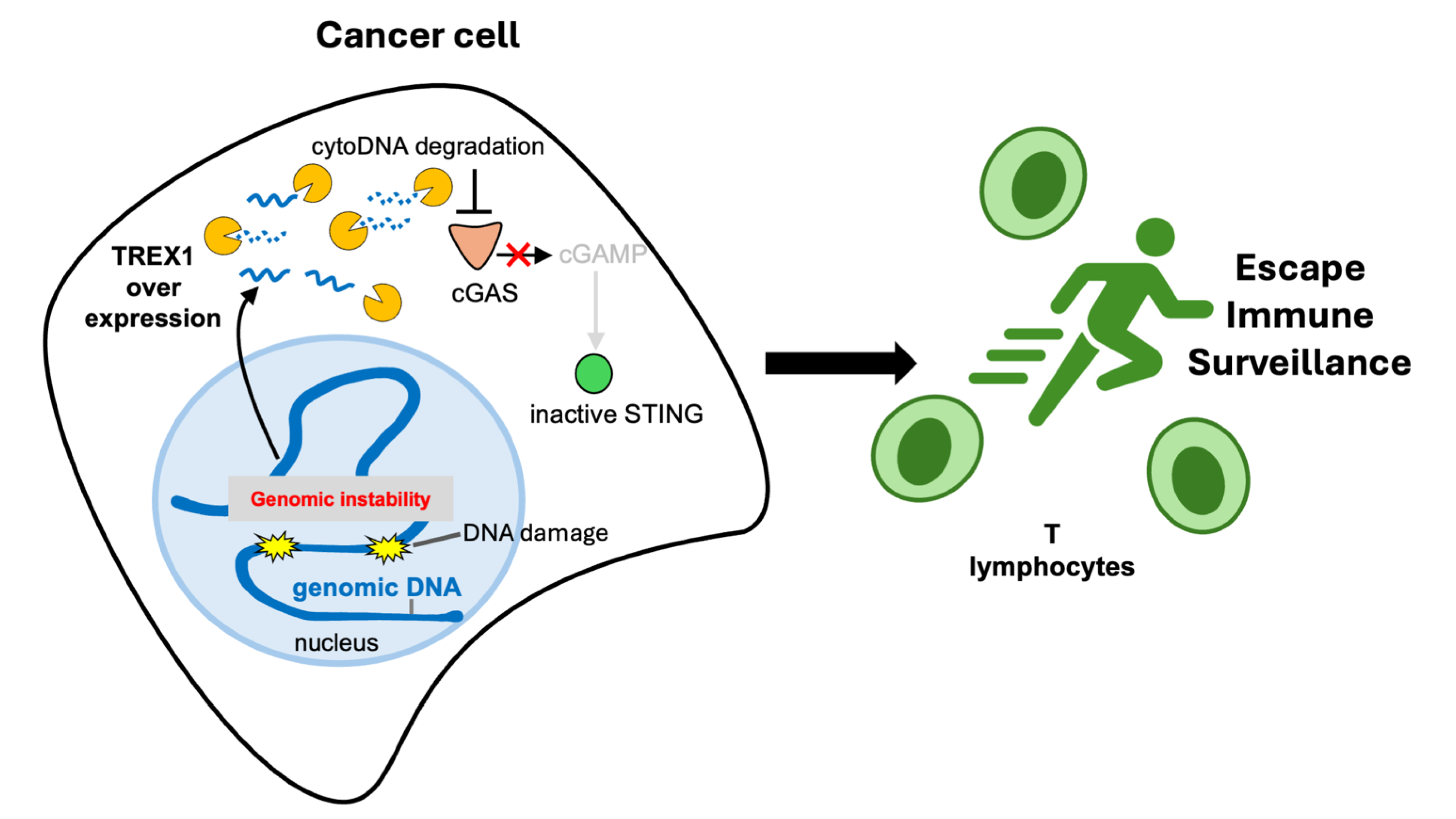
Cancer cells exhibit uncontrolled rates of cellular divisions and therefore are prone to DNA damage and breaks, notably during DNA replication [6]. Some types of cancer cells show a spontaneous accumulation of cytoDNA and elevated markers of cGAS-STING activation [12,13,14,15]. The presence of high levels of cytoDNA can presumably lead to two opposite outcomes: the induction of cGAS-STING-IFN signaling or TREX1-mediated degradation of cytosolic DNA, which in turn limits cGAS-mediated cytoDNA sensing (Figure 1). Based on these presumptions, it has been recently shown that TREX1 inactivation in different cancer models can promote anti-cancer immunity and therefore provides the proof-of-concept that targeting TREX1 can offer a new line of treatment in cancer therapy [23,24,25]. We describe these recent findings in more detail in the next sections. Unexpectedly, some cancers show an upregulation of TREX1 expression that prevents cGAS-STING activation. It is now more evident that either cGAS-STING downregulation or TREX1 upregulation are routes taken by some cancers to escape immune surveillance [23,24,25,26] (see Figure 2).

1.3. Impact of TREX1 on Genomic Stability

TREX1 absence, loss of exonuclease activity, or mis-localization have all been linked to genomic instability [27].
Loss of TREX1 exonuclease activity in different cell-types, caused by mutations of the N-terminal part of the protein or deletion of TREX1 gene, as described in familial chilblain lupus (FCL) or Aicardi–Goutière Syndrome (AGS), is characterized by an increase in chromosomal instability and senescence [28,29,30,31]. This senescence phenotype is the consequence of persistent DNA damage and sustained inflammatory signaling. For instance, it has been shown that human fibroblasts overexpressing the FCL-causing dominant-negative mutation, TREX1-D18N, have an increased frequency of micronuclei and accumulate 53BP1 foci (a DNA break marker), suggesting an accumulation of DNA damage and chromosomal instability driving senescence [28]. Mutations in the TREX1 gene have been linked to several autoinflammatory diseases [16]. These diseases are characterized by chronic inflammation, but the pathological impact of DNA damage and chromosomal instability is still poorly understood in this context [27]. Supplementing human fibroblasts with recombinant INF-β for several days altered cell proliferation, increased DNA damage and led to senescence [28]. Similar results were obtained when challenging cancer cells with INF-β, notably with phosphorylation of the DNA replication checkpoint kinase CHK1 [32]. These results suggest that the interferon response in the absence of functional TREX1 can promote the destabilization of genomic DNA.
Additionally, C-terminal truncations in TREX1 are known to cause autosomal dominant retinal vasculopathy with cerebral leukodystrophy (RVCL), which is associated with a functional exonuclease activity but a mis-localization of TREX1 protein into the nucleus [33,34]. This mis-localization is hypothesized to cause DNA damage through degradation of exposed nicks in the nucleus contributing in turn to the pathology [35]. This hypothesis has been supported by recent findings showing in different species that TREX1 C-terminal truncations, mimicking the RVCL causing mutations, can promote the accumulation of DNA damage [33]. It is not the first time that TREX1 entry into the nucleus has been shown to generate DNA damage and subsequently senescence. Physical compression of cells has been shown to lead to nuclear envelope shattering. Breakage of nuclear envelope upon this physical constraint led to TREX1 entry into the nucleus, subsequent accumulation of DNA damage, and a senescent phenotype [35]. Inflammation is not involved in RVCL [33].

1.4. Cancer Immunotherapy

A significant advancement in cancer therapy has been the development of immunotherapies, which aim at promoting the inherent capacity of the immune system to recognize and to eliminate cancerous cells [5,36]. Immune cells and cancer cells express cell surface receptors that act as immune regulators, known as immune checkpoints [37,38]. PD1 (Programmed Cell Death Protein 1) is a checkpoint protein expressed on T cells, which acts like an “off switch” that keeps the active T cells from attacking cells when it is bound to its ligand PD-L1 (Programmed Death-Ligand 1), expressed by normal and some cancer cells. This PD1/PD-L1 immune checkpoint acts as a regulatory pathway that suppresses the immune system’s activity, notably of T-cells [37,38,39]. PD1 and PD-L1 interaction can be blocked with monoclonal antibodies targeting their domain of contact, constituting a class of therapies that are known as the immune checkpoint blockade (ICB). In brief, ICB by blocking PD1/PD-L1 binding allows immune cells to recognize and eliminate cancer cells, in a phenomenon known as immune-cell death (ICD) and anti-tumor immunity. ICB therapies overcome the inhibition of immune cells mediated by cancerous cells that express on their surface PD1, or other types of negative regulators of immunity, and therefore promote tumor rejection [36].
In some cancers, these ICB therapies are known to be ineffective, notably because of a poor infiltration of immune cells into the solid tumor mass [39,40,41,42,43]. One promising strategy to overcome this limitation is to leverage the inflammatory signaling to promote immunogenicity of cancerous tissues to induce ICD. This can be achieved by increasing the production of type I interferons (IFN-I) within the tumor microenvironment [44]. In this review, we discuss recent advances made in pre-clinical cancer models to promote anti-tumor immunity by increasing the cGAS-STING signaling and IFN-I response, focusing on TREX1 ablation. We also describe how immunotherapies and pro-inflammatory signaling offer new lines of treatment that add to the panel of conventional chemotherapies or radiotherapies, already in use in clinics.

2. Inactivation of TREX1 Promotes Type I Interferon Response Unleashing Tumor Immunogenicity

2.1. Cancer Rejection through Activation of the cGAS-STING Pathway

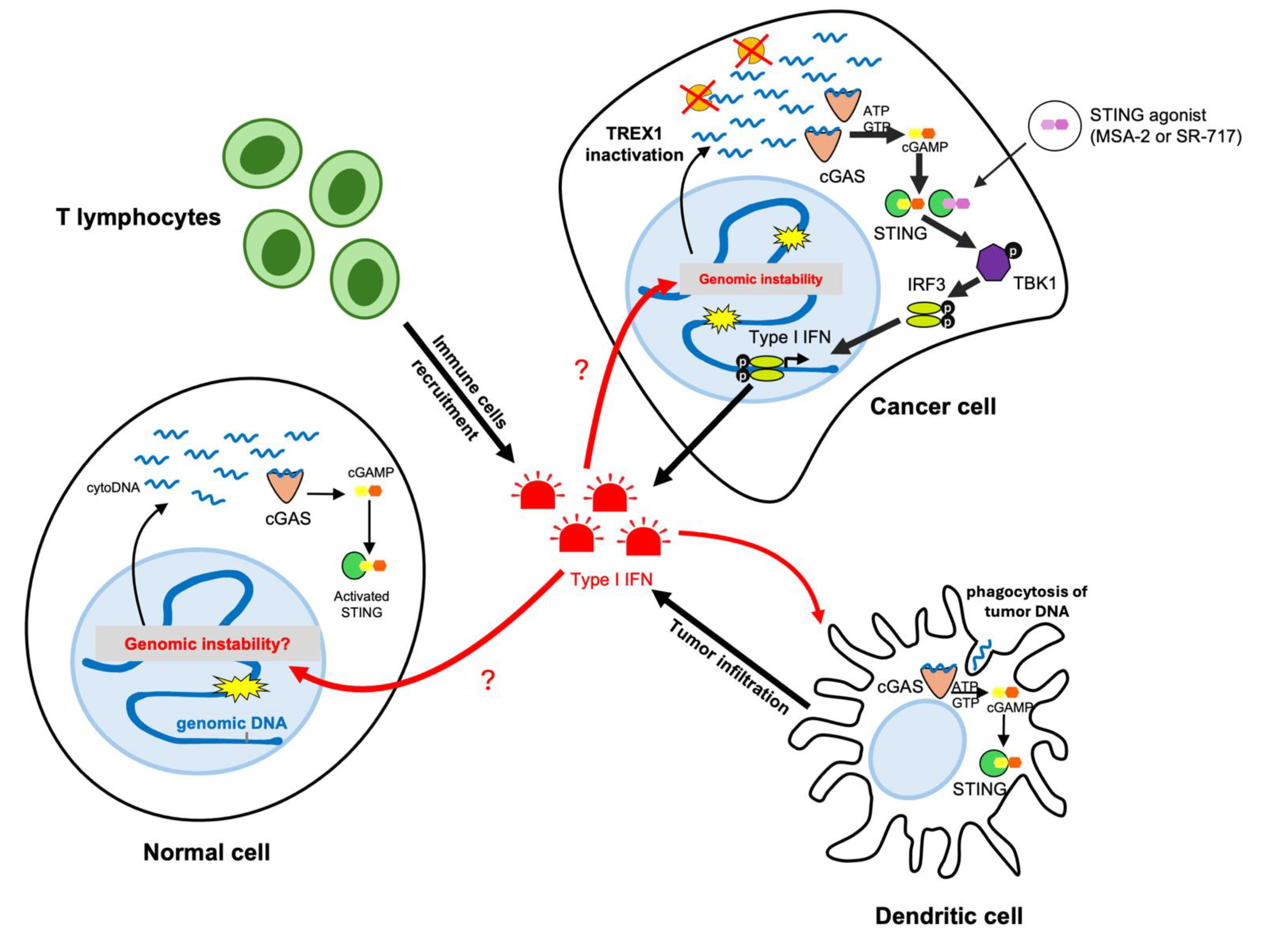
It is remarkable that conventional chemotherapies, which are DNA damage-inducers (e.g., PARP or CHK1 inhibitors) or mitotic poisons (MPS1i), exert their anti-tumor effect in a cGAS- and STING-dependent manner in immuno-competent mice models [9,11,14,45]. These results show that treatments that promote cGAS-STING signaling can favor tumor shrinkage. Recently, forced induction of STING signaling has been shown to leverage an inflammatory response, therefore promoting the recruitment of immune cells inside the tumor mass and its shrinkage in relevant immune-competent mice models [46,47]. This result has been achieved by extensive screening of small molecules that can induce IFN-I response in human cells, leading to the discovery of STING agonists (STINGa). STINGas are non-nucleotide molecules that bind to STING and induce its activation. Reduction of tumor growth was observed when using such STINGas, namely MSA-2 or SR-717, in different cancer types [46,47]. STINGas represent a promising path forward and demonstrate that cGAS-STING-IFN-I signaling can be used to promote cancer rejection.

2.2. TREX1 Induction by Chemo- and Radio-Therapies

Recent works show that chromosomal instability in cancer cells induces TREX1 expression, notably when using mitotic poisons (e.g., MPS1i, BAY1217389) or as a consequence of endogenous DNA damage in cancer cells [23,24]. TREX1 induction serves as an adaptive response by cancer cells to limit the magnitude of downstream IFN-I production (Figure 2). Hence, TREX1 induction restrains the cGAS-STING signal amplification, thus diminishing the inflammatory response and anti-cancer immunity. This mechanism of TREX1 induction has been previously shown to impact radiotherapy. When irradiation is delivered at high doses, DNA damage is introduced into the cancer cells, resulting in elevated release of dsDNA fragments into the cytosol, leading to inflammatory signaling accompanied by an increase of TREX1 expression [48,49]. High levels of TREX1 upon irradiation limits in time and amplitude the inflammatory signaling necessary to produce a potent anti-cancer immunity. When administrated at a lower dose, radiotherapy did not induce TREX1 expression and therefore improved anti-tumor immunity [48,49]. Thus, depletion or inactivation of TREX1 could serve as a promising approach in combination with radiotherapy or chemotherapies. This again demonstrates TREX1′s importance as a vital regulator of anti-tumor immunity. Inactivation of TREX1 has thus been proposed to have the potential to boost the anti-tumor immunity in cGAS-STING proficient cancers [50].

2.3. Promising Efficacy of TREX1 Inactivation in Pre-Clinical Cancer Models

As explained above, TREX1 inactivation results in the accumulation of cytoDNA and subsequent IFN-I pro-inflammatory response through cGAS activation. Inactivation of TREX1 in cancer cells (achieved by knock-out of TREX1 by CRISPR-Cas9 in different mice cancer models) has been recently shown to promote anti-cancer immunity and tumor shrinkage in a cGAS-STING-IFN-I-dependent manner in immuno-competent mice models [23,24,25]. It has been shown by deleting IRF3 or blocking IFN-receptors that cytoDNA sensing and subsequent inflammatory response are responsible for cancer rejection [23,24,25]. This dependence is further underscored by experiments demonstrating that concomitant loss of TREX1 and of cGAS (in the injected cancer cells), or TREX1 and STING, fully restored tumors growth, as well as erasing the survival benefits associated with intratumoral TREX1 deletion. The effect of TREX1 knock-out (KO) in cancer cells was notably very effective to control tumor growth in the colon carcinoma model CT26 [23,24,25]. It is of note that the CT26 colon cancer cells are proficient for the cGAS-STING pathway. A reduction of tumor growth was also observed with TREX1 KO in the melanoma model B16F10 [23] and the breast cancer model EO771.LMB (luminal B mammary cancer) [24]. It is important to emphasize here that TREX1 inactivation can be effective only if the cGAS-STING pathway is functional and thus generating an IFN-I response upon detection of cytoDNA [51] (e.g., when cancer cells are transfected with exogenous DNA). On the other hand, deletion of TREX1 in 4T1 cells that are unable to activate a proper IFN-I response had no impact on tumor growth [24]. TREX1 KO induced an IFN-I response even in low STING-expressing cancer cells, but not in absence of cGAS or STING [23]. Altogether, these data show that TREX1 targeting would be efficient in principle but limited in use for cGAS-STING-proficient cancers [23,24,25].

2.4. Depletion of TREX1 Remodels the Immune Cell Population in the Tumor Microenvironment

The build-up of cytoplasmic DNA upon TREX1 inactivation stimulates the cGAS-STING pathway and thus the subsequent IFN-I production by tumor cells [23,24,25]. Indeed, the initiation of an interferon response in one or a few tumor cells is hypothetically sufficient to be spread locally in the microenvironment and to different tissues and organs because of the paracrine nature of the inflammatory signaling. We picture inflammatory signaling like a wildfire that can initiate in a small bush and then spread to an entire forest. (Figure 3). This is caused by the binding of type I interferons to their corresponding receptors through an autocrine or paracrine pathway that activates STAT1 signaling. STAT1 instigates the induction of STING expression through competing with the H3K27me3 and/or DNA methylation marks that epigenetically silence STING expression in cancer cells. This in turn increases STING signaling and therefore further promotes the amplification of the interferon response between tumor cells [23]. In addition, IFN-I is also produced by several cell types in the tumor microenvironment, such as dendritic, macrophages, and endothelial cells, when stimulated by tumor-derived DNA, for example (Figure 3). Myeloid cell populations including monocytes and dendritic cells as well as lymphoid CD8+ effector T cells are recruited by the interferon signaling in the tumor microenvironment [23,25]. Additionally, CD4+ T cells, CD19+ B cells, and CD45+ cells levels were reported to increase significantly in tumors that are TREX1-deficient [24]. NK (Natural Killer) cells infiltration was also significantly increased within such tumors, leading to a marked increase in apoptosis of TREX1 KO tumor cells compared to control tumors, attributed to IFNγ-mediated mechanisms [25]. Although some discrepancies were reported by these three recent papers on which sub-type of immune cells is detected in the tumors, we think that most of these differences stem from the methods and markers used in each laboratory to identify such cell types. However, the main finding reported by these studies is that the tumor microenvironment is different when TREX1 is lacking, enabling innate and adaptative anti-cancer immunity. Thus, TREX1 loss amplifies tumor-intrinsic STING-IFN-I signaling, which thereby induces an increase in the infiltration rates of tumor-reactive immune cells within the tumor microenvironment.

3. Combining TREX1 Inactivation with Immune Checkpoint Blockade Shows Promising Potential for Immune Clearance of Cancer Cells

TREX1 inactivation through inflammatory signaling may foster the efficiency of immunotherapies, such as ICB [50,52]. TREX1 inactivation in genomically unstable cancer cells, may overcome the lack of efficacy of immunotherapies in some cancer types, while sparing non-tumorigenic cells that generally do not produce high levels of cytoDNA [5,48,53,54].
The combination of TREX1 targeting with ICB, specifically anti-PD-1 therapy, shows significant promise for enhancing immune responses against tumors [50]. It has been demonstrated that in TREX1 KO tumors, anti-PD1 treatment leads to reduced tumor growth and increased infiltration of CD8+ T cells, CD3+ T cells, and NK cells [23,24,25]. This is accompanied with upregulated ISGs expression, which is further amplified by anti-PD1 therapy across various tumor models. Therefore, the combination of both TREX1 inactivation and ICB will allow the recruitment of effector immune cells to the tumor site and their activation. It is important to highlight here that similar results were obtained when combining conventional DNA-damaging chemotherapies (e.g., PARP inhibitor) with ICB in pre-clinical mice models [9,11]. These chemotherapies induce both DNA damage and cGAS-STING signaling through the accumulation of cytoDNA [2,5]. These results strongly suggest that cGAS-STING signaling can benefit therapeutical settings, through accumulation of cytoDNA. TREX1 inactivation, DNA damaging agents, or mitotic poisons are all possible means to induce the accumulation of cytoDNA and subsequently boost anti-cancer immunity.
However, cancer cells can silence or inactivate cGAS or STING as a way to render the innate immune system blind to them (mechanism of evasion). As stated above, this invalidates the possibility of targeting TREX1 in such cGAS-STING-deficient cancers and may explain in some cases the lack of efficacy of conventional chemotherapies when anti-tumor immunity cannot be promoted by pro-inflammatory signaling. Prognostic and functional assays are yet to be designed to assess cGAS-STING-IFN-I status from patient samples in order to inform on which patient could benefit from TREX1 targeting or inflammation-based therapy.

4. Limitations and Future Perspectives for TREX1 Targeting in Cancer Therapy

Three remarkable articles have shown that TREX1 inactivation in cancer therapies has a promising potential in repressing tumor growth via a cGAS-STING-induced inflammatory response [23,24,25]. However, TREX1 inactivation is still limited in its translation to the clinics for the following reasons.
First, there is no TREX1 inhibitor available yet, therefore the inactivation of TREX1 is for now limited to “proof-of-concept” studies using knockouts of TREX1. This current lack of TREX1 inhibitors can be overcome thanks to published structures of TREX1 and by establishing functional and cell-based assays that are amenable to high-throughput screenings to identify and then to validate the potency of candidate molecules to inhibit TREX1 [50,55]. A recent work used a patented TREX1 inhibitor [56] to test its efficacy on suppressing TREX1-induced DNA damage in RVCL cell models [33]. This TREX1 inhibitor has been recently validated in cell lysates and cell-based assays to inhibit human TREX1 [55,57]. We are looking forward to seeing if such a compound would have an effect on cancer pre-clinical models. From a drug development perspective, these TREX1 inhibitors should be assessed in immunocompetent cancer mouse models. If the effect of TREX1 ablation on tumor growth is recapitulated with TREX1 inhibitors, and if these molecules are well-tolerated when administrated to mice, then clinical trials should start by assessing first the safety and then the effectiveness in different cancer settings.
Second, as described previously the effect of TREX1 inactivation is dependent on two crucial parameters: the presence of a functional cGAS-STING pathway in the targeted tumor and a proficient host immune system. Some tumors silence the expression of cGAS or STING as a form of resistance to immune surveillance thus rendering TREX1 targeting ineffective. It would be important to account for cGAS-STING-IFN-I status (expression and function) when deciding which patient would benefit from an inflammation-based therapy, such as TREX1 targeting. It has been shown that cGAS expression is high in different cancer types, and high cGAS expression correlates with a poor prognosis (including liver hepatocellular carcinoma, pancreatic adenocarcinoma, skin cutaneous melanoma, and uveal melanoma) [58]. Expression of cGAS may therefore be used as a biomarker to evaluate the overall survival of patients. Assessing cGAS-STING status in cancers might be challenging in practice because of the heterogeneous nature of some cancers, and because tumor biopsies may contain a mixture of different non-cancerous cell types that are present in the tumor microenvironment, notably immune cells. It has been shown that long non-coding RNA (lncRNA), lncRNA RP11-770J1.4 is an immune-regulator impacting the cGAS-STING pathway in glioma. Downregulation of lncRNA RP11-770J1.4 correlated with an increase expression of the cGAS-STING pathway. Whether this lncRNA may be used as a biomarker to inform on cGAS-STING status in glioma and other cancers has yet to be evaluated [59]. Moreover, if the cGAS-STING pathway was indeed functional, an intact immune system of the patient is critical. As described in studies, tumor clearance is achieved through the infiltration of different immune cells, emphasizing that in the absence of an immune system, tumor growth is not expected to be affected significantly. It is to be considered that most conventional and common chemotherapies impact the patient’s immunity through their myelosuppressive side-effects [60,61]. Hence, we raise the question of whether TREX1 depletion in an immunocompromised patient, due to chemotherapies, may result in unproductive inflammation with possible side effects. Treatments can be combined and administrated sequentially. It would be important to test if TREX1 ablation improves cancer rejection when combined with different chemotherapies or used sequentially. In this context, it would be interesting to assess if TREX1 inactivation followed by DNA-damaging agents would induce an inflammatory state that would enhance the efficiency of chemotherapies or radiotherapies. A recent article has shown that chemoresistance in small-cell lung cancers correlate with an elevated expression of TREX1 [62]. Ablation of TREX1 in chemoresistant cancer models improved the sensitivity to chemotherapies, suggesting that TREX1 inhibition would improve the response to treatment in these types of lung cancers [62]. It remains to be determined if TREX1 high expression would indicate chemoresistance in cancer patients, and if alternative therapies can be proposed in such cases.
Third, different studies show that IFN signaling and TREX1 inactivation can lead to DNA damage and senescence [28,29,30,32,63]. These results suggest that the interferon response in the absence of functional TREX1 can promote the destabilization of genomic DNA. TREX1 inhibition could therefore promote the generation of DNA lesions in cancer cells and normal cells (Figure 3). We thus wonder if TREX1 inhibition may lead to unwanted side effects, impacting cancer treatment outcome. Indeed, inflammation is an autocrine and paracrine phenomenon. An inflammatory signature has been correlated to cancer progression into metastasis, especially in highly genomically unstable cancers [15]. It will be important to further investigate the possible impact of such TREX1 inactivation on healthy tissues, as well as long-term effects, such as relapse or secondary malignancies.
Oncogenic stress or loss of tumor suppressors or changes in the physical properties of cancerous cells can damage the nuclear envelope, allowing nuclear DNA to mix-up with cytoplasmic content [35,64,65]. This has been observed upon YAP/TAZ deficiency; breaches in the nuclear envelope expose chromatin into the cytoplasm, leading to the formation of large cGAS foci at the level of nuclear envelope breakage [65]. Whether nuclear fragility could be manipulated in cancer therapy to promote the accumulation of TREX1-dependent DNA damage needs further investigation.

5. Conclusions

TREX1 is a key regulator of the cGAS-STING signaling cascade and therefore an essential factor in controlling inflammation. TREX1 expression is induced in some cancers to limit the induction of an interferon response and hence inflammation-induced immune-surveillance of cancer cells. This in turn allows cancer cells to benefit from a highly unstable genome while escaping immune detection through a repressed interferon production. Therefore, TREX1 inactivation emerged as a promising target in combination with well-established immunotherapies such as anti-PD1. Such a combination of two immune checkpoint blockade therapies resulted in significant tumor regression through the recruitment of various effector immune cells to the tumor microenvironment, resulting in better overall survival in tumor mouse models. Future studies, especially high-throughput screenings to define potent TREX1 inhibitors, and clinical trials are necessary. These future clinical trials should notably aim at testing the most potent combinations and sequences of therapies that would be defined in relevant pre-clinical models.

Author Contributions

K.H., S.K. and H.T.: writing and editing of the manuscript and figures. H.T: planned and supervised the work. All authors have read and agreed to the published version of the manuscript.

Funding

H.T.’s lab has been supported by the French government through the France 2030 investment plan managed by the National Research Agency (ANR), as part of the Initiative of Excellence Université Côte d’Azur (IdEx), under reference number ANR-15-IDEX-01. H.T.’s research is supported by a Chair of Professor Junior from the Université Côte d’Azur (Projet ANR-23-CPJ1-0025-01). H.T. has received a co-funding for the installation of new teams from IdEx of Université Côte d’Azur (ANR-15-IDEX-01).

Acknowledgments

H.T. wishes to express his gratitude to Université Côte d’Azur, IRCAN, Miguel Godinho Ferreira and Dmitry Bulavin for providing intellectual support, space, and resources for his research. K.H. and H.T. thank Mona Diab Assaf for her support and suggestions during the writing of this manuscript. K.H. wishes to express his gratitude to the Lebanese University and Mona Diab Assaf for providing educational support and guidance during his master’s research. The authors thank the reviewers for their meaningful comments that improved the manuscript.

Conflicts of Interest

The authors declare no conflicts of interest.

References

  1. Li, T.; Chen, Z.J. The CGAS–CGAMP–STING Pathway Connects DNA Damage to Inflammation, Senescence, and Cancer. J. Exp. Med. 2018, 215, 1287–1299. [Google Scholar] [CrossRef] [PubMed]
  2. Samson, N.; Ablasser, A. The CGAS–STING Pathway and Cancer. Nat. Cancer 2022, 3, 1452–1463. [Google Scholar] [CrossRef] [PubMed]
  3. Pathare, G.R.; Decout, A.; Glück, S.; Cavadini, S.; Makasheva, K.; Hovius, R.; Kempf, G.; Weiss, J.; Kozicka, Z.; Guey, B.; et al. Structural Mechanism of CGAS Inhibition by the Nucleosome. Nature 2020, 587, 668–672. [Google Scholar] [CrossRef] [PubMed]
  4. Volkman, H.E.; Cambier, S.; Gray, E.E.; Stetson, D.B. Tight Nuclear Tethering of CGAS Is Essential for Preventing Autoreactivity. Elife 2019, 8, e47491. [Google Scholar] [CrossRef]
  5. Chabanon, R.M.; Rouanne, M.; Lord, C.J.; Soria, J.C.; Pasero, P.; Postel-Vinay, S. Targeting the DNA Damage Response in Immuno-Oncology: Developments and Opportunities. Nat. Rev. Cancer 2021, 21, 701–717. [Google Scholar] [CrossRef]
  6. Técher, H.; Pasero, P. The Replication Stress Response on a Narrow Path Between Genomic Instability and Inflammation. Front. Cell Dev. Biol. 2021, 9, 702584. [Google Scholar] [CrossRef] [PubMed]
  7. Yamazaki, T.; Kirchmair, A.; Sato, A.; Buqué, A.; Rybstein, M.; Petroni, G.; Bloy, N.; Finotello, F.; Stafford, L.; Navarro Manzano, E.; et al. Mitochondrial DNA Drives Abscopal Responses to Radiation That Are Inhibited by Autophagy. Nat. Immunol. 2020, 21, 1160–1171. [Google Scholar] [CrossRef]
  8. Erdal, E.; Haider, S.; Rehwinkel, J.; Harris, A.L.; McHugh, P.J. A Prosurvival DNA Damage-Induced Cytoplasmic Interferon Response Is Mediated by End Resection Factors and Is Limited by Trex1. Genes Dev. 2017, 31, 353–369. [Google Scholar] [CrossRef]
  9. Kitai, Y.; Kawasaki, T.; Sueyoshi, T.; Kobiyama, K.; Ishii, K.J.; Zou, J.; Akira, S.; Matsuda, T.; Kawai, T. DNA-Containing Exosomes Derived from Cancer Cells Treated with Topotecan Activate a STING-Dependent Pathway and Reinforce Antitumor Immunity. J. Immunol. 2017, 198, 1649–1659. [Google Scholar] [CrossRef]
  10. Dou, Z.; Ghosh, K.; Vizioli, M.G.; Zhu, J.; Sen, P.; Wangensteen, K.J.; Simithy, J.; Lan, Y.; Lin, Y.; Zhou, Z.; et al. Cytoplasmic Chromatin Triggers Inflammation in Senescence and Cancer. Nature 2017, 550, 402–406. [Google Scholar] [CrossRef]
  11. Sen, T.; Rodriguez, B.L.; Chen, L.; Della Corte, C.M.; Morikawa, N.; Fujimoto, J.; Cristea, S.; Nguyen, T.; Diao, L.; Li, L.; et al. Targeting DNA Damage Response Promotes Antitumor Immunity through STING-Mediated T-Cell Activation in Small Cell Lung Cancer. Cancer Discov. 2019, 9, 646–661. [Google Scholar] [CrossRef] [PubMed]
  12. Shen, Y.J.; LeBert, N.; Chitre, A.A.; Koo, C.X.E.; Nga, X.H.; Ho, S.S.W.; Khatoo, M.; Tan, N.Y.; Ishii, K.J.; Gasser, S. Genome-Derived Cytosolic DNA Mediates Type I Interferon-Dependent Rejection of B Cell Lymphoma Cells. Cell Rep. 2015, 11, 460–473. [Google Scholar] [CrossRef] [PubMed]
  13. Ho, S.S.W.; Zhang, W.Y.L.; Tan, N.Y.J.; Khatoo, M.; Suter, M.A.; Tripathi, S.; Cheung, F.S.G.; Lim, W.K.; Tan, P.H.; Ngeow, J.; et al. The DNA Structure-Specific Endonuclease MUS81 Mediates DNA Sensor STING-Dependent Host Rejection of Prostate Cancer Cells. Immunity 2016, 44, 1177–1189. [Google Scholar] [CrossRef] [PubMed]
  14. Guan, J.; Lu, C.; Jin, Q.; Lu, H.; Chen, X.; Tian, L.; Zhang, Y.; Ortega, J.; Zhang, J.; Siteni, S.; et al. MLH1 Deficiency-Triggered DNA Hyperexcision by Exonuclease 1 Activates the CGAS-STING Pathway. Cancer Cell 2021, 39, 109–121.e5. [Google Scholar] [CrossRef] [PubMed]
  15. Bakhoum, S.F.; Ngo, B.; Laughney, A.M.; Cavallo, J.A.; Murphy, C.J.; Ly, P.; Shah, P.; Sriram, R.K.; Watkins, T.B.K.; Taunk, N.K.; et al. Chromosomal Instability Drives Metastasis through a Cytosolic DNA Response. Nature 2018, 553, 467–472. [Google Scholar] [CrossRef]
  16. Crow, Y.J.; Manel, N. Aicardi–Goutières Syndrome and the Type I Interferonopathies. Nat. Rev. Immunol. 2015, 15, 429–440. [Google Scholar] [CrossRef]
  17. Yang, Y.G.; Lindahl, T.; Barnes, D.E. Trex1 Exonuclease Degrades SsDNA to Prevent Chronic Checkpoint Activation and Autoimmune Disease. Cell 2007, 131, 873–886. [Google Scholar] [CrossRef] [PubMed]
  18. Stetson, D.B.; Ko, J.S.; Heidmann, T.; Medzhitov, R. Trex1 Prevents Cell-Intrinsic Initiation of Autoimmunity. Cell 2008, 134, 587–598. [Google Scholar] [CrossRef] [PubMed]
  19. Fye, J.M.; Orebaugh, C.D.; Coffin, S.R.; Hollis, T.; Perrino, F.W. Dominant Mutation of the TREX1 Exonuclease Gene in Lupus and Aicardi-Goutieres Syndrome. J. Biol. Chem. 2011, 286, 32373–32382. [Google Scholar] [CrossRef]
  20. Lehtinen, D.A.; Harvey, S.; Mulcahy, M.J.; Hollis, T.; Perrino, F.W. The TREX1 Double-Stranded DNA Degradation Activity Is Defective in Dominant Mutations Associated with Autoimmune Disease. J. Biol. Chem. 2008, 283, 31649–31656. [Google Scholar] [CrossRef]
  21. Huang, K.W.; Wu, C.Y.; Toh, S.I.; Liu, T.C.; Tu, C.I.; Lin, Y.H.; Cheng, A.J.; Kao, Y.T.; Chu, J.W.; Hsiao, Y.Y. Molecular Insight into the Specific Enzymatic Properties of TREX1 Revealing the Diverse Functions in Processing RNA and DNA/RNA Hybrids. Nucleic Acids Res. 2023, 51, 11927–11940. [Google Scholar] [CrossRef] [PubMed]
  22. Lee-Kirsch, M.A.; Gong, M.; Chowdhury, D.; Senenko, L.; Engel, K.; Lee, Y.A.; De Silva, U.; Bailey, S.L.; Witte, T.; Vyse, T.J.; et al. Mutations in the Gene Encoding the 3′-5′ DNA Exonuclease TREX1 Are Associated with Systemic Lupus Erythematosus. Nat. Genet. 2007, 39, 1065–1067. [Google Scholar] [CrossRef] [PubMed]
  23. Tani, T.; Mathsyaraja, H.; Campisi, M.; Li, Z.H.; Haratani, K.; Fahey, C.G.; Ota, K.; Mahadevan, N.R.; Shi, Y.; Saito, S.; et al. TREX1 Inactivation Unleashes Cancer Cell STING-Interferon Signaling and Promotes Antitumor Immunity. Cancer Discov. 2024, 14, 752–765. [Google Scholar] [CrossRef] [PubMed]
  24. Toufektchan, E.; Dananberg, A.; Striepen, J.; Hickling, J.H.; Shim, A.; Chen, Y.; Nichols, A.; Paez, M.A.D.; Mohr, L.; Bakhoum, S.F.; et al. Intratumoral TREX1 Induction Promotes Immune Evasion by Limiting Type I IFN. Cancer Immunol. Res. 2024, 12, 673–686. [Google Scholar] [CrossRef]
  25. Lim, J.; Rodriguez, R.; Williams, K.; Silva, J.; Gutierrez, A.G.; Tyler, P.; Baharom, F.; Sun, T.; Lin, E.; Martin, S.; et al. The Exonuclease TREX1 Constitutes an Innate Immune Checkpoint Limiting CGAS/STING-Mediated Antitumor Immunity. Cancer Immunol. Res. 2024, 12, 663–672. [Google Scholar] [CrossRef]
  26. Lau, A.; Gray, E.E.; Brunette, R.L.; Stetson, D.B. DNA Tumor Virus Oncogenes Antagonize the CGAS-STING DNA-Sensing Pathway. Science 2015, 350, 568–571. [Google Scholar] [CrossRef]
  27. Técher, H. T-Rex Escaped from the Cytosolic Park: Re-Thinking the Impact of TREX1 Exonuclease Deficiencies on Genomic Stability. BioEssays 2024, 46, 2400066. [Google Scholar] [CrossRef] [PubMed]
  28. Técher, H.; Gopaul, D.; Heuzé, J.; Bouzalmad, N.; Leray, B.; Vernet, A.; Mettling, C.; Moreaux, J.; Pasero, P.; Lin, Y.-L. MRE11 and TREX1 Control Senescence by Coordinating Replication Stress and Interferon Signaling. Nat. Commun. 2024, 15, 5423. [Google Scholar] [CrossRef] [PubMed]
  29. Du, H.; Xiao, N.; Zhang, S.; Zhou, X.; Zhang, Y.; Lu, Z.; Fu, Y.; Huang, M.; Xu, S.; Chen, Q. Suppression of TREX1 Deficiency-Induced Cellular Senescence and Interferonopathies by Inhibition of DNA Damage Response. iScience 2023, 26, 107090. [Google Scholar] [CrossRef]
  30. Giordano, A.M.S.; Luciani, M.; Gatto, F.; Alezz, M.A.; Beghè, C.; Della Volpe, L.; Migliara, A.; Valsoni, S.; Genua, M.; Dzieciatkowska, M.; et al. DNA Damage Contributes to Neurotoxic Inflammation in Aicardi-Goutières Syndrome Astrocytes. J. Exp. Med. 2022, 219, e20211121. [Google Scholar] [CrossRef] [PubMed]
  31. Wolf, C.; Rapp, A.; Berndt, N.; Staroske, W.; Schuster, M.; Dobrick-Mattheuer, M.; Kretschmer, S.; König, N.; Kurth, T.; Wieczorek, D.; et al. RPA and Rad51 Constitute a Cell Intrinsic Mechanism to Protect the Cytosol from Self DNA. Nat. Commun. 2016, 7, 11752. [Google Scholar] [CrossRef] [PubMed]
  32. Abt, E.R.; Le, T.M.; Dann, A.M.; Capri, J.R.; Poddar, S.; Lok, V.; Li, L.; Liang, K.; Creech, A.L.; Rashid, K.; et al. Reprogramming of Nucleotide Metabolism by Interferon Confers Dependence on the Replication Stress Response Pathway in Pancreatic Cancer Cells. Cell Rep. 2022, 38, 110236. [Google Scholar] [CrossRef] [PubMed]
  33. Chauvin, S.D.; Ando, S.; Holley, J.A.; Sugie, A.; Zhao, F.R.; Poddar, S.; Kato, R.; Miner, C.A.; Nitta, Y.; Krishnamurthy, S.R.; et al. Inherited C-Terminal TREX1 Variants Disrupt Homology-Directed Repair to Cause Senescence and DNA Damage Phenotypes in Drosophila, Mice, and Humans. Nat. Commun. 2024, 15, 4696. [Google Scholar] [CrossRef] [PubMed]
  34. Richards, A.; Maagdenberg, A.M.J.M.v.D.; Jen, J.C.; Kavanagh, D.; Bertram, P.; Spitzer, D.; Liszewski, M.K.; Barilla-Labarca, M.L.; Terwindt, G.M.; Kasai, Y.; et al. C-Terminal Truncations in Human 3′-5′ DNA Exonuclease TREX1 Cause Autosomal Dominant Retinal Vasculopathy with Cerebral Leukodystrophy. Nat. Genet. 2007, 39, 1068–1070. [Google Scholar] [CrossRef] [PubMed]
  35. Nader, G.P.d.F.; Agüera-Gonzalez, S.; Routet, F.; Gratia, M.; Maurin, M.; Cancila, V.; Cadart, C.; Palamidessi, A.; Ramos, R.N.; San Roman, M.; et al. Compromised Nuclear Envelope Integrity Drives TREX1-Dependent DNA Damage and Tumor Cell Invasion. Cell 2021, 184, 5230–5246.e22. [Google Scholar] [CrossRef]
  36. Sharma, P.; Goswami, S.; Raychaudhuri, D.; Siddiqui, B.A.; Singh, P.; Nagarajan, A.; Liu, J.; Subudhi, S.K.; Poon, C.; Gant, K.L.; et al. Immune Checkpoint Therapy—Current Perspectives and Future Directions. Cell 2023, 186, 1652–1669. [Google Scholar] [CrossRef]
  37. Sharma, P.; Allison, J.P. Dissecting the Mechanisms of Immune Checkpoint Therapy. Nat. Rev. Immunol. 2020, 20, 75–76. [Google Scholar] [CrossRef]
  38. Chamoto, K.; Yaguchi, T.; Tajima, M.; Honjo, T. Insights from a 30-Year Journey: Function, Regulation and Therapeutic Modulation of PD1. Nat. Rev. Immunol. 2023, 23, 682–695. [Google Scholar] [CrossRef]
  39. Pang, K.; Shi, Z.D.; Wei, L.Y.; Dong, Y.; Ma, Y.Y.; Wang, W.; Wang, G.Y.; Cao, M.Y.; Dong, J.J.; Chen, Y.A.; et al. Research Progress of Therapeutic Effects and Drug Resistance of Immunotherapy Based on PD-1/PD-L1 Blockade. Drug Resist. Updates 2023, 66, 100907. [Google Scholar] [CrossRef]
  40. De Martino, M.; Vanpouille-Box, C.; Galluzzi, L. Immunological Barriers to Immunotherapy in Primary and Metastatic Breast Cancer. EMBO Mol. Med. 2021, 13, e14393. [Google Scholar] [CrossRef]
  41. de Miguel, M.; Calvo, E. Clinical Challenges of Immune Checkpoint Inhibitors. Cancer Cell 2020, 38, 326–333. [Google Scholar] [CrossRef] [PubMed]
  42. Sun, Q.; Hong, Z.; Zhang, C.; Wang, L.; Han, Z.; Ma, D. Immune Checkpoint Therapy for Solid Tumours: Clinical Dilemmas and Future Trends. Signal Transduct. Target. Ther. 2023, 8, 320. [Google Scholar] [CrossRef] [PubMed]
  43. Binnewies, M.; Roberts, E.W.; Kersten, K.; Chan, V.; Fearon, D.F.; Merad, M.; Coussens, L.M.; Gabrilovich, D.I.; Ostrand-Rosenberg, S.; Hedrick, C.C.; et al. Understanding the Tumor Immune Microenvironment (TIME) for Effective Therapy. Nat. Med. 2018, 24, 541–550. [Google Scholar] [CrossRef] [PubMed]
  44. Zitvogel, L.; Galluzzi, L.; Kepp, O.; Smyth, M.J.; Kroemer, G. Type I Interferons in Anticancer Immunity. Nat. Rev. Immunol. 2015, 15, 405–414. [Google Scholar] [CrossRef]
  45. Kitajima, S.; Tani, T.; Springer, B.F.; Campisi, M.; Osaki, T.; Haratani, K.; Chen, M.; Knelson, E.H.; Mahadevan, N.R.; Ritter, J.; et al. MPS1 Inhibition Primes Immunogenicity of KRAS-LKB1 Mutant Lung Cancer. Cancer Cell 2022, 40, 1128–1144.e8. [Google Scholar] [CrossRef]
  46. Pan, B.S.; Perera, S.A.; Piesvaux, J.A.; Presland, J.P.; Schroeder, G.K.; Cumming, J.N.; Wesley Trotter, B.; Altman, M.D.; Buevich, A.V.; Cash, B.; et al. An Orally Available Non-Nucleotide STING Agonist with Antitumor Activity. Science 2020, 369, eaba6098. [Google Scholar] [CrossRef]
  47. Chin, E.N.; Yu, C.; Vartabedian, V.F.; Jia, Y.; Kumar, M.; Gamo, A.M.; Vernier, W.; Ali, S.H.; Kissai, M.; Lazar, D.C.; et al. Antitumor Activity of a Systemic STING-Activating Non-Nucleotide CGAMP Mimetic. Science 2020, 369, 993–999. [Google Scholar] [CrossRef] [PubMed]
  48. Vanpouille-Box, C.; Alard, A.; Aryankalayil, M.J.; Sarfraz, Y.; Diamond, J.M.; Schneider, R.J.; Inghirami, G.; Coleman, C.N.; Formenti, S.C.; Demaria, S. DNA Exonuclease Trex1 Regulates Radiotherapy-Induced Tumour Immunogenicity. Nat. Commun. 2017, 8, 15618. [Google Scholar] [CrossRef]
  49. Diamond, J.M.; Vanpouille-Box, C.; Spada, S.; Rudqvist, N.P.; Chapman, J.R.; Ueberheide, B.M.; Pilones, K.A.; Sarfraz, Y.; Formenti, S.C.; Demaria, S. Exosomes Shuttle TREX1-Sensitive IFN-Stimulatory DsDNA from Irradiated Cancer Cells to DCs. Cancer Immunol. Res. 2018, 6, 910–920. [Google Scholar] [CrossRef]
  50. Hemphill, W.O.; Simpson, S.R.; Liu, M.; Salsbury, F.R.; Hollis, T.; Grayson, J.M.; Perrino, F.W. TREX1 as a Novel Immunotherapeutic Target. Front. Immunol. 2021, 12, 660184. [Google Scholar] [CrossRef]
  51. Deng, L.; Liang, H.; Xu, M.; Yang, X.; Burnette, B.; Arina, A.; Li, X.D.; Mauceri, H.; Beckett, M.; Darga, T.; et al. STING-Dependent Cytosolic DNA Sensing Promotes Radiation-Induced Type I Interferon-Dependent Antitumor Immunity in Immunogenic Tumors. Immunity 2014, 41, 843–852. [Google Scholar] [CrossRef] [PubMed]
  52. Lira, M.C.; Vanpouille-Box, C.; Galluzzi, L. Adaptive Inhibition of CGAS Signaling by TREX1. Trends Cancer 2024, 10, 177–179. [Google Scholar] [CrossRef] [PubMed]
  53. Won, J.K.; Bakhoum, S.F. The Cytosolic DNA-Sensing CGAS-STING Pathway in Cancer. Cancer Discov. 2020, 10, 26–39. [Google Scholar] [CrossRef]
  54. Mohr, L.; Toufektchan, E.; von Morgen, P.; Chu, K.; Kapoor, A.; Maciejowski, J. ER-Directed TREX1 Limits CGAS Activation at Micronuclei. Mol. Cell 2021, 81, 724–738.e9. [Google Scholar] [CrossRef] [PubMed]
  55. Huynh, K.; Partch, C.L. Substituted Pyrrolo [1,2-a]Pyrazines and Pyrrolo [1,2-a][1,4]Diazepines as TREX1 Inhibitors. U.S. Patent 11,583,538, 21 February 2023. [CrossRef]
  56. Letourneau, J.J.; Elamparuthi, K.S.; Huang, C.-Y.; Venkata, V.B. NOVEL CYCLIC TREX1 INHIBITORS 2020.
  57. Flowers, S.; Petronella, B.A.; McQueney, M.S.; Fanelli, B.; Eisenberg, W.; Uveges, A.; Roden, A.L.; Salowe, S.; Bommireddy, V.; Letourneau, J.J.; et al. A Novel TREX1 Inhibitor, VB-85680, Upregulates Cellular Interferon Responses. PLoS ONE 2024, 19, e0305962. [Google Scholar] [CrossRef] [PubMed]
  58. Chen, P.; Yang, X.; Wang, P.; He, H.; Chen, Y.; Yu, L.; Fang, H.; Wang, F.; Huang, Z. Systematic Pan-Cancer Analysis Identifies CGAS as an Immunological and Prognostic Biomarker. Ann. Transl. Med. 2023, 11, 121. [Google Scholar] [CrossRef] [PubMed]
  59. Zhuang, Q.; Liu, C.; Hu, Y.; Liu, Y.; Lyu, Y.; Liao, Y.; Chen, L.; Yang, H.; Mao, Y. Identification of RP11-770J1.4 as Immune-related LncRNA Regulating the CTXN1–CGAS–STING Axis in Histologically Lower-grade Glioma. MedComm 2023, 4, e458. [Google Scholar] [CrossRef]
  60. Epstein, R.S.; Roy, U.K.B.; Aapro, M.; Salimi, T.; Moran, D.; Krenitsky, J.; Leone-Perkins, M.L.; Girman, C.; Schlusser, C.; Crawford, J. Cancer Patients’ Perspectives and Experiences of Chemotherapy-Induced Myelosuppression and Its Impact on Daily Life. Patient Prefer. Adherence 2021, 15, 453–465. [Google Scholar] [CrossRef]
  61. Epstein, R.S.; Aapro, M.S.; Basu Roy, U.K.; Salimi, T.; Krenitsky, J.A.; Leone-Perkins, M.L.; Girman, C.; Schlusser, C.; Crawford, J. Patient Burden and Real-World Management of Chemotherapy-Induced Myelosuppression: Results from an Online Survey of Patients with Solid Tumors. Adv. Ther. 2020, 37, 3606–3618. [Google Scholar] [CrossRef]
  62. Murayama, T.; Mahadevan, N.R.; Meador, C.B.; Ivanova, E.V.; Pan, Y.; Knelson, E.H.; Tani, T.; Nakayama, J.; Ma, X.; Thai, T.C.; et al. Targeting TREX1 Induces Innate Immune Response in Drug-Resistant Small-Cell Lung Cancer. Cancer Res. Commun. 2024, 4, 2399–2414. [Google Scholar] [CrossRef]
  63. Raso, M.C.; Djoric, N.; Walser, F.; Hess, S.; Schmid, F.M.; Burger, S.; Knobeloch, K.P.; Penengo, L. Interferon-Stimulated Gene 15 Accelerates Replication Fork Progression Inducing Chromosomal Breakage. J. Cell Biol. 2020, 219, e202002175. [Google Scholar] [CrossRef] [PubMed]
  64. Kidiyoor, G.R.; Li, Q.; Bastianello, G.; Bruhn, C.; Giovannetti, I.; Mohamood, A.; Beznoussenko, G.V.; Mironov, A.; Raab, M.; Piel, M.; et al. ATR Is Essential for Preservation of Cell Mechanics and Nuclear Integrity during Interstitial Migration. Nat. Commun. 2020, 11, 4828. [Google Scholar] [CrossRef] [PubMed]
  65. Sladitschek-Martens, H.L.; Guarnieri, A.; Brumana, G.; Zanconato, F.; Battilana, G.; Xiccato, R.L.; Panciera, T.; Forcato, M.; Bicciato, S.; Guzzardo, V.; et al. YAP/TAZ Activity in Stromal Cells Prevents Ageing by Controlling CGAS–STING. Nature 2022, 607, 790–798. [Google Scholar] [CrossRef] [PubMed]
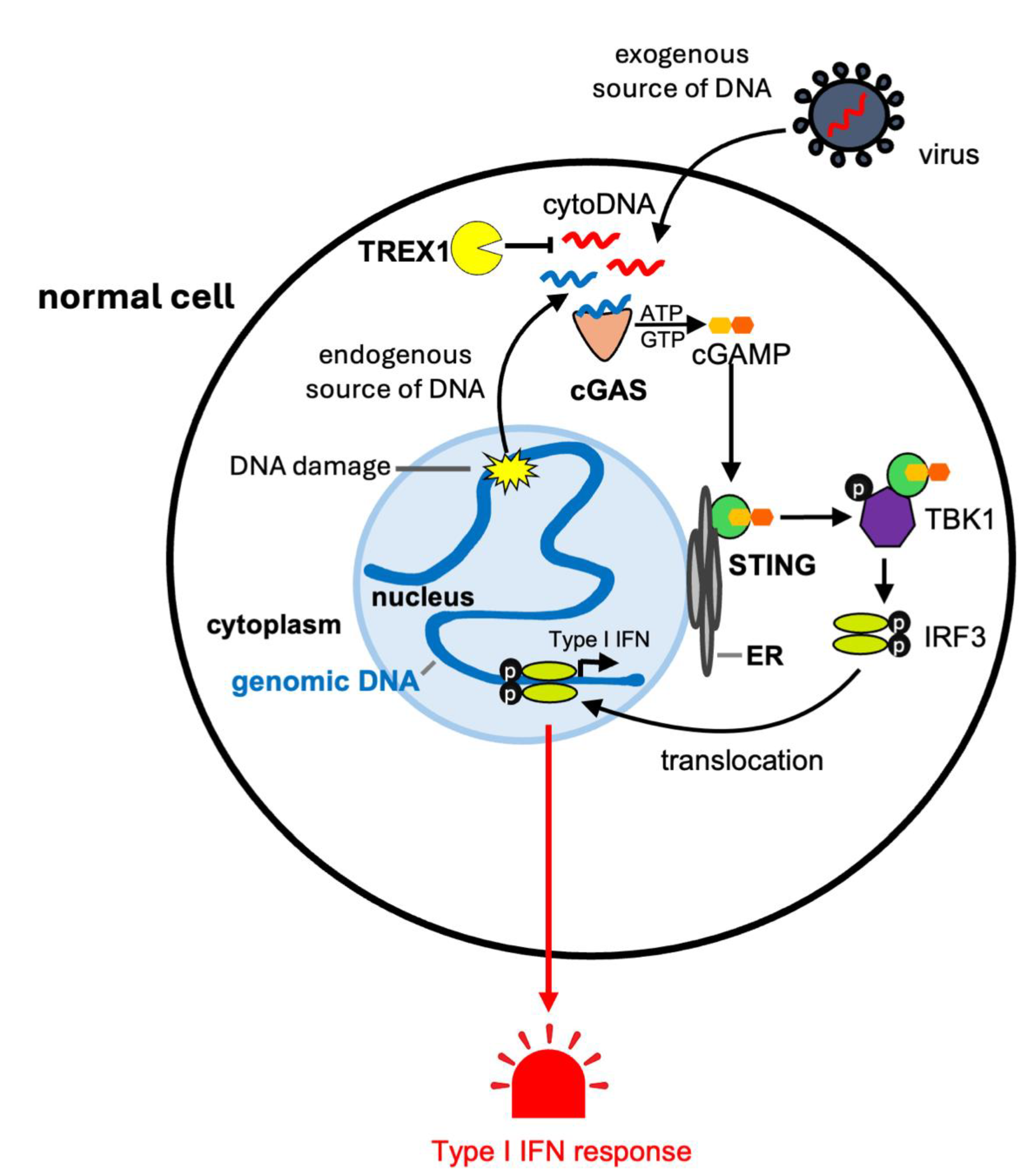
Figure 1. cGAS-STING signaling pathway. The DNA sensor cGAS recognizes double-stranded DNA from exogenous and endogenous (self) origin. Exogenous DNA can for instance be exposed in the cytosol following viral infection. Endogenous DNA can accumulate in the cytosol as a consequence of DNA damage. cGAS synthesizes the cyclic dinucleotide cGAMP that subsequently binds to and activates STING (stimulator of interferon genes), which is anchored to the endoplasmic reticulum (ER). STING activation is followed by its translocation to the Golgi and sequential phosphorylation of TBK1 and IRF3, which in turn translocates into the nucleus to induce the expression of type I interferons (type I IFN). TREX1 is an exonuclease that is mainly localized into the cytoplasm and that negatively regulates cGAS-STING-IFN signaling by degrading cytosolic DNA (cytoDNA). See the main text for details and references.
Onco 04 00022 g001
Figure 2. Cancer cells upregulate TREX1 expression to escape immune detection. Some cancer cells increase the expression of TREX1 exonuclease. Although cancer cells experience DNA damage and have unstable genomes, cytosolic DNA (cytoDNA) is constantly degraded by TREX1. As a result, high TREX1 expression in cancer cells suppresses the cGAS-STING signaling. Inactive STING favors the escape to immune surveillance, notably by T cells. See main text for details and references.
Onco 04 00022 g002
Figure 3. The impact of TREX1 inactivation and of an interferon response on the tumor microenvironment. Inactivation of TREX1 in cancer cells promotes the accumulation of cytosolic DNA resulting in cGAS-STING-IFN-I signaling. The pro-inflammatory response, the type I IFN response, notably induced by TREX1 inactivation leads to the attraction and recruitment of innate and adaptative immune cells. Either TREX1 ablation or STING agonists (MSA-2 and SR-717) have been shown to boost anti-cancer immunity notably by remodeling the tumor microenvironment, with infiltration of dendritic cells and activation of T lymphocytes. Immune cells and normal cells may then spread this inflammatory response. For instance, dendritic cells can activate STING signaling upon phagocytosis of tumor DNA. Prolonged type I IFN response could promote genomic instability in both cancer and normal cells. See main text for details and references.
Onco 04 00022 g003
Disclaimer/Publisher’s Note: The statements, opinions and data contained in all publications are solely those of the individual author(s) and contributor(s) and not of MDPI and/or the editor(s). MDPI and/or the editor(s) disclaim responsibility for any injury to people or property resulting from any ideas, methods, instructions or products referred to in the content.