Article

Effectiveness of an Assistive Instrument for Physical Therapists with Visual Impairment to Conduct the Muscle Tightness Test

1
Doctoral Program in Sports Medicine, Graduate School of Comprehensive Human Sciences, University of Tsukuba, Tsukuba 305-8577, Japan
2
Clinical Center for Medicine, Welfare and Health, Chiba 263-0021, Japan
3
Kobe City School for the Visually Impaired, Kobe 650-0044, Japan
4
Tottori Prefectural School for the Visually Impaired, Tottori 680-0151, Japan
5
Acupuncture and Physical Therapy Teacher Training School, University of Tsukuba, Bunkyo 112-0012, Japan
6
Department of Disability Sciences, Faculty of Human Sciences, University of Tsukuba, Tsukuba 305-8577, Japan
*
Author to whom correspondence should be addressed.
Disabilities 2021, 1(4), 471-480; https://doi.org/10.3390/disabilities1040033
Submission received: 6 October 2021 / Revised: 26 November 2021 / Accepted: 6 December 2021 / Published: 10 December 2021

Abstract

:
Disability sports have been globally promoted to achieve an inclusive society. However, the current opportunities for people with disabilities are few in terms of participating in supporting positions, such as athletic trainers. In Japan, many people with visual impairment (VI) work as physical therapists; thus, they can become active as medical staff in the sports field. An example of a medical assessment is the muscle tightness test (MTT); however, conducting it is expected to be difficult for people with VI. To improve this difficulty, the current study developed an assistive instrument (AsI) and examined its effectiveness. We recruited 22 physical therapists with VI. The measurement for MTT targeted the hamstrings, iliopsoas, gastrocnemius, and quadriceps. The participants performed the MTT using a universal goniometer (UG) and the AsI and filled up a questionnaire. AsI was compared against UG based on three aspects, namely, ease, accuracy, and measurement time. In terms of ease, significant differences were observed in 9 out of 12 questionnaire items. The mean (SD) score for “Comprehensive ease of the MTT” improved from 2.8 (1.0) to 3.9 (0.8). For accuracy, the AsI yielded a decrease in the total error of the measurement. Lastly, the measurement time for AsI was longer than that for the UG. Therefore, the AsI was effective in terms of ease and accuracy, whereas measurement time remained as an issue.

1. Introduction

The achievement of an inclusive society is one of the current issues worldwide. Toward this goal, disability sports (including adapted or para-sports) have been promoted to increase the opportunities for people with disabilities to participle in society [1]. In general, participation in sports can be categorized into three dimensions, namely, doing, watching, and supporting. Nowadays, several studies examined the enhancement of the performance of para-athletes [2] or the improvement of accessibility to sports events for people with disability [3]. Thus, opportunities to participate in doing and watching positions have been improved. However, there are still few opportunities for people with disabilities to participate in the supporting position, such as medical staff.
A typical example of supporting positions is athletic trainers who play a role in conditioning and rehabilitating athletes [4]. In the case of Japan, many athletic trainers obtain licenses of physical therapy, such as acupuncture, moxibustion, and massage [5]. In addition, the history of vocational education for people with visual impairment (VI) as physical therapists is extensive [6,7]. Thus, Japan is suitable for demonstrating a pilot case of the training of physical therapists with VI as athletic trainers.
Medical assessments are necessary for athletic trainers in evaluating the condition of athletes. One such method is the muscle tightness test (MTT). Hansberger et al. [8] defined muscle tightness as a restriction of the range of motion (ROM) with a feeling of resistance when the muscle is passively stretched [8]. Higher muscle tightness involves many types of sports injuries and disorders [9,10,11]. Thus, the appropriate assessment of muscle tightness is important for conditioning athletes. Moreover, several studies reported that acupuncture and massage therapy can relieve muscle tightness [12,13]. Therefore, the proper administration of the MTT is crucial for physical therapists in supporting athletes.
Although medical care should be based on appropriate assessment, accurately conducting the MTT may be difficult for physical therapists with VI. The reason for this notion is that the MTT was developed from the ROM measurement, which poses difficulty for physical therapists with VI [14]. In particular, two points hinder the appropriate administration of the MTT. First, the basic axis of measurement should be adjusted vertically on the floor. VI limits one’s sense of equilibrium [15]; thus, adjusting the vertical axis accurately may lead to difficulty. Second, both hands are occupied during the MTT. Behnoush et al. [16] reported that measuring the ROM using a universal goniometer (UG) causes a lack of touch on the body part, which can lead to inaccuracy in measurements. This issue is presumed to be more obvious especially for people with VI, who mainly depend on the tactile sense.
To address these issues, we developed an assistive instrument (AsI) and hypothesized that the inverted-T shape can help to adjust the vertical axis in an easy and accurate manner. Furthermore, a Velcro tape was secured to the vertical axis to attach the UG at any height. This enables physical therapists to conduct the MTT without holding the UG, and thus they can pay attention to other operations. In this regard, we expected that therapists can conduct the MTT smoothly within a shorter time frame. The present study aimed to examine the effectiveness related to the use of AsI among physical therapists with VI in conducting the MTT.

2. Materials and Methods

2.1. Participants

The study recruited 22 physical therapists with VI. Table 1 summarizes the characteristics of the participants. During the recruitment process, the researchers verified with the participants that they lack previous knowledge about the MTT to avoid bias in experience.

2.2. Procedure

Prior to the study, each participant received a video with an audio description that demonstrated the methods of the MTT. The reason for teaching by video was to equalize the knowledge level about the MTT by controlling the information received by the participants. This video was created by individuals with blindness and low vision to help the participants with various types of VI to understand. In addition, we instructed them to refrain from practicing prior to the experiment to minimize the effects of habituation and to standardize the skill levels. Immediately before the experiment, we requested the participants to conduct MTT once to ensure that they understand the correct method. Nearly all participants displayed understanding of the video and performed the MTT correctly. If they performed incorrectly, we corrected them as few times as possible. After verifying, the participants were instructed to conduct the MTT using the UG and AsI (Figure 1). After the experiment, the participants answered the questionnaire described below.

2.3. Method of Muscle Tightness Test
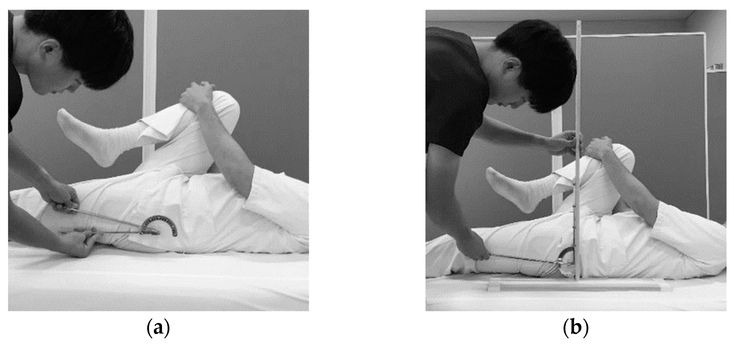
The methods of the MTT were based on a previous study [17]. The objects of measurement are the hamstrings, iliopsoas, gastrocnemius, and quadriceps. We prepared the UG, which is attached a screw for every 5° on the dial face to enable the participants with VI to read by touching. Muscle tightness was considered high when the angle was larger for the hamstrings and iliopsoas but smaller for the gastrocnemius and quadriceps.

2.3.1. Hamstrings

Figure 2 displays the methods of the MTT for the hamstrings. The model is in supine position. The physical therapist extends the model’s knee joint and keeps the measurement at the side hip joint to a flexing of 90°. The angle of the knee joint is measured when tension in the muscle is initially recognized. The basic axis demotes the vertical line on the floor, whereas the moving axis refers to the tibia.

2.3.2. Iliopsoas

Figure 3 shows the methods of the MTT for the iliopsoas. The model is in supine position. The physical therapist instructs the model to flex the non-measurement side hip joint as much as possible. The angle of the hip joint is measured. The horizontal line on the floor denotes the basic axis, whereas the femur serves as the moving axis.

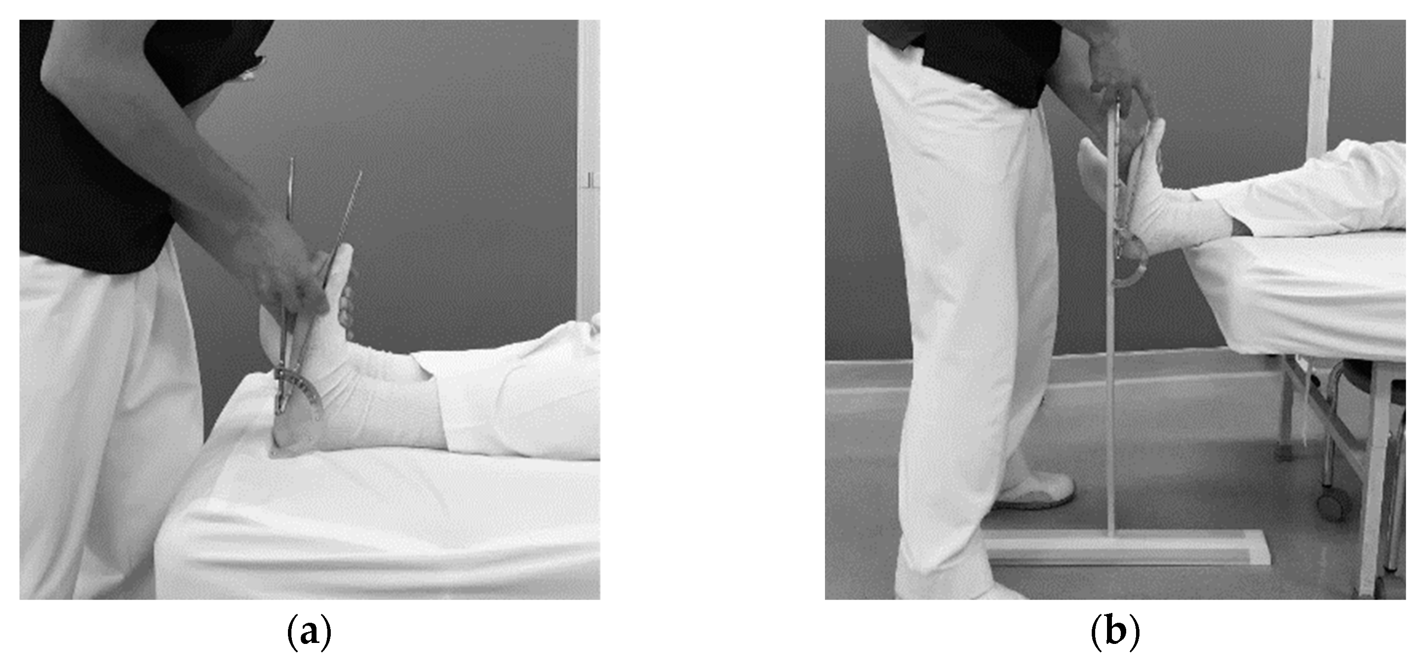
2.3.3. Gastrocnemius

Figure 4 demonstrates the methods of the MTT for the gastrocnemius. The model is in supine position. The physical therapist keeps the knee joint extended, whereas the ankle joint is dorsiflexed. The angle of the ankle joint is measured when it reaches the maximum dorsiflex. The vertical line on the floor serves as the basic axis, whereas the fifth metatarsal is referred to as the moving axis.

2.3.4. Quadriceps

Figure 5 presents the methods of the MTT for the quadriceps. The model is in prone position. The physical therapist flexes the model’s knee joint and measures the angle of the knee joint when tension in the muscle is recognizable or when trick motions (hip-lifts or lateral rotation of the hip joint) occur. The vertical line on the floor constitutes the basic axis, whereas the tibia is considered the moving axis.

2.4. Examining the Effectiveness of the Assistive Instrument

This study evaluated the effectiveness of the AsI on the basis of the following three aspects.

2.4.1. Ease

A questionnaire was constructed following the previous report [14]. The questionnaire was created by classifying the operations required for the ROM measurement; thus, we added several operations specific to MTT. The questions focused on 12 aspects, namely, (1) operability of the goniometer, (2) accurate placement of the angle meter with the basic axis, (3) accurate placement of the angle meter with the moving axis, (4) verification of the measurement position, (5) maintenance of the measurement position, (6) measurement of the joint angle as the position is maintained, (7) readability of the scales of the angle meter, (8) verification of muscle tightness, (9) verification of trick motion, (10) accurate verification of the basic (vertical) axis, (11) comprehensive ease of the MTT, and (12) confidence in performing the MTT accurately. Items were rated using a 5-point scale ranging from 5 = very easy to 1 = very difficult. In addition, the participants were asked to provide suggestions for improving the MTT in terms of performance. Moreover, they were encouraged to report whether UG or AsI is better for use in the MTT of the abovementioned muscles or whether they make no difference.

2.4.2. Accuracy

Accuracy was analyzed by comparing between errors in the measurement of the participants and an expert. The MTT is performed by stretching muscles; thus, a possibility exists that muscle tightness may change during repeated stretching. Hence, an expert licensed as an athletic trainer measured the MTT before the participants before each test.

2.4.3. Measurement Time

The time required to perform the MTT was measured from the time the researcher says start up to the time that the participant calls out the measurement result.

2.5. Statistical Analysis

Data were expressed as mean (standard deviation [SD]). Statistical analyses were performed using SPSS Statistics Version 26 (IBM, Armonk, NY, USA). Wilcoxon’s signed-rank test was used to compare between the UG and AsI for all aspects. p-value < 0.05 was considered to indicate a statistically significant difference in all tests.

3. Results

3.1. Ease

Table 2 presents the results of the questionnaire on the ease of conducting the MTT. All items displayed higher values for AsI, and 9 out of 12 items exhibited significant differences (p < 0.05). In particular, (2) accurate placement of the angle meter with the basic axis (p < 0.001), (6) measurement of the joint angle as the position is maintained (p < 0.001), and (10) accurate verification of the basic (vertical) axis (p < 0.001) demonstrated major improvements.
Table 3 displays the suggestions offered by the physical therapists for improving the MTT. Many answered that they should (a) practice and (b) gather advice and that (c) the instrument should be developed or improved.
Table 4 presents the responses to whether UG or AsI is better for conducting the MTT for the measured muscles or to whether they make no difference. The participants tended to select the AsI. For the iliopsoas, however, the number of votes for UG and AsI was the same, with the most common answer being “no difference”.

3.2. Accuracy

Figure 6 depicts the total error of all measurement parts. Although statistical significance was observed only for the gastrocnemius (p < 0.01), the error for AsI was smaller for all parts. The differences in the mean (SD) of the measurement errors between the UG and AsI were as follows: 0.5 (9.8)°, 1.6 (4.6)°, 3.4 (5.1)°, and 2.7 (10.4)° for the hamstrings, iliopsoas, gastrocnemius, and quadriceps, respectively.

3.3. Measurement Time

Figure 7 presents the result of the measurement time for each muscle. The required time was significantly longer for AsI for all muscles (p < 0.05). The differences in the mean (SD) of measurement time between the UG and AsI were as follows: 17.4 (16.4) s, 11.1 (13.0) s, 20.3 (21.9) s, and 12.2 (13.4) s for the hamstrings, iliopsoas, gastrocnemius, and quadriceps, respectively.

4. Discussion

The present study examined the effectiveness and issues associated with using the AsI, which was developed for physical therapists with VI in performing the MTT from the three abovementioned aspects. The results of the comparison between the AsI and UG indicated a significant difference in 9 out of 12 items that centered on the comprehensive ease of the MTT. Moreover, although statistical significance was observed only for the gastrocnemius from the aspect of accuracy, the error for AsI was smaller for all measure muscles. In terms of measurement time, the AsI required a longer time.
Initially, we examined which aspects caused difficulty in conducting the MTT. Focusing on the result of UG in Table 2, the score for (11) comprehensive ease of the MTT was nearly similar to that for “Comprehensive ease of the ROM”, as previously reported [14]. Thus, the study inferred that conducting the MTT was also difficult for physical therapists with VI. In addition, the scores for (6) measurement of the joint angle as the position is maintained and (10) accurate verification of the basic (vertical) axis were lower than those for the other aspects. Therefore, the study inferred that these two points were the main factors that led to the difficulty associated with the use of the MTT, which we expected.
We discuss the effectiveness of AsI based on the abovementioned results. In terms of ease, the use of AsI provided significant improvement in 9 out of 12 items. Moreover, the AsI made less errors for all muscles compared with those of the UG. The reason for these improvements can be attributed to the solutions to the two aforementioned aspects, namely, (6) measurement of the joint angle as the position is maintained and (10) accurate verification of the basic (vertical) axis. For item (6), attaching the angle meter to the AsI solved the problem previously pointed out [16], where both hands are occupied with using the UG. Thus, our solution enabled the participants to touch the required body part, which may have resulted in the positive effect on ease and accuracy. For item (10), the AsI has set the vertical axis, such that the participants were only required to attach the angle meter on the appropriate height of the axis when adjusting the basic axis vertically. These effects were reflected in the particularly large improvement in the two aspects (Table 2). Therefore, the AsI may be more effective in terms of ease and accuracy.
Alternatively, measurement time remained an issue for the use of the AsI. We propose that two factors underlie this problem. The first is that the participants were inexperienced in using the AsI. The participants were refrained from practicing to minimalize the learning effect, whereas people with VI basically require a longer time to acquire new skills due to the difficulty in observing by sight [18]. To address this issue, practice was considered as the first solution. Table 3 indicates that nearly all participants answered that they required practice to improve the difficulty they experienced in conducting the MTT. In addition, Kimura et al. [19] stated that VI renders several specific motions challenging. Therefore, identifying difficult motions and practicing them repeatedly may be effective in reducing the measurement time in conducting the MTT.
The second is the operability of the AsI. We used a velcro tape to fix the angle meter on the vertical axis due to its availability and fixability. However, we realized a disadvantage, i.e., adjusting the height of the angle meter after fixing the tape is difficult. As a possible option for improving operability, we suggest the use of magnets instead of velcro tapes, because magnets are also available and suitable for attaching to metal objects, such as angle meters. In addition, Behnoush et al. [16] used smartphones to measure the ROM instead of the UG. This technology may be available for people with VI due to the voice guide feature of smartphones.
This study points out two major limitations. The first is that the effect of the AsI may differ according to individual characteristics, such as levels of disability. For example, VI includes various patterns of sight according to visual acuity, visual field, and other factors. In addition, individual potentials may influence applied movements, such as the MTT. This notion is similar to strong and weak points in sports. In the experiments, we observed that several participants conducted the MTT appropriately with UG alone, whereas others demonstrated substantial improvement using the AsI. Second, the AsI is still in the process of development. As this study illustrated the first attempt, we infer that the AsI is by nature a prototype. We expect that there are many potential ideas for improvement of the AsI, and thus its effectiveness can be improved further by updating the materials or implementing new technologies.
For clinical applications, we note that the AsI may not be effective for all cases. For example, the AsI may not benefit people with low levels of VI. In addition, we expect that effectiveness is dependent on the targeted muscle. In fact, the participants answered that the usability of the AsI and UG are not different in measuring the iliopsoas. An important aspect to consider in the application of the AsI is the unique characteristics of each individual.
Incidentally, the fact that minimal assistance may enable people with VI to participate in supporting positions in sports underlies the finding of this study. If people with VI can participate in highly specialized roles, such as athletic trainers, then many people with various types of disability may play supportive roles in sports. In fact, in the Tokyo 2020 Olympic and Paralympic Games, people with disabilities were encouraged to participate as volunteer staff [20]. Such attempts will be made worldwide in the future; thus, further studies are required to examine better or other ideas for increasing the opportunities for participation among people with disabilities in supporting positions. We believe that this minimal assistance can lead to major progress in achieving an inclusive society.

5. Conclusions

The present study developed the AsI for physical therapists with VI to enable them to conduct the MTT with ease. Moreover, we examined its use on the basis of three aspects, namely, ease, accuracy, and measurement time. The results suggested that the AsI was effective in terms of ease and accuracy. However, the lengthy measurement time was considered an issue. To address it, two points should be improved. The first is sufficient practice of the MTT, and the second to the revision of the AsI. These findings suggested the possibility that physical therapists with VI can perform the MTT similar to athletic trainers given that the identified problems are solved.

Author Contributions

Conceptualization, S.T.; methodology, S.T., S.K., T.Y. and T.W.; validation, S.T., S.K., T.Y. and T.W.; investigation, S.T., S.K. and T.Y.; data curation, S.T. and T.W.; writing—original draft preparation, S.T.; writing—review and editing, S.K. and T.Y.; supervision, T.W. All authors have read and agreed to the published version of the manuscript.

Funding

This research received no external funding.

Institutional Review Board Statement

The study was conducted according to the guidelines of the Declaration of Helsinki and approved by the Research Ethics Committee of the University of Tsukuba in Tokyo Campus (Tou 29-69).

Informed Consent Statement

The potential participants were informed in writing and verbally, about the purpose and method of the study, including its possible risks. Written informed consent for participation to this study and publication of their details was obtained from all participants. None complained of discomfort or physical condition during the survey.

Data Availability Statement

The data presented in this study are available on request from the corresponding author.

Conflicts of Interest

The authors declare no conflict of interest.

References

  1. Blauwet, C.; Willick, S.E. The Paralympic Movement: Using sports to promote health, disability rights, and social inte-gration for athletes with disabilities. PM R J. Inj. Funct. Rehabil. 2012, 4, 851–856. [Google Scholar] [CrossRef] [PubMed]
  2. Shimony, N.D.; Lidor, R.; Ziv, G. The effectiveness of bandwidth knowledge of results on a throwing task in goalball players with visual impairments. Eur. J. Adapt. Phys. Act. 2020, 13. [Google Scholar] [CrossRef]
  3. Dickson, T.J.; Darcy, S.; Johns, R.; Pentifallo, C. Inclusive by design: Transformative services and sport-event accessibility. Serv. Ind. J. 2016, 36, 532–555. [Google Scholar] [CrossRef]
  4. Duncan, K.M.; Wright, K.E. A National Survey of Athletic Trainer Roles and Responsibilities in the Allied Clinical Setting. J. Athl. Train. 1992, 27, 311–316. [Google Scholar]
  5. Japan Sport Association. The 1st Professional Survey of Trainers in Japan. 2018. Available online: https://www.japan-sports.or.jp/Portals/0/data/ikusei/doc/AT/Report/Japan_trainer_report.pdf (accessed on 17 January 2020).
  6. Wong, M.E. Higher education or vocational training inriryoka? Post-school choices of visually-impaired students. Part 2, Japan. Br. J. Vis. Impair. 2004, 22, 59–65. [Google Scholar] [CrossRef]
  7. Kondo, H.; Fujii, R. A survey of visually impaired anma-massage-shiatsu practitioners in Japanese hospitals. NTUT Educ. Disabil. 2014, 12, 24–27. [Google Scholar]
  8. Hansberger, B.L.; Loutsch, R.; Hancock, C.; Bonser, R.; Zeigel, A.; Baker, R.T. Evaluating the relationship between clinical assess-ments of apparent hamstring tightness: A correlational analysis. Int. J. Sports Phys. Ther. 2019, 14, 253–263. [Google Scholar] [CrossRef] [PubMed] [Green Version]
  9. Bahr, R.; Holme, I. Risk factors for sports injuries—a methodological approach. Br. J. Sports Med. 2003, 37, 384–392. [Google Scholar] [CrossRef] [PubMed]
  10. Reis, F.J.J.; Macedo, A. Influence of Hamstring Tightness in Pelvic, Lumbar and Trunk Range of Motion in Low Back Pain and Asymptomatic Volunteers during Forward Bending. Asian Spine J. 2015, 9, 535–540. [Google Scholar] [CrossRef] [PubMed] [Green Version]
  11. Endo, Y.; Sakamoto, M. Correlation of shoulder and elbow injuries with muscle tightness, core stability, and balance by longitu-dinal measurements in junior high school baseball players. J. Phys. Ther. Sci. 2014, 26, 689–693. [Google Scholar] [CrossRef] [PubMed] [Green Version]
  12. Huguenin, L.; Brukner, P.D.; McCrory, P.; Smith, P.; Wajswelner, H.; Bennell, K. Effect of dry needling of gluteal muscles on straight leg raise: A randomised, placebo controlled, double blind trial. Br. J. Sports Med. 2005, 39, 84–90. [Google Scholar] [CrossRef] [PubMed]
  13. Zink, K.; Chini, B.; Cowens, J.; Kremer, L.; Lin, L. Improving Clinical Outcomes and Quality of Life with Massage Therapy in Youth and Young Adults with Cystic Fibrosis: A Pilot Study. Int. J. Ther. Massage Bodywork Res. Educ. Pract. 2019, 12, 4–15. [Google Scholar]
  14. Tamai, S.; Kuroda, S.; Numoto, N.; Yabe, T.; Wada, T. Examination of problems when the visually impaired measure the range of motion. Bull. Acupunct. Phys. Ther. Teach. Train. School Univ. Tsukuba 2019, 4, 5–12. [Google Scholar]
  15. Klavina, A.; Jekabsone, I. Static Balance of Persons with Intellectual Disabilities, Visual Impairment and without Disabilities. Eur. J. Adapt. Phys. Act. 2014, 7, 50–57. [Google Scholar] [CrossRef] [Green Version]
  16. Behnoush, B.; Tavakoli, N.; Bazmi, E.; Fard, F.N.; Shahi, M.H.P.; Okazi, A.; Mokhtari, T. Smartphone and Universal Goniometer for Measurement of Elbow Joint Motions: A Comparative Study. Asian J. Sports Med. 2016, 7, e30668. [Google Scholar] [CrossRef] [PubMed] [Green Version]
  17. Abe, H.; Watanabe, H.; Tojima, M.; Torii, S.; Shigeta, K.; Tsukimura, T.; Abe, H. Intrarater and interrater reliability of the muscle tightness test. J. Jpn. Soc. Clin. Sports Med. 2012, 20, 336–343. [Google Scholar]
  18. Lieberman, L.J.; Lepore, M.; Lepore-Stevens, M.; Ball, L. Physical Education for Children with Visual Impairment or Blindness. J. Phys. Educ. Recreat. Dance 2018, 90, 30–38. [Google Scholar] [CrossRef]
  19. Kimura, K.; Miyaji, H.; Hanaoka, Y.; Kadota, T.; Sato, T.; Wada, T.; Miyamoto, T. Examination of problems when the visually impaired conduct low frequency electroacupuncture. Bull. Acupunct. Phys. Ther. Teach. Train. School Univ. Tsukuba 2018, 3, 5–13. [Google Scholar]
  20. The Nippon Foundation Volunteer Support Center. Volunteer Participation Practice Report Meeting for the Visually Impaired Tokyo Olympic and Paralympic Games and beyond Has Been Held! 2021. Available online: https://www.volasapo.tokyo/column/report/3988/ (accessed on 5 October 2021).
Figure 1. The assistive instrument. An inverted T-shaped instrument with a Velcro tape on the vertical axis to fix the universal goniometer. Regarding its development, we paid attention to two points, namely, availability and portability. The instrument is made of wood due to its affordability, durability, lightness, and ease of processing. Moreover, we formulated a simple design, such that it is easily reproducible and could be carried in the sports field. Incidentally, the vertical axis can be removed from the base plate to enable the instrument to be carried compactly to the sports field.
Disabilities 01 00033 g001
Figure 2. Methods of the muscle tightness test for the hamstrings with the universal goniometer (a) and with the assistive instrument (b).
Disabilities 01 00033 g002
Figure 3. Methods of the muscle tightness test for the iliopsoas with the universal goniometer (a) and with the assistive instrument (b).
Disabilities 01 00033 g003
Figure 4. Methods of the muscle tightness test for the gastrocnemius with the universal goniometer (a) and with the assistive instrument (b).
Disabilities 01 00033 g004
Figure 5. Methods of the muscle tightness test for the quadriceps with the universal goniometer (a) and with the assistive instrument (b).
Disabilities 01 00033 g005
Figure 6. Comparison between the measurement errors for each muscle. Data were expressed as mean (SD). Wilcoxon’s signed-ranked test was used for statistical analysis. Significant differences were expressed as ** p < 0.01. UG: universal goniometer; AsI: assistive instrument.
Disabilities 01 00033 g006
Figure 7. Comparison between the measurement times for all muscles. Data were expressed as mean (SD). Wilcoxon’s signed-ranked test was used for statistical analysis. Significant differences were expressed as * p < 0.05 and ** p < 0.01. UG: universal goniometer, AsI: assistive instrument.
Disabilities 01 00033 g007
Table 1. Characteristics of Participants.
CharacteristicsMean (SD)/Number (%)
Sex (men:women)18:4
Age (year)31.1 (7.5)
Age of diagnosis of VI (year)17.5 (10.5)
Duration of VI (year)13.9 (7.4)
Visual acuity0.24 (0.27)
Visual field constriction19 (86.3%)
Central scotoma5 (22.7%)
Duration of license of physical therapists (year)1.4 (1.4)
The breakdown of eye diseases was as follows: retinitis pigmentosa: 12; glaucoma: 4; macular dystrophy: 1; cone dystrophy: 1; optic nerve atrophy: 1; uveitis: 1; and unknown: 2. VI: Visual impairment.
Table 2. Results of the questionnaire on ease of conducting the MTT.
ItemsUGAsIp-Value
(1) Operability of the goniometer3.0 (1.0)3.9 (0.8)0.001
(2) Accurate placement of the angle meter with the basic axis2.8 (0.8)4.3 (0.6)<0.001
(3) Accurate placement of the angle meter with the moving axis2.7 (1.0)3.7 (0.8)0.001
(4) Verification of the measurement position3.3 (1.0)3.6 (0.7)0.109
(5) Maintenance of the measurement position2.9 (1.1)3.3 (1.0)0.058
(6) Measurement of the joint angle as the position is maintained2.3 (1.1)3.8 (0.8)<0.001
(7) Readability of the scales of the angle meter2.5 (1.1)2.9 (1.1)0.046
(8) Verification of muscle tightness3.3 (0.9)3.6 (0.6)0.083
(9) Verification of trick motion2.7 (0.6)3.2 (0.6)0.013
(10) Accurate verification of the basic (vertical) axis2.5 (1.0)4.1 (0.8)<0.001
(11) Comprehensive ease of the MTT2.8 (1.0)3.9 (0.8)<0.001
(12) Confidence in performing the MTT accurately2.1 (1.0)3.1 (0.8)0.001
Data were expressed as mean (SD). Wilcoxon’s signed-ranked test was used for statistical analysis. UG: universal goniometer; AsI: assistive instrument; and MTT: muscle tightness test.
Table 3. Suggestions for improving the performance of the MTT.
ItemsUGAsI
(a) Practice2122
(b) Gather advice1917
(c) Develop or improve the instrument76
(d) No need for improvement00
(e) Others *11
* The therapist who answered “Others” mentioned that sight is required to perform the MTT. UG: universal goniometer; AsI: assistive instrument; and MTT: muscle tightness test.
Table 4. Answers for whether the UG or AsI is better.
Measurement PartsUGAsINo Difference
(I) Hamstrings1201
(II) Iliopsoas778
(III) Gastrocnemius4144
(IV) Quadriceps1201
UG: universal goniometer; AsI: assistive instrument.
Publisher’s Note: MDPI stays neutral with regard to jurisdictional claims in published maps and institutional affiliations.

