Article

Improved Measurement Method of Human Skin Temperature Based on Human Skin-like Gradient Standard Radiation Source

1
National Institute of Metrology of China, Beijing 100029, China
2
Beijing Key Laboratory of Performance Guarantee on Urban Rail Transit Vehicles, Beijing University of Civil Engineering and Architecture, Beijing 100044, China
3
School of Materials Science and Physics, China University of Mining and Technology, Xuzhou 221116, China
*
Author to whom correspondence should be addressed.
Thermo 2025, 5(4), 38; https://doi.org/10.3390/thermo5040038
Submission received: 11 August 2025 / Revised: 29 September 2025 / Accepted: 30 September 2025 / Published: 2 October 2025

Abstract

Infrared thermography for human skin temperature measurement, when calibrated with standard blackbodies, suffers from errors due to the mismatch in emissivity between a blackbody and human skin. This study introduces a novel calibration method utilizing a human skin-like gradient radiation source to enhance measurement accuracy. A custom radiation source with six temperature points and skin-like emissivity was developed. Thermal imagers were calibrated using this source, and their performance was compared against traditional blackbody calibration. The proposed method reduced the calibration error to 0.04 °C, a significant improvement over the 0.15 °C error obtained with blackbody calibration. Calibration with a skin-like radiation source proves superior to the blackbody method, enabling high-accuracy (less than 0.1 °C) human skin temperature measurement for improved fever screening.

1. Introduction

Temperature measurement is attracting growing attention, with a growing emphasis on the performance metrics of temperature measurement equipment, particularly the demand for heightened accuracy [1]. Radiation temperature measurement boasts a broad temperature range, rapid response speed, and non-contact advantages, making it widely applicable in military, security, firefighting, medical, industrial testing, electric power industry, and other fields. In typically crowded place like airports, stations, docks, hospitals, and shopping malls, infrared radiation temperature measurement is almost the only way to measure temperature quickly.
However, the non-contact nature of radiation temperature measurement renders it susceptible to various external factors during practical use. These factors include temperature measurement distance, measurement angle, ambient temperature, atmospheric absorption and reflection along the propagation path, object surface roughness, background temperature, and the surrounding objects’ reflected radiation [2,3,4]. The accuracy of measurements is affected, and this method can only conduct preliminary screening of human body temperature and is unable to achieve precise measurement, potentially leading to the oversight of suspicious cases.
To address these challenges, the calibration of radiation thermometry equipment emerges as a crucial step. Effectively calibrating such equipment enhances measurement accuracy and enables fine screening of human body temperature. This, in turn, improves the efficiency of epidemic protection measures and reduces unnecessary resource consumption [5]. In the context of the pandemic in previous years, the FDA (Food and Drug Administration) has performed extensive research on the use of thermography for traceable, non-contact human temperature screening, made some progress, and issued guidance documents and international standards on pandemic protection [6].
The conventional method for calibrating a thermal imaging camera involves blackbody calibration [7,8], as illustrated in Figure 1. A commonly employed blackbody radiation source is equipped with a digital display controller to regulate the temperature. Precision Resistance Temperature Detectors (RTDs) and thermocouples are placed inside the blackbody radiation source to enhance its accuracy and repeatability. Additionally, PID thermostats (PID stands for Proportion, Integral, and Differential coefficient, which represent three types of control algorithms) are utilized to enhance the temperature resolution of the blackbody. The blackbody radiation source incorporates heat-resistant and stable thermal insulation materials, ensuring a long lifespan and rapid temperature stabilization. V. Rastogi and V. Kumar used digital holography to measure the temperature of human skin (on the index finger and palm of the hand) and the radiation temperature of a flame [9,10,11].
In an alternative approach to calibrating thermal cameras, the captured images are displayed on a computer screen, allowing real-time detection of temperature changes in the tested object [12].
Zhang et al. [13] introduced the equivalent blackbody method, aligning ambient radiation with blackbody radiation at the ambient temperature, making it particularly suitable for graybody objects. Building upon this, Cui et al. [14] enhanced the equivalent blackbody calibration method. They employed two calibration functions at different ambient temperatures to separate target and ambient radiation superimposed on calibration data, constructing an equivalent blackbody calibration function. This refinement reduced the maximum measurement error from ±6.6 °C to ±1.8 °C. In comparison to conventional environment-compensated calibration functions, the results from the equivalent blackbody calibration function proved more satisfactory. Further improvements were made by Cui et al. [15], who proposed a data processing method better suited for specific scenarios involving non-Lambertian bodies in non-uniform environments. Assuming the heat source around the measured object to be Lambertian, they derived a data processing method for non-Lambertian bodies in non-uniform environments based on equations from the classical method and the angle between the heat source and the measured substance. This method has higher accuracy than the classical method in non-uniform environments. Zhang et al. [16] proposed a temperature measurement method incorporating voltage compensation to enhance the accuracy of temperature measurements of an uncooled infrared camera, particularly in relation to internal radiation effects within the camera. The method involves converting the radiation from the measured object into a voltage value, which is then transformed into a measurable grayscale value. This establishes the relationship between the radiation of the measured object and the grayscale value. Subsequently, a temperature measurement model for the uncooled infrared camera is developed based on the established relationship between the radiation and grayscale values. To determine the coefficients in the temperature measurement formula, the true temperature of the object surface was calculated using the formula, combined with the acquisition of infrared image data from a standard blackbody. The resulting maximum error in temperature measurement by the uncooled infrared camera was reduced to 0.7 °C, with an average error of 0.34 °C. This method proved effective in significantly reducing the relative error in temperature measurement by the camera.
Romain et al. [17]. employed two-color pyrometry for precise temperature measurements of liquid metal surfaces. However, this method, relying on the ratio of two monochromatic images, introduces two types of errors that impact the accuracy of temperature measurements. The first error is directly attributed to optical distortions caused by the lens and variations due to the beam splitter. To address this, the authors propose a geometric correction method aiming to mitigate distortions induced by the optical system. The second source of error arises from differences in the spectral response between the two cameras, a discrepancy that is rectified through a spectral calibration procedure.
Gao et al. [18] conducted a comparative analysis of errors in mono-spectral, colorimetric, and multi-spectral temperature measurements of targets in high-temperature environments. Building on this comparison, they proposed a multi-wavelength temperature measurement method incorporating reflection correction. This method employs the surface element approach to partition the high-temperature background surface into many isothermal surfaces. By calculating radiative heat transfer between each isothermal surface and the object being measured, the reflected radiation is determined, thereby reducing the impact of the high-temperature environment on temperature measurements. This approach effectively decreases the uncertainty of multispectral temperature measurements from 4.16% to 0.26%.
Addressing a related challenge, Jian Zhu and Bo-Tao Wang proposed a high-precision correction method for temperature measurement errors induced by reflected radiation in high-temperature backgrounds [19]. The correction theory for temperature measurement errors caused by reflected radiation was deduced from principles, and a physical field model for the temperature measurement of ceramic blades in a high-temperature furnace was established [20]. Experimental results demonstrated a significant reduction in errors for monochromatic and colorimetric temperature measurements, decreasing from 13.5% and 7.5% to 0.3% and 0.5%, respectively. This method proved highly effective in minimizing temperature measurement errors resulting from reflected radiation. While both methods were specifically designed for high-temperature objects, the experimental approach has potential value in measuring human skin temperature.
To accurately measure the temperature of human skin, this paper proposes a novel method for calibrating a thermal imaging camera. Instead of using a blackbody, the calibration process utilizes a human skin-like gradient standard radiation source which comprises a series of emissivity standard plate whose emissivity is similar to that of human skin, and six individual temperature control modules. The human skin-like gradient standard radiation source includes six high-precision equal temperature difference infrared radiation sources. This allows the thermal imager to be calibrated to multiple temperatures simultaneously, eliminating the need to heat the blackbody to various temperatures. Additionally, this approach avoids the linearity issues associated with single-point calibration after measuring other temperature points [21].

2. Theoretical Analysis

Planck’s formula is the basic formula in the blackbody radiation field. According to Planck’s formula, the basic principle of radiation thermometry, supposing an absolute temperature T and wavelength λ , C1 and C2 are constant, and the spectral radiance at the opening of the ideal blackbody is L ( λ , T ) , then
L λ , T = C 1 λ 5 1 e C 2 λ T 1
Therefore, to measure the temperature of an object [22], a thermal camera needs to detect the radiance of the object L ( λ , T ) , which can be inverted to the actual temperature of the object T .
For the human body, assume that the ambient radiance is L am , the human skin reflectance is ρ ( λ ) , and ε ( λ ) is the emissivity of the human forehead skin, where L ( λ , T ) is the radiance of an ideal blackbody at temperature T , and R ( λ ) is the responsivity of the thermal imaging camera. The signal, S , measured by the thermal camera at a certain wavelength can be expressed by
S = S e m i t t e d + S r e f l e c t e d = L ( λ , T ) ε ( λ ) R ( λ ) + L am ρ ( λ ) R ( λ )
Assuming that the operating wavelength of the camera is located at 7–14 μm, the total signal obtained by a thermal camera is S 1 , hence S 1 can be calculated by integrating S over its operating region [23].
S 1 = 7 14 ( L ( λ , T ) ε ( λ ) + L am ρ ( λ ) ) R λ d λ
For opaque objects, according to Kirchhoff’s law, it is generally accepted that ε ( λ ) = 1 ρ ( λ ) , hence Formula (3) can be changed to Formula (4).
S 1 = 7 14 ( L ( λ , T ) ε ( λ ) + L am L am ε ( λ ) ) R ( λ ) d λ
Moreover, the emissivity of an ideal blackbody is 1 and reflectance is 0, so Formula (4) can be simplified as
S bb = 7 14 L ( λ , T ) R ( λ ) d λ
For an object with an actual unknown temperature T x , the result measured by the thermal camera can be expressed by
S x = 7 14 ( L ( λ , T x ) ε ( λ ) + L am L am ε ( λ ) ) R ( λ ) d λ
Furthermore, if the thermal camera is calibrated by blackbody, the output temperature T m e a s of the camera is (within an extremely small temperature range where the temperature variation is less than 5K, and S and T are approximately linear):
T m e a s = S x S bb T = 7 14 ( L ( λ , T x ) ε ( λ ) + L am L am ε ( λ ) ) R ( λ ) d λ 7 14 L ( λ , T ) R ( λ ) d λ T
If the thermal camera is calibrated by a human skin-like standard radiation source instead of by blackbody, the temperature T n e w can be calculated by Formula (8):
T n e w = S x S hum T = 7 14 ( L ( λ , T x ) ε ( λ ) + L am L am ε ( λ ) ) R ( λ ) d λ 7 14 ( L ( λ , T ) ε ( λ ) + L am L am ε ( λ ) ) R ( λ ) d λ T
In the simplest scenario, the emissivity and the response sensitivity of the thermal imaging camera are constant and both independent of wavelength (in the actual calibration, the calibration is carried out near the temperature of the measured temperature point, so T x is approximately equal to T). If this is the case, then
T m e a s = S x S bb T = 7 14 ( L ( λ , T x ) ε ( λ ) + L am L am ε ( λ ) ) d λ 7 14 L ( λ , T x ) d λ T > T
T n e w = S x S hum T = 7 14 ( L ( λ , T x ) ε ( λ ) + L am L am ε ( λ ) ) d λ 7 14 ( L ( λ , T x ) ε ( λ ) + L am L am ε ( λ ) ) d λ T = T
At room temperature, according to Equations (9) and (10), a thermal camera calibrated by a human skin-like standard radiation source is more accurate than one calibrated by blackbody.
Theoretical deductions indicate that human skin-like radiation source calibration offers superior accuracy for measuring human skin temperature compared to traditional blackbody calibration. However, some thermal imaging cameras can be calibrated with a blackbody emissivity of 1 and used in conjunction with a system emissivity of 0.98 to mitigate system errors arising from differences between blackbody emissivity and human skin emissivity; this correction also has its limitations.
What is more important is that the nonlinear relationship between the measurement signal and blackbody radiation is very complicated. While this correction may enhance accuracy at a single temperature point, its effectiveness diminishes at other temperature points due to nonlinear and other factors, resulting in less satisfactory outcomes.

3. Experimental

3.1. Experimental Setup

The experimental setup can be divided into a measurement unit and a calibration unit. A representative infrared thermal imager (FOTRIC 628C, manufacturer: Shanghai Thermal Image Technology Corp., Ltd., Shanghai, China) was selected as the measurement unit to validate the proposed calibration methodology. This model is widely used for human temperature screening, and its specifications (e.g., uncooled microbolometer detector and 7.5–13 μm spectral range) are representative of common thermal imaging systems deployed in such applications. The focus of this study was to establish a proof of concept for a skin-like calibration source; future work will involve testing it with a broader range of camera models.
The key specifications of the camera are as follows: a spectral range of 7.5–13 μm, a thermal sensitivity (NETD) of ≤0.04 °C at 30 °C, a spatial resolution (IFOV) of 1.36 mrad, and a measurement accuracy of reading of ±0.5 °C or ±1%. The detector array is 384 × 288 pixels, with a standard lens providing a field of view (FOV) of 17.1° × 12.8°. The camera was selected for its suitability for human skin temperature measurement and its provided software development kit (SDK), which allowed for customized data acquisition and temperature correction. The calibration unit consists of a human skin-like radiation source (as shown in Figure 2). The calibration unit comprises a temperature detector, a temperature control board, a thermally conductive aluminum plate, and a reflectance standard board. Given that the emissivity of human skin is approximately 0.98, the primary function of the emissivity standard board is to simulate the emissivity of human skin. Both the human skin-like radiation source and the emissivity standard board have emissivity values close to 0.98, aiming to minimize errors in radiation temperature measurement and enhance measurement accuracy and reliability.
The emissivity standard board is crucial for simulating human skin emissivity, and its surface must be kept clean to ensure accurate measurement results. The emissivity standard plate is similar to a blackbody radiation source and requires similar maintenance: 1. Regularly store it under a sealed protective cover. 2. Light dust can be blown off using an ear syringe. 3. For more serious contamination, the plate can be replaced with a new one or washed with purified water, but it must be recalibrated afterward.
The temperature control system consists of a TEC (Thermoelectric Cooler, manufacturer: Zhejiang Fuxin Thermo-electric Technology Co., Ltd., Foshan City, China) temperature control board used to simulate human body temperature. The temperature control precision reaches the order of 0.01 °C. Six different temperatures are set on the temperature control board from left to right and top to bottom, labeled as 1# to 6#: 35 °C, 36 °C, 37 °C, 38 °C, 39 °C, and 40 °C. The heat generated by the temperature control board is then conducted through the thermally conductive aluminum plate to the surface of the emissivity standard board. The gaps between the temperature control board, aluminum plate, and emissivity standard board are filled with thermally conductive silicone grease to expedite heat conduction. It is essential to ensure uniform and appropriate application of silicone grease. Finally, the radiant temperature of the emissivity standard board is measured using the infrared thermal imager.
The human skin-like radiation source is a device designed to replicate both the surface emissivity of human skin and different human body temperatures. During calibration, aligning the infrared imaging camera lens with the human skin-like radiation source is necessary. For each temperature, the temperature of the infrared imaging camera should be calibrated separately, enabling subsequent radiant temperature measurement of human skin. Additionally, simultaneous pointing of the infrared imaging camera at both the person being tested and the human skin-like radiation source is feasible. This allows for radiant temperature measurement of human skin during the calibration process, significantly enhancing measurement efficiency and accuracy.
From Equations (9) and (10), it is evident that the closer the emissivity of the object is to the emissivity of human skin during calibration, the more accurate the calibrated thermal imaging camera will be in measuring the temperature of human skin. The primary source of error between the measured temperature and the real temperature of the object is mainly attributed to the emissivity of the object. Therefore, obtaining the emissivity of the object allows for the elimination of errors caused by emissivity in radiant temperature measurement [24]. However, the accuracy of object emissivity testing is not always high, and the measurement process can be complex, lacking wide adaptability and flexibility [25]. The measurement of human skin emissivity also poses certain challenges and is rarely reported on in the literature.
To address these challenges, a series of human skin radiation sources was established to calibrate the measuring instrument, reducing the error between the measured temperature of human skin and the actual temperature. This calibration approach aimed to meet the specific requirements of human radiation temperature measurement.
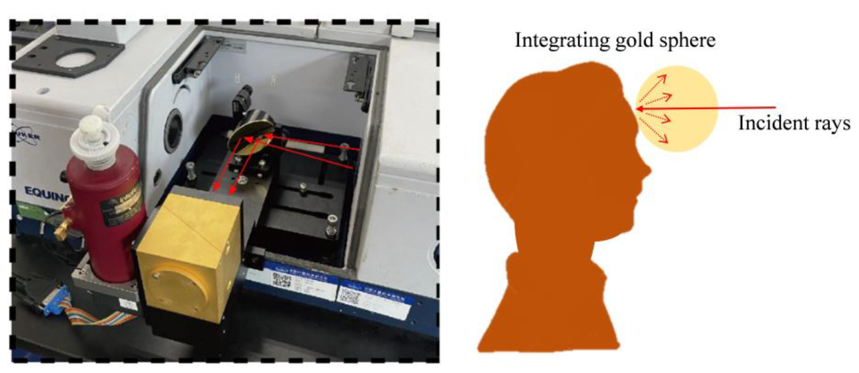
We constructed an experimental setup to measure the emissivity of human skin. We designed an external gold integrating sphere attachment based on the Fourier transform infrared spectrometer (Bruker company, Billerica, MA, USA, Type: E55) for measuring the reflectance of human skin, shown in Figure 3. According to Kirchhoff’s law, we can calculate the emissivity of human skin using ε ( λ ) = 1 ρ ( λ ) , shown in Figure 3.
To achieve a standard plate with an emissivity closely resembling human skin, an extensive literature review led us to develop a coating with an emissivity very similar to that of human skin. This coating was based on 3M’s NEXTEL VELVET COATING 811-21 (Hamburg, Germany) material, with the addition of keratin and casein thoroughly mixed together. To better simulate the characteristics of human skin, considering that the outer layer of human skin contains a lot of organic matter, keratin and casein are typically representative and have no sharp absorption peaks, which is similar to the emissivity of human skin. So, we chose to add these two components. By experimenting with different proportions of these materials, we found an appropriate ratio that makes the emissivity of the standard plate similar to that of the human skin surface. The final formulation and process flow were as follows: 8.0 g solid precipitate of NEXTEL-811, 1.0 g keratin, and 1.0 g casein were fully mixed, 10 mL butyl chloroacetate was taken as diluent and fully diluted by stirring, and ultrasonic shock was performed for 12 h to form a uniform bright black suspension. After the above operation was completed, the prepared precursor was smeared on the aluminum plate, and the mixture was completely hardened after about 24 h. A coating of human skin-like material was applied to the aluminum plate with a thickness of around 100 μm. Emissivity measurements were then performed using a Fourier Transform Infrared Spectrometer(Bruker company, Billerica, MA, USA, Type: E55).
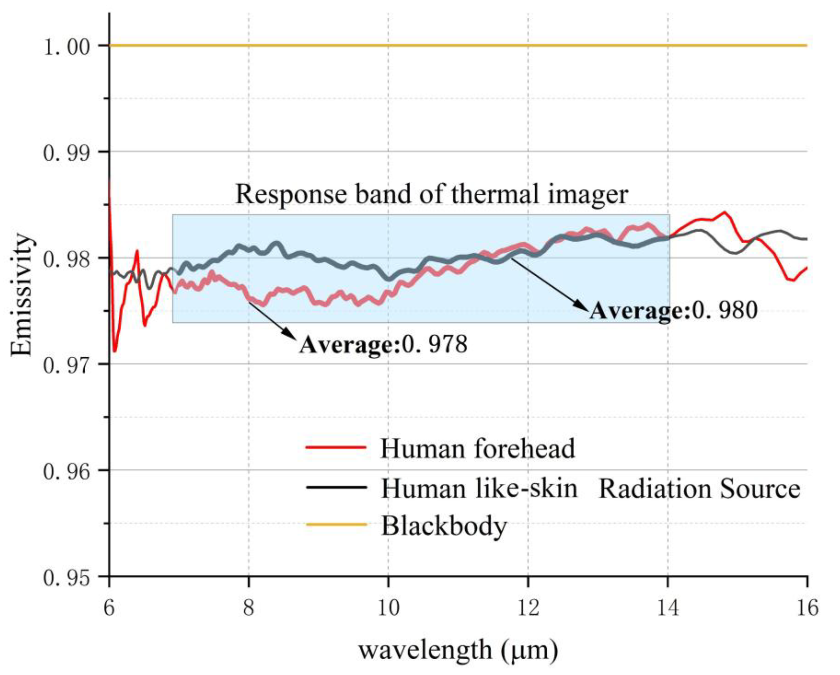
The emissivity characteristics of the human skin-like radiation source and the emissivity of human forehead skin are depicted in Figure 4. It is notable that the emissivity of the human forehead skin and the reflectance standard plate almost coincide in the wavelength range between 10 μm~16 μm. This is a significant difference from the emissivity of the blackbody, which remains close to 1. Furthermore, the response band of the thermal imaging camera falls between 7 μm and 14 μm, revealing a close match between the emissivity of the reflectance ratio standard plate and human forehead skin within the working band of the thermal imaging camera. Figure 4 shows that the emissivity of the standard board developed in this study and the emissivity of the human body are within the 7 μm–14 μm range, with an error as low as 0.002. This design can be applied to almost any infrared thermal imaging camera.

3.2. Accuracy

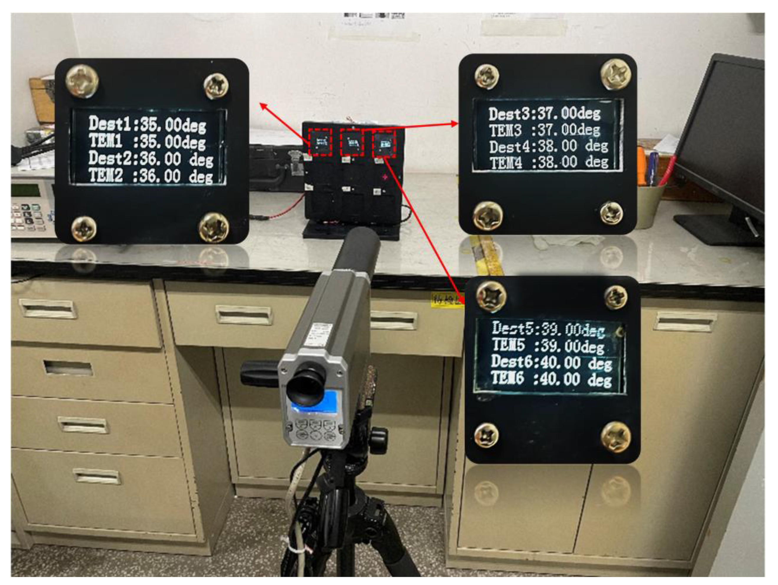
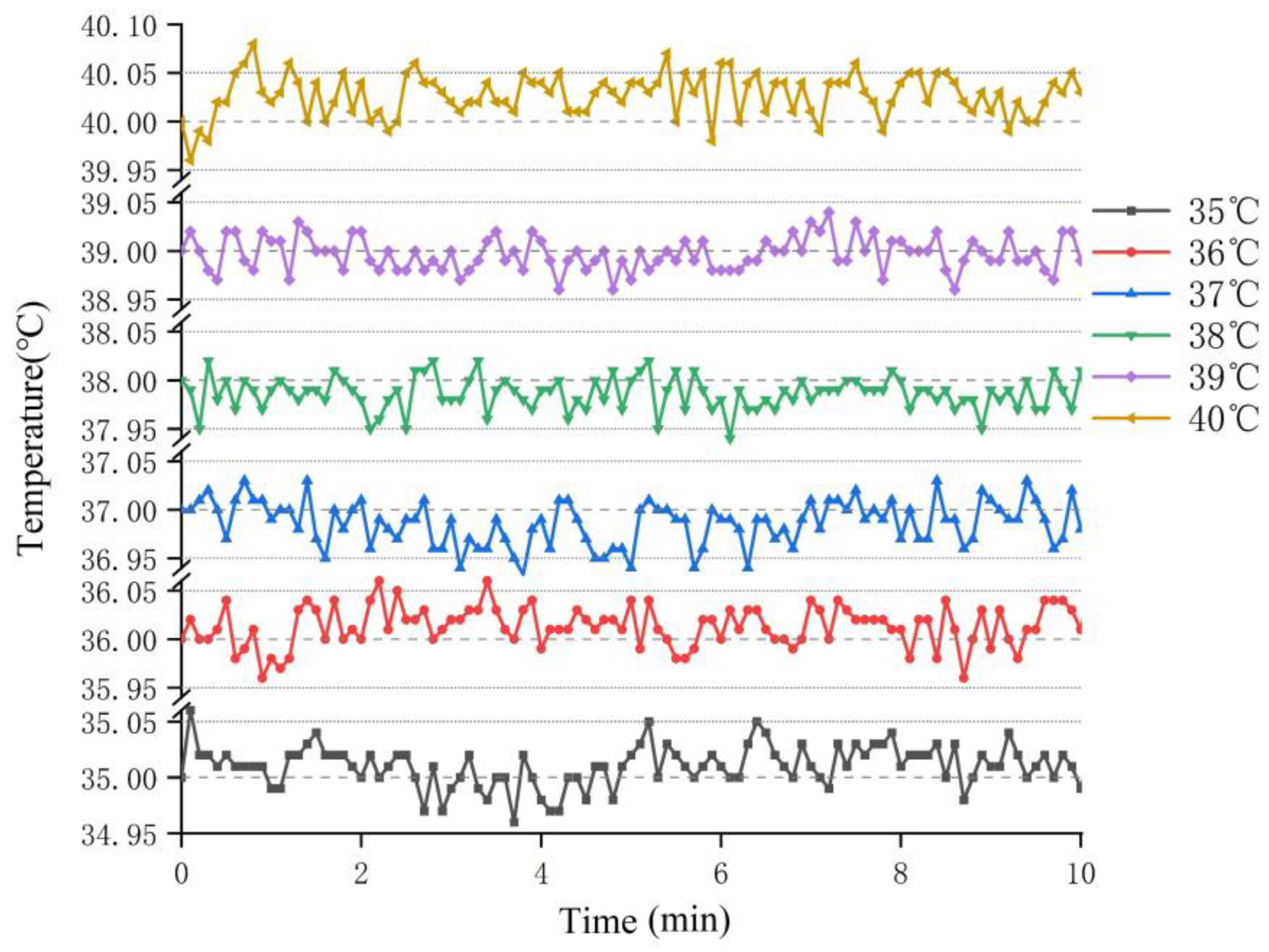
The measurement device used is a radiation thermometer from Heitronics (Heitronics GmbH, Wiesbaden, Hesse, Germany, Type: TRT IV.82) capable of detecting minimum temperature differences ranging from 0.02 °C to 0.06 °C. The device has been calibrated by the National Institute of Metrology, Beijing, China, ensuring the accuracy and reliability of the measurement results. It is designed to operate in an environment with a temperature range of 23 °C ± 3 °C, aligning with the requirements of the laboratory room temperature. Before measurement, the emissivity of the TRT IV.82 radiation thermometer was set to 0.98, and the ambient temperature was set to 23 °C (room temperature). The human skin-like radiation source was powered on and off in a manner similar to the actual measurement conditions. Once its temperature stabilized, temperature measurements were taken at the center of the gradient standard radiation source’s surface at 10 s intervals using the TRT IV.82 radiation thermometer, as shown in Figure 5. The recorded temperatures are illustrated in Figure 6. This process was conducted continuously for 10 min, and the averaged results are presented in Table 1.
As observed from Table 1, the majority of the differences between the actual temperature and the set temperature were approximately 0.2 °C. The discrepancies represent only about 0.7% of the set temperature, with the largest difference occurring for the human skin-like radiation source set at 37 °C, which is 0.5 °C.
Although these temperature errors were quite large, the temperature stability of the radiation source was very good, and we could calibrate the radiation temperature by adding a setting error constant from data in the temperature controlling system. Then, in order to measure the change in temperature over time, we needed as much data as possible, so we adjusted the interval to 6 s with acceptable minimal loss of accuracy. Sata was collected continuously for 10 min, resulting in 600 data points. The obtained data was then recorded. The results are depicted in Figure 6.
The temperature fluctuations of the stabilized human skin-like radiation source are illustrated in Figure 6. These fluctuations are on the order of 10−2 °C (standard deviation). After calibration, it was found that the average temperature system error was nearly 0.03 °C.
It should be emphasized that the temperature resolution (NETD) of the TRT IV.82 radiation thermometer can just reach 0.02 °C~0.06 °C, while the temperature resolution of the gradient standard radiation source can reach 0.01 °C and can also reach up to 0.02 °C per hour, demonstrating long-term stability (using a TEC heating element to heat standard plates with different gradients and placing a temperature sensor between the standard plate and the heating element to monitor the temperature change in real time. Based on the temperature measured by the sensor, PID control technology is then employed to maintain the stability of the temperature). For this reason, we tend to consider that the temperature fluctuations measured by the radiometer were due to the resolution of the radiation thermometer (TRT IV.82) itself and can be ignored.

3.3. Uniformity

During the calibration process of the thermal imaging camera, maintaining alignment with the center of the human skin-like gradient standard radiation source was not always guaranteed. Therefore, the uniformity of the radiation source significantly influenced calibration results. To assess uniformity, the radiation thermometer was positioned 20 cm (in some regulations for domestic thermal imager calibration, 20 cm is the recommended value, which is also in line with the actual working condition) away from the human skin-like radiation source, with the lens of the radiation thermometer perpendicular to the source. After a 30 min wait, during which the temperature monitor of the human skin-like radiation source stabilized and the heat from the thermostat was fully transferred to the emissivity standard plate on the surface and reached a stable state, then it was measured by a radiation thermometer. Temperature data from five different zones were compared, as shown in Figure 7, to determine homogeneity.
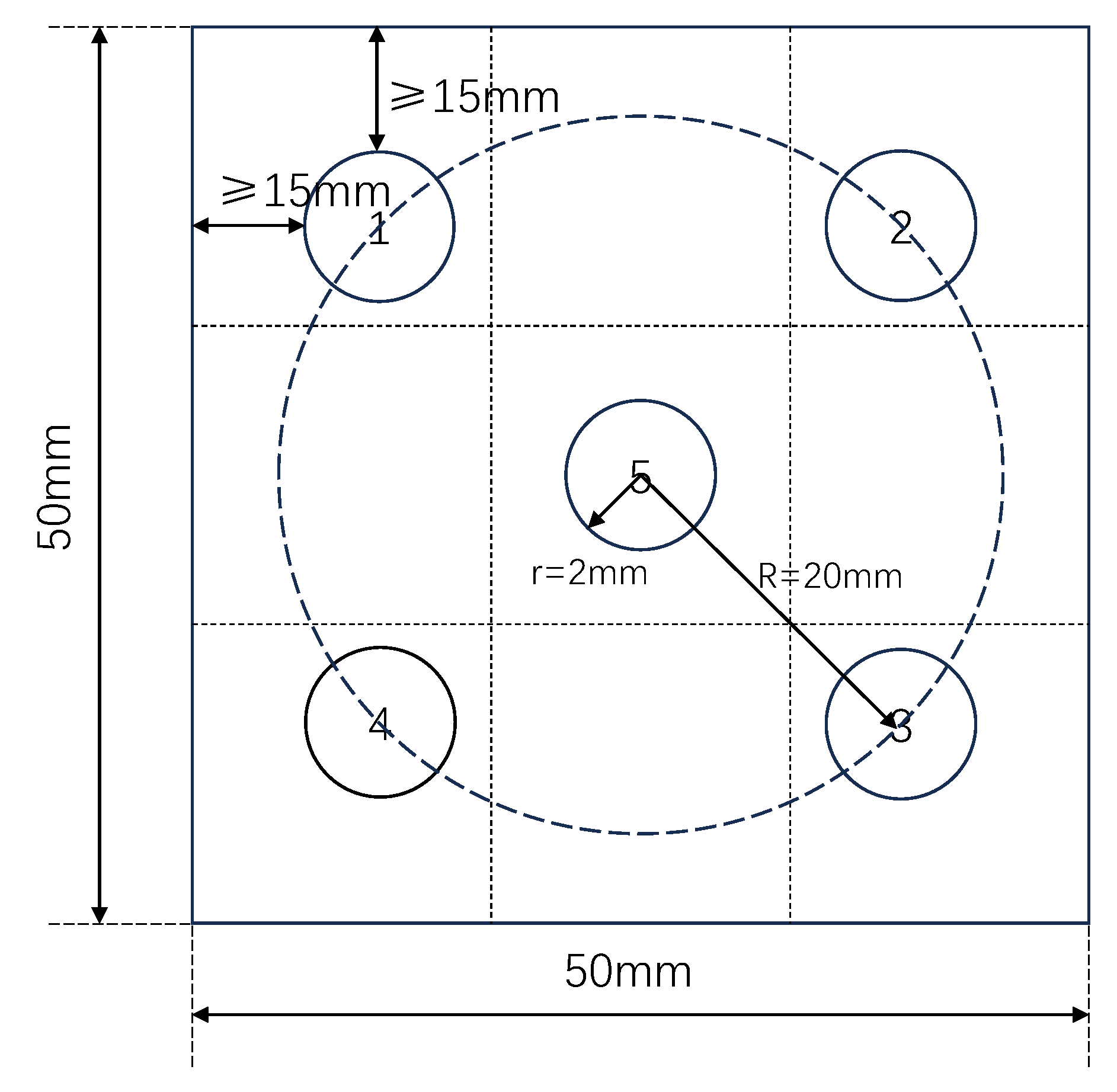
The surface of the human skin-like radiation source includes six 50 mm × 50 mm emissivity standard plates; each standard surface can be divided into five identical measurement areas, see Figure 7.
Once the equipment was stabilized, the temperature at the center position (position 5) was measured 3 times and averaged. Subsequently, positions 1 to 4 were measured sequentially, and the obtained results were compared with the averaged data from position 5. The comparative results are presented in Table 2 and Figure 8.
As shown in Table 2, after the temperature of the center point was calibrated, the deviation between the measured result and the set temperature was small. However, the temperature difference between different locations on the same radiation source was still different. Combined with Figure 8, the surface uniformity at different locations of the same radiation source was around 0.3 °C at maximum and 0.01 °C at minimum. This is because the temperature measurement point of the radiation source was generally located in the center of the radiation source (Position 5), see Figure 7; there was no temperature measurement point around the radiation source. This, coupled with the uneven application of thermal grease, the stability time not being long enough, and other reasons, caused the difference in radiation temperatures at different locations.
Considering that, in the application process below, the resolution of the thermal imager is sufficient to distinguish the different positions of the radiation source, the influence of the uniformity of the radiation source surface on it is almost negligible when only the central position of the radiation source is used.
In the process of use, if the spatial resolution of the thermal imager is not enough, or the distance between the thermal imager and the target object is too far, resulting in the need for the average value of the entire radiant surface, the error caused by the surface uniformity must be considered.

3.4. Repeatability

Repeatability is crucial for testing reliability of equipment. To assess the repeatability of the equipment, the radiation thermometer was used once again for temperature measurements of the human skin-like radiation source.
To assess repeatability, the entire measurement process (stabilization and 10 min data acquisition) was repeated six times over different days. The combined standard uncertainty for each setpoint, calculated from the repeatability measurements, was found to be on the order of 0.02 °C (see Table 3). This value is comparable to the resolution of the reference radiation thermometer (TRT IV.82, 0.02–0.06 °C), indicating excellent repeatability that meets the requirements for thermal imager calibration.
As observed from the figure, the standard deviations are also on the order of 10−2 °C, with a maximum value of 0.02362 °C and a minimum of 0.01696 °C. Although these indicators are not too high, they are sufficient for the measurement applications of thermal imagers.

3.5. Spatial Distribution

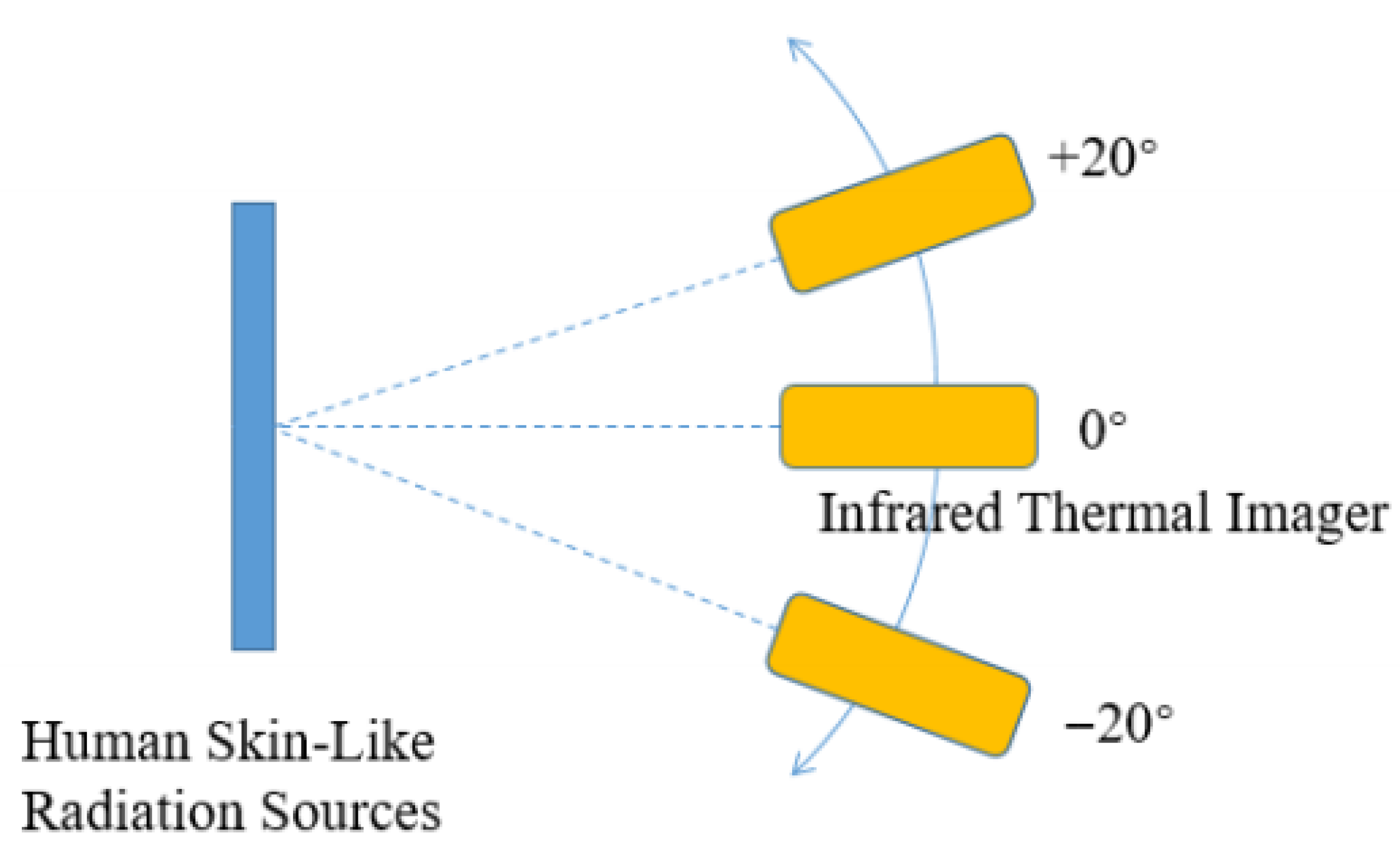
To investigate the impact of different distributions of devices and thermal imaging cameras in practical application spaces on measurement results, we conducted a comparative analysis based on varying angles and distances between the two devices, as illustrated in Figure 9.
By keeping the human skin-like radiation source fixed and changing the angle of the thermal imaging camera, the angles between the two devices were set to −45°, −25°, −5°, 5°, 25°, and 45°. The results are shown in Figure 10.
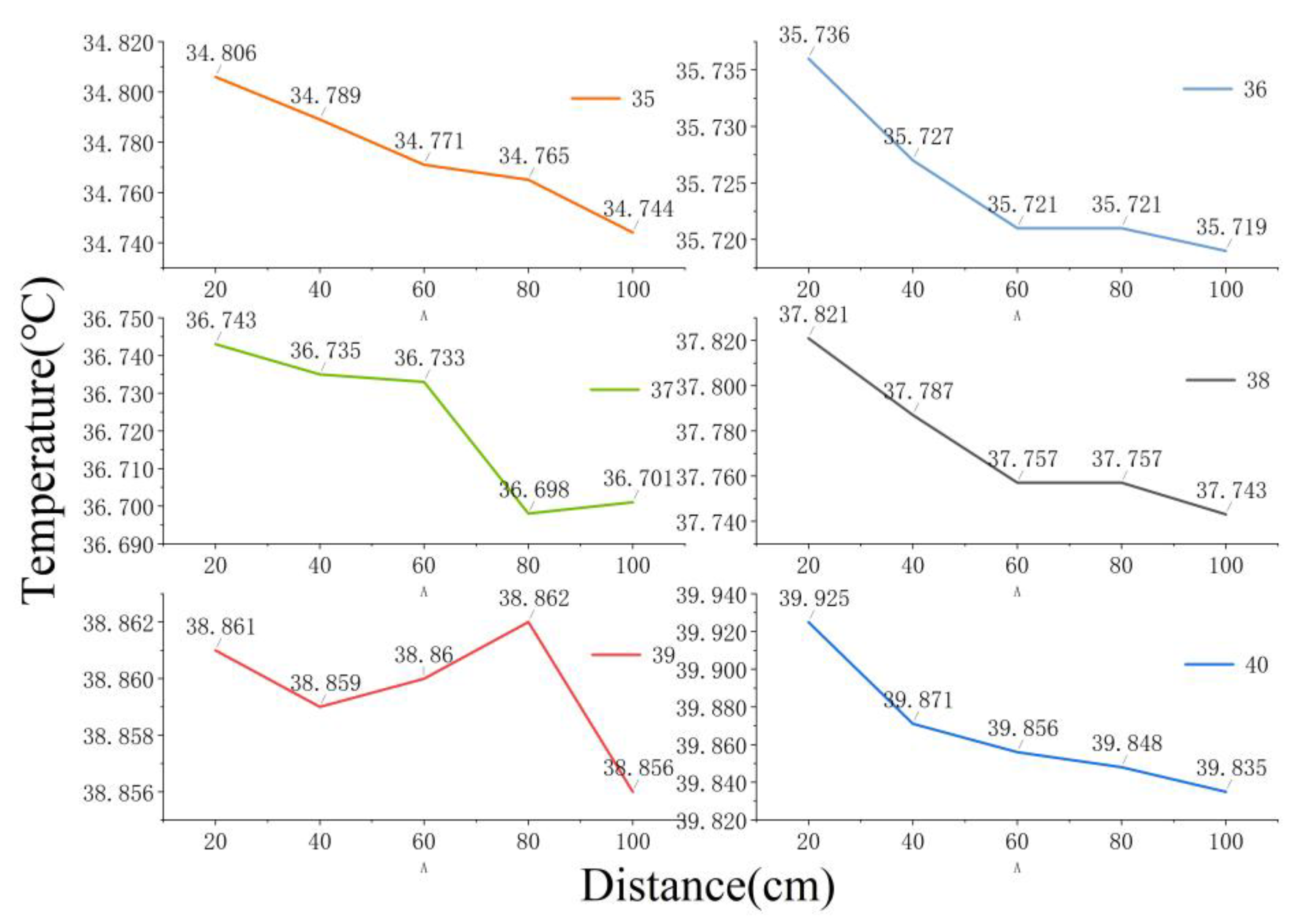
Following the same procedure, by keeping the human skin-like radiation source fixed and varying the distance between the thermal imaging camera and the human skin-like radiation source, the distances were set to 20 cm, 40 cm, 60 cm, and 80 cm. The results are shown in Figure 11.
As can be seen from Figure 11, there is a slight decrease in temperature measurements as the distance increases. At closer distances (less than 1 m), the difference between different distances for measurement results is small; all of them are less than 0.1 °C. So, if we only consider the radiation temperature measurement at a distance near 20 cm, the error caused by the distance is negligible.
As can be seen from Figure 10, there is little difference in temperature variation within the observation angle range of ±25°, which is less than 0.1 °C. Ideally, if the sample surface approximates Lambertian properties, the observations should vary little from angle to angle. In fact, different samples showed slightly different trends. We believe that this is because surface processing of each material was still unable to achieve ideal uniformity. Considering that in our practical application, we generally approximate the normal direction, which is 0°, we think that this change can be ignored.

3.6. Uncertainty

According to the Guide to the Expression of Uncertainty in Measurement promoted by JCGM (Joint Committee for Guides in Metrology), uncertainty measurements are classified into two types: (A) assessed by statistical methods and (B) assessed by other methods. In practice, type A uncertainty pertains to the difference between individual results, while type B uncertainty reflects the precision of each measurement. The total system uncertainty is detailed in Table 3.
The combined standard uncertainty u C was calculated as the root sum of squares of all individual standard uncertainty components. The expanded uncertainty (U) was obtained by multiplying u C by a coverage factor of k = 2, providing a confidence level of approximately 95%. For example, at 37 °C, the reported temperature is 37.00 °C ± 0.10 °C.

4. Practical Applications

In order to reduce the influence of other heat sources on the measurement results, we carefully arranged the heat source distribution in the field of view. The heat source of the same temperature was in the same plane perpendicular to the direction of the thermal camera field of view, so as to reduce the mutual influence between them.
To validate the accuracy of the human skin-like radiation source in calibrating thermal imaging cameras for practical applications, its results were compared with those obtained from thermal imaging cameras calibrated with a blackbody and a platinum resistance temperature sensor (PT100).
Initially, the actual temperature of a human forehead was measured to be 33.85 °C using a platinum resistor (during the process of measuring forehead temperature using a platinum resistance thermometer, a layer of highly thermally conductive metal tape was adhered to the forehead at the measurement location, followed by attaching the platinum resistance thermometer to the corresponding position). Given the platinum resistor’s high sensitivity to temperature and its connection using the four-wire method to eliminate resistance errors in the connection lines, the measured data was considered more accurate. Therefore, we consider the temperature measured by the platinum resistor as the actual temperature of the human body.
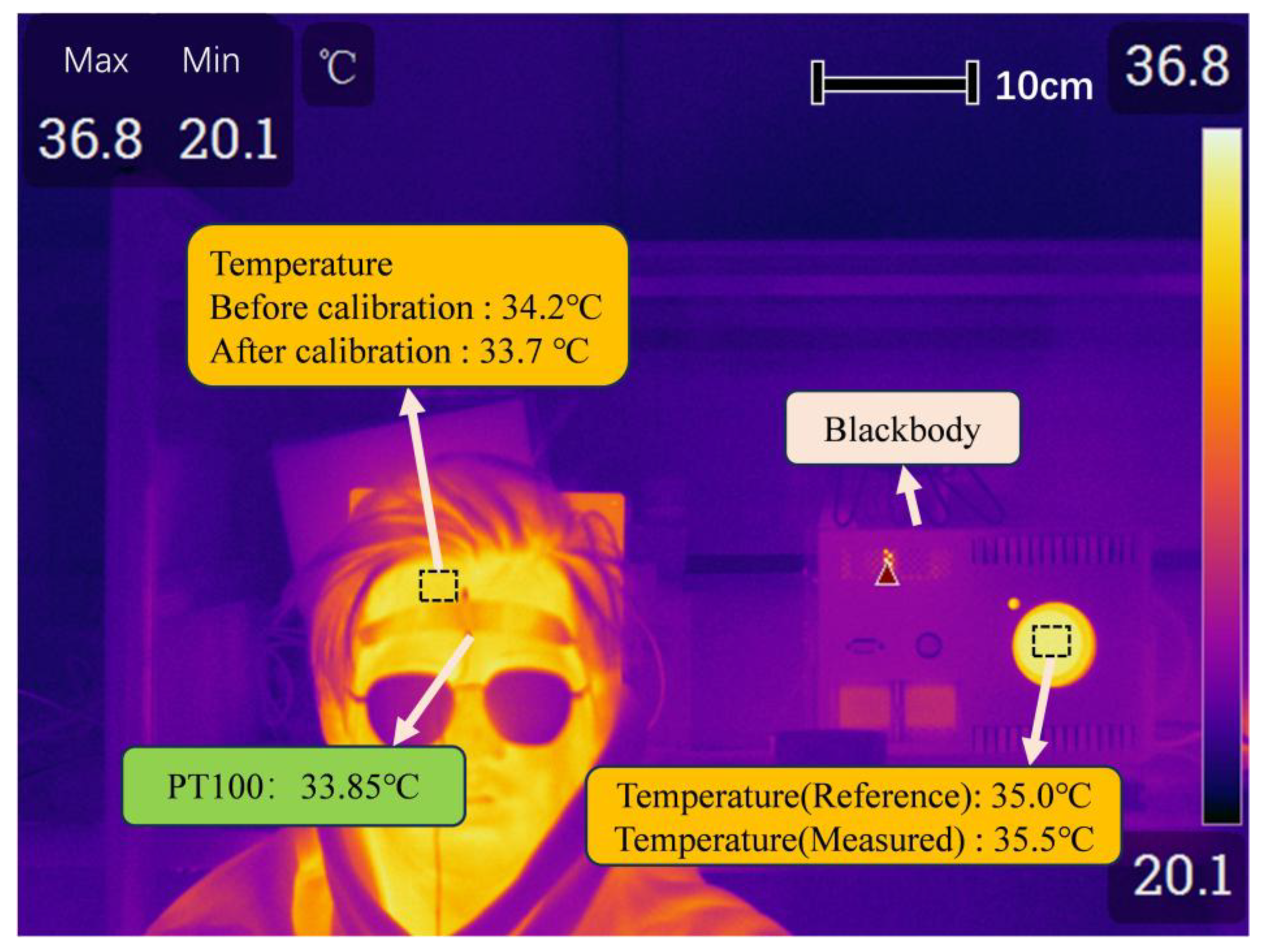
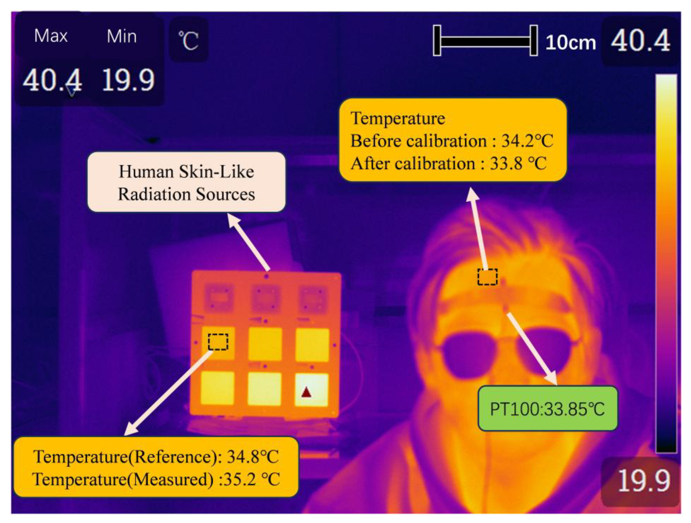
Subsequently, the temperature of Isotech’s Hyperion R Portable 982 blackbody furnace was set to 35 °C, with an emissivity of 0.995. Using the thermal imaging camera (with the target emissivity set to 0.98 and ambient temperature set to 20 °C; the detector pixels were 384 × 288, the field of view angle was 17.1° × 12.8°, and the pixel size was 0.00078 m × 0.00078 m), the temperatures of the blackbody cavity and the human forehead were measured. The temperature of the blackbody cavity at 35 °C was measured to be 35.5 °C, and the temperature of the human forehead was 34.2 °C, as shown in Figure 12. Therefore, the temperature of the human forehead measured by the calibrated thermal imaging camera after blackbody furnace calibration was 33.7 °C, with a difference of 0.15 °C from the actual temperature of the human forehead.
As the human body temperature measured by the platinum resistor was 33.85 °C, and the emissivity of the standard plate of the human skin-like radiation source at 35 °C was closest to this value, the thermal imaging camera was used to measure the temperatures of the 35 °C standard plate and the human forehead. Before calibration, the measured result by the thermal imaging camera was 34.2 °C (denoted as A) and 35.2 °C (denoted as B), respectively. Since the temperature of the 35 °C standard radiation source measured in Section 3.2 was 34.8 °C (suppose C), the temperature of the human forehead measured by the calibrated thermal imaging camera after human skin-like radiation source calibration was A−(B−C) = 33.81 °C (to maintain accuracy, online real-time calibration is required. Moreover, since the human skin-like radiation source and the measured object were in the same environment, the method could offset the influence of environmental factors on the measurement results), as shown in Figure 13. This value differed by 0.04 °C from the actual temperature of the human forehead.
The calibration error for the traditional blackbody method was 0.15 °C, while the error for the human skin-like radiation source method was 0.04 °C, resulting in an improvement in measurement accuracy of 0.11 °C. This fulfills the requirements for high-precision radiation temperature measurement of the human body.
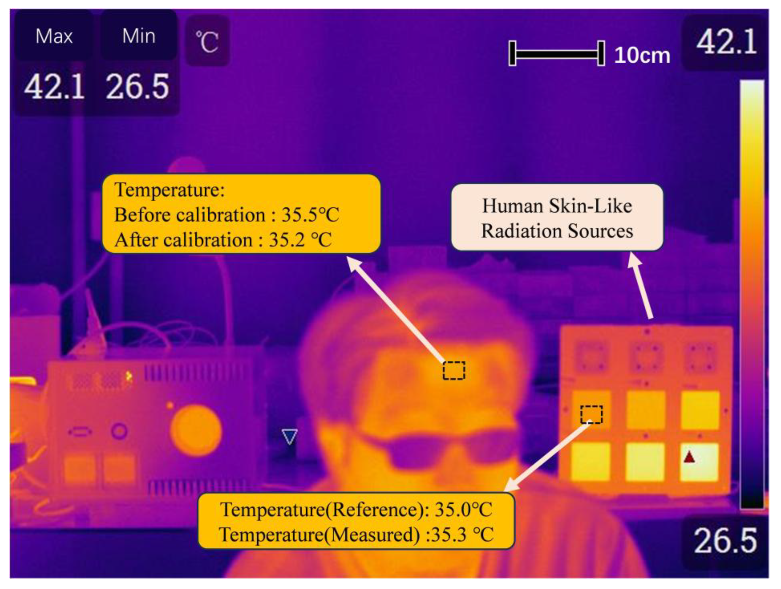
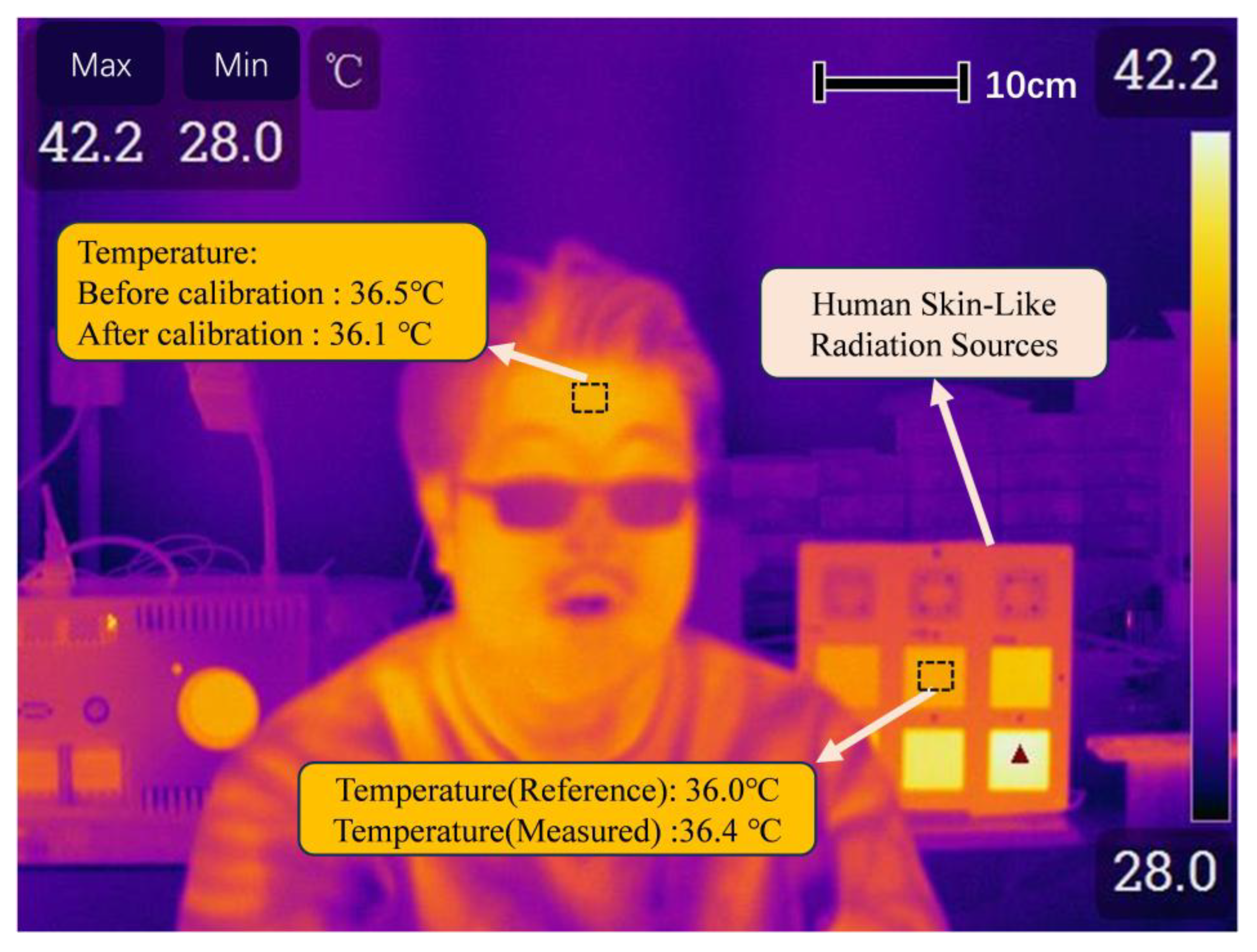
To demonstrate the application of this method and provide an initial assessment of the method’s robustness, the calibration and measurement procedures were repeated across multiple temperature setpoints (35 °C and 36 °C sources) and on two human subjects with different baseline forehead temperatures (approximately 33.85 °C and 35.2 °C). For each condition, the measurement was repeated five times to ensure consistency. The consistent improvement in accuracy observed across these different scenarios suggests the method’s potential generalizability, although validation on a larger scale is acknowledged as necessary. The results are shown in Figure 14 and Figure 15.
When measuring the human forehead temperature with an uncalibrated thermal imager, it was found that the temperature was around 35 °C. Therefore, the 35 °C human skin-like radiation source was used to calibrate the thermal imager. After calibration, the thermal imager measured the human forehead temperature as 35.2 °C.
Continuing the aforementioned operation, the uncalibrated thermal imager recorded data that was closer to 36 °C. Therefore, a 36 °C human skin-like radiation source was used to calibrate the thermal imager. After calibration, the thermal imager measured the human forehead temperature as 36.1 °C.

5. Conclusions

In order to accurately screen individuals with fever, achieve precise measurement of human skin temperature, and improve the temperature measurement accuracy of infrared thermal imagers, a gradient standard source simulating human skin radiation is proposed for calibrating infrared thermal imagers. By comparing and analyzing the temperature measurement results with traditional blackbody calibration methods and platinum resistance temperature sensors, it was found that the temperature measurement accuracy was improved (less than 0.1 °C), proving the effectiveness of the proposed method. It provides a method for high-precision human body radiation temperature measurement in the future.
Although the principle of this new method is clear and the operation is feasible, there are still some unavoidable problems. The first problem is that the standard uncertainty of the skin-like gradient radiation source can only reach the level of 0.051 °C to 0.219 °C, which is more than that of the blackbody. Moreover, surface uniformity of the human skin-like gradient standard source is an important problem that needs to be further improved. The second problem is that the measurement repeatability level of the current thermal imager is only around 0.1 °C. Although the laboratory measurement can obtain better data through averaging a large number of measurements, the measurement speed still faces challenges in practical application, especially in crowded places.
To conclusively demonstrate the reproducibility and generalization of the results, future work will focus on the following:
Multi-device validation: Testing the calibration method with a variety of thermal imager models from different manufacturers, featuring different detector types, resolutions, and internal calibration algorithms.
Large-scale testing: Conducting a comprehensive study with a larger cohort of human subjects to statistically quantify the accuracy improvement under diverse physiological and environmental conditions.
Long-term stability assessment: Evaluating the long-term stability and durability of the skin-like radiation source itself.
Despite these limitations, the significant and consistent reduction in measurement error achieved under controlled conditions provides strong preliminary evidence for the effectiveness of the proposed method and warrants further investigation.

Author Contributions

Methodology, G.F.; software, Z.Z.; formal analysis, T.L. and Z.H.; investigation, Z.Z.; writing—original draft preparation, Z.Z.; writing—review and editing, G.F. and X.C. All authors have read and agreed to the published version of the manuscript.

Funding

This research work was supported by the National Key R&D Projects, China (2021YFF0600204), Natural Science Foundation of Beijing Municipality (8202015), the Excellent Researcher Award Program from Ministry of Beijing (No. 2017000020124G005), the Fundamental Research Funds for Beijing Universities (No. X18253), Open Research Fund Program of Beijing Advanced Innovation Center for Future Urban Design (Grant No. UDC2017033022), and Open Research Fund Program of Beijing Key Laboratory of Performance Guarantee on Urban Rail Transit Vehicles (No. 06080915001).

Data Availability Statement

The raw data are available on request to the authors.

Acknowledgments

The authors would like to thank Haiyong Gan for providing research topics. The authors are grateful to Jinhui Wang and Chenyu Bai for the help with the temperature calibration in this paper.

Conflicts of Interest

The authors declare no conflicts of interest.

References

  1. Gao, S.; Zhang, X.; Chen, L.; Cui, Y.; Jiang, J.; Zhang, Z.; Yu, P.; Wang, C. Review: Radiation temperature measurement methods for engine turbine blades and environment influence. Infrared Phys. Technol. 2022, 123, 104204. [Google Scholar] [CrossRef]
  2. Tian, X.Y.; Fang, L.; Liu, W.W. The influencing factors and an error correction method of the use of infrared thermography in human facial skin temperature. Build. Environ. 2023, 244, 110736. [Google Scholar] [CrossRef]
  3. Lundstrom, H.; Mattsson, M. Radiation influence on indoor air temperature sensors: Experimental evaluation of measurement errors and improvement methods. Exp. Therm. Fluid Sci. 2020, 115, 110082. [Google Scholar] [CrossRef]
  4. Shin, D.H.; Kim, M.; Kim, J.S.; Lee, B.J.; Lee, J. Precise infrared thermometry with considering background radiation for gas turbine air cooling application. Int. J. Therm. Sci. 2020, 158, 106534. [Google Scholar] [CrossRef]
  5. Tung, C.L.; Wang, C.H.; Su, Y.L. Real-time Face Mask-Wearing Detection and Temperature Measurement based on a Deep Learning Model. J. Imaging Sci. Technol. 2022, 66, 010403. [Google Scholar] [CrossRef]
  6. Ghassemi, P.; Pfefer, T.J.; Casamento, J.P.; Simpson, R.; Wang, Q. Best practices for standardized performance testing of infrared thermographs intended for fever screening. PLoS ONE 2018, 13, e0203302. [Google Scholar] [CrossRef]
  7. Li, L.; Dai, C.H.; Wu, Z.F.; Wang, Y.F. Laboratory Calibration and Temperature Research of Spectral Radiometric Instruments. Spectrosc. Spectr. Anal. 2019, 39, 1965–1969. [Google Scholar]
  8. Mlacnik, V.; Pusnik, I. A Traceable Spectral Radiation Model of Radiation Thermometry. Appl. Sci. 2023, 13, 4973. [Google Scholar] [CrossRef]
  9. Rastogi, V.; Kumar, V.; Dubey, S.K.; Khan, G.S.; Shakher, C. Noncontact temperature measurement of human hand skin using volume phase holographic optical element based digital holographic interferometer. Opt. Lasers Eng. 2022, 151, 106886. [Google Scholar] [CrossRef]
  10. Rastogi, V.; Agarwal, S.; Kumar, V.; Shakher, C. Holographic optical element based digital holographic interferometer for the study of macro flames, micro flames and their temperature instability. Opt. Lasers Eng. 2019, 122, 29–36. [Google Scholar] [CrossRef]
  11. Kumar, V.; Rastogi, V.; Agarwal, S.; Shakher, C. Investigation of temperature profile and temperature stability of micro diffusion flame under the influence of magnetic field by use of a holo-shear lens-based interferometer. Opt. Eng. 2020, 59, 064107. [Google Scholar] [CrossRef]
  12. González-Chávez, O.; Cárdenas-Garcia, D.; Karaman, S.; Lizarraga, M.; Salas, J. Radiometric Calibration of Digital Counts of Infrared Thermal Cameras. IEEE Trans. Instrum. Meas. 2019, 68, 4387–4399. [Google Scholar] [CrossRef]
  13. Zhang, F.C.; Sun, X.G.; Xing, J. Research of Multi Points and Multi Spectral Calibration in Two-Dimensional Temperature Field Reconstruction Based on CCD. Spectrosc. Spectr. Anal. 2017, 37, 2283–2287. [Google Scholar]
  14. Cui, S.L.; Xing, J. Research on Calibration Method of Infrared Temperature Measurement System Near Room Temperature Field. Front. Phys. 2022, 9, 786443. [Google Scholar] [CrossRef]
  15. Cui, S.L.; Sun, X.G.; Wang, Y.; Wei, Y.; Xing, J. A data processing method on infrared temperature measurement of non-lambert body in a non-uniform environment. Infrared Phys. Technol. 2022, 123, 104187. [Google Scholar] [CrossRef]
  16. Zhang, Y.-C.; Chen, Y.-M.; Luo, C. A method for improving temperature measurement precision on the uncooled infrared thermal imager. Measurement 2015, 74, 64–69. [Google Scholar] [CrossRef]
  17. Monier, R.; Thumerel, F.; Chapuis, J.; Soulié, F.; Bordreuil, C. Liquid metals surface temperature fields measurements with a two-colour pyrometer. Measurement 2017, 101, 72–80. [Google Scholar] [CrossRef]
  18. Gao, S.; Zhao, C.H.; Chen, L.W.; Jiang, J.; Yu, P.; Zhang, Z.; Wang, C. Error analysis and reflection correction for radiation temperature measurements at high background temperatures. Meas. Sci. Technol. 2021, 32, 055003. [Google Scholar] [CrossRef]
  19. Martinez, I.; Otamendi, U.; Olaizola, I.G.; Solsona, R.; Maiza, M.; Viles, E.; Fernandez, A.; Arzua, I. A novel method for error analysis in radiation thermometry with application to industrial furnaces. Measurement 2022, 190, 110646. [Google Scholar] [CrossRef]
  20. Qi, P.Y.; Wang, G.; Gao, Z.; Liu, X.; Liu, W. Measurements of Temperature Distribution for High Temperature Steel Plates Based on Digital Image Correlation. Materials 2019, 12, 3322. [Google Scholar] [CrossRef]
  21. Xing, W.H.; Ju, X.P.; Yan, C.X.; Yang, B.; Zhang, J. Self-correction of alignment errors and retardations for a channeled spectropolarimeter. Appl. Opt. 2018, 57, 7857–7864. [Google Scholar] [CrossRef]
  22. Hisaka, M. Noncontact localized internal infrared radiation measurement using an infrared point detector. Opt. Rev. 2018, 25, 397–402. [Google Scholar] [CrossRef]
  23. Poissenot-Arrigoni, C.; Marcon, B.; Rossi, F.; Fromentin, G. Fast and easy radiometric calibration method integration time insensitive for infrared thermography. Infrared Phys. Technol. 2023, 133, 104741. [Google Scholar] [CrossRef]
  24. Lisiecka, E. Reduction of the impact of emissivity on high temperature measurements in non-contact thermometric devices. Opt. Appl. 2017, 47, 373–381. [Google Scholar]
  25. Owda, A.Y.; Salmon, N.; Harmer, S.W.; Shylo, S.; Bowring, N.J.; Rezgui, N.D.; Shah, M. Millimeter-wave emissivity as a metric for the non-contact diagnosis of human skin conditions. Bioelectromagnetics 2017, 38, 559–569. [Google Scholar] [CrossRef]
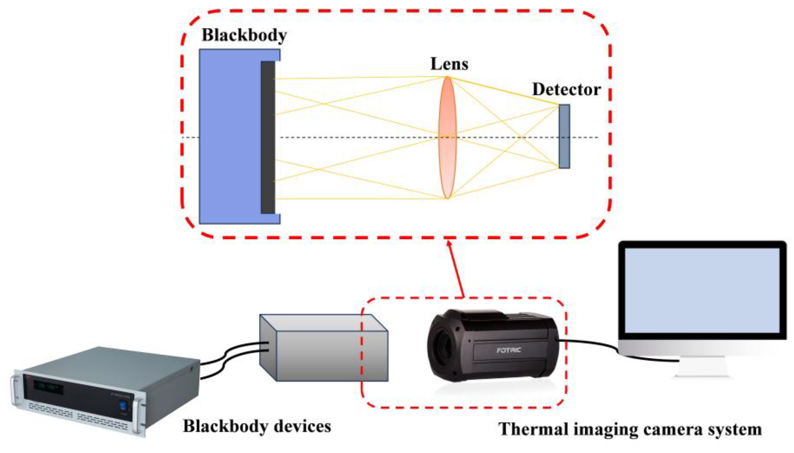
Figure 1. Schematic representation of blackbody calibration.
Thermo 05 00038 g001
Figure 2. Calibration process schematic for human skin-like radiation sources and composition of human skin-like radiation sources.
Thermo 05 00038 g002
Figure 3. Reflectance measurement device for human skin.
Thermo 05 00038 g003
Figure 4. Comparison of emissivity between human skin-like radiation source and human forehead.
Thermo 05 00038 g004
Figure 5. Experimental setup for accuracy testing.
Thermo 05 00038 g005
Figure 6. Stability of human skin-like radiation sources.
Thermo 05 00038 g006
Figure 7. Schematic diagram of uniformity measurement for human skin-like radiation source.
Thermo 05 00038 g007
Figure 8. Repeatability of radiation source uniformity measurements for human skin-like radiation source.
Thermo 05 00038 g008
Figure 9. Schematic diagram of measurements at different angles.
Thermo 05 00038 g009
Figure 10. Temperature change in each standard plate at different observation angle.
Thermo 05 00038 g010
Figure 11. Temperature change in each human skin-like radiation source at different distances.
Thermo 05 00038 g011
Figure 12. Calibration of thermal imaging camera using blackbody furnace.
Thermo 05 00038 g012
Figure 13. Calibration of thermal imaging camera using human skin-like radiation source.
Thermo 05 00038 g013
Figure 14. Calibration of thermal imaging camera using 35 °C human skin-like radiation source.
Thermo 05 00038 g014
Figure 15. Calibration of thermal imaging camera using 36 °C human skin-like radiation source.
Thermo 05 00038 g015
Table 1. Accuracy of human skin-like radiation sources (before being calibrated).
Area IDSetting/°CMeasured/°CMeasured-Setting/°C
1#35.0034.81−0.19
2#36.0035.71−0.29
3#37.0036.50−0.50
4#38.0037.76−0.24
5#39.0038.84−0.16
6#40.0039.73−0.27
Table 2. Results of source uniformity measurements of human skin-like radiation source.
NoSetting (°C)Position 5 (°C)Position 1Position 2Position 3Position 4
Position X-Position 5 (°C)
135.0034.970.050.08−0.21−0.16
236.0036.000.020.03−0.02−0.03
337.0037.000.010.12−0.04−0.10
438.0037.97−0.060.14−0.17−0.29
539.0039.04−0.040.05−0.11−0.11
640.0039.990.100.00−0.32−0.31
Table 3. Total uncertainty value for human skin-like gradient radiation sources.
Source of Uncertainty35 °C36 °C37 °C38 °C39 °C40 °C
Temperature accuracy u 1 0.030<0.010<0.0100.0300.0400.010
Temperature stability u 2 0.0180.0200.0240.0170.0170.022
Surface uniformity u 3 0.1460.0290.0930.1830.0760.215
Uncertainty of radiation thermometers u 4 0.0350.0350.0350.0350.0350.035
Difference between standard plate emissivity and human forehead skin emissivity u 5 0.0100.0100.0100.0100.0100.010
Uncertainty of spatial distribution u 6 0.0400.0450.0520.0430.0500.052
Other imponderables u 7 0.0050.0050.0050.0050.0050.005
Total standard uncertainty u C = u 1 2 + u 2 2 + u 3 2 + u 4 2 + u 5 2 + u 6 2 + u 7 2 0.1600.0510.1030.1900.0950.219
Disclaimer/Publisher’s Note: The statements, opinions and data contained in all publications are solely those of the individual author(s) and contributor(s) and not of MDPI and/or the editor(s). MDPI and/or the editor(s) disclaim responsibility for any injury to people or property resulting from any ideas, methods, instructions or products referred to in the content.