Article

Tracking Photoinduced Charge Redistribution in a Cu(I) Diimine Donor–Bridge–Acceptor System with Time-Resolved Infrared Spectroscopy

1
Chemical Sciences and Engineering Division, Argonne National Laboratory, Lemont, IL 60439, USA
2
Department of Chemistry, University of Washington, Seattle, WA 98195, USA
3
Department of Chemistry, Northwestern University, Evanston, IL 60208, USA
*
Authors to whom correspondence should be addressed.
Photochem 2025, 5(2), 16; https://doi.org/10.3390/photochem5020016
Submission received: 18 May 2025 / Revised: 9 June 2025 / Accepted: 13 June 2025 / Published: 19 June 2025
(This article belongs to the Special Issue Feature Papers in Photochemistry, 3rd Edition)

Abstract

:
Understanding electron density migration along excited-state pathways in photochemical systems is critical for optimizing solar energy conversion processes. In this study, we investigate photoinduced electron transfer (PET) in a covalently linked donor–bridge–acceptor (D-B-A) system, where [Cu(I)-bis(1,10-phenanthroline)]+ acts as an electron donor, and anthraquinone, tethered to one of the phenanthroline ligands via a vibrationally active ethyne bridge, behaves as an electron acceptor. Visible transient absorption spectroscopy revealed the dynamic processes occurring in the excited state, including PET to the acceptor species. This was indicated by the spectral features of the anthraquinone radical anion that appeared on a timescale of 30 ps in polar solvents. Time-resolved infrared (TRIR) spectroscopy of the alkyne vibration (CC stretch) of the ethyne bridge provided insight into electronic structural changes in the metal-to-ligand charge transfer (MLCT) state and along the PET reaction coordinate. The observed spectral shift and enhanced transition dipole moment of the CC stretch demonstrated that there was already partial delocalization to the anthraquinone acceptor following MLCT excitation, verified by DFT calculations. An additional excited-state TRIR signal unrelated to the vibrational mode highlighted delocalization between the phenanthroline ligands in the MLCT state. This signal decayed and the CC stretch narrowed and shifted towards the ground-state frequency following PET, indicating a degree of localization onto the acceptor species. This study experimentally elucidates charge redistribution during PET in a Cu(I) diimine D-B-A system, yielding important information on the ligand design for optimizing PET reactions.

Graphical Abstract

1. Introduction

With significant technological advancement comes an even greater demand for energy. For example, current initiatives in quantum computing [1] and data centers for cloud computing, storage, and artificial intelligence have significant energy demands. Thus, alternative renewable energy sources, such as solar energy, must be further developed for continuous and sustainable technological growth, while mitigating the economic burdens of these demands. Transition metal complexes are commonly utilized in solar energy conversion processes, such as photovoltaics and photocatalysis. Ruthenium-based photosensitizers, like [Ru(II)-tris(2,2′-bipyridine)]2+ and its molecular derivatives, are a standard in dye-sensitized solar cells because of their high molar absorptivity in the visible region, and long excited-state lifetimes that enable the charge separation processes vital for solar energy conversion [2]. However, there has been a long-standing search for an adequate replacement due to the rarity and, therefore, cost of ruthenium and other relevant precious metals. This has driven research on earth-abundant, first-row transition metals and the photophysics of their complexes [3,4,5].
The primary challenge with first-row transition metals are their low energy metal-centered electronic states that rapidly quench the excited state before useful chemistry can occur [3,4]. With a full d10 electronic configuration, Cu(I) complexes have no metal-centered states and circumvent this problem. Four-coordinate Cu(I) diimine complexes are suitable candidates for solar energy applications due to their intense metal-to-ligand charge transfer (MLCT) transitions and their strong reducing potential following excitation [6,7,8]. Early experiments showed that Cu(I) complexes can act as donors in intermolecular photoinduced electron transfer (PET) to viologen-based acceptors [6,9,10,11] and, later, in intramolecular PET to methyl viologen and fullerene acceptors [7,12,13]. Further studies showed that Cu(I) complexes were able to sensitize semiconductor materials and generate a photocurrent, demonstrating their feasibility for solar energy conversion [14,15,16].
Cu(I) diimine complexes also exhibit structure-dependent photophysics that may be leveraged for optimal photochemistry. Specifically, the complex undergoes a pseudo Jahn–Teller flattening distortion after transiently oxidizing the Cu(I) species to an open-shell, d9 configuration via photoexcitation [17,18,19]. This distortion leads to a change from pseudo-D2d to D2 symmetry that decreases spin–orbit coupling and therefore reduces the rate of intersystem crossing (ISC) [20]. Additionally, it increases the susceptibility of the Cu center to coordinating solvents that can stabilize the MLCT state, reducing the excited-state lifetime [21,22,23]. Significant progress has been made towards the molecular control of the photophysics of [Cu(I)-bis(1,10-phenanthroline)]+ (Cu-bisphen) complexes [18,24,25]. The use of steric encumbrance to limit structural distortions as well as exciplex formation with coordinating solvents has notably enhanced the excited-state lifetime [18,24,25]. The vast knowledge of phenanthroline chemistry [26] has also allowed for investigations of inductive effects [27,28] and extended π-conjugation [29,30], which can impact the photophysics of these complexes. Moreover, the recent HETPHEN synthetic scheme has enabled the formation of heteroleptic Cu–bisphen complexes, allowing for new asymmetric macromolecular systems, including donor–acceptor (D-A) and donor–bridge–acceptor (D-B-A) systems, which can be utilized to control the directionality of electron migration in the MLCT states [27,30,31,32,33].
Computational reports on the electronic structure of the MLCT state in Cu–bisphen complexes show that the charge is delocalized between the phenanthroline ligands [27,30]. In heteroleptic systems, the charge can be directed to a specific ligand through structural modifications, such as the addition of electron-donating or -withdrawing groups [27]. In a heteroleptic Cu–bisphen D-A system, the charge distribution following excitation and the location of the acceptor were shown to have significant implications on the observed PET rate [34]. When the MLCT transition preferentially directs charge to the acceptor-bearing ligand, faster PET can occur by avoiding convoluted pathways. Furthermore, for a fused dipyridophenazine/anthraquinone acceptor ligand, evidence of fast PET to the anthraquinone (Anq) following excitation into a partially delocalized MLCT state was observed through visible transient absorption spectroscopy and resonance Raman spectroscopy [35]. Specifically, resonance Raman spectroscopy observed the amplified vibrational modes of Anq, implying that the hole orbital of the MLCT transition has some orbital contribution from Anq due to conjugation with the bridging ligand. These studies highlight that deeper insight into electronic localization in the excited-state pathway is needed to fully understand PET in Cu–bisphen D-A systems.
Here, we study PET in a D-B-A system, where the donor is a heteroleptic Cu–bisphen complex and the acceptor is Anq tethered to the phenanthroline ligand using an ethynyl bridge moiety (Scheme 1). Specifically, we aim to investigate the charge redistribution in the excited state using both visible transient absorption (TA) spectroscopy and time-resolved infrared spectroscopy (TRIR) of the ethyne stretch. With recent interest in the vibrational control of PET through IR-active bridging moieties [36,37,38,39], we also aim to characterize the feasibility of this system for examining this phenomenon. TRIR experiments give electronic structural information from the spectral shifts in vibrational modes that arise with the new equilibrium nuclear geometry. They provide time-resolved information on the charge distribution across the ligands of the Cu(I) complex, which complements resonance Raman spectroscopy, which only yields information on the electronic character of the optically excited state. A comparison of the TA and TRIR experimental responses of Cu-CC and Cu-CC-Anq, with validation from density functional theory (DFT) calculations, clearly elucidates the time-dependent electronic structural changes that take place in these systems following photoexcitation.

2. Experimental Methods

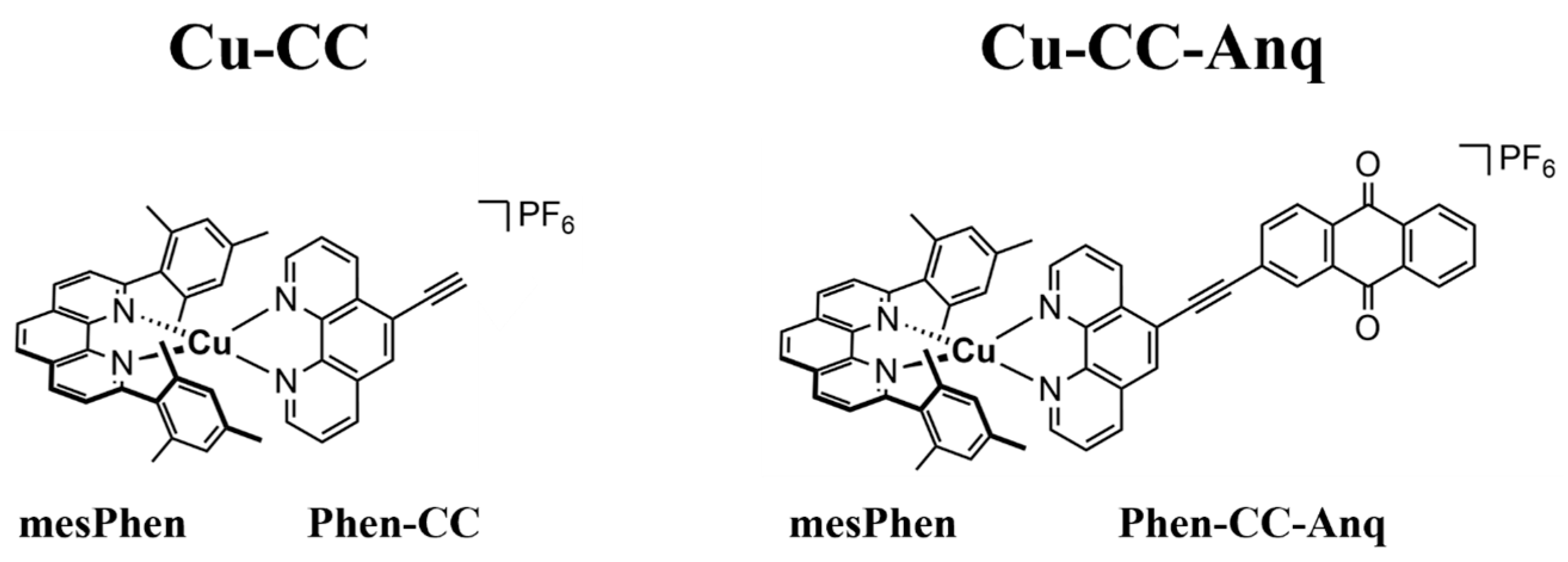
2.1. Sample Preparation

The synthesis of the Phen-CC-Anq ligand was performed using Sonagashira coupling of the previously reported 5-ethyne-1,10-phenanthroline and 2-bromoanthraquinone [40]. Both Cu-CC and Cu-CC-Anq were prepared using the HETPHEN synthetic approach for pure asymmetrically coordinated Cu–bisphen complexes, originally designed by the Schmittel group [31,32] and used by our group [33,34,41], as well as many others [30,42,43,44], to design various Cu(I) HETPHEN complexes. The full synthetic details of Cu-CC and Cu-CC-Anq are provided in Section S1 of the Supplementary Materials.
Acetonitrile (MeCN), acetonitrile-d3 (MeCN-d3), nitromethane (NM), and dichloromethane (DCM) were used for spectroscopic measurements. All solvents had purity > 99% and were used as received from commercial suppliers (Sigma-Aldrich, St. Louis, MO, USA, and Thermo Scientific, Waltham, MA, USA). All optical and infrared spectroscopic measurements (steady-state and time-resolved) were performed using a Harrick cell with 2 mm CaF2 windows and PTFE spacers of variable thicknesses (Harrick Scientific, Pleasantville, NY, USA). Optical measurements were taken with a pathlength of 950 µm (unless otherwise stated), while infrared measurements varied from 390 to 950 µm, depending on the solvent.

2.2. Spectroscopic Characterization

Steady-state UV/Vis absorption spectra were recorded from 300 to 800 nm (1 nm resolution) using a DU800 spectrophotometer (Beckman Coulter, Brea, CA, USA). FT-IR absorption spectra were recorded from 500 to 6500 cm−1 (1 cm−1 resolution) using a Thermo Scientific Nicolet 6700 FTIR spectrometer. Femtosecond transient absorption spectroscopy measurements were performed using a Pharos Yb:KGW regenerative amplifier (Light Conversion, Vilnius, Lithuania; 1030 nm, 10 W at 10 kHz) that was split to generate the visible pump, visible probe, and mid-infrared (mid-IR) probe pulses. A second harmonic generation crystal (2 mm thick BBO, Newlight Photonics, Toronto, ON, Canada) was used to generate a visible pump pulse (515 nm) with a pulse duration of ~150 fs. A 3 mm thick YAG crystal was used to generate a white light continuum spanning 520–850 nm. An Orpheus-One-HP optical parametric amplifier (Light Conversion) was used to generate a mid-IR probe pulse centered at 2160 cm−1, with FWHM of ~125 cm−1. The polarization of the visible pump pulse was set at magic angle (54.7°) to both the mid-IR and visible probe pulses and had a pulse energy of ~300 nJ. The delay between pump and probe pulses was scanned using an XMS160-S mechanical delay stage (Newport, Irvine, CA, USA). The time window of the experiments was limited to 500 ps. The visible probe was detected using a Kymera 328i spectrometer (Oxford Instruments, Belfast, UK) and a Zyla-5.5 sCMOS camera (Oxford Instruments). The mid-IR probe was detected using an iHR320 spectrometer (Horiba, Kyoto, Japan) and a 32 × 2 pixel mercury cadmium telluride array detector (Infrared Associates, Stuart, FL, USA) with a frequency resolution of ~2 cm−1. Every 2 pixels were averaged to reduce the pixel to pixel noise in the observed spectra.

2.3. Computational Methods

All electronic structure calculations were performed using a developmental version of Gaussian quantum chemistry software package (Developmental Version Revision J.14+) [45]. Each density function theory (DFT) calculation used the PBE0 functional [46], and Grimme’s dispersion with Becke-Johnson damping (GD3BJ) was applied [47]. The accuracy of numerical integrations was at the ultrafine grid (99,590) level. The def2TZVP basis set was chosen to describe the Cu and N atoms, and the def2SVP basis set was chosen for all other atoms [48]. The polarizable continuum model (PCM) was included to simulate the effects of NM solvent [49]. The ground-state geometries of Cu-CC and Cu-CC-Anq were optimized using restricted Kohn–Sham (RKS) DFT.
The excited-state energies of Cu-CC and Cu-CC-Anq were calculated using linear response time-dependent DFT (TDDFT). Natural transition orbitals (NTOs) were visualized using VMD software (Version 1.9.4a53) [50]. TDDFT was also used to optimize excited-state structures from the S0 reference state using the same dispersion and solvent corrections. Spin–orbit (SO) coupling matrix elements were computed within the TDDFT using the one-electron Breit–Pauli Hamiltonian alongside the screened nuclear spin–orbit approximation to capture two-electron SO effects [51,52,53].

3. Results and Discussion

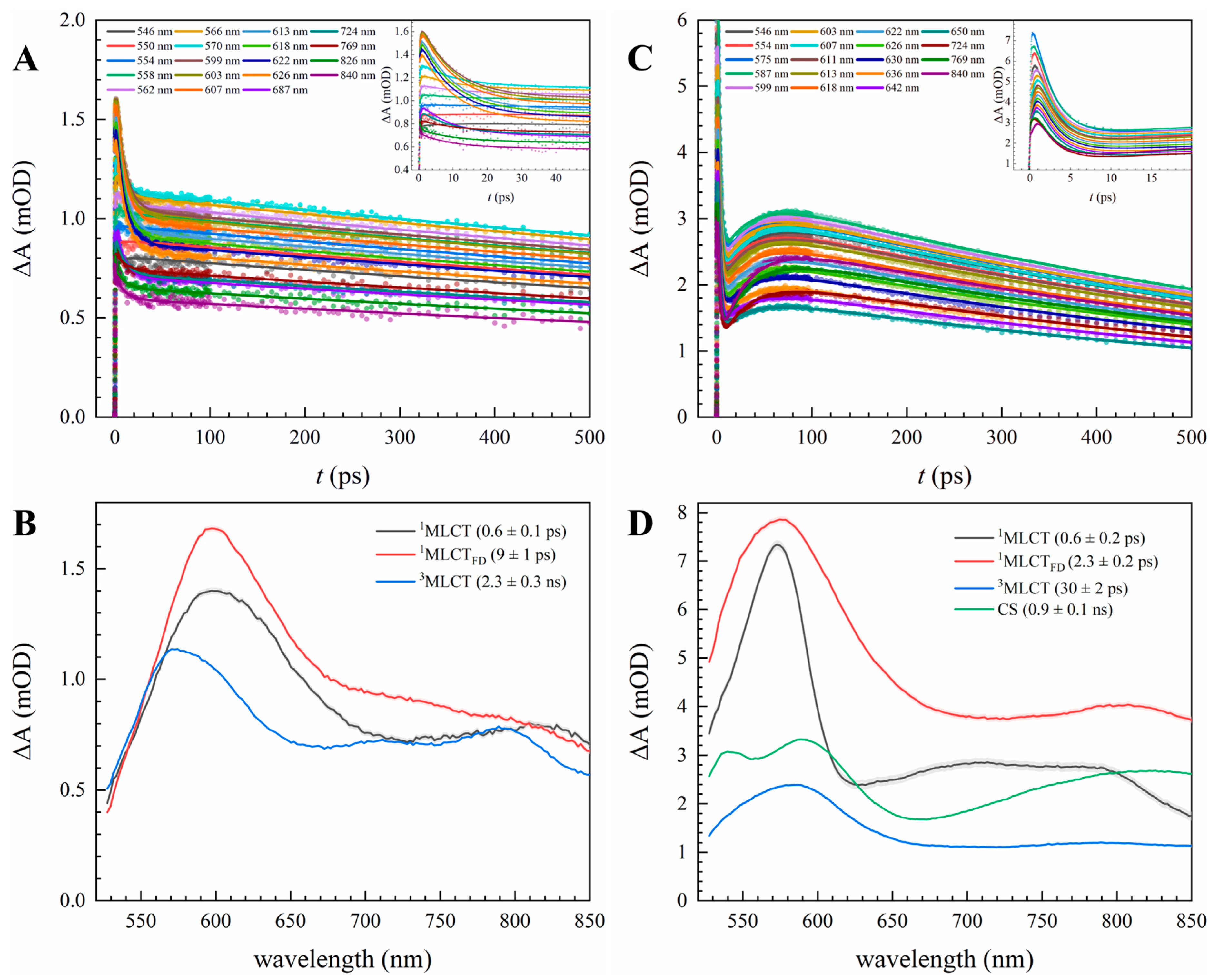
3.1. Optical Characterization of Excited State and PET Dynamics

The experimental UV/Vis absorption spectra of Cu-CC and Cu-CC-Anq in NM are shown in Figure 1A. The simulated spectra from the TDDFT calculations, shown in Figure 1B, match well with the experimental spectra, barring a slight red shift in two absorption features present in Cu-CC-Anq around 300–400 nm. The natural transition orbitals (NTOs) of these transitions, shown in Figure S6, indicate that they are localized on the Phen-CC-Anq ligand and are unique to Cu-CC-Anq. The lowest energy features of these complexes correspond to MLCT transitions from the Cu center to the phenanthroline ligands. In the experimental spectra, the main MLCT transition is centered at 470 nm and features a low energy shoulder that extends to ~600 nm. The shoulder is a well-known MLCT transition in Cu–bisphen complexes that is symmetry-forbidden but gains oscillator strength when the tetrahedral, ground-state geometry is distorted [18,20]. The MLCT transitions show no differences when comparing the two complexes, implying that the nature of these transitions is unaffected by the addition of Anq. The NTOs corresponding to the MLCT transitions of Cu-CC and Cu-CC-Anq (Figure 1C,D and Figure S6C,D) agree with this observation, with similar hole and particle orbitals for both systems. From the NTOs, the electron is delocalized between the mesPhen and Phen ligands in both systems. We note that there is negligible electron density on the Anq acceptor of the Phen-CC-Anq ligand in both the MLCT transitions. However, there is an additional, lower-energy transition from the TDDFT calculations of Cu-CC-Anq at 591 nm (inset of Figure 1B) with near zero oscillator strength, where the electron density is solely on the Phen-CC-Anq ligand and is extended to Anq (Figure 1E).
The pump wavelength for the TA and TRIR experiments was 515 nm, exciting the transitions depicted in Figure 1C,D for Cu-CC and Cu-CC-Anq. The excited-state kinetics of Cu-CC in NM from the TA experiment are best fit using a three-state sequential model and are shown for characteristic wavelengths in Figure 2A. The spectral features of each state along the excited-state pathway are captured by the resulting species-associated difference spectra (SADS), as shown in Figure 2B. An excited-state absorption (ESA) band is observed at 598 nm with a weaker ESA feature spanning from 650 nm to the near-infrared region (experiment measured to 850 nm). The main ESA band showed an initial increase in absorption with time (shown in inset of Figure 2A), followed by a shift to a lower wavelength. The growth occurs on a timescale of 600 fs, resulting from the pseudo Jahn–Teller flattening distortion (FD) that arises from the transient oxidation of the Cu center [18,19,20]. The angle between the two phenanthroline ligand planes from the optimized electronic structures shows flattening from 81° (near tetrahedral) in the ground state (S0) to 56° in the first excited state (S1). The blue shift in the band occurs on a timescale of ~10 ps, likely resulting from intersystem crossing (ISC) to the 3MLCT state [18,19,23]. The blue shift is also accompanied by the appearance of vibronic progression in the ESA region, which is best seen from 650 to 850 nm in the 3MLCT spectrum (blue spectrum in Figure 2B). The vibronic progression has spacing of ~1500 cm−1, corresponding to the in-plane ring stretching motions of both the mesPhen and Phen ligands from ground-state DFT calculations (Figure S10).
The observed SADS and kinetic time constants of Cu-CC from TA experiments using MeCN and DCM are also reported in Figure S7 and Table S1. While the timescales for FD and ISC hardly change, the excited-state lifetime is significantly solvent-dependent, with Cu-CC returning to the ground state in 0.12 ± 0.01 ns, 2.3 ± 0.3 ns, and 6.9 ± 1.7 ns in MeCN, NM, and DCM, respectively. The lifetime of Cu–bisphen complexes is known to depend on the coordinating ability of the solvent, as well as the solvent accessibility to the metal center [24,25]. Solvent coordination stabilizes the MLCT state and, by the energy gap law [54,55], speeds up ground-state recovery. Unlike DCM, MeCN can coordinate to the Cu center, strongly quenching the excited-state lifetime. While NM and MeCN have similar polarities, NM has weaker coordinating ability, resulting in a less substantial change in lifetime in comparison to MeCN [56]. The reduced coordinating ability of NM likely plays a role in the broadening of the ESA features observed in comparison to DCM and MeCN, as shown in Figure S8. The loose coordination leads to a range of solvent configurations around the Cu center, causing the inhomogeneous broadening of the ESA feature. While MeCN severely quenches the excited-state lifetime of Cu-CC, the addition of the alkyne bridge to the phenanthroline ligand also has a significant effect on the lifetime. Ground-state recovery of the complex without the alkyne group occurs in 770 ps in MeCN [33], marking a ~6-fold reduction in the lifetime (770 ps vs. 120 ps) with the addition of the alkyne on the Phen ligand. Barring the unlikely first-coordination effects that result from the addition of the alkyne (out of the scope of this work), the significant reduction in the lifetime possibly stems from further electronic coupling to lower energy states because of increased conjugation.
Compared to the three-state sequential kinetics in Cu-CC, Cu-CC-Anq requires an additional, fourth state to fully capture the excited-state trajectory. The kinetics and the resulting SADS from fitting to a four-state sequential model are shown in Figure 2C,D. The main ESA band is centered around 575 nm, with weaker ESA features from 650 to 850 nm. The timescales and spectral changes in the first two transitions are comparable to what was observed in Cu-CC and are therefore assigned to FD and ISC. Following ISC, new ESA features grow at 540 and ~800 nm on a ~30 ps timescale before recovering to the ground state. These features align with the absorption spectrum of the singly reduced Anq radical anion (Anq•−) [57], indicating that charge separation has occurred, where the electron from the Cu center has localized on Anq. The dependence of the excited-state kinetics on the solvent was again explored for Cu-CC-Anq and provided in Figure S9. These spectral features appear in MeCN at a similar rate, while no comparable features are observed in DCM. This is likely due to the low polarity of DCM compared to MeCN and NM, which does not facilitate charge separation. A comparable observation was made in a similar ruthenium-based D-A species [58].
The excited-state lifetime of Cu-CC-Anq is comparable to that of Cu-CC in DCM (4.9 ± 2.2 ns for D-B-A and 6.9 ± 1.7 ns for D-B), but is a factor of two faster in NM (0.9 ± 0.1 ns), and, inversely, over an order of magnitude slower in MeCN (4.8 ± 0.8 ns). Full kinetics are provided in Table S1. Like Cu-CC, solvent coordination likely occurs when Cu-CC-Anq is excited to the MLCT state, with minimal, weak, and complete coordination observed in DCM, NM, and MeCN, respectively. We note that these complexes decay to the ground state from two different electronic states: Cu-CC decays from the 3MLCT state, while Cu-CC-Anq decays from a charge-separated (CS) state. While the trends in the excited-state lifetime of the 3MLCT state are guided by solvent accessibility and coordinating ability, the CS state of Cu–bisphen D-A systems has shown clear deviation from these trends. Phelan et al. reported a reduction in the excited-state lifetime, with increasing steric bulk around the Cu center in a Cu–dipyridophenazine/Anq D-A system, which is the opposite of what is expected when considering solvent accessibility and the energy gap law [35]. Hayes et al. also reported a reverse of the solvent coordinating ability trend in a Cu–bisphen-NDI D-A complex [34]. Both studies highlight the importance of considering electron transfer theory when rationalizing ground-state recovery from the CS state. Since there is no evidence of PET in Cu-CC-Anq in DCM, ground-state recovery is likely to occur from the 3MLCT state, resulting in a similar lifetime to Cu-CC. However, differences in the lifetime of the CS state in MeCN and NM must be considered from the perspective of electron transfer theory. MeCN likely stabilizes the CS state more than NM as it is better at coordinating to the Cu center. This could be seen from the previously discussed lifetimes of Cu-CC. Stabilizing the CS state reduces the driving force for charge recombination. Since a slower rate of charge recombination is observed with reduced driving force in MeCN, this implies that this ET process occurs in the normal region (assuming no other changes in the Marcus equation) [59]. However, it is also possible that the solvent reorganization energy of NM may be more favorable for charge recombination than that of MeCN due to weaker coordination. Specifically, the ground- and excited-state solvent configurations are more likely to be similar in NM compared to those of MeCN, possibly speeding up the back electron transfer process.
The TA experiments clearly outlined the excited-state processes taking place in these complexes using prior knowledge of the spectral features of Cu–bisphen and Anq•− species. For Cu-CC-Anq, it provided evidence that charge separation from the Cu center to Anq occurs on a ~30 ps timescale in polar solvents. However, detailed information on the charge distribution in these electronic states is absent from the transient visible spectra. In the next section, TRIR experiments are utilized to explore the electronic structural changes in the system within this Cu–bisphen D-B-A system.

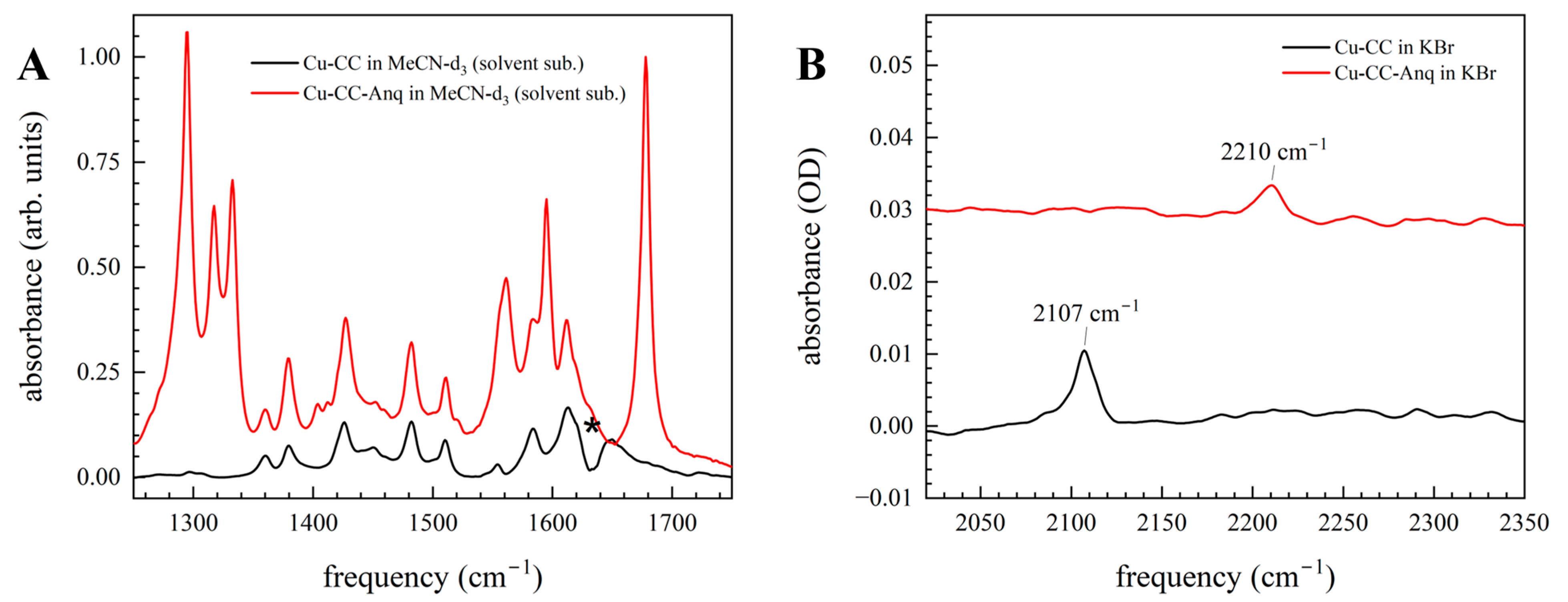
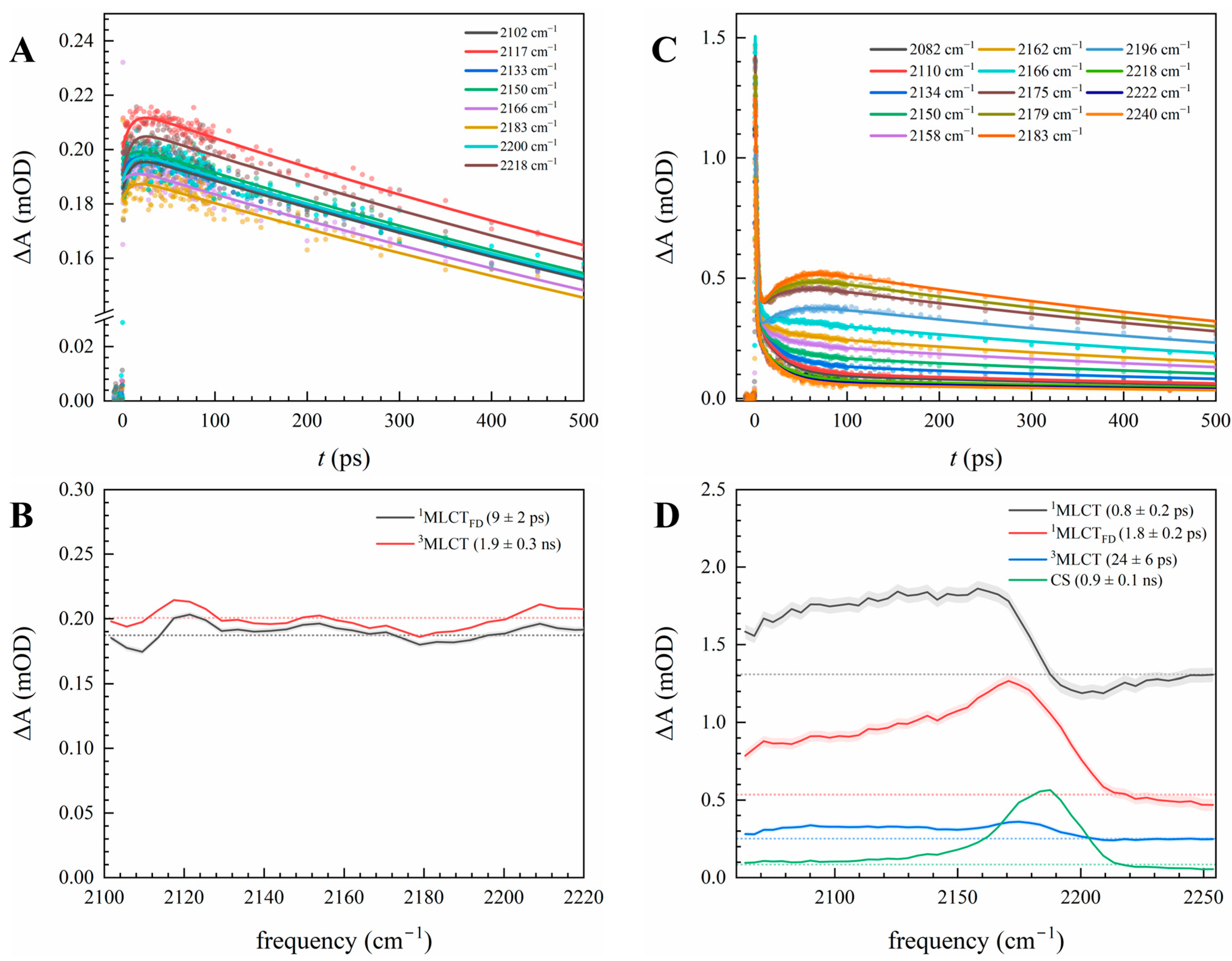
3.2. Electron Delocalization Probed by Infrared Spectroscopy of the Bridge

The linear IR absorption spectra of Cu-CC and Cu-CC-Anq are shown in Figure 3. MeCN-d3 was used to reduce contributions from solvent vibrational modes over the spectral range of 1250–1750 cm−1 (Figure 3A). A comparison of the linear spectra of Cu-CC and Cu-CC-Anq at lower frequencies highlights vibrational modes from the covalent linking of the Anq at 1280–1320 cm−1, 1540–1620 cm−1, and 1680 cm−1. The first two ranges are attributed to ring stretching vibrations, while the absorption feature at 1680 cm−1 corresponds to anti-symmetric and symmetric carbonyl stretching motions. Phen and mesPhen vibrations appear from 1350 to 1540 cm−1. These assignments were verified by ground-state DFT calculations (Figure S10).
Solution IR spectra of the ethyne stretching mode (CC stretch) proved challenging due to the weak transition dipole of the vibrational mode and significant solvent absorption in MeCN (and MeCN-d3). The CC stretch of Cu-CC and Cu-CC-Anq in KBr are shown in Figure 3B (full spectra in KBr are shown in Figure S11). The mode is observed at 2107 cm−1 and 2210 cm−1 in Cu-CC and Cu-CC-Anq, respectively. The CC stretch of the terminal alkyne (present in Cu-CC) typically appears at lower frequencies than that of the internal alkyne (present in Cu-CC-Anq) [60]. The use of NM, to extend the observable spectral range to ~2230 cm−1, and a high concentration of Cu-CC-Anq in collecting solution IR spectra yielded results in agreement with the solid-state measurement (Figure S12).
The excited-state kinetics and SADS of Cu-CC and Cu-CC-Anq in NM from TRIR experiments in the CC stretching region are shown in Figure 4. The SADS of Cu-CC (Figure 4B) showed no clear ground-state bleach or ESA features corresponding to vibrational modes from 2100 to 2230 cm−1. Rather, a broad, low-amplitude ESA feature (visualized for the readers using dotted lines) was observed with kinetics like those observed in the TA experiment (Section 3.1). Time constants of 9 ± 2 ps and 1.9 ± 0.3 ns were extracted from a two-state sequential model and can be correlated to ISC and ground-state recovery. The shape of the spectral feature was largely unchanged after ISC, but there was a slight rise in signal intensity after the transition (indicated in the SADS and early-time kinetics). Similar spectra were observed in DCM and MeCN (Figure S13). Full kinetic components are presented in Table S2.
Broad, featureless TRIR signals have been previously reported in symmetric multi-branched D-A systems and arise due to electronic delocalization between different branches [61,62]. The observation of this signal from the excited Cu-CC indicates that the MLCT transition is delocalized between the two Phen ligands, in agreement with the calculated NTOs depicted in Figure 1 and previous findings on Cu–bisphen systems [27]. The absence of a transient signal from the CC stretch suggests that there was no significant change in the vibrational mode upon MLCT excitation. This implies that the electronic structural changes arising from the excitation had a minimal effect on the CC bond of the Phen-CC ligand, and highlight only partial delocalization on this ligand. The particle NTO shows little orbital contribution at the 5 and 6 positions of the Phen-CC ligand, and the calculated CC stretch frequencies in the S0 and S1 states also show little change, as shown in Table 1, verifying the experimental observations.
The SADS of Cu-CC-Anq, shown in Figure 4B, are significantly different from those of Cu-CC. MLCT excitation generates an ESA feature that is centered at ~2170 cm−1 and shifts to higher frequencies as it progresses through its excited-state pathway. Again, the TRIR kinetics captured similar timescales to the TA experiments, corresponding to the flattening distortion, ISC, charge separation, and charge recombination. Like Cu-CC, there is an underlying, broad ESA feature that also decays with Cu-CC-Anq excited-state kinetics. The broad feature is illustrated using a dotted line for SADS in Figure 4B. The main ESA feature corresponds to the CC stretch and is at a lower frequency relative to the ground-state absorption at 2210 cm−1 (Figure 2B). The experimental absence of a ground-state bleach feature in the TRIR spectra (due to weak absorption) indicates that the transition dipole of the CC stretch in the excited state is significantly enhanced. This implies that the degree of polarization across the CC bond substantially changes after electronic excitation. The lower vibrational frequency of the mode in the excited state implies that there is a lower bond order relative to the ground state and a weakening and lengthening of the bond.
The frequency change in the CC stretch, captured by the SADS when transitioning between excited states, offers insight into electron density changes throughout the excited-state pathway. The vibrational frequency of each excited state is given in Table 2. The 1MLCT state has the lowest frequency observed for the CC stretch and, therefore, the lowest bond order. The broad linewidth, enhanced transition dipole, and −40 cm−1 shift relative to the ground state imply that the MLCT transition places an electron into a molecular orbital that is largely delocalized across Phen-CC-Anq. DFT calculations of the vibrational frequency of the CC stretch and NTOs of the excited states provide further evidence of this delocalization. Unlike the experimentally observed 1MLCT state, the S2 state shows negligible changes in the CC stretch frequency, intensity, and bond length compared to those of the ground state (Table 2). The NTOs for the S2 transition (Figure 2D) show that the electron is delocalized between the mesPhen orbitals and Phen orbitals of the Phen-CC-Anq ligand, but not delocalized across the CC bond. However, the CC stretch in the S1 state is shifted −75 cm−1, and the IR intensity of the mode is significantly larger than that of the ground state. This is in better agreement with experimental observations for the 1MLCT state. The CC bond also lengthens in accordance with the shift to lower frequencies. The particle NTO (Figure 1E) shows that the electron lies solely on Phen-CC-Anq and is completely delocalized across the CC bond. Additionally, as discussed for Cu-CC, the presence of the underlying, broad TRIR signal also suggests that there is also some delocalization of the electron between the mesPhen and Phen ligands. From all these observations, it can be inferred that the electronic character of the 1MLCT state is delocalized between the phenanthroline ligands, like that of Cu-CC (and calculated S2 state), but with extended delocalization of the electron across the CC bond towards the Anq acceptor species (like the S1 state). The 3MLCT state shows a minor +4 cm−1 shift compared to the 1MLCT state, and the broad TRIR signal is maintained. This implies that only very subtle electronic structural changes occur following ISC.
After charge separation, there is a narrowing of the CC stretch ESA feature, and a +14 cm−1 shift relative to the 1MLCT state towards the ground-state absorption. The enhancement of the transition dipole strength relative to the ground state is also maintained in this electronic state. These observations indicate that there is a reduction in the electron delocalization across the CC bond. The broad TRIR signal present following excitation (illustrated by dotted lines in Figure 4B) has mostly vanished in the CS state, which is similar to what is observed in excited-state symmetry breaking processes, and indicates electron migration away from mesPhen and towards the Phen-CC-Anq ligand [61,62]. These spectral changes in the vibrational spectrum also happen concomitantly with the appearance of Anq•− spectral features in the visible spectrum (Section 3.1). These observations point towards the localization of charge on Anq; however, it is partial localization, since there is still a notable shift in the vibrational frequency compared to the ground state.

4. Conclusions

Full understanding of the excited-state properties of first-row transition metal complexes like Cu(I) complexes is critical for solar energy conversion and achieving both current and future energy demands. The kinetics and electronic structural changes that occur along the excited-state pathway of a Cu–bisphen D-B-A system were mapped out using TA and TRIR experiments in tandem with DFT calculations. Spectral changes observed in the TA spectra of Cu-CC and Cu-CC-Anq were assigned to known excited-state processes analogous to those in Cu–bisphen complexes. A key finding was the appearance of spectral features unique to the Anq•−, which provided evidence of intramolecular PET between the Cu center and Anq. In polar solvents, charge separation was found to occur in 30 ps, while charge recombination was solvent dependent. The excited-state lifetime of Cu-CC-Anq showed complicated trends that deviated from those of typical Cu–bisphen systems [18,24,25]. Notably, the lifetime was much shorter in NM than in MeCN, two polar solvents that stabilize the CS state. These were rationalized through the low coordinating ability of NM, which raises the driving force for charge recombination, and possibly offers a more favorable outer-sphere reorganization energy for this electron transfer process when compared to MeCN.
TRIR spectroscopy in the CC stretch region provided insight into the electronic structural changes that occur. Following MLCT excitation, the near-instantaneous, broad TRIR signal in the heteroleptic Cu–bisphen complexes highlighted the delocalized nature of the MLCT excited state, with the electronic character split between both phenanthroline ligands [61,62]. In Cu-CC-Anq, a red-shifted absorption of the CC stretch with enhanced transition dipole strength was overlaid on the broad signal, indicating further delocalization towards Anq attached to the phenanthroline ligand. After charge separation, the broad TRIR signal had mostly decayed, akin to what was observed in the excited-state symmetry breaking of multibranched donor–acceptor systems. This represented a transition from being delocalized between two ligands to localization on a single ligand. Blue-shifting and narrowing of the CC stretch also occurred, further depicting localization to Anq.
The alkyne bridge between the donor and acceptor species acted as a spectroscopic probe for tracking the electronic changes. It also served to electronically couple Anq to the MLCT transition, resulting in transition dipole enhancement of the CC stretch. The spectral shifting of the CC stretch that is observed with electron transfer is indicative of changes in the CC bond length from its ground-state equilibrium position. The lengthening of the CC bond in the CS state clearly shows that the nuclear displacement corresponding to the CC stretch aligns with the electron transfer reaction coordinates and likely contributes to the electron transfer rate through vibronic coupling [63,64,65]. This means that the CC stretch is not only a great reporter of electron transfer, but also a key vibrational mode that could be used to modulate electron transfer via an additional IR perturbation. We hope to conduct future experiments on these Cu(I) D-alkyne-A systems to further investigate vibrational control over electron transfer.

Supplementary Materials

The following supporting information can be downloaded at: https://www.mdpi.com/article/10.3390/photochem5020016/s1. Scheme S1: Synthesis of 2-((1,10-phenanthrolin-5-yl)ethynyl)anthracene-9,10-dione (Phen-CC-Anq); Scheme S2: Synthesis of heteroleptic Cu(I)-bis(1,10-phenanthroline) complexes Cu-CC and Cu-CC-Anq; Figure S1: 500 MHz 1H NMR spectra of Phen-CC-Anq in CDCl3; Figure S2: 500 MHz 1H NMR spectra of Cu-CC in CDCl3; Figure S3: Positive mode ESI-MS of Cu-CC; Figure S4: 500 MHz 1H NMR spectra of Cu-CC-Anq in CDCl3; Figure S5: Positive mode ESI-MS of Cu-CC-Anq; Figure S6: NTOs of Phen-CC-Anq-localized electronic transitions and main MLCT transition of Cu-CC and Cu-CC-Anq; Figure S7: SADS of Cu-CC in MeCN and DCM from the TA experiments; Figure S8: Comparison of SADS of the 1MLCTFD electronic state of Cu-CC in NM and MeCN from TA experiments; Figure S9: SADS of Cu-CC-Anq in MeCN and DCM from the TA experiments; Table S1: Observed time constants from fits to TA kinetics of Cu-CC and Cu-CC-Anq in various solvents; Figure S10: experimental IR absorption spectrum of Cu-CC-Anq and Cu-CC and the calculated vibrational modes; Figure S11: Experimental IR absorption spectra of Cu-CC and Cu-CC-Anq in KBr; Figure S12: Solvent subtraction of the IR absorption spectrum of a concentrated solution of Cu-CC-Anq; Figure S13: SADS of Cu-CC in MeCN and DCM from TRIR experiments; and Table S2: Observed time constants from fits to TRIR kinetics of Cu-CC and CU-CC-Anq in various solvents.

Author Contributions

Conceptualization, S.A.R., P.K., K.L.M. and L.X.C.; methodology, S.A.R., W.C.H., P.K., X.L. and K.L.M.; software, S.A.R., M.T., P.K. and X.L.; validation, S.A.R., M.T., P.K., J.T.Y., X.L., K.L.M. and L.X.C.; formal analysis, S.A.R. and M.T.; investigation, S.A.R., W.C.H., M.T. and J.T.Y.; resources, X.L., K.L.M. and L.X.C.; data curation, S.A.R. and M.T.; writing—original draft preparation, S.A.R.; writing—review and editing, S.A.R., W.C.H., M.T., P.K., K.L.M. and L.X.C.; visualization, S.A.R. and M.T.; supervision, P.K., X.L., K.L.M. and L.X.C.; project administration X.L., K.L.M. and L.X.C.; funding acquisition, X.L., K.L.M. and L.X.C. All authors have read and agreed to the published version of the manuscript.

Funding

This work was funded by the Chemical Sciences, Geosciences, and Biosciences Division, Basic Energy Science, Office of Science, the U.S. Department of Energy under Contract No. DE-AC02-06CH11357. M.T. was supported in part by the state of Washington through the University of Washington Clean Energy Institute Graduate Fellowship.

Data Availability Statement

The data supporting the conclusions of this article will be made available by the authors on request.

Acknowledgments

FT-IR spectroscopy was performed at the Center for Nanoscale Materials, Argonne National Laboratory under user proposal 83199. Work performed at the Center for Nanoscale Materials, a U.S. Department of Energy Office of Science User Facility, was supported by the U.S. DOE, Office of Basic Energy Sciences, under Contract No. DE-AC02-06CH11357. We thank Colby Bell for assistance with IR measurements in KBr, and Tyler Haddock for helpful discussions. Computations were facilitated through advanced computational, storage, and networking infrastructure provided by the Hyak supercomputer system at the University of Washington, funded by the Student Technology Fee.

Conflicts of Interest

The authors declare no conflicts of interest.

References

  1. Auffèves, A. Quantum Technologies Need a Quantum Energy Initiative. PRX Quantum 2022, 3, 020101. [Google Scholar] [CrossRef]
  2. Hagfeldt, A.; Boschloo, G.; Sun, L.; Kloo, L.; Pettersson, H. Dye-Sensitized Solar Cells. Chem. Rev. 2010, 110, 6595–6663. [Google Scholar] [CrossRef]
  3. Wenger, O.S. Photoactive Complexes with Earth-Abundant Metals. J. Am. Chem. Soc. 2018, 140, 13522–13533. [Google Scholar] [CrossRef]
  4. McCusker, J.K. Electronic structure in the transition metal block and its implications for light harvesting. Science 2019, 363, 484–488. [Google Scholar] [CrossRef]
  5. Housecroft, C.E.; Constable, E.C. Solar energy conversion using first row d-block metal coordination compound sensitizers and redox mediators. Chem. Sci. 2022, 13, 1225–1262. [Google Scholar] [CrossRef] [PubMed]
  6. Ruthkosky, M.; Castellano, F.N.; Meyer, G.J. Photodriven Electron and Energy Transfer from Copper Phenanthroline Excited States. Inorg. Chem. 1996, 35, 6406–6412. [Google Scholar] [CrossRef] [PubMed]
  7. Ruthkosky, M.; Kelly, C.A.; Zaros, M.C.; Meyer, G.J. Long-Lived Charge-Separated States Following Light Excitation of Cu(I) Donor−Acceptor Compounds. J. Am. Chem. Soc. 1997, 119, 12004–12005. [Google Scholar] [CrossRef]
  8. McMillin, D.R.; Buckner, M.T.; Ahn, B.T. A light-induced redox reaction of bis(2,9-dimethyl-1,10-phenanthroline)copper(I). Inorg. Chem. 1977, 16, 943–945. [Google Scholar] [CrossRef]
  9. Sakaki, S.; Koga, G.; Ohkubo, K. Successful photocatalytic reduction of methylviologen (MV2+) with [Cu(NN)(PPh3)2]+ (NN = 2,9-dimethyl-1,10-phenanthroline or 4,4’,6,6’-tetramethyl-2,2’-bipyridine) upon near-UV-light irradiation and a novel solvent effect on its catalytic activity. Inorg. Chem. 1986, 25, 2330–2333. [Google Scholar] [CrossRef]
  10. Sakaki, S.; Koga, G.; Hinokuma, S.; Hashimoto, S.; Okubo, K. Improvement of the photochemical reactivity of [Cu(dmp)P2]+ (dmp = 2,9-dimethyl-1,10-phenanthroline; P = tertiary phosphine) by substituting triphenylphosphine with larger phosphines: Application to photoreduction of methylviologen. Inorg. Chem. 1987, 26, 1817–1819. [Google Scholar] [CrossRef]
  11. Ruthkosky, M.; Kelly, C.A.; Castellano, F.N.; Meyer, G.J. Electron and energy transfer from Cu(I) MLCT excited states. Coord. Chem. Rev. 1998, 171, 309–322. [Google Scholar] [CrossRef]
  12. Listorti, A.; Accorsi, G.; Rio, Y.; Armaroli, N.; Moudam, O.; Gégout, A.; Delavaux-Nicot, B.; Holler, M.; Nierengarten, J.-F. Heteroleptic Copper(I) Complexes Coupled with Methano[60]fullerene: Synthesis, Electrochemistry, and Photophysics. Inorg. Chem. 2008, 47, 6254–6261. [Google Scholar] [CrossRef]
  13. Kirner, S.V.; Guldi, D.M.; Megiatto, J.J.D.; Schuster, D.I. Synthesis and photophysical properties of new catenated electron donor–acceptor materials with magnesium and free base porphyrins as donors and C60 as the acceptor. Nanoscale 2015, 7, 1145–1160. [Google Scholar] [CrossRef] [PubMed]
  14. Breddels, P.A.; Blasse, G.; Casadonte, D.J.; McMillin, D.R. Photoelectrochemical Studies of Bis(triphenylphosphine)biquinoline Copper(I), Bis(biquinoline) Copper(I) and Bis(2,9-dimethyl-1,10-phenanthroline) Copper(I) on SnO2 Electrodes. Ber. Bunsenges. Phys. Chem. 1984, 88, 572–578. [Google Scholar] [CrossRef]
  15. Alonso-Vante, N.; Nierengarten, J.-F.; Sauvage, J.-P. Spectral sensitization of large-band-gap semiconductors (thin films and ceramics) by a carboxylated bis(1,10-phenanthroline)copper(I) complex. J. Chem. Soc. Dalton Trans. 1994, 1649–1654. [Google Scholar] [CrossRef]
  16. Sakaki, S.; Kuroki, T.; Hamada, T. Synthesis of a new copper(i) complex, [Cu(tmdcbpy)2]+ (tmdcbpy = 4,4′,6,6′-tetramethyl-2,2′-bipyridine-5,5′-dicarboxylic acid), and its application to solar cells. J. Chem. Soc. Dalton Trans. 2002, 840–842. [Google Scholar] [CrossRef]
  17. Cunningham, C.T.; Moore, J.J.; Cunningham, K.L.H.; Fanwick, P.E.; McMillin, D.R. Structural and Photophysical Studies of Cu(NN)2+ Systems in the Solid State. Emission at Last from Complexes with Simple 1,10-Phenanthroline Ligands. Inorg. Chem. 2000, 39, 3638–3644. [Google Scholar] [CrossRef]
  18. Mara, M.W.; Fransted, K.A.; Chen, L.X. Interplays of excited state structures and dynamics in copper(I) diimine complexes: Implications and perspectives. Coord. Chem. Rev. 2015, 282–283, 2–18. [Google Scholar] [CrossRef]
  19. Iwamura, M.; Takeuchi, S.; Tahara, T. Ultrafast Excited-State Dynamics of Copper(I) Complexes. Acc. Chem. Res. 2015, 48, 782–791. [Google Scholar] [CrossRef]
  20. Siddique, Z.A.; Yamamoto, Y.; Ohno, T.; Nozaki, K. Structure-Dependent Photophysical Properties of Singlet and Triplet Metal-to-Ligand Charge Transfer States in Copper(I) Bis(diimine) Compounds. Inorg. Chem. 2003, 42, 6366–6378. [Google Scholar] [CrossRef]
  21. McMillin, D.R.; Kirchhoff, J.R.; Goodwin, K.V. Exciplex quenching of photo-excited copper complexes. Coord. Chem. Rev. 1985, 64, 83–92. [Google Scholar] [CrossRef]
  22. Chen, L.X.; Shaw, G.B.; Novozhilova, I.; Liu, T.; Jennings, G.; Attenkofer, K.; Meyer, G.J.; Coppens, P. MLCT State Structure and Dynamics of a Copper(I) Diimine Complex Characterized by Pump−Probe X-ray and Laser Spectroscopies and DFT Calculations. J. Am. Chem. Soc. 2003, 125, 7022–7034. [Google Scholar] [CrossRef] [PubMed]
  23. Shaw, G.B.; Grant, C.D.; Shirota, H.; Castner, E.W.; Meyer, G.J.; Chen, L.X. Ultrafast Structural Rearrangements in the MLCT Excited State for Copper(I) bis-Phenanthrolines in Solution. J. Am. Chem. Soc. 2007, 129, 2147–2160. [Google Scholar] [CrossRef]
  24. Eggleston, M.K.; McMillin, D.R.; Koenig, K.S.; Pallenberg, A.J. Steric Effects in the Ground and Excited States of Cu(NN)2+ Systems. Inorg. Chem. 1997, 36, 172–176. [Google Scholar] [CrossRef]
  25. Castellano, F.N.; Rosko, M.C. Steric and Electronic Influence of Excited-State Decay in Cu(I) MLCT Chromophores. Acc. Chem. Res. 2024, 57, 2872–2886. [Google Scholar] [CrossRef]
  26. Queffélec, C.; Pati, P.B.; Pellegrin, Y. Fifty Shades of Phenanthroline: Synthesis Strategies to Functionalize 1,10-Phenanthroline in All Positions. Chem. Rev. 2024, 124, 6700–6902. [Google Scholar] [CrossRef] [PubMed]
  27. Wang, L.; Xie, Z.-L.; Phelan, B.T.; Lynch, V.M.; Chen, L.X.; Mulfort, K.L. Changing Directions: Influence of Ligand Electronics on the Directionality and Kinetics of Photoinduced Charge Transfer in Cu(I)Diimine Complexes. Inorg. Chem. 2023, 62, 14368–14376. [Google Scholar] [CrossRef]
  28. Rosko, M.C.; Espinoza, E.M.; Arteta, S.; Kromer, S.; Wheeler, J.P.; Castellano, F.N. Employing Long-Range Inductive Effects to Modulate Metal-to-Ligand Charge Transfer Photoluminescence in Homoleptic Cu(I) Complexes. Inorg. Chem. 2023, 62, 3248–3259. [Google Scholar] [CrossRef]
  29. Garakyaraghi, S.; McCusker, C.E.; Khan, S.; Koutnik, P.; Bui, A.T.; Castellano, F.N. Enhancing the Visible-Light Absorption and Excited-State Properties of Cu(I) MLCT Excited States. Inorg. Chem. 2018, 57, 2296–2307. [Google Scholar] [CrossRef]
  30. Rosko, M.C.; Wheeler, J.P.; Alameh, R.; Faulkner, A.P.; Durand, N.; Castellano, F.N. Enhanced Visible Light Absorption in Heteroleptic Cuprous Phenanthrolines. Inorg. Chem. 2024, 63, 1692–1701. [Google Scholar] [CrossRef]
  31. Schmittel, M.; Ganz, A. Stable mixed phenanthroline copper(I) complexes. Key building blocks for supramolecular coordination chemistry. Chem. Commun. 1997, 999–1000. [Google Scholar] [CrossRef]
  32. Schmittel, M.; Lüning, U.; Meder, M.; Ganz, A.; Michel, C.; Herderich, M. Synthesis of Sterically Encumbered 2,9-Diaryl Substituted Phenanthrolines. Key Building Blocks for the Preparation of Mixed (Bis-Heteroleptic) Phenanthroline Copper(I) Complexes. Heterocycl. Commun. 1997, 3, 493–498. [Google Scholar] [CrossRef]
  33. Kohler, L.; Hayes, D.; Hong, J.; Carter, T.J.; Shelby, M.L.; Fransted, K.A.; Chen, L.X.; Mulfort, K.L. Synthesis, structure, ultrafast kinetics, and light-induced dynamics of CuHETPHEN chromophores. Dalton Trans. 2016, 45, 9871–9883. [Google Scholar] [CrossRef]
  34. Hayes, D.; Kohler, L.; Chen, L.X.; Mulfort, K.L. Ligand Mediation of Vectorial Charge Transfer in Cu(I)diimine Chromophore–Acceptor Dyads. J. Phys. Chem. Lett. 2018, 9, 2070–2076. [Google Scholar] [CrossRef]
  35. Phelan, B.T.; Xie, Z.-L.; Liu, X.; Li, X.; Mulfort, K.L.; Chen, L.X. Photodriven electron-transfer dynamics in a series of heteroleptic Cu(I)–anthraquinone dyads. J. Chem. Phys. 2024, 160, 144905. [Google Scholar] [CrossRef] [PubMed]
  36. Lin, Z.; Lawrence, C.M.; Xiao, D.; Kireev, V.V.; Skourtis, S.S.; Sessler, J.L.; Beratan, D.N.; Rubtsov, I.V. Modulating Unimolecular Charge Transfer by Exciting Bridge Vibrations. J. Am. Chem. Soc. 2009, 131, 18060–18062. [Google Scholar] [CrossRef]
  37. Delor, M.; Keane, T.; Scattergood, P.A.; Sazanovich, I.V.; Greetham, G.M.; Towrie, M.; Meijer, A.J.H.M.; Weinstein, J.A. On the mechanism of vibrational control of light-induced charge transfer in donor–bridge–acceptor assemblies. Nat. Chem. 2015, 7, 689–695. [Google Scholar] [CrossRef]
  38. Yue, Y.; Grusenmeyer, T.; Ma, Z.; Zhang, P.; Schmehl, R.H.; Beratan, D.N.; Rubtsov, I.V. Electron transfer rate modulation in a compact Re(i) donor–acceptor complex. Dalton Trans. 2015, 44, 8609–8616. [Google Scholar] [CrossRef]
  39. Delor, M.; Archer, S.A.; Keane, T.; Meijer, A.J.H.M.; Sazanovich, I.V.; Greetham, G.M.; Towrie, M.; Weinstein, J.A. Directing the path of light-induced electron transfer at a molecular fork using vibrational excitation. Nat. Chem. 2017, 9, 1099–1104. [Google Scholar] [CrossRef]
  40. Wintergerst, P.; Mengele, A.K.; Nauroozi, D.; Tschierlei, S.; Rau, S. Impact of Alkyne Functionalization on Photophysical and Electrochemical Properties of 1,10-Phenanthrolines and Their RuII Complexes. Eur. J. Inorg. Chem. 2019, 2019, 1988–1992. [Google Scholar] [CrossRef]
  41. Xie, Z.-L.; Liu, X.; Valentine, A.J.S.; Lynch, V.M.; Tiede, D.M.; Li, X.; Mulfort, K.L. Bimetallic Copper/Ruthenium/Osmium Complexes: Observation of Conformational Differences Between the Solution Phase and Solid State by Atomic Pair Distribution Function Analysis. Angew. Chem. Int. Ed. 2022, 61, e202111764. [Google Scholar] [CrossRef]
  42. Pellegrin, Y.; Sandroni, M.; Blart, E.; Planchat, A.; Evain, M.; Bera, N.C.; Kayanuma, M.; Sliwa, M.; Rebarz, M.; Poizat, O.; et al. New Heteroleptic Bis-Phenanthroline Copper(I) Complexes with Dipyridophenazine or Imidazole Fused Phenanthroline Ligands: Spectral, Electrochemical, and Quantum Chemical Studies. Inorg. Chem. 2011, 50, 11309–11322. [Google Scholar] [CrossRef] [PubMed]
  43. Sandroni, M.; Kayanuma, M.; Planchat, A.; Szuwarski, N.; Blart, E.; Pellegrin, Y.; Daniel, C.; Boujtita, M.; Odobel, F. First application of the HETPHEN concept to new heteroleptic bis(diimine) copper(i) complexes as sensitizers in dye sensitized solar cells. Dalton Trans. 2013, 42, 10818–10827. [Google Scholar] [CrossRef] [PubMed]
  44. Singh, Z.; Kamal, S.; Majewski, M.B. Copper(I) Donor–Chromophore–Acceptor Assembly for Light-Driven Oxidation on a Zinc Oxide Nanowire Electrode. J. Phys. Chem. C 2022, 126, 16732–16743. [Google Scholar] [CrossRef]
  45. Gaussian Developmental, Version Rev. J.14+; Gaussian, Inc.: Wallingford, CT, USA, 2020.
  46. Adamo, C.; Barone, V. Toward reliable density functional methods without adjustable parameters: The PBE0 model. J. Chem. Phys. 1999, 110, 6158–6170. [Google Scholar] [CrossRef]
  47. Grimme, S.; Ehrlich, S.; Goerigk, L. Effect of the damping function in dispersion corrected density functional theory. J. Comput. Chem. 2011, 32, 1456–1465. [Google Scholar] [CrossRef]
  48. Weigend, F.; Ahlrichs, R. Balanced basis sets of split valence, triple zeta valence and quadruple zeta valence quality for H to Rn: Design and assessment of accuracy. Phys. Chem. Chem. Phys. 2005, 7, 3297–3305. [Google Scholar] [CrossRef]
  49. Mennucci, B. Polarizable continuum model. WIREs Comput. Mol. Sci. 2012, 2, 386–404. [Google Scholar] [CrossRef]
  50. Humphrey, W.; Dalke, A.; Schulten, K. VMD: Visual molecular dynamics. J. Mol. Graph. 1996, 14, 33–38. [Google Scholar] [CrossRef]
  51. Boettger, J.C. Approximate two-electron spin-orbit coupling term for density-functional-theory DFT calculations using the Douglas-Kroll-Hess transformation. Phys. Rev. B 2000, 62, 7809–7815. [Google Scholar] [CrossRef]
  52. Liao, C.; Kasper, J.M.; Jenkins, A.J.; Yang, P.; Batista, E.R.; Frisch, M.J.; Li, X. State Interaction Linear Response Time-Dependent Density Functional Theory with Perturbative Spin–Orbit Coupling: Benchmark and Perspectives. JACS Au 2023, 3, 358–367. [Google Scholar] [CrossRef] [PubMed]
  53. Ehrman, J.; Martinez-Baez, E.; Jenkins, A.J.; Li, X. Improving One-Electron Exact-Two-Component Relativistic Methods with the Dirac–Coulomb–Breit-Parameterized Effective Spin–Orbit Coupling. J. Chem. Theory Comput. 2023, 19, 5785–5790. [Google Scholar] [CrossRef]
  54. Englman, R.; Jortner, J. The energy gap law for radiationless transitions in large molecules. Mol. Phys. 1970, 18, 145–164. [Google Scholar] [CrossRef]
  55. Jortner, J.; Rice, S.A.; Hochstrasser, R.M. Radiationless Transitions in Photochemistry. In Advances in Photochemistry; Wiley: Hoboken, NJ, USA, 1969; pp. 149–309. [Google Scholar]
  56. Gutmann, V. Principles of Coordination Chemistry in Non-Aqueous Solutions; Springer: New York, NY, USA, 1968. [Google Scholar]
  57. Büschel, M.; Stadler, C.; Lambert, C.; Beck, M.; Daub, J. Heterocyclic quinones as core units for redox switches: UV–vis/NIR, FTIR spectroelectrochemistry and DFT calculations on the vibrational and electronic structure of the radical anions. J. Electroanal. Chem. 2000, 484, 24–32. [Google Scholar] [CrossRef]
  58. Larsen, C.B.; Farrow, G.A.; Smith, L.D.; Appleby, M.V.; Chekulaev, D.; Weinstein, J.A.; Wenger, O.S. Solvent-Mediated Activation/Deactivation of Photoinduced Electron-Transfer in a Molecular Dyad. Inorg. Chem. 2020, 59, 10430–10438. [Google Scholar] [CrossRef]
  59. Marcus, R.A. Chemical and Electrochemical Electron-Transfer Theory. Annu. Rev. Phys. Chem. 1964, 15, 155–196. [Google Scholar] [CrossRef]
  60. Smith, B.C. An IR spectral interpretation potpourri: Carbohydrates and alkynes. Spectroscopy 2017, 32, 18–24. [Google Scholar]
  61. Vauthey, E. Watching Excited-State Symmetry Breaking in Multibranched Push–Pull Molecules. J. Phys. Chem. Lett. 2022, 13, 2064–2071. [Google Scholar] [CrossRef] [PubMed]
  62. Dereka, B.; Svechkarev, D.; Rosspeintner, A.; Aster, A.; Lunzer, M.; Liska, R.; Mohs, A.M.; Vauthey, E. Solvent tuning of photochemistry upon excited-state symmetry breaking. Nat. Commun. 2020, 11, 1925. [Google Scholar] [CrossRef]
  63. Chaudhuri, S.; Hedström, S.; Méndez-Hernández, D.D.; Hendrickson, H.P.; Jung, K.A.; Ho, J.; Batista, V.S. Electron Transfer Assisted by Vibronic Coupling from Multiple Modes. J. Chem. Theory Comput. 2017, 13, 6000–6009. [Google Scholar] [CrossRef]
  64. Ulstrup, J.; Jortner, J. The effect of intramolecular quantum modes on free energy relationships for electron transfer reactions. J. Chem. Phys. 1975, 63, 4358–4368. [Google Scholar] [CrossRef]
  65. Jortner, J.; Bixon, M. Intramolecular vibrational excitations accompanying solvent-controlled electron transfer reactions. J. Chem. Phys. 1988, 88, 167–170. [Google Scholar] [CrossRef]
Scheme 1. Chemical structures of heteroleptic Cu(I)–bisphenanthroline complexes studied (ligands labeled).
Photochem 05 00016 sch001
Figure 1. (A) Normalized, experimental UV/Vis spectra of Cu-CC and Cu-CC-Anq in NM (150 µm pathlength was used to limit NM absorption in UV region). (B) Calculated electronic transitions (bars) and peak-broadened, simulated UV/Vis spectra (lines). Natural transition orbitals of the lowest energy transitions in (C) Cu-CC and (D,E) Cu-CC-Anq.
Photochem 05 00016 g001
Figure 2. Excited-state kinetics and species-associated difference spectra (with error indicated by colored areas) of Cu-CC (A,B) and Cu-CC-Anq (C,D) in NM from the TA experiments. Time constants for sequential transitions between each electronic state are given in parentheses.
Photochem 05 00016 g002
Figure 3. Background-subtracted, linear IR absorption spectra of Cu-CC (black) and Cu-CC-Anq (red) in (A) MeCN-d3 from 1250 to 1750 cm−1 and (B) KBr from 2020 to 2350 cm−1. Asterisk indicates error from solvent subtraction in spectrum of Cu-CC.
Photochem 05 00016 g003
Figure 4. Excited-state kinetics and species-associated difference spectra (with error indicated by colored areas) of Cu-CC (A,B) and Cu-CC-Anq (C,D) in NM from the TRIR experiments. Time constants for sequential transitions between electronic states provided in parentheses. Dotted lines are added in B and D to illustrate the broad transient ESA feature underlying the TRIR spectra.
Photochem 05 00016 g004
Table 1. Vibrational frequency of the CC stretch of Cu-CC in NM from DFT calculations, and linear and time-resolved IR experiments in the ground (S0) and first excited state (S1).
State Frequency (cm−1) IR Intensity (arb. Units) Bond Length (Å)
DFT
S022301.01.214
S1 (531 nm)22330.91.214
Experiment
S02107.1 ± 0.1
S12107.1 ± 0.1 1
1 Inferred from FTIR and TRIR spectra.
Table 2. Vibrational frequency of the CC stretch of Cu-CC-Anq in NM from DFT calculations, and linear and time-resolved IR experiments in the ground (S0) and excited states.
State Frequency (cm−1) IR Intensity (arb. Units) Bond Length (Å)
DFT
S0233234.41.219
S1 (590 nm)225712,830.01.226
S2 (531 nm)23350.21.218
Experiment
S02211.5 ± 0.1
1MLCT2170.2 ± 0.4
3MLCT2173.6 ± 0.4
CS2183.7 ± 0.3
Disclaimer/Publisher’s Note: The statements, opinions and data contained in all publications are solely those of the individual author(s) and contributor(s) and not of MDPI and/or the editor(s). MDPI and/or the editor(s) disclaim responsibility for any injury to people or property resulting from any ideas, methods, instructions or products referred to in the content.

