Review

Biotextiles for Biomedical Applications: A Review

by
Heitor Luiz Ornaghi Júnior
* and
Julia Pradella Garavatti
Postgraduate in Process Engineering and Technologies Program, University of Caxias do Sul, Francisco Getúlio Vargas St., 1130, Caxias do Sul 95070-560, RS, Brazil
*
Author to whom correspondence should be addressed.
Textiles 2025, 5(2), 19; https://doi.org/10.3390/textiles5020019
Submission received: 14 January 2025 / Revised: 9 April 2025 / Accepted: 18 April 2025 / Published: 12 May 2025
(This article belongs to the Special Issue Advances of Medical Textiles: 2nd Edition)

Abstract

:
The demand for healthcare and medical devices increases as the population ages. With the advance of textile science and technology, new products have been developed to replace the traditional ones, including biotextiles. This review has the objective of presenting biotextiles for biomedical applications, specifically drug delivery systems, medical implants, and regenerative medicine, showing the scientific progress in the respective fields, some relevant scientific studies, and commercially available products. The aim is to present to readers a quick overview guide for reference, including future trends and challenges.

1. Introduction

Textiles have been crucial in human existence since ancient civilizations, often going unnoticed in everyday life [1,2]. They not only protect us from environmental factors, but also contribute to our visual appeal. Various cultures have articulated their identities through distinct languages, artistic expressions, and clothing styles. Using natural fibers in textile manufacturing has a profound historical significance, showcasing a wide range of applications [3]. These fibers, sourced mainly from plants and animals, are becoming increasingly important in modern society, especially in light of technological innovations. Their uses have expanded beyond conventional applications, such as clothing, carpets, and home décor, to include specialized sectors like medical and healthcare textiles (including antimicrobial materials, bandages, and wound dressings), protective equipment (such as firefighter uniforms, stab-resistant apparel, and bulletproof vests), smart textiles (or electronic textiles), automotive components (like tires and road markings), and geotextiles (for embankment reinforcement and agricultural safeguarding). The term ‘textile’ and its plural ‘textiles’ refer to fibers, filaments, yarns, and most fabrics produced from these raw materials [4]. Biotextiles are medical devices that engage with living organisms [5]. With advancements in technology and scientific research, these products exhibit customized characteristics that enable their application across various biomedical fields. Notably, attributes such as increased surface area and biocompatibility are well-established; however, bioactivity has recently garnered significant attention from both industry and academic researchers [6,7,8].
Natural fibers provide a renewable supply [9] which is capable of regenerating over time, either through biological reproduction or other naturally occurring processes. These fibers are typically identified by several essential properties, such as air permeability, hygroscopicity, dyeability, moisture absorption and release capabilities, reduced flammability, biodegradability, biocompatibility, and the absence of harmful substances that could adversely affect the environment or human health, in contrast to synthetic fibers [10,11,12]. Although synthetic fibers may offer enhanced durability and strength, along with lower production costs, they pose considerable challenges, including non-degradability, non-renewability, and the generation of toxic byproducts during decomposition [13].
Natural textile materials, encompassing woven, non-woven, knitted, and composite fibers sourced from renewable origins, play a crucial role in various applications and are essential to our everyday experiences in the field of textile science and technology. Their importance is attributed to their unique textile characteristics and enhanced surface functionalities when compared to synthetic alternatives. As a result, the surfaces of natural textile fibers present opportunities for functional modifications that maintain their core attributes, thereby catering to specific requirements across multiple applications. Beyond their common uses, surface modifications improve the properties of these textiles and foster innovative design solutions [13]. The range of applications of textiles has increased substantially in the last few years, particularly in the fields of ecology/environmental protection, military/aerospace, and medical/health [9,10,11,12,13]. Figure 1 shows some examples of technical areas for the application of textiles.
Biotextiles are increasingly utilized in the domains of medicine, due to their remarkable characteristics, including regulated mechanical strength, substantial porosity conducive to cellular proliferation, and the capacity for modification. Over the past decade, there has been a surge in advancements related to textile structures and manufacturing technologies, establishing textiles as suitable materials for biomaterials and tissue engineering. Key features of biotextiles, such as adjustable mechanical strength, enhanced surface area, flexibility, and adaptability, render them highly attractive for these applications. In addition to their biocompatibility, there is a growing focus among researchers on bioactivity, a trait conferred by natural polymers. Biotextiles are favored for their superior biocompatibility, safe biodegradability, and non-toxic nature, distinguishing them from synthetic alternatives. A variety of fabrication techniques, including weaving, knitting, braiding, and spinning, have been employed to create both resorbable and non-resorbable biotextile structures that are functionally and mechanically effective [14,15].
The global market for biomedical textiles is projected to reach USD 20.7 billion by 2027, with a compound annual growth rate (CAGR) of 6.3% throughout the forecast period. Key factors driving the growth of the biotextile sector include an increasing elderly population and rising healthcare expenditures. According to the Centers for Disease Control and Prevention (CDC), emergency departments in the United States treat approximately 2.7 million elderly patients annually due to falls. As individuals age, their natural ability to seal and heal wounds diminishes, largely due to declining cellular function. Additionally, chronic conditions such as pressure ulcers are prevalent among older adults. Consequently, a significant proportion of individuals requiring sutures, implants, and wound care products are from this demographic. The demand for these medical products is expected to rise in tandem with the growing aging population. Furthermore, the escalating number of surgical procedures worldwide presents substantial opportunities for market growth, fostering the development of advanced biomedical textiles designed to resist various pathogens, fire, stains, and water. However, the pursuit of high-quality fabric production entails considerable research and development costs, driven by labor, technology, and other associated expenses. The manufacturing of raw materials, including polyester, cotton, polypropylene, polyurethane, and vinyl, involves intricate processes that necessitate significant investments. Additionally, the production of these materials requires sophisticated machinery, cutting-edge technology, and sufficient raw materials. Antimicrobial biomedical textiles are composed of a range of fibers and antimicrobial agents, such as silver, copper, and quaternary ammonium compounds, all of which are notably expensive. This complexity further underscores the challenges faced by market participants [16].
Textile materials are essential in the creation of appropriate structures within the medical and healthcare textile industries. Medical textiles are classified as a specialized segment of the larger category of technical textiles. These materials and their associated products must demonstrate particular characteristics, including functionality, durability, biological degradability, biocompatibility, non-toxicity, and the capacity for sterilization. Natural-based textile materials, along with their derived products, exhibit similar properties suitable for medical applications, allowing them to serve as alternatives for human organs or biological structures, thus addressing challenges related to organ failure and structural inadequacies [17,18]. Medical textile materials include both implantable and non-implantable options, extracorporeal devices, and various healthcare and hygiene products. The production of these items typically employs methods such as weaving, knitting, braiding, and other advanced processing techniques, utilizing a wide range of both natural and synthetic textile materials. The demand for medical textiles and healthcare products has increased significantly, expanding beyond hospitals and healthcare facilities to encompass hotels, residences, and other settings where hygiene is critical. As a result, this sector has emerged as a high-value industry with considerable market potential. With the growing consumer awareness of health issues, medical textile materials that emphasize human health present a promising avenue for the evolution and improvement of conventional textile manufacturing practices [19].
This mini review has as its objective the presentation of biotextiles for biomedical applications, specifically drug delivery systems, medical implants, and regenerative medicine, showing the scientific progress in the respective fields, some relevant scientific studies, and commercially available products. The aim is to present to readers an overview guide for reference, including future trends.

2. Drug Delivery Systems

Drug delivery systems (DDSs) are widely employed in the pharmaceutical and medical sectors. According to the Fortune Business Insights website [20], a compound annual growth rate (CAGR) of almost 10% is expected in this market through 2032, reaching nearly USD 100 billion. DDSs can be simply described as technologies that carry drugs into or throughout the body [21], and different types and sizes of particles can be used. Figure 2A shows different drug delivery systems used in the human body, while Figure 2B illustrates the types of drug delivery systems.
DDSs can be administered via different routes and delivered depending on the target, as shortly described below.
Buccal administration can resolve some problems involving conventional drug administration, such as presystemic metabolism and drug depletion in the digestive tract. The structures come into direct contact with both the absorbent surface and the mucus membrane, enhancing the therapeutic effects of the medication administered. Certain drawbacks must be taken into account, such as the substantial loss of medication through salivation and swallowing, as well as the limited delivery areas [23].
Sublingual—used when a rapid onset of action is required owing to its greater bioavailability and better patient compliance [24].
Vaginal—encompasses many forms of solids, including gels, suppositories, tablets, and nanoparticles [25,26,27].
Rectal—used in case of gastric irritation, when low enzymatic activity and localized activity are required [28].
Nasal—suitable for both local and systemic use, addressing disadvantages such as stabilization of the drug and bioavailability [29,30].
Ocular—indicated for the treatment of anterior segment diseases. It is considered the most challenging type of DDSs by experts [31].
Gastrointestinal—this mode is the most convenient for patients. However, the absorption of drugs is limited by the individual characteristics of each patient [32].
The different nanoparticles used for drug delivery systems over time can be easily visualized in Table 1, which shows the evolution of nanoparticles from 1991 to 2022, along with their applications, diseases, characterization, and other characteristics.
Briefly, according to the table above, specific nanoparticles are better applied in specific treatments. As an example, Gao et al. [99] developed DDSs for cancer therapy. The authors developed abortifacient mifepristone (MIF)-loaded mesoporous silica nanoparticles coated with epithelial cell adhesion molecule antibodies. The main results point out that the nanoparticles inhibit the hetero-adhesion between cancer cells and endothelial cells, with an effective MIF delivery to inhibit lung metastasis.
Summarily, drug delivery systems are used to maximize the effect of drugs while minimizing side effects. It is always interesting to propose an antibacterial ointment or a vaccine for localized infection instead of other, more invasive treatments. For both proposals, the drug must be packaged until it reaches its target. Some examples of biotextiles can be found in the literature. Gencturk et al. [100] developed donepezil hydrochloride-containing polyurethane/hydroxypropyl cellulose (PU/HPC) nanofibers for transdermal drug delivery. The nanofibers were prepared by the electrospinning method, and the main results indicated that the PU/HPC mat was successfully applied to the skin, not promoting irritation. Youssef et al. [101] developed a gastroretentive metronidazole floating raft system (FRS) for targeting Helicobacter pylori. The authors studied FRSs containing 3, 4, 5, 0.5, 0.75, and 1% w/v sodium alginate (Alg) and gellan gum (G), respectively, and 0.25% w/v sodium citrate and calcium carbonate (C). Lipids such as glyceryl monostearate (GMS), Precirol®, and Compritol® were incorporated into G-based formulations (G1%C1%). The Mz:lipid ratio was 1:1, except for Mz:GMS, with ratios of 1:1.5 and 1:2 also investigated. Viscosity, in vitro gelation, in vitro floating, in vitro drug release, in vitro release parameters, kinetics, and stability were evaluated. The authors reached the conclusion that the recommended FRS formulations could effectively preserve the minimum inhibitory concentration of the drug for a sufficient period, enabling local eradication. This strategy is likely to result in enhanced therapeutic effectiveness, increased patient compliance, decreased costs, and minimized side effects, typically associated with immediate-release dosage forms. Yu et al. [102] investigated the enhancement of dissolution for acetaminophen through the use of electrospun nanofiber-based solid dispersions (SDs). The study involved the preparation of poorly water-soluble SDs utilizing a continuous web structure, specifically in the form of a non-woven membrane. These were then compared to three conventional SD methods: freeze-drying, vacuum drying, and heat drying. The authors conducted a series of evaluations, including microscopy, thermal analysis, structural assessments, and biological testing, concluding that their proposed method significantly enhanced the drug’s dissolution rate.
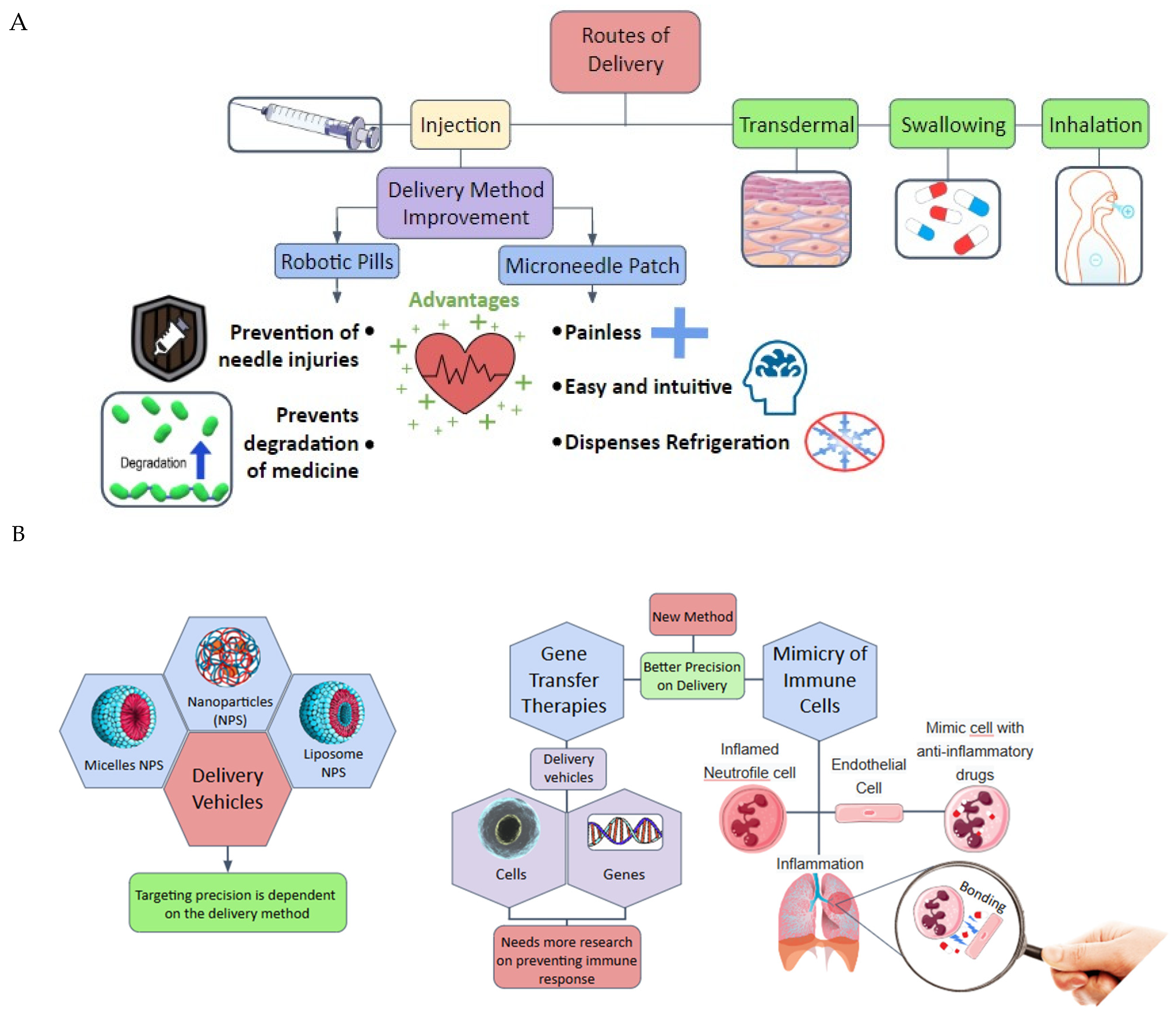
Current research on drug delivery systems can be grouped into two broad categories: routes of delivery and delivery vehicles, as demonstrated in Figure 3A,B. It is shown that the delivery vehicles represent a field with more potential to be explored than the routes of delivery.
The use of fiber (and biotextiles) in DDSs can be divided into three stages that date back to ancient times: (i) inhalation or infusion of active ingredients from textile bags or packets containing herbal drugs, (ii) production of man-made fibers (synthetic fibers) in the form of bandages, wound dressings, or for specific diseases, and (iii) biotextiles to treat systemic diseases directly due to the evolution of textile technologies (such as the electrospinning process, for example) [103].
The primary benefits of drug-embedded fibers include straightforward production processes, an extensive surface area conducive to drug release, and a diverse array of structural possibilities. Drug-loaded polymer nanofibers produced through electrospinning typically exhibit diameters ranging from a few nanometers to over 1 μm, with a more common range of 50–500 nm. These nanofibers are characterized by an exceptionally high surface area relative to their mass; for example, nanofibers with a diameter of approximately 100 nm can achieve a specific surface area of around 1000 m2/g. Additionally, they demonstrate significant porosity, superior mechanical properties, high axial strength, remarkable flexibility, low weight, and cost-effectiveness compared to alternative nanomaterials utilized in pharmaceuticals. These attributes can be leveraged in the design and advancement of innovative drug delivery systems (DDSs). The integration of biotextiles into DDSs has the potential to enhance efficiency and patient compliance, ultimately improving the overall patient experience—key objectives within the pharmaceutical domain. The exploration of new DDSs derived from biotextiles is poised to become a crucial area of research for both the pharmaceutical and technical textile industries [104].
According to Zhu and Yu [103], there are two different approaches to the incorporation of active ingredients in biotextiles: (i) the fiber is treated with a drug, and (ii) the drug can be incorporated into the fabrics. Due to the poor sustained-release profile of the former, the latter is the most popular method (schematically represented in Figure 4).

2.1. Applications

The incorporation of drugs into biotextiles is achieved by using polymers as a bridge between biotextiles and drugs. Biotextiles designed for drug delivery systems have been developed in various forms, including woven fabrics, non-woven fabrics, and non-woven electrospun fabrics. In the case of woven fabrics, drug incorporation occurs through methods such as physical adsorption, coating, encapsulation, or covalent bonding. These woven systems are commonly utilized in applications such as bioactive bandages, artificial skin grafts, wound dressings, scaffolds for tissue repair or regeneration, aromatherapy, and antimicrobial purposes. Non-woven fabrics involve drug encapsulation on the textile surface, achieved by aggregating small fibers into sheets or web-like structures through mechanical, thermal, and chemical processes. These non-woven fabrics are particularly suitable for controlled and sustained-release applications. In the realm of non-woven electrospun fabrics, drugs are integrated into the textile surfaces via techniques such as spinning, coating, encapsulation, and bioconjugation (including plasma treatment, chemical activation, and grafting). These fabrics are applicable for creating scaffolds used in tumor treatment for cancer therapy, tissue engineering, and various other medical uses [105,106]. Encapsulated antiseptic medications play a crucial role in preventing infections at wound sites following surgical procedures or implantations. However, it is essential to assess the compatibility of the drug with the encapsulating polymer to ensure optimal outcomes. In the context of drug delivery systems, a variety of therapeutic agents can be administered, including antibiotics, plant extracts, antimicrobial peptides or proteins, antifungal agents, anti-inflammatory compounds, antioxidants, vitamins, hormones, essential oils, and nanoparticles [107,108,109]. According to Zhu et al. [103], polymers should have three specific properties for use in biotextiles: the ability to be spun, elasticity, and biocompatibility. Also, biotextiles can be designed for different dosage forms (transdermal, oral, implantable) in different release profiles (sustained, time-controlled, targeted).
Kim et al. [110] developed an adhesive composite hydrogel patch (composed of a double network of polyacrylamide and polydopamine embedded with silica nanoparticles) for sustained transdermal drug delivery to treat atopic dermatitis (AD) (Figure 5). A sustained delivery of dexamethasone (DEX) was achieved by loading DEX into the pores of the silica nanoparticles in the hydrogels, as opposed to a rapid release when DEX was directly incorporated in hydrogels. The adhesive showed potential to be used for inflammatory skin diseases due to some characteristics, such as restoration of the thickened epidermal layer, decreased inflammatory cell infiltration in the skin, recovery of collagen deposition, and reduced levels of immunoglobulin.
Cheng et al. [111] developed textile triboelectric nanogenerators (TENGs), which provide highly sensitive, self-powered, and low-cost biotechnology for pulse-to-electricity conversion, while maintaining the advantageous features of textiles, such as superior comfort and stability. TENGs can be connected to terminals to enable real-time, personalized healthcare monitoring and the collection of clinical information, such as heart rate, pulse wave velocity, and blood pressure.
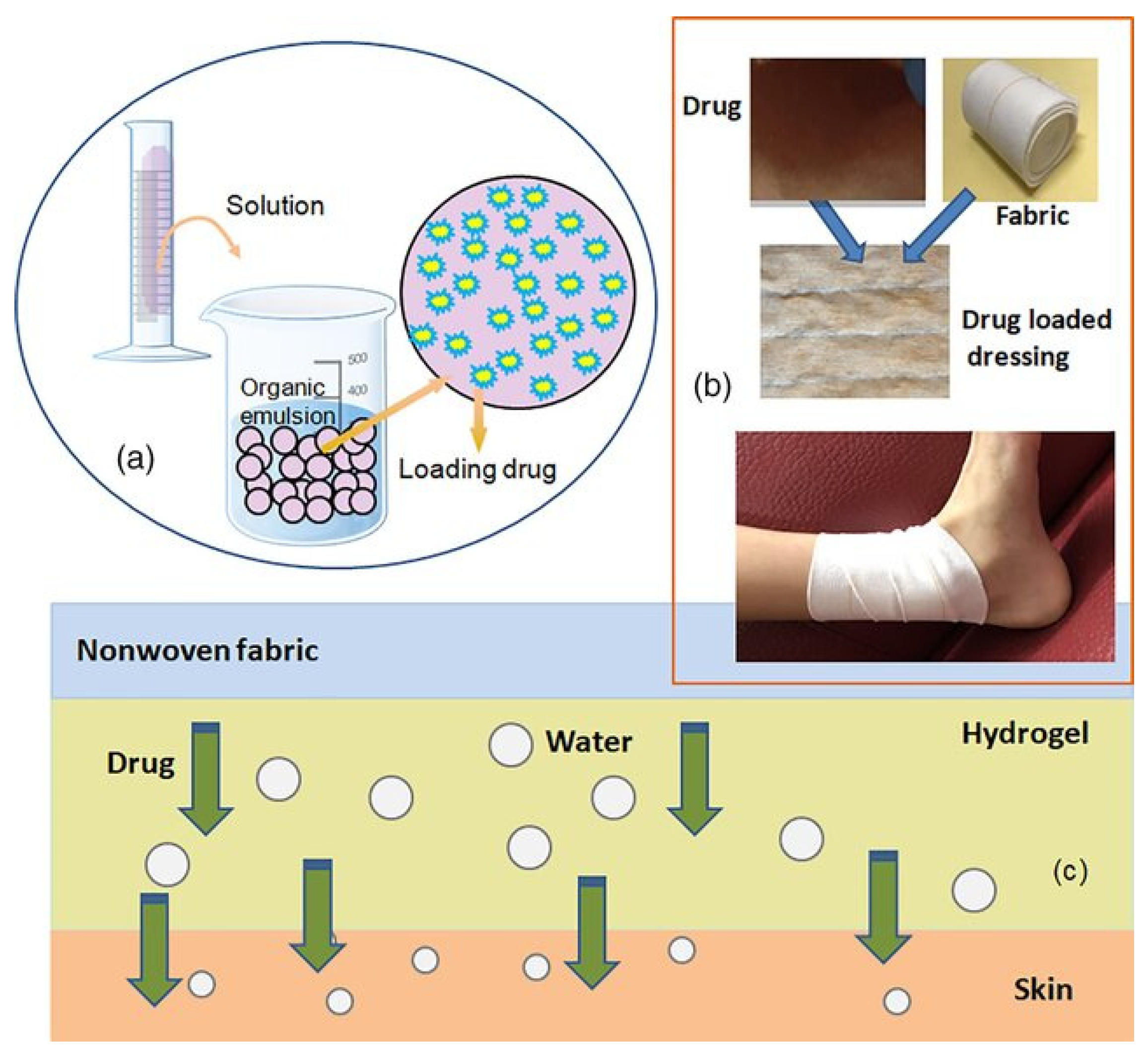
Wu et al. [112] presented a review of several therapeutic compression materials and wound dressings for chronic venous insufficiency. Figure 6a presents the preparation of a drug-loaded polymer matrix, while Figure 6b,c show the schematic diagram of drug-releasing dressings with dual delivery systems for ulcer management.
A nicotine transdermal system patch [113] is used to eliminate frequent administrations to maintain constant drug delivery. Another popular product that has been popularly increasing in demand since 2019 is nicotine pouches, which the user holds between the lip and gum in the mouth. These pouches consist of 80–90% water and microcrystalline cellulose [114]. They can be commercially found under brands such as ZYN, Rogue, BPN, NIIN, ON!, Velo, FRE, and Juice Head, among others. Figure 7A,B show an example of a nicotine pouch and how it is usually commercially sold, respectively.
Bhutto et al. [115] fabricated and characterized vitamin-B5-loaded poly (l-lactide-co-caprolactone) silk-fiber-aligned electrospun nanofibers for Schwann cell proliferation. The vitamin-loaded nanofibers showed a hydrophilic surface with significant higher cell proliferation. Additionally, a sustained release of B5 was observed to be higher in P(LLA-CL)/Vt nanofibers (80% in 24 h).
Lee et al. [116] studied a poly(l-lactic acid)/gelatin fibrous scaffold loaded with a simvastatin/beta-cyclodextrin-modified hydroxyapatite inclusion complex for bone tissue regeneration. The authors concluded that simvastatin-loaded hydroxyapatite-β-cyclodextrin (HAp-βCD) coatings were effectively developed on the surfaces of fibrous scaffolds, demonstrating significant potential for application in bone tissue engineering. The findings indicate that the incorporation of βCD into HAp results in enhanced coating and simvastatin loading compared to HAp alone. The HAp-SIM-loaded poly(L-lactic acid)/gelatin/adipose-derived (PLLA/gelatin/AD) fibrous scaffolds exhibited markedly increased alkaline phosphatase (ALP) activity and mineralization when compared to control fibers under physiological conditions. Both the simvastatin-loaded and hydroxyapatite-only coatings on fibrous scaffolds facilitated improved growth and osteogenic differentiation of human adipose-derived stem cells (hADSCs). Fibrous scaffolds modified on their surface and embedded with drugs create favorable microenvironments that promote the proliferation and differentiation of human adipose-derived stem cells (hADSCs). This indicates the potential utility of simvastatin-loaded hydroxyapatite-coated fibrous scaffolds in the field of bone tissue engineering. Figure 8 illustrates the schematic representation of the production processes alongside the scanning electron microscopy (SEM) images of the three distinct specimens.
Grimaudo et al. [117] produced crosslinked hyaluronan electrospun nanofibers for ferulic acid ocular delivery, an innovative ophthalmic insert composed of hyaluronan (HA) nanofibers for the dual delivery of an antioxidant (ferulic acid, FA) and an antimicrobial peptide (ε-polylysine, ε-PL). Polyvinylpyrrolidone (PVP) was incorporated to enhance the electrospinning process. Fibers with diameters of approximately 100 nm were produced using mixtures of PVP 5%–HA 0.8% w/v and PVP 10%-HA 0.5% w/v in a solvent system of ethanol and water at a ratio of 4:6 v/v. An increase in PVP concentration to 20% w/v, both with and without HA, resulted in the fibers measuring around 1 µm. The PVP 5%-HA 0.8% w/v fibers were found to contain 83.3 ± 14.0 µg of ferulic acid (FA) per mg. Following the crosslinking of the nanofibers with ε-PL, both blank and FA-loaded inserts exhibited average thicknesses of 270 ± 21 µm and 273 ± 41 µm, respectively. Under sink conditions, both types of inserts completely released ε-PL within 30 min, while the FA-loaded inserts released the antioxidant within 20 min. The authors concluded that the HA nanofibers were successfully produced in the presence of PVP, demonstrating appropriate dimensions and effective loading of ferulic acid. The crosslinked inserts, created by crosslinking the nanofibers with ε-PL, were assessed for potential ocular applications. These inserts displayed a suitable thickness, release profiles, in vitro biocompatibility, and antibacterial properties. Further ex vivo and in vivo investigations (Figure 9), particularly regarding the retention time of the formulation on the sclera/conjunctiva and the permeability of FA, could provide insights into the viability of the crosslinked inserts for ocular use.
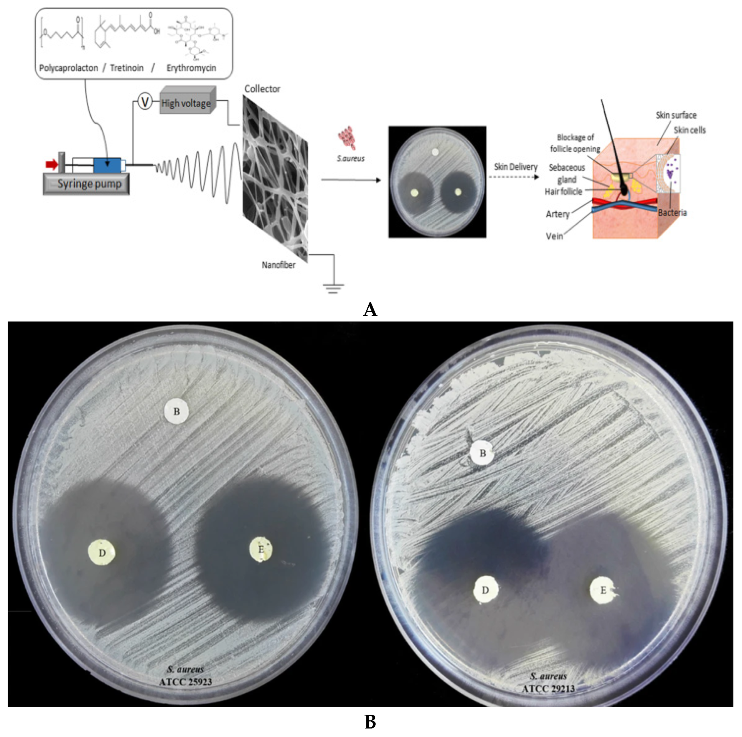
Khoshbakht et al. [107] developed nanofibers loaded with tretinoin intended for topical application in skin treatment, aiming to create a drug delivery system utilizing nanofibers infused with tretinoin for acne management. Through the process of electrospinning, uniform fibers devoid of drug crystals were successfully produced. The formulations were assessed for their drug release profiles, stability, and antimicrobial efficacy, particularly in conjunction with erythromycin. The electrospun nanofibers demonstrated a sustained release of tretinoin. Notably, the stability of these formulations was found to be greater at refrigerated temperatures than at room temperature. The authors anticipate that the straightforward fabrication process, cost-effectiveness, and reduced dosing frequency of the reported construct will facilitate its adaptation for on-demand tretinoin delivery, whether used alone or in combination with erythromycin. Future investigations should focus on modifying formulations and incorporating additional pharmaceutical excipients to enhance drug stability and release characteristics. Figure 10A,B illustrate the schematic representation of the nanofiber fabrication process and the in vitro antibacterial activity of the studied nanofibers.

2.2. Future Trends

In spite of the significant advancements in the field of biotextiles for drug delivery, several challenges remain, including systems that require combined applications, drug-loading capabilities, and understanding the interactions between the polymer and other components during the fabrication process. Additional areas requiring improvements include the development of more accurate mathematical models for the prediction of drug release, understanding the structure-release profile relationships, a creating more controllable structures for drug release. Moreover, systems are needed for the incorporation of delicate active ingredients, such as peptides, proteins, RNA, and DNA, and water-insoluble ingredients, as well as for use in tissue engineering. The development of new fabrication methods and new biopolymers will allow for the integration of conventional electrospinning techniques with advanced methods like nanotechnology and rapid prototyping.
A preliminary phase of polymer science and advanced manufacturing technologies is required for the creation of DDS biotextiles before they can be used in clinical settings. The development of novel biomaterials using effective additive manufacturing techniques is a crucial field of research, as the majority of biotextiles are made using the electrospinning process. For biotextile-based drug delivery systems with intelligent drug delivery features, this includes novel biodegradable polymers, dendrimers, electroactive polymers, modified C-60 fullerenes, and environment-sensitive hydrogels. By combining textile technologies with cutting-edge techniques like rapid prototyping and nanotechnology, the unfavorable conditions often encountered during the spinning process in the creation of medicated fibers are lessened. Another relevant concern is the use of sensitive active components, such as proteins, peptides, and DNA or RNA vaccines.
To effectively utilize the potential of biotextiles in the investigation and creation of novel DDSs, research must primarily focus on the remaining important and intricate problems in the pharmaceutical industry. Examples include DDSs for water-insoluble active ingredients, oral DDSs for proteins and peptides, multifunctional systems for hybrid therapeutic and diagnostic uses, and DDSs for tissue engineering. Medicated fiber topologies (microcrystal, amorphous, particles), drug-loading capacity, release controllability, and possible drug–polymer interactions during spinning are additional problems that still require resolution. Other concerns include how drugs are released from biotextiles, how the structure of biomedical fibers affects their release characteristics, and how to predict drug release using mathematical models.
The drug delivery market is currently expanding globally. Already, efforts are underway to design, formulate, and engineer next-generation medicines. Researchers are actively paralleling material design and synthesis to capture novel therapeutic findings in an attempt to meet clinical demands. It is challenging to develop polymer libraries that are highly responsive to physical, biological, and chemical stimuli. One important factor is the range of environmental factors that substances encounter inside the human body. Patient variability is a continuous challenge in the design of living materials. Enhancing the biocompatibility of implantable or injectable materials is a never-ending endeavor, and, as recent advancements have demonstrated, there are still some unsolved problems that must be addressed in order to increase our understanding of this area. As the physiological mechanisms underlying both healthy and diseased states continue to be clarified, we will be better equipped to create responsive and flexible drug delivery materials. Designs for living materials will advance in sophistication over time, as molecular immunology and our fundamental understanding of these biological environments improve.
The ability to fabricate optimal structures with controlled, continuous drug release properties requires further advancements in design and synthesis procedures, made possible by recent developments in nanofabrication technology. Biotextiles are increasingly offering opportunities to engineer scaffolds and tissue-like structures with controlled micro-architecture, cellular distribution, and drug release rate, as they become lighter, stronger, thinner, and more malleable in terms of flexibility, porosity, and functionalization. It is a fascinating field of science that is growing steadily and quickly, which is why many companies are investing substantial sums of money in the development of untested products and technologies. Therefore, wearable and flexible electronics and sensors will benefit significantly from biotextile advancements. The development of textile-based implantable and biodegradable sensors that incorporate electronics into tissue scaffolds to track or stimulate cellular activity will advance this process. For textile technology to reach its full potential, despite the many advantages it offers, a number of issues must be resolved. Combining cutting-edge textile equipment, innovative biomaterials, and biological advancements represents the primary challenges inherent in the use of biotextiles. It is noteworthy to mention that the future of biotextiles in DDSs undoubtedly depends on our growing understanding of the material and how it interacts with particular biomolecules, cells, and tissues.

3. Biotextiles for Medical Implants and Regenerative Medicine

Cotton, silk, cellulose, and other natural polymers have been used for medical applications throughout history. With advances in technology and science, these materials have undergone modifications (regulation of quality and purification) for improved performance. As an example, silk can be applied to surgical sutures, wound dressings, nerve guidance conduits, or scaffolds for bone and cartilage regeneration due to some characteristics such as low inflammatory properties, excellent tensile and mechanical properties, and high oxygen and vapor permeability properties [118]. Chitosan is used in hemostatic wound dressing, tissue engineering, bone and cartilage regeneration, and drug delivery due to its naturally antimicrobial properties, abundance, and hemostatic characteristics [118]. Carboxymethyl cellulose, due to its easy availability, low cost, and biocompatibility, is often applied to wound dressings and drug delivery systems [119]. Many natural polymers are modified to add some advantages, such as improved cell adhesion or bioactivity. The surface properties of natural polymers can be treated with plasma to introduce new functional groups or can be embedded with metallic nanoparticles to promote antibacterial activity [120].
To produce textile structures, unique or combined fabrication methods can be used, depending on the complexity of the final product [121]. Composite tissue engineering scaffolds and implant materials have been used in vascular graft fabrication [122] by combining electrospinning and knitting technologies.

3.1. Applications

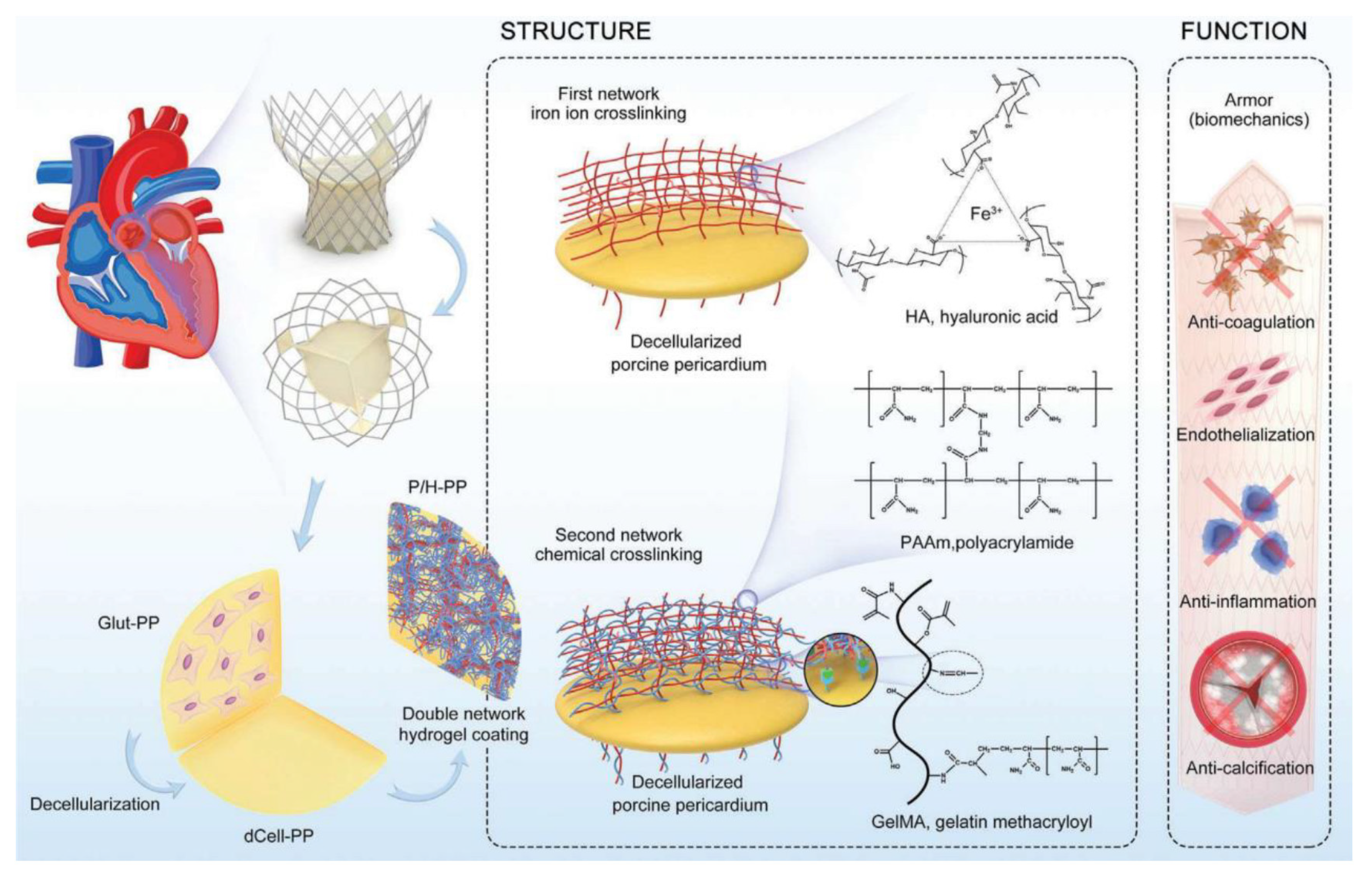
Biotextiles can be applied to different systems [123]. For tissue engineering, some examples can be found in the literature. Tamai et al. [124] developed PLLA stents as an alternative to metallic stents. The authors implanted a total of 25 stents in fifteen patients for coronary artery stenosis. Angiographic success was achieved in all procedures, with no stent thrombosis or major cardiac event occurring within 30 days. In this period, the stents were shown to be feasible, safe, and effective in humans. Nakazawa et al. studied the molecular dynamics of silk fibroin/polyurethane composites [125], aiming to design small-diameter arterial prostheses for artificial vascular grafts. Cheng et al. [126] developed a polyacrylamide/HA hydrogel hybrid PP bioprosthetic heart valve, as shown in Figure 11. The main results indicated satisfactory endothelialization, biocompatibility, and anticalcification properties in cell experiments and rat subcutaneous implantation. Additionally, excellent biomechanical and biological properties were achieved after 200,000,000 cycles.
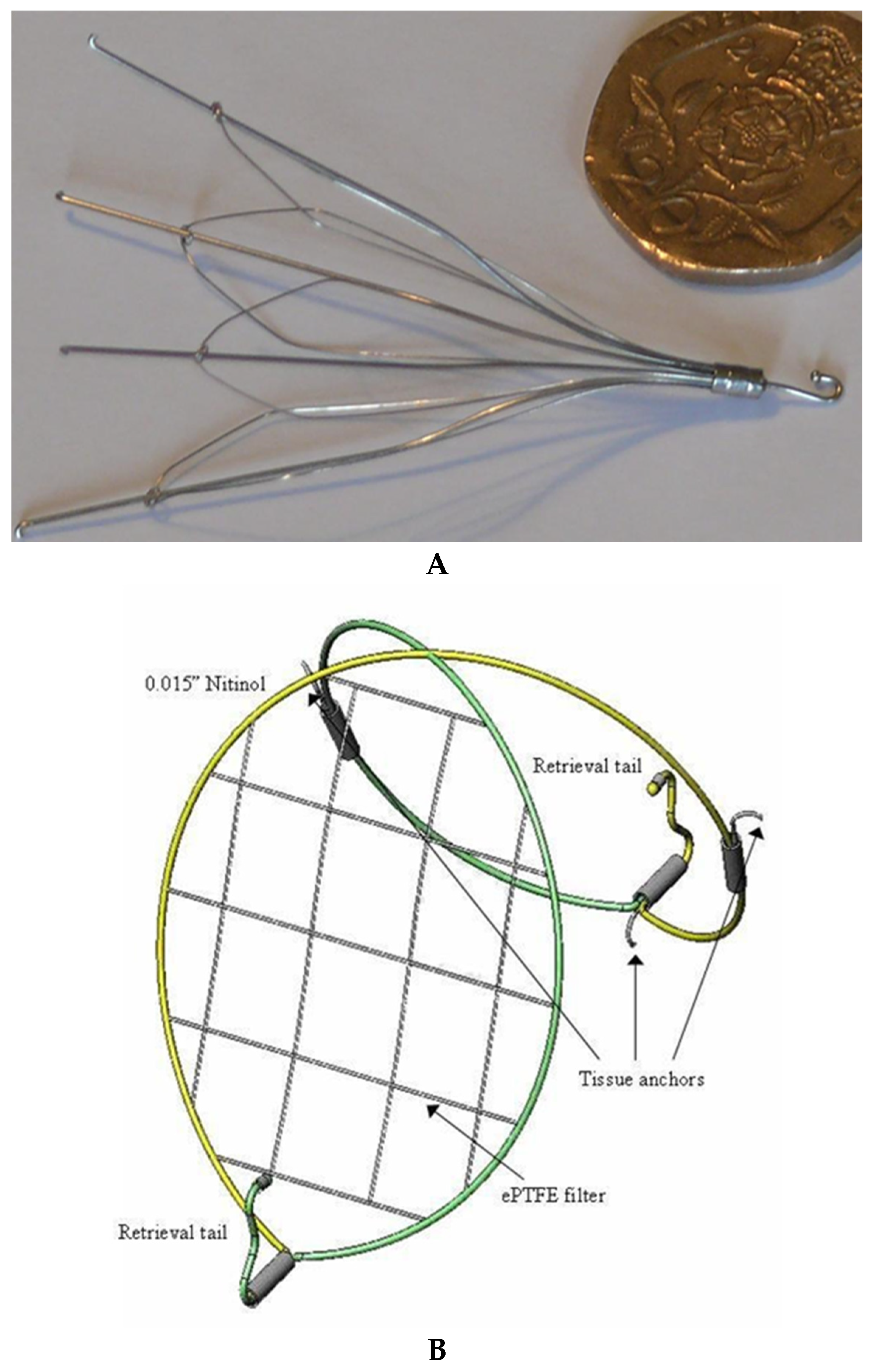
In their research, Yoon et al. [127] developed an innovative prototype textile structure designed for a vena cava filter, aimed at replacing those currently employed in clinical practice. The motivation for this investigation arose from numerous complications which have been documented in the literature since the 1960s, such as recurrent pulmonary embolism, tilting, and structural failures of existing filters. These issues underscored the urgent need for a new device that meets specific criteria: it must be non-thrombogenic, exhibit high filtration efficiency for emboli of at least 5 mm, ensure unobstructed blood flow with minimal turbulence, mitigate thrombosis at the access site, facilitate easy insertion and retrieval, provide secure fixation within the vena cava, cause minimal damage to the luminal wall, prevent perforation of the vena cava wall, be compatible with MR imaging, and be cost-effective. The authors designed a filter made from nitinol wire, arranged in a figure-eight configuration. Additionally, one lobe of this figure-eight structure featured an open mesh of PTFE yarns with specific pore sizes, ensuring reliable clot-trapping performance (Figure 12B). The authors concluded that the proposed filter is effective in preventing pulmonary embolism and represents a viable alternative to previously utilized filters (Figure 12A).
A biomedical company [128] developed Dacron and Patch for vascular anastomosis, the approach consisting of a surgical procedure that entails arterial reconstruction, replacement, and bypass procedures (connection between two blood vessels, allowing adequate blood circulation). The Dacron graft, knitted and impregnated with bovine collagen, is used for vascular prostheses in the aortofemoral, iliofemoral, and femoropopliteal regions above the knee joint, as well as in the extracranial arteries and for extra-anatomical bypasses. Collagen impregnation enables implantation without precoagulation, since the vascular prosthesis wall is impermeable at the time of implantation. The main advantages include readiness for implantation, resistance and flexibility, radiopacity, and low porosity. The Dacron-knitted vascular patch consists of polyester filaments created using knitted technology, with one side impregnated with bovine collagen, which is gradually absorbed by the body. It is indicated for use in plastic surgeries or in any arteries or prostheses where repair is necessary, offering format versatility, gamma ray sterilization, and low porosity.
Conventional bandages composed of wool or cotton have been supplanted by advanced wound dressing materials, including hydrogels, sponges, and, notably, textile structures. Significant progress has been achieved in this area. Textile structures offer considerable advantages as wound dressing materials due to their high surface area, flexibility, durability, and adaptability. Russell et al. [129] developed a new method to measure fluid transmission in non-woven wound dressings, highlighting the correlation between anisotropic fluid transmission and fiber orientation and distribution, helping readers and developers create new materials by controlling fiber direction. To produce natural polymers as fibers for wound dressing, the most commonly employed method is electrospinning, despite it not always being feasible for wide-scale industrial applications. Examples exist of some commercial products (HemCon GuardaCare®Pro, HemCon Patch®Pro, ChitoFlex®, QuikClot®, ACS+TM, and CELOXTM) based on chitosan to induce hemostatic, antimicrobial, and coagulant properties, as well as some alginate-based commercial products for use in ulcers and wounds with a high number of exudates. Devlin et al. [130] compared the equivalency of ChitoFlex® dressing, QuikClot® ACS+™ dressing, and CELOX™ with standard gauze regarding their effectiveness in controlling bleeding from non-cavitary groin wounds. According to the authors, all alternatives proved to be effective, but no agent was superior to the standard gauze.
A warp-knitted mesh composed of poly(L-lactide-co-glycolide) (PLGA) was integrated with collagen–chitosan scaffolds (CCS). This combination was further enhanced by the addition of a polyurethane (PU) membrane, resulting in the creation of a bi-layered dermal scaffold. In vivo evaluations were conducted to compare this dermal substitute with the commercially available artificial dermis PELNACTM [131]. The findings indicated that the dermal substitute exhibited reduced wound contraction, improved mechanical strength and porosity, as well as organized neotissue formation. Previous studies have demonstrated that dermal scaffolds perform optimally when designed with microstructures featuring pore sizes of approximately 100 μm.
Wu et al. [132] prepared bacterial cellulose nanocrystals with regenerated chitin to produce a yarn of 30 loaded fibers for surgical sutures (Figure 13). The study demonstrated that the mechanical properties of the bacterial cellulose nanocrystals increased with the addition of regenerated chitin. It also demonstrated good biodegradability, with no cytotoxicity of the produced yarn, as well as wound healing without obvious adverse effects.
Confluent Company [133] produces Sports Med Sutures and assemblies, joint and ligament fixation and reconstruction devices, and spinal fixation tethers for the orthopedic field, among several other applications in cardiovascular and tissue engineering.
The nervous system can always find applications in nanofibrous nerve guidance conduits, which require the production of tubes with optimal flexibility, minimal swelling and degradation behavior, permeability to nutrients and oxygen, ease of handling, and stability. A commercial product developed by Deltamed demonstrates these characteristics. According to the website, NeuraGen® Nerve Guide is a soft, bendable, unbreakable, porous collagen tube that is easy to use when moistened, while NeuraWrapTM, designed to be an interface between the nerve and the surrounding tissue, is a soft, bendable, unbreakable, porous collagen channel that is easy to use when moistened [134].
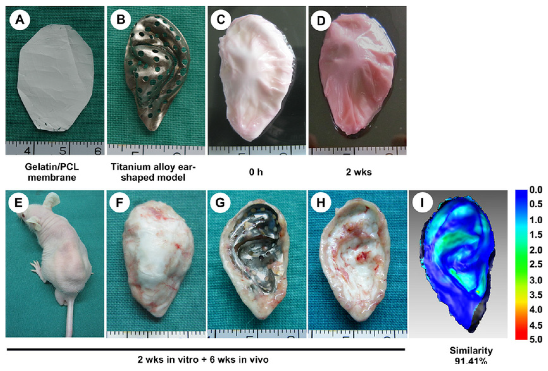
Other examples include fibrous cartilaginous implants and anterior cruciate ligament (ACL) prostheses for the skeletal system. Chitosan is one of the most promising materials for implants or scaffolds for cartilage regeneration. Xue et al. [135] engineered 3D ear-shaped cartilage using electrospun fibrous membranes of gelatin/polycaprolactone. According to the authors, the membranes were seeded with chondrocytes in the sandwich model, followed by in vitro and in vivo cultivation using an ear-shaped titanium alloy mold. The authors claimed that a shape similarity of 91.41% was maintained after 2 weeks of culture in vitro and 6 weeks of subcutaneous incubation in vivo, as demonstrated in Figure 14.
Zhi et al. [136] developed a hybrid material composed of polyethylene terephthalate (PET) and silk ligament, which was subsequently evaluated in a canine model of anterior cruciate ligament reconstruction. This hybrid construct utilized silk fibers in the weft direction and PET fibers in the warp direction, and it was compared against a control model made entirely of PET in both yarn orientations. The findings indicate that both hybrid and control models were adequate for the reconstruction of the canine anterior cruciate ligament. Notably, the incorporation of silk facilitated enhanced ligamentization of the PET ligament, resulting in a greater amount of regenerated autologous tissue and collagen when compared to the PET-only model.
Li et al. [137] conducted a comprehensive review examining a dynamic and hierarchically structured composite, emphasizing that the native extracellular matrix (ECM) not only provides essential mechanical support for embedded cells but also modulates various cellular functions through its interactions. According to the ECM-mimetic principle, the ideal scaffolds for tissue engineering and regenerative medicine must meet two fundamental criteria: excellent biocompatibility and suitable mechanical properties. The review highlights the effectiveness of certain fibers and tubes in enhancing the structural integrity of scaffolds used in these fields. It is organized into three sections: the effects of fiber or tube incorporation on scaffold properties, the application of fiber or tube-reinforced scaffolds in soft tissue repair, and their use in hard tissue repair. The findings indicate a significant improvement in tissue repair and regeneration outcomes due to the reinforcement provided by fibers or tubes. Furthermore, the review suggests that these reinforcing materials can enhance both the biocompatibility and biodegradability of scaffolds in many instances. Nonetheless, the authors also address several concerns, including the uniformity of structure and composition within the reinforced scaffolds, the adhesive strength between the matrix and the reinforcing fibers or tubes, and the potential cytotoxicity of nanoscale reinforcing agents, which are discussed in the conclusions and perspectives section. Figure 15 exemplifies a radiograph taken 15 days after surgery for PLLA samples.
Dehari et al. [138] conducted a comprehensive review of research carried out over the last decade, highlighting the increasing significance of antimicrobial treatments in the textile industry. Antimicrobial resistance (AMR), also referred to as multi-drug resistance (MDR), has emerged as a pressing global health issue in recent years. After a prolonged period of relative inattention, AMR has now captured international attention. The emergence of antibiotic-resistant strains poses a significant threat to the advancements made in science and medicine, as these strains undermine the efficacy of conventional antimicrobial therapies. Various methods for fiber and textile production can be employed to create fibrous materials essential for targeted and controlled drug delivery systems, thereby enhancing strategies to combat AMR. A range of antimicrobial agents, including quaternary ammonium compounds (QACs), polybiguanides, triclosan, metals and their oxides, natural dyes, natural polymers, herbal extracts, and essential oils, have been integrated with textile fibers. The specialized healthcare textile sector is experiencing significant growth and innovation, particularly due to advancements in technology, especially nanotechnology. This evolution is particularly evident in applications such as wound and tissue repair, personal protective equipment (PPE), sterile gauze, surgical hosiery, and implantable textiles (e.g., vascular grafts, surgical sutures, and resorbable polymers). Furthermore, it is imperative that any antimicrobial treatment applied to textiles is non-toxic to both human health and the environment to effectively combat microbial threats.
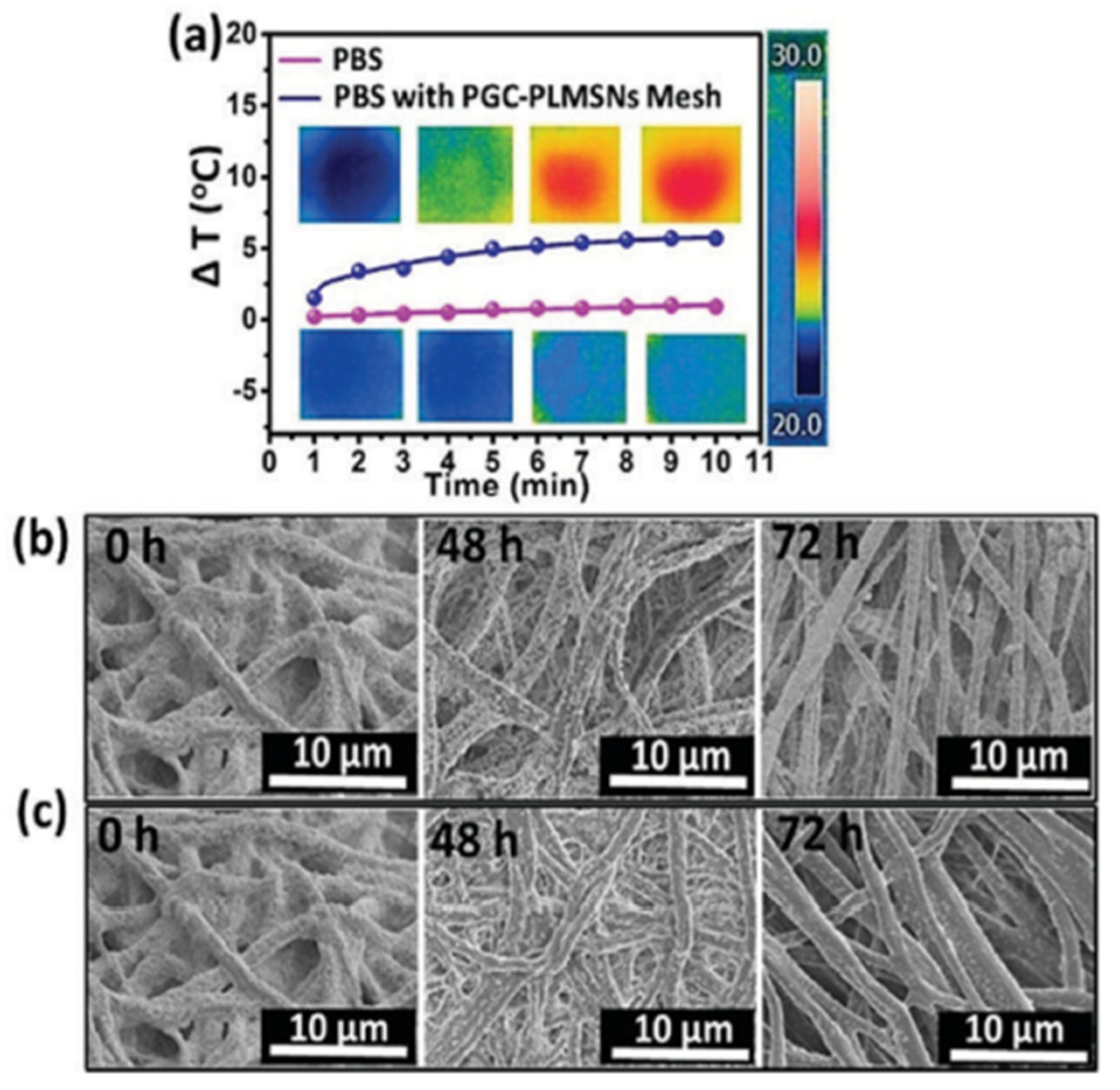
Li et al. [139] investigated the challenges faced by nanoparticle-based drug delivery systems (DDSs), particularly regarding the low efficiency of intracellular delivery. In this study, a localized drug delivery device was developed which comprised photoluminescent mesoporous silica nanoparticles (PLMSNs) integrated with a photothermal fibrous matrix. The PLMSNs were modified with a pH-sensitive polydopamine (PDA) ‘gatekeeper’ that functioned as a carrier for doxorubicin (DOX), allowing for the release of DOX upon uptake by cancer cells. The PLMSNs were electrostatically assembled onto the surface of an electrospun biodegradable fibrous mesh made from poly(ε-caprolactone)/gelatin, which was also embedded with photothermal carbon nanoparticles (CNPs). This resulted in an implantable patch designed for localized drug delivery. In comparison to conventional free particulate DDSs, this composite patch demonstrated significantly enhanced cell uptake and improved in vitro therapeutic efficacy against tumor cells. Specifically, under near-infrared irradiation, the photothermal effect (Figure 16) of the CNPs in the patch diminished the electrostatic interactions between the PLMSNs and the poly(ε-caprolactone)/gelatin/CNP fibrous mesh, facilitating the controlled release of the PLMSNs and their subsequent internalization into tumor cells, thereby promoting more effective cancer cell destruction. This innovative therapeutic device may serve as a model for advancing localized cancer treatment strategies.

3.2. Future Trends

The studies discussed in this review show that, besides biocompatibility, other properties have been incorporated, such as regeneration and bioactivity. Textile structures have higher mechanical properties compared to analogous yarns or filaments, and hence, much research can be focused on this field. With the advancement of science, new technologies and processes have been incorporated into the fabrication of 3D multidirectional structures, as in the case of four-dimensional 3D printing. Additionally, the manufacturing of smart devices to respond to different environmental stimuli is of great importance in human body applications. Multifunctional textile constructions with antibacterial, cell adhesion, biocompatible, and cell-regenerating capabilities are being emphasized. Nanofibers for wound healing applications have been created using the Arg-Gly-Asp (RGD) peptide, which stimulates cell proliferation, and the antibiotic doxycycline. To design nanofibrous textile structures that encourage cell regeneration, it may be essential to comprehend cell–nanofiber interactions and cell-signaling pathways. Technologies for textile manufacture are being combined and altered to create three-dimensional structures that guarantee cell alignment. New developments in 3D printing and additive manufacturing (AM) have created opportunities for a better and more efficient version of the multidirectional textile framework. The fabrication process has several restrictions in terms of production complexity, even though three-dimensional structures can be created using conventional textile techniques. This procedure has been mechanized using additive manufacturing, which also guarantees reduced energy loss. In addition to the traditional layer-by-layer deposition technique, this technology has been further improved to enable one-step 3D printing utilizing computed axial lithography (CAL). It is also possible to use four-dimensional 3D printing to produce “smart” gadgets that can react to environmental cues and perform at their best when responding to them.
For appropriate use in biomedical applications, fibers from alternative natural sources—especially plant-based sources—are being produced in addition to the existing and well-established natural fibers. Numerous cell types have shown encouraging growth and proliferation in response to these fibers; some even multiplied without any alterations. Furthermore, tensile tests have demonstrated that palm fibers are just as strong and elastic as tendons. Palm fibers used in applications such as tendon tissue engineering or hernia repair meshes may be a viable and sustainable choice. Another strategy is to employ naturally occurring cellulose fibers from plant sources, like sisal (Agave sisalana) and coconut (Cocos nucifera L.), as a possible suture material. Although more research is required for widespread use, they can be used as an alternative strategy to lower the risk of surgical site infections.

Author Contributions

Conceptualization, H.L.O.J.; validation, H.L.O.J. and J.P.G.; formal analysis, H.L.O.J.; investigation, H.L.O.J.; writing—original draft preparation, H.L.O.J. and J.P.G.; writing—review and editing, H.L.O.J. and J.P.G.; visualization, H.L.O.J. and J.P.G.; supervision, H.L.O.J.; project administration, H.L.O.J. All authors have read and agreed to the published version of the manuscript.

Funding

This research received no external funding.

Data Availability Statement

No data are available.

Conflicts of Interest

The authors declare no conflicts of interest.

References

  1. Cherenack, K.; van Pieterson, L. Smart textiles: Challenges and opportunities. J. Appl. Phys. 2012, 112, 091301. [Google Scholar] [CrossRef]
  2. van Langenhove, L.; Hertleer, C. Smart clothing: A new life. Int. J. Cloth. Sci. Technol. 2004, 16, 63–72. [Google Scholar] [CrossRef]
  3. Karimah, A.; Ridho, M.R.; Munawar, S.S.; Adi, D.S.; Damayanti, R.; Subiyanto, B.; Fatriasari, W.; Fudholi, A. A review on natural fibers for development of eco-friendly bio-composite: Characteristics, and utilizations. J. Mater. Rese. Technol. 2021, 13, 2442–2458. [Google Scholar] [CrossRef]
  4. Xing, T.; He, A.; Huang, Z.; Luo, Y.; Zhang, Y.; Wang, M.; Shi, Z.; Ke, G.; Bai, J.; Zhao, S.; et al. Silk-based flexible electronics and smart wearable Textiles: Progress and beyond. Chem. Eng. J. 2023, 474, 145534. [Google Scholar] [CrossRef]
  5. Williams, D.F. Definitions in Biomaterials: Proceedings of a Consensus Conference of the European Society for Biomaterials; Elsevier Science Limited: London, UK, 1987; pp. 3–5. [Google Scholar]
  6. Rostamitabar, M.; Abdelgawad, A.M.; Jockenhoevel, S.; Ghazanfari, S. Drug-eluting medical textiles: From fiber production and textile fabrication to drug loading and delivery. Macromol. Biosci. 2021, 21, 2100021. [Google Scholar] [CrossRef]
  7. Libanori, A.; Chen, G.; Zhao, X.; Zhou, Y.; Chen, J. Smart textiles for personalized healthcare. Nat. Electron. 2022, 5, 142–156. [Google Scholar] [CrossRef]
  8. Cesarelli, G.; Donisi, L.; Coccia, A.; Amitrano, F.; D’Addio, G.; Ricciardi, C. The E-textile for biomedical applications: A systematic review of literature. Diagnostics 2021, 11, 2263. [Google Scholar] [CrossRef]
  9. Ornaghi, H.L., Jr.; Neves, R.M.; Monticelli, F.M.; Dall Agnol, L. Smart Fabric Textiles: Recent advances and challenges. Textiles 2022, 2, 582–605. [Google Scholar] [CrossRef]
  10. Ornaghi, H.L., Jr.; Bianchi, O. Temperature-dependent shape-memory textiles: Physical principles and applications. Textiles 2023, 3, 257–274. [Google Scholar] [CrossRef]
  11. Kubley, A.; Chitranshi, M.; Hou, X.; Schulz, M. Manufacturing and characterization of customizable flexible carbon nanotube fabrics for smart wearable applications. Textiles 2021, 1, 534–546. [Google Scholar] [CrossRef]
  12. Islam, M.R.; Golovin, K.; Dolez, P.I. Clothing thermophysiological Comfort: A textile science perspective. Textiles 2023, 3, 353–407. [Google Scholar] [CrossRef]
  13. Dolez, P.I.; Marsha, S.; McQueen, R.H. Fibers and textiles for personal protective equipment: Review of recent progress and perspectives on future developments. Textiles 2022, 2, 349–381. [Google Scholar] [CrossRef]
  14. Mondal, M.I.H. Medical Textiles from Natural Resources, 1st ed.; Woodhead Publishing: Amsterdam, The Netherlands, 2022; p. 934. [Google Scholar]
  15. da Silva, C.J.G.; de Medeiros, A.D.M.; de Amorim, J.D.P.; do Nascimento, H.A.; Converti, A.; Costa, A.F.S.; Sarubbo, L.A. Bacterial celulose biotextiles for the future of sustainable fashion: A review. Environ. Chem. Lett. 2021, 19, 2967–2980. [Google Scholar] [CrossRef]
  16. Biomedical Textiles Market Worth $20.7 Billion by 2027—Exclusive Report by Markets and Markets™. Available online: https://www.prnewswire.com/news-releases/biomedical-textiles-market-worth-20-7-billion-by-2027--exclusive-report-by-marketsandmarkets-301698086.html (accessed on 11 November 2024).
  17. Indira, M.; Sudarsini, B.; Sumalatha, B. Advancements in Implantable Medical Textile Materials. In Textile Materials for Good Health and Wellbeing: Design and Applications; Springer Nature: Singapore, 2024; pp. 197–229. [Google Scholar]
  18. Baptista-Silva, S.; Borges, S.; Brassesco, M.E.; Ezequiel, R.C.; Oliveira, A.L.; Pintado, M. Research, development and future trends for medical textile products. In Medical Textiles from Natural Resources; Woodhead Publishing: Cambridge, UK, 2022; pp. 795–828. [Google Scholar]
  19. Bazaka, K.; Jacob, M.V. Implantable devices: Issues and challenges. Electronics 2012, 2, 1–34. [Google Scholar] [CrossRef]
  20. Drug Delivery Systems Market Size, Share & Industry Analysis, By Type (Inhalation, Transdermal, Injectable, and Others), By Device Type (Conventional and Advanced), By Distribution Channel (Hospital Pharmacies, Retail Pharmacies, and Others), and Regional Forecast, 2024–2032. Available online: https://www.fortunebusinessinsights.com/drug-delivery-systems-market-103070 (accessed on 1 July 2024).
  21. Drug Delivery Systems. Available online: https://www.nibib.nih.gov/science-education/science-topics/drug-delivery-systems-getting-drugs-their-targets-controlled-manner (accessed on 2 July 2024).
  22. Pei, J.; Yan, Y.; Palanisamy, C.P.; Jayaraman, S.; Natarajan, P.M.; Umapathy, V.R.; Gopathy, S.; Roy, J.R.; Sadagopan, J.C.; Thalamati, D.; et al. Materials-based drug delivery approaches: Recent advances and future perspectives. Green Process. Synth. 2024, 13, 20230094–20230123. [Google Scholar] [CrossRef]
  23. Hussain, M.; Hussain, M.S. A brief review on buccal drug delivery system: Advantages, limitations, and impact on healthcare system. World J. Pharm. Res. 2021, 10, 558–576. [Google Scholar]
  24. Nayak, B.S.; Sourajit, S.; Palo, M.; Behera, S. Sublingual drug delivery system: A novel approach. Int. J. Pharm. Drug Anal. 2017, 5, 399–405. [Google Scholar]
  25. Osmalek, T.; Froelich, A.; Jadach, B.; Tatarek, A.; Gadziński, P.; Falana, A.; Gralińska, K.; Ekert, M.; Puri, V.; Wrotyńska-Barzyńska, J.; et al. Recent advances in polymer-based vaginal drug delivery systems. Pharmaceutics 2021, 13, 884. [Google Scholar] [CrossRef]
  26. Das Neves, J.; Sarmento, B. Precise engineering of dapivirine-loaded nanoparticles for the development of anti-HIV vaginal microbicides. Acta Biomater. 2015, 18, 77–87. [Google Scholar] [CrossRef]
  27. Gu, J.; Yang, S.; Ho, E.A. Biodegradable film for the targeted delivery of siRNA-loaded nanoparticles to vaginal immune cells. Mol. Pharm. 2015, 12, 2889–2903. [Google Scholar] [CrossRef]
  28. Rathi, R.; Sanshita, M.; Kumar, A.; Vishvakarma, V.; Huanbutta, K.; Singh, I.; Sangnim, T. Advancements in rectal drug delivery systems: Clinical trials, and patents perspective. Pharmaceutics 2022, 17, 2210. [Google Scholar] [CrossRef] [PubMed]
  29. Katare, P.; Medhe, T.P.; Nadkarni, A.; Deshpande, M.; Tekade, R.T.; Benival, D.; Jain, A. Nasal drug delivery system and devices: An overview on health effects. ACS Chem. Health Saf. 2024, 31, 127–143. [Google Scholar] [CrossRef]
  30. Laffleur, F.; Bauer, B. Progress in nasal drug delivery systems. Int. J. Pharm. 2021, 607, 120994. [Google Scholar] [CrossRef] [PubMed]
  31. Patel, A.; Cholkar, K.; Agrahari, V.; Mitra, A.K. Ocular drug delivery systems: An overview. World J. Pharmacol. 2013, 2, 47–64. [Google Scholar] [CrossRef]
  32. Chu, J.N.; Traverso, G. Foundations of gastrointestinal-based drug delivery and future developments. Nat. Rev. Gastroenterol. Hepatol. 2022, 19, 219–238. [Google Scholar] [CrossRef]
  33. Afzal, O.; Altamimi, A.S.A.; Nadeem, M.S.; Alzarea, S.I.; Almalki, W.H.; Tariq, A.; Mubeen, B.; Murtaza, B.N.; Iftikhar, S.; Riaz, M.; et al. Nanoparticles in drug delivery: From history to therapeutic applications. Nanomaterials 2022, 12, 4494. [Google Scholar] [CrossRef]
  34. Torchilin, V.P. Recent advances with liposomes as pharmaceutical carriers. Nat. Rev. Drug Discov. 2005, 4, 145–160. [Google Scholar] [CrossRef]
  35. Couvreur, P.; Vauthier, C. Poly alkyl cyanoacrylate nanoparticles as drug carrier: Present state and perspectives. J. Control. Release 1991, 17, 187–198. [Google Scholar] [CrossRef]
  36. Vauthier, C.; Dubernet, C.; Chauvierre, C.; Brigger, I.; Couvreur, P. Drug delivery to resistant tumors: The potential of poly (alkyl cyanoacrylate) nanoparticles. J. Control. Release 2003, 93, 151–160. [Google Scholar] [CrossRef]
  37. Shinto, Y.; Uchida, A.; Korkusuz, F.; Araki, N.; Ono, K. Calcium hydroxyapatite ceramic used as a delivery system for antibiotics. J. Bone Jt. Surg. Br. 1992, 74, 600–604. [Google Scholar] [CrossRef]
  38. Esterhai, J.L., Jr.; Bednar, J.; Kimmelman, C.P. Gentamicin-induced ototoxicity complicating treatment of chronic osteomyelitis. Clin. Orthop. 1986, 209, 185–188. [Google Scholar] [CrossRef]
  39. Couvreur, P.; Puisieux, F. Nano-and microparticles for the delivery of polypeptides and proteins. Adv. Drug Deliv. Rev. 1993, 10, 141–162. [Google Scholar] [CrossRef]
  40. Hwang, S.R.; Byun, Y. Advances in oral macromolecular drug delivery. Expert Opin. Drug Deliv. 2014, 11, 1955–1967. [Google Scholar] [CrossRef] [PubMed]
  41. Müller, J.J.; Lukowski, G.; Kröber, R.; Damaschun, G.; Dittgen, M. Acrylic acid copolymer nanoparticles for drug delivery: Structural characterization of nanoparticles by small-angle X-ray scattering. Colloid Polym. Sci. 1994, 272, 755–769. [Google Scholar] [CrossRef]
  42. Lukowski, G.; Müller, R.H.; Müller, B.W.; Dittgen, M. Acrylic acid copolymer nanoparticles for drug delivery. Part II: Characterization of nanoparticles surface-modified by adsorption of ethoxylated surfactants. Colloid Polym. Sci. 1993, 271, 100–105. [Google Scholar] [CrossRef]
  43. Fresta, M.; Puglisi, G.; Giammona, G.; Cavallaro, G.; Micali, N.; Furneri, P.M. Pefloxacine mesilate-and ofloxacin-loaded poly ethyl cyanoacrylate nanoparticles: Characterization of the colloidal drug carrier formulation. J. Pharm. Sci. 1995, 74, 895–902. [Google Scholar] [CrossRef]
  44. Cavallaro, G.; Fresta, M.; Giammona, G.; Puglisi, G.; Villari, A. Entrapment of β-lactams antibiotics in polyethylcyanoacrylate nanoparticles: Studies on the possible in vivo application of this colloidal delivery system. Int. J. Pharm. 1994, 111, 31–41. [Google Scholar] [CrossRef]
  45. Pardridge, W.M. Physiologic-based strategies for protein drug delivery to the brain. J. Control. Release 1996, 39, 281–286. [Google Scholar] [CrossRef]
  46. Partridge, W.M. Drug and gene targeting to the brain via blood–brain barrier receptor-mediated transport systems. Int. Congr. Ser. 2005, 1277, 49–62. [Google Scholar] [CrossRef]
  47. Labhasetwar, V.; Song, C.; Levy, R.J. Nanoparticle drug delivery system for restenosis. Adv. Drug Deliv. Rev. 1997, 24, 63–85. [Google Scholar] [CrossRef]
  48. Labhasetwar, V.; Underwood, T.; Schwendeman, S.P.; Levy, R.J. Iontophoresis for modulation of cardiac drug delivery in dogs. Proc. Natl. Acad. Sci. USA 1995, 92, 2612–2616. [Google Scholar] [CrossRef] [PubMed]
  49. Kwon, G.S. Diblock copolymer nanoparticles for drug delivery. Crit. Rev. Ther. Drug Carr. Syst. 1998, 5, 481–512. [Google Scholar] [CrossRef]
  50. Kataoka, K.; Harada, A.; Nagasaki, Y. Block copolymer micelles for drug delivery: Design, characterization and biological significance. Adv. Drug Deliv. Rev. 2012, 64, 37–48. [Google Scholar] [CrossRef]
  51. Fernández-Urrusuno, R.; Calvo, P.; Remuñán-López, C.; Vila-Jato, J.L.; Alonso, M.J. Enhancement of nasal absorption of insulin using chitosan nanoparticles. Pharm. Res. 1999, 16, 1576–1581. [Google Scholar] [CrossRef]
  52. Zhang, X.; Zhang, H.; Wu, Z.; Wang, Z.; Niu, H.; Li, C. Nasal absorption enhancement of insulin using PEG-grafted chitosan nanoparticles. Eur. J. Pharm. Biopharm. 2008, 68, 526–534. [Google Scholar] [CrossRef]
  53. Kong, G.; Braun, R.D.; Dewhirst, M.W. Hyperthermia enables tumor-specific nanoparticle delivery: Effect of particle size. Cancer Res. 2000, 60, 4440–4445. [Google Scholar]
  54. May, J.P.; Li, S.-D. Hyperthermia-induced drug targeting. Expert Opin. Drug Deliv. 2013, 10, 511–527. [Google Scholar] [CrossRef]
  55. Calvo, P. PEGylated poly cyanoacrylate nanoparticles as vector for drug delivery in prion diseases. J. Neurosci. Methods 2001, 111, 151–155. [Google Scholar] [CrossRef]
  56. Collinge, J. Molecular neurology of prion disease. J. Neurol. Neurosurg. Psychiatry 2005, 76, 906–919. [Google Scholar] [CrossRef]
  57. Qian, Z.M.; Li, H.; Sun, H.; Ho, K. Targeted drug delivery via the transferrin receptor-mediated endocytosis pathway. Pharmacol. Rev. 2002, 54, 561–587. [Google Scholar] [CrossRef]
  58. Ulbrich, K.; Hekmatara, T.; Herbert, E.; Kreuter, J. Transferrin-and transferrin-receptor-antibody6modified nanoparticles enable drug delivery across the blood–brain barrier (BBB). Eur. J. Pharm. Biopharm. 2009, 71, 251–256. [Google Scholar] [CrossRef] [PubMed]
  59. Shankar, S.S.; Ahmad, A.; Pasricha, R.; Sastry, M. Bio reduction of chloroaurate ions by geranium leaves and its endophytic fungus yields gold nanoparticles of different shapes. J. Mater. Chem. 2003, 13, 1822–1826. [Google Scholar] [CrossRef]
  60. Panyam, J.; Labhasetwar, V. Targeting intracellular targets. Curr. Drug Deliv. 2004, 1, 235–247. [Google Scholar] [CrossRef]
  61. Ashihara, H.; Suzuki, T. Distribution and biosynthesis of caffeine in plants. Front. Biosci. 2005, 9, 1864–7336. [Google Scholar] [CrossRef]
  62. Paciotti, G.F.; Kingston, D.G.; Tamarkin, L. Colloidal gold nanoparticles: A novel nanoparticle platform for developing multifunctional tumor-targeted drug delivery vectors. Drug Dev. Res. 2006, 67, 47–54. [Google Scholar] [CrossRef]
  63. Hattori, Y.; Maitani, Y. Folate-linked lipid-based nanoparticle for targeted gene delivery. Curr. Drug Deliv. 2005, 2, 243–252. [Google Scholar] [CrossRef]
  64. Lu, Y.; Low, P.S. Folate-mediated delivery of macromolecular anticancer therapeutic agents. Adv. Drug Deliv. Rev. 2002, 54, 675–693. [Google Scholar] [CrossRef]
  65. Xiao, S. Preparation of folate-conjugated starch nanoparticles and its application to tumor-targeted drug delivery vector. Chin. Sci. Bull. 2006, 51, 1693–1697. [Google Scholar] [CrossRef]
  66. Yu, D.; Xiao, S.; Tong, C.; Chen, L.; Liu, X. Dialdehyde starch nanoparticles: Preparation and application in drug carrier. Chin. Sci. Bull. 2007, 52, 2913–2918. [Google Scholar] [CrossRef]
  67. Han, G.; Ghosh, P.; Rotello, V.M. Multi-Functional Gold Nanoparticles for Drug Delivery. In Bio-Applications of Nanoparticles; Chan, W.C.W., Ed.; Springer: New York, NY, USA, 2007; pp. 48–56. [Google Scholar]
  68. Ghosh, P.; Han, G.; De, M.; Kim, C.K.; Rotello, V.M. Gold nanoparticles in delivery applications. Adv. Drug Deliv. Rev. 2008, 60, 1307–1315. [Google Scholar] [CrossRef]
  69. Kim, H.S.; Lee, D.Y. Near-infrared-responsive cancer photothermal and photodynamic therapy using gold nanoparticles. Polymers 2018, 10, 961. [Google Scholar] [CrossRef] [PubMed]
  70. Cheng, Y.; Samia, A.C.; Meyers, J.D.; Panagopoulos, I.; Fei, B.; Burda, C. Highly efficient drug delivery with gold nanoparticle vectors for in vivo photodynamic therapy of cancer. J. Am. Chem. Soc. 2008, 130, 10643–10647. [Google Scholar] [CrossRef] [PubMed]
  71. Gazori, T.; Khoshayand, M.R.; Azizi, E.; Yazdizade, P.; Nomani, A.; Haririan, I. Evaluation of Alginate/Chitosan nanoparticles as antisense delivery vector: Formulation, optimization and in vitro characterization. Carbohydr. Polym. 2009, 77, 599–606. [Google Scholar] [CrossRef]
  72. Sarmento, B.; Ribeiro, A.J.; Veiga, F.; Ferreira, D.C.; Neufeld, R.J. Insulin-loaded nanoparticles are prepared by alginate ionotropic pre-gelation followed by chitosan polyelectrolyte complexation. J. Nanosci. Nanotechnol. 2007, 7, 2833–2847. [Google Scholar] [CrossRef]
  73. Rosenholm, J.M.; Peuhu, E.; Bate-Eya, L.T.; Eriksson, J.E.; Sahlgren, C.; Lindén, M. Cancer-Cell-Specific Induction of Apoptosis Using Mesoporous Silica Nanoparticles as Drug-Delivery Vectors. Small 2010, 6, 1234–1241. [Google Scholar] [CrossRef]
  74. Kohler, N.; Sun, C.; Wang, J.; Zhang, M. Methotrexate-modified superparamagnetic nanoparticles and their intracellular uptake into human cancer cells. Langmuir 2005, 21, 8858–8864. [Google Scholar] [CrossRef]
  75. Alhaddad, A. Nanodiamond as a vector for siRNA delivery to Ewing sarcoma cells. Small 2011, 21, 3087–3095. [Google Scholar] [CrossRef]
  76. Mengesha, A.E.; Youan, B.C. Nano diamonds for drug delivery systems. In Diamond-Based Materials for Biomedical Applications; Elsevier: Amsterdam, The Netherlands, 2013; pp. 186–205. [Google Scholar]
  77. Arjunan, N.K.; Murugan, K.; Rejeeth, C.; Madhiyazhagan, P.; Barnard, D.R. Green Synthesis of Silver Nanoparticles for the Control of Mosquito Vectors of Malaria, Filariasis, and Dengue. Vector-Borne Zoonotic Dis. 2012, 12, 262–268. [Google Scholar] [CrossRef]
  78. Jadoun, S.; Arif, R.; Jangid, N.K.; Meena, R.K. Green synthesis of nanoparticles using plant extracts: A review. Environ. Chem. Lett. 2021, 19, 355–374. [Google Scholar] [CrossRef]
  79. Brown, P.K.; Qureshi, A.T.; Moll, A.N.; Hayes, D.J.; Monroe, W.T. Silver Nanoscale Antisense Drug Delivery System for Photoactivated Gene Silencing. ACS Nano 2013, 7, 2948–2959. [Google Scholar] [CrossRef]
  80. Minelli, C.; Lowe, S.B.; Stevens, M.M. Engineering nanocomposite materials for cancer therapy. Small 2010, 21, 2336–2357. [Google Scholar] [CrossRef] [PubMed]
  81. Rajeshkumar, S. Synthesis of silver nanoparticles using fresh bark of Pongamia pinnata and characterization of its antibacterial activity against gram positive and gram negative pathogens. Resour.-Effic. Technol. 2016, 2, 30–35. [Google Scholar]
  82. Beg, M. Green synthesis of silver nanoparticles using Pongamia pinnata seed: Characterization, antibacterial property, and spectroscopic investigation of interaction with human serum albumin. J. Mol. Recognit. 2017, 30, e2565. [Google Scholar] [CrossRef] [PubMed]
  83. Urbán, P.; Ranucci, E.; Fernàndez-Busquets, X. Polyamidoamine nanoparticles as nanocarriers for the drug delivery to malaria parasite stages in the mosquito vector. Nanomed 2015, 10, 3401–3414. [Google Scholar] [CrossRef]
  84. Chamundeeswari, M.; Jeslin, J.; Verma, M.L. Nanocarriers for drug delivery applications. Environ. Chem. Lett. 2019, 17, 849–865. [Google Scholar] [CrossRef]
  85. Kulbacka, J. Electroporation and lipid nanoparticles with cyanine IR-780 and flavonoids as efficient vectors to enhanced drug delivery in colon cancer. Bioelectrochemistry 2016, 110, 19–31. [Google Scholar] [CrossRef]
  86. Lamichhane, T.N.; Raiker, R.S.; Jay, S.M. Exogenous DNA loading into extracellular vesicles via electroporation is size-dependent and enables limited gene delivery. Mol. Pharm. 2015, 12, 3650–3657. [Google Scholar] [CrossRef]
  87. Ju, Z.; Sun, W. Drug delivery vectors based on filamentous bacteriophages and phage-mimetic nanoparticles. Drug Deliv. 2017, 24, 1898–1908. [Google Scholar] [CrossRef]
  88. Jahromi, M.A.M.; Zangabad, P.S.; Basri, S.M.M.; Zangabad, K.S.; Ghamarypour, A.; Aref, A.R. Recent progress in targeted delivery vectors based on biomimetic nanoparticles. Signal Transduct. Target. Ther. 2021, 6, 225. [Google Scholar]
  89. Aljabali, A.A. Innovative Applications of Plant Viruses in Drug Targeting and Molecular Imaging—A Review. Curr. Med. Imaging 2021, 17, 491–506. [Google Scholar] [CrossRef]
  90. Slita, A.; Egorova, A.; Casals, E.; Kiselev, A.; Rosenholm, J.M. Characterization of modified mesoporous silica nanoparticles as vectors for siRNA delivery. Asian J. Pharm. Sci. 2018, 13, 592–599. [Google Scholar] [CrossRef] [PubMed]
  91. Wu, S.H.; Mou, C.Y.; Lin, H.P. Synthesis of mesoporous silica nanoparticles. Chem. Soc. Rev. 2013, 42, 3862–3875. [Google Scholar] [CrossRef] [PubMed]
  92. Garg, U.; Chauhan, S.; Nagaich, U.; Jain, N. Current Advances in Chitosan Nanoparticles Based Drug Delivery and Targeting. Adv. Pharm. Bull. 2019, 9, 195–204. [Google Scholar] [CrossRef] [PubMed]
  93. Pathak, C.; Vaidya, F.U.; Pandey, S.M. Mechanism for development of nanobased drug delivery system. Appl. Target. Nano Drugs Deliv. Syst. 2019, 1, 35–67. [Google Scholar]
  94. Ghaz-Jahanian, M.A.; Abbaspour-Aghdam, F.; Anarjan, N.; Berenjian, A.; Jafarizadeh-Malmiri, H. Application of chitosan-based nanocarriers in tumor-targeted drug delivery. Mol. Biotechnol. 2015, 157, 201–218. [Google Scholar] [CrossRef]
  95. Assa, F. Chitosan magnetic nanoparticles for drug delivery systems. Crit. Rev. Biotechnol. 2017, 37, 492–509. [Google Scholar] [CrossRef]
  96. Li, Y.; Wang, S.; Song, F.X.; Zhang, L.; Yang, W.; Wang, H.X. A pH-sensitive drug delivery system based on folic acid-targeted HBP-modified mesoporous silica nanoparticles for cancer therapy. Colloids Surf. Physicochem. Eng. Asp. 2020, 590, 124470. [Google Scholar] [CrossRef]
  97. Shafiei, N.; Nasrollahzadeh, M.; Iravani, S. Green Synthesis of Silica and Silicon Nanoparticles and Their Biomedical and Catalytic Applications. Comments Inorg. Chem. 2021, 41, 317–372. [Google Scholar] [CrossRef]
  98. Shariatinia, Z. Inorganic Material-Based Nanocarriers for Delivery of Biomolecules. Nanoeng. Biomater. Biomed. Appl. 2022, 2, 245–293. [Google Scholar]
  99. Gao, Y.; Gu, S.; Zhang, Y.; Xie, X.; Yu, T.; Lu, Y.; Zhu, Y.; Chen, W.; Zhang, H.; Dong, H.; et al. The architecture and function of monoclonal antibody-functionalized mesoporous silica nanoparticles loaded with mifepristone: Repurposing abortifacient for cancer metastatic chemoprevention. Small 2016, 12, 2595–2608. [Google Scholar] [CrossRef]
  100. Gencturk, A.; Kahraman, E.; Güngör, S.; Özhan, G.; Özsoy, Y.; Sarac, A.S. Polyurethane/hydroxypropyl cellulose electrospun nanofiber mats as potential transdermal drug delivery system: Characterization studies and in vitro assays. Artif. Cells Nanomed. Biotechnol. 2017, 45, 655–664. [Google Scholar] [CrossRef]
  101. Youseff, N.A.H.A.; Kassem, A.A.; El-Massik, M.A.E.; Boraie, N.A. Development of gastroretentive metronidazole floating raft system for targeting Helicobacter pylori. Int. J. Pharm. 2015, 486, 297–305. [Google Scholar] [CrossRef]
  102. Yu, D.-G.; Branford-White, C.; White, K.; Li, X.-L.; Zhu, L.-M. Dissolution improvement of electrospun nanofiber-based solid dispersions for acetaminophen. AAPS PharmSciTech 2010, 11, 809–817. [Google Scholar] [CrossRef]
  103. Zhu, L.-M.; Yu, D.G. Drug delivery systems using biotextiles. In Biotextiles as Medical Implants; Woodhead Publishing Series in Textiles; Woodhead Publishing: Cambridge, UK, 2013; pp. 213–231. [Google Scholar]
  104. Zilberman, M. Novel composite fiber structures to provide drug/protein delivery for medical implants and tissue regeneration. Acta Biomat. 2007, 3, 51–57. [Google Scholar] [CrossRef]
  105. Sikareepaisan, P.; Suksamrarn, A.; Supaphol, P. Electrospun gelatin fiber mats containing a herbal—Centella asiatica—Extract and release characteristic of asiaticoside. Nanotechnology 2007, 19, 015102. [Google Scholar] [CrossRef]
  106. Suwantong, O.; Opanasopit, P.; Ruktanonchai, U.; Supaphol, P. Electrospun cellulose acetate fiber mats containing curcumin and release characteristic of the herbal substance. Polymer 2007, 48, 7546–7557. [Google Scholar] [CrossRef]
  107. Khoshbakht, S.; Asghari-Sana, F.; Fathi-Azarbayjani, A.; Sharifi, Y. Fabrication and characterization of tretinoin-loaded nanofiber for topical skin delivery. Biomat. Res. 2020, 24, 8. [Google Scholar] [CrossRef]
  108. Garg, A.; Alfatease, A.; Hani, U.; Haider, N.; Akbar, M.J.; Talath, S.; Angolkar, M.; Paramshetti, S.; Osmani, R.A.M.; Gundawar, R. Drug eluting protein and polysaccharides-based biofunctionalized fabric textiles- pioneering a new frontier in tissue engineering: An extensive review. Int. J. Biol. Macromol. 2024, 268, 131605. [Google Scholar] [CrossRef]
  109. Arik, B. Smart bio-textiles for medicine and healthcare applications. In Smart Textiles from Natural Resources; Woodhead Publishing: Cambridge, UK, 2024; pp. 495–537. [Google Scholar]
  110. Kim, Y.E.; Jung, H.Y.; Park, N.; Kim, J. Adhesive composite hydrogel patch for sustained transdermal drug delivery to treat atopic dermatitis. Chem. Mater. 2023, 35, 1209–1217. [Google Scholar] [CrossRef]
  111. Chen, G.; Au, C.; Chen, J. Textile triboelectric nanogenerators for wearable pulse wave monitoring. Trends Biotechnol. 2021, 39, 1078–1092. [Google Scholar] [CrossRef]
  112. Wu, X.; Liu, R.; Lao, T.T. Therapeutic compression materials and wound dressings for chronic venous insufficiency: A comprehensive review. J. Biomed. Mater. Res. B Appl. Biomater. 2020, 108, 892–909. [Google Scholar] [CrossRef]
  113. Szunerits, S.; Boukherroub, R. Heat: A highly efficient skin enhancer for transdermal drug delivery. Front. Bioeng. Biotechnol. 2018, 6, 15. [Google Scholar] [CrossRef]
  114. Jackson, J.M.; Weke, A.; Holliday, R. Nicotine pouches: A review for the dental team. Br. Dent. J. 2023, 235, 643–646. [Google Scholar] [CrossRef]
  115. Bhutto, M.A.; Wu, T.; Sun, B.; Ei-Hamshary, H.; Al-Deyab, S.S.; Mo, X. Fabrication and characterization of vitamin B5 loaded poly (l-lactide-co-caprolactone)/silk fiber aligned electrospun nanofibers for schwann cell proliferation. Colloids Surf. B Biointerfaces 2016, 144, 108–117. [Google Scholar] [CrossRef]
  116. Lee, J.B.; Kim, J.E.; Balikov, D.A.; Bae, M.S.; Heo, D.N.; Lee, D.; Rim, H.J.; Lee, D.-W.; Sung, H.-J.; Kwon, I.K. Poly (l-lactic acid)/gelatin fibrous scaffold loaded with simvastatin/beta-cyclodextrin-modified hydroxyapatite inclusion complex for bone tissue regeneration. Macromol. Biosci. 2016, 16, 1027–1038. [Google Scholar] [CrossRef]
  117. Grimaudo, M.A.; Concheiro, A.; Alvarez-Lorenzo, C. Crosslinked hyaluronan electrospun nanofibers for ferulic acid ocular delivery. Pharmaceutics 2020, 12, 274. [Google Scholar] [CrossRef]
  118. Kalantari, K.; Afifi, A.M.; Jahangirian, H.; Webster, T.J. Biomedical applications of chitosan electrospun nanofibers as a green polymer e review. Carbohydr. Polym. 2019, 207, 588–600. [Google Scholar] [CrossRef]
  119. Hemamalini, T.; Giri Dev, V.R. Comprehensive review on electrospinning of starch polymer for biomedical applications. Int. J. Biol. Macromol. 2018, 106, 712–718. [Google Scholar] [CrossRef]
  120. Kuwabara, M.; Sato, Y.; Ishihara, M.; Takayama, T.; Nakamura, S.; Fukuda, K.; Murakami, K.; Yokoe, H.; Kiyosawa, T. Healing of Pseudomonas aeruginosa-infected wounds in diabetic db/db mice by weakly acidic hypochlorous acid cleansing and silver nanoparticle/chitin-nanofiber sheet covering. Wound Med. 2020, 28, 100183. [Google Scholar] [CrossRef]
  121. Miranda, C.S.; Ribeiro, A.R.M.; Homem, N.C.; Felgueiras, H.P. Spun Biotextiles in tissue engineering and biomolecules delivery systems. Antibiotics 2020, 9, 174. [Google Scholar] [CrossRef]
  122. Xie, Y.; Guan, Y.; Kim, S.H.; King, M.W. The mechanical performance of weft-knitted/ electrospun bilayer small diameter vascular prostheses. J. Mech. Behav. Biomed. Mater. 2016, 61, 410–418. [Google Scholar] [CrossRef]
  123. Akram, S.N.B.; Jahangir, M.U.; Mondal, M.I.H.; Arafat, M.T. Biotextiles for medical implants and regenerative medicine. In Medical Textiles from Natural Resources; Woodhead Publishing: Cambridge, UK, 2022; pp. 169–211. [Google Scholar]
  124. Tamai, H.; Igaki, K.; Kyo, E.; Kosuga, K.; Kawashima, A.; Matsui, S.; Komori, H.; Tsuji, T.; Motohara, S.; Uehata, H. Initial and 6-month results of biodegradable poly-I-lactic acid coronary stents in humans. Circulation 2000, 102, 399–404. [Google Scholar] [CrossRef]
  125. Nakazawa, Y.; Asano, A.; Nakazawa, C.T.; Tsukatani, T.; Asakura, T. Structural characterization of silk-polyurethane composite material for biomaterials using solid-state NMR. Polym. J. 2012, 44, 802–807. [Google Scholar] [CrossRef]
  126. Cheng, S.; Liu, X.; Qian, Y.; Maitusong, M.; Yu, K.; Cao, N.; Fang, J.; Liu, F.; Chen, J.; Xu, D.; et al. Double-Network Hydrogel Armored Decellularized Porcine Pericardium as Durable Bioprosthetic Heart Valves. Adv. Healthc. Mater. 2022, 11, 2102059. [Google Scholar] [CrossRef]
  127. Yoon, J.; King, M.W.; Johnson, E. Designing vena cava filters with textile structures. Medical and Healthcare Textiles. In Medical Textiles from Natural Resources; Woodhead Publishing: Cambridge, UK, 2010; pp. 334–341. [Google Scholar]
  128. Dacron e Patch na Anastomose Vascular. Available online: https://biomedical.com.br/home-en/ (accessed on 17 August 2024).
  129. Russel, S.J.; Mao, N. Anisotropic fluid transmission in nonwoven wound dressings. In Medical Textiles; Woodhead Publishing: Cambridge, UK, 2001; pp. 156–163. [Google Scholar]
  130. Devlin, J.J.; Kircher, S.; Kozen, B.G.; Littlejohn, L.F.; Johnson, A.S. Comparison of ChitoFlex®, CELOX™, and QuikClot® in control of hemorrhage. J. Emerg. Med. 2011, 41, 237–245. [Google Scholar] [CrossRef]
  131. PELNACTM. Available online: https://www.gunze.co.jp/medical/e/products/item_pn.html (accessed on 27 August 2024).
  132. Wu, H.; Williams, G.R.; Wu, J.; Wu, J.; Niu, S.; Li, H.; Wang, H.; Zhu, L. Regenerated chitin fibers reinforced with bacterial cellulose nanocrystals as suture biomaterials. Carbohydr. Polym. 2018, 180, 304–313. [Google Scholar] [CrossRef]
  133. Biomedical Textiles. Available online: https://confluentmedical.com/capabilities/biomedical-textiles (accessed on 25 August 2024).
  134. Deltamed Products. Available online: https://www.deltamed.com.tr/en/urunler/nerve-regeneration-grafts-012670478267976604/neuragen-08106615366558876 (accessed on 27 August 2024).
  135. Xue, j.; Feng, B.; Zheng, R.; Lu, Y.; Zhou, G.; Liu, W.; Cao, Y.; Zhang, Y.; Zhang, W.J. Engineering ear-shaped cartilage using electrospun fibrous membranes of gelatin/polycaprolactone. Biomaterials 2013, 34, 2624–2631. [Google Scholar] [CrossRef]
  136. Zhi, Y.; Jiang, J.; Zhang, P.; Chen, S. Silk enhances the ligamentization of the polyethylene terephthalate artificial ligament in a canine anterior cruciate ligament reconstruction model. Artif. Organs 2018, 43, E94–E108. [Google Scholar] [CrossRef]
  137. Li, X.; Yang, Y.; Fan, Y.; Feng, Q.; Cui, F.-Z.; Watari, F. Biocomposites reinforced by fibers or tubes as scaffolds for tissue engineering or regenerative medicine. J. Biomed. Mater. Res. Part A 2014, 102, 1580–1594. [Google Scholar] [CrossRef]
  138. Dehari, D.; Chaudhuri, A.; Kumar, D.N.; Nath, G.; Agrawal, A.K. Fiber and textile in drug delivery to combat multidrug resistance microbial infection. In Fiber and Textile Engineering in Drug Delivery Systems; Woodhead Publishing: Cambridge, UK, 2023; pp. 359–387. [Google Scholar]
  139. Li, Y.; Fu, Y.; Ren, Z.; Li, X.; Mao, C.; Han, G. Enhanced cell uptake of fluorescent drug-loaded nanoparticles via an implantable photothermal fibrous patch for more effective cancer cell killing. J. Mater. Chem. B 2017, 5, 7504–7511. [Google Scholar] [CrossRef]
Figure 1. Examples of common and technical applications of textiles. The images were sourced from the Internet under the Creative Commons License.
Textiles 05 00019 g001
Figure 2. (A) Types of nanoparticles used in drug delivery systems, and (B) different drug delivery systems. Figure 2A was used under the CC BY 4.0 from [22].
Textiles 05 00019 g002
Figure 3. Schematic representation of different (A) routes of drug delivery and (B) delivery vehicles. The images were sourced from the Internet under the Creative Commons License.
Textiles 05 00019 g003
Figure 4. Preparation and applications of biotextiles using co-blending methods. The image is reproduced with kind permission from [103].
Textiles 05 00019 g004
Figure 5. Effectiveness of the adhesive hydrogel in the treatment of atopic dermatitis. This figure was reused under kind permission from [110].
Textiles 05 00019 g005
Figure 6. Schematic procedure of drug-loaded polymer matrix and dual functional delivery system of drug-based dressings. (a) Drug loaded with the polymer. (b) Application of the drug-loaded fabrics to a patient’s wound. (c) Working mechanism of the dual functional delivery system of drug-based dressings. The same legend was maintained from the original study. This figure is reproduced with kind permission from [112].
Textiles 05 00019 g006
Figure 7. (A) Example of how a nicotine pouch is inserted into the mouth and (B) nicotine pouches as commercially available (container with 20 pouches). This figure is reused under the Creative Commons License 4.0 from [114].
Textiles 05 00019 g007
Figure 8. Schematic illustration of a hydroxyapatite-coated PLLA/gelatin/AD fibrous scaffold for osteogenic differentiation. Step I: the synthesis of β-cyclodextrin grafted hydroxyapatite. Step II: preparation of the hydroxyapatite-coated PLLA/gelatin fibrous scaffold. SEM images of (a) PLLA/gelatin (PG), (b) HAp-coated PLLA/gelatin/adamantane (PGA-H), and (c) HAp-βCD-coated PLLA/gelatin/adamantane (PGA-HB) fibrous scaffold. The legend was maintained from the original study. The images are reproduced with kind permission from [116].
Textiles 05 00019 g008
Figure 9. Inhibition zones of Staphylococcus aureus (first row) and Pseudomonas aeruginosa (second row) caused by blank and FA-loaded inserts. The image is reused under the Creative Commons Attribution (CC BY) license [117].
Textiles 05 00019 g009
Figure 10. In vitro antibacterial activity of formulations B (tretinoin 1% w/v), D (tretinoin 1% w/v and erythromycin 0.7% w/v), and E (erythromycin 0.7% w/v) against S. aureus (ATCC® 25,923™) and S. aureus (ATCC® 29,213™). For formulations D and E, there is no significant difference in the zone of inhibition. Creative Commons Attribution 4.0 International License [107].
Textiles 05 00019 g010
Figure 11. A schematic overview presenting the fabrication process, structure, and function of the decellularized porcine pericardium coated with polyacrylamide/hyaluronic acid dual-network hydrogel. This figure is reproduced with kind permission from [126].
Textiles 05 00019 g011
Figure 12. Conventional vena cava filter (A) and the proposed prototype IVC filter (B) based on a textile construction. Image A was sourced from the Internet under the Creative Commons License, while image B is reproduced with kind permission from [127].
Textiles 05 00019 g012
Figure 13. Use of bacterial cellulose and chitin fibers on sutures. Theis figures is reproduced with kind permission from [132].
Textiles 05 00019 g013
Figure 14. Engineering ear-shaped cartilage. (A): Tailored electrospun GT/PCL membrane; (B): Titanium alloy ear-shaped mold. (C): Gross view of ear-shaped cellescaffold constructs immediately after stacking (0 h). (D): Cellescaffold constructs after 2 weeks of culture in vitro (2 wks). (E): Subcutaneous implantation in nude mice; (FH): Engineered ear-shaped cartilage after 6 weeks of in vivo incubation. (I): The similarity analysis of engineered ear compared to the titanium mold. The legend was maintained the same from the original study. This figure is reproduced with kind permission from [135].
Textiles 05 00019 g014
Figure 15. Radiographs from the defect group at 15 weeks after surgery (a); pure PLLA group at 5 weeks (b1), at 10 weeks (b2), and at 15 weeks (b3); nHACP group at 5 weeks (c1), at 10 weeks (c2), and at 15 weeks (c3); the reinforced nHACP group at 5 weeks (d1), at 10 weeks (d2), and 15 weeks (d3). The legend was maintained from the original study. This figure is reproduced with kind permission from [137].
Textiles 05 00019 g015
Figure 16. Photothermal behaviors of PGC-PLMSNs. (a) Photothermal heating curves of PBS solution containing PGC-PLMSN composite meshes and pure PBS solution under irradiation with 808 NIR at 0.6 W.cm−2. (b,c) SEM images of the PGC-PLMSN fibers after immersion in PBS solution (pH = 7.4) without (b) and with (c) NIR laser irradiation. This figure is reproduced with kind permission from [139].
Textiles 05 00019 g016
Table 1. Evolution of nanoparticles. This table was reused under the Creative Commons Attribution (CC BY) license [33].
YearTypes of NPsDrug Delivery ApproachesDiseasesApplicationsCharacterization
1991 [34,35]Poly-alkyl-cyanoacrylate nanoparticlesA carrier that delivers drugs to target specific site.CancerCancer chemotherapy and intracellular antibiotherapyScanning electron microscope (SEM)
1992 [36,37]Calcium hydroxyapatite ceramic (CHC)Drug gentamicin placed in porous blocks of calcium hydroxyapatite antibiotics (CHA).Chronic osteomyelitis (animal model)Bactericidal activity retained, with effective resultsNo in vivo experiments performed
1993 [38,39]Nano- and microparticlesMicroparticulate system used for drug administration.Enhancement of oral immune system (immunization)In vitro experiments performedNo characterization provided
1994 [40,41]Acrylic acid copolymer NPsNot specifiedNot specifiedNot specifiedNot specified
1995 [42,43]Poly-alkyl-cyanoacrylate (PECA) nanoparticlesAcrylic acid, acrylic amide, acrylic-butyl ester, and methacrylic methyl ester are used as copolymers in drug delivery. OFX and PFX systems compared.Bacterial diseasesOFX system showing greater efficient than the PFX systemNo
1996 [44,45]Protein and peptide-based NPsMonoclonal antibodies, recombinant proteins transported to the BBB by the chimeric Alzheimer’s disease peptide approach.Brain diseasesFluoroquinolone-loaded nanoparticles, enhanced antimicrobial activity. Avidin conjugated with BBB vector for protein transport across BBBIn vitro experiments performed; small angle X-ray scattering, freeze fracture electron microscopy, physicochemical characterization
1997 [46,47]NanoparticlesNanoparticles as carriers to deliver the drug to the intra-arterial localization system.Restenosis (arterial reobstruction)Catheter-based deliveryNo
1998 [48,49]Diblock copolymer nanoparticlesNot specifiedNot specifiedNot specifiedNot specified
1999 [50,51]Chitosan nanoparticlesMicelles and nanosphere carry genes and hydrophobic drugs to target site.DiabetesPotential of chitosan nanoparticles to improve insulin absorption through nasal cavityMicroAB assay to determine insulin loading and release
2000 [52,53]Liposome with hyperthermia nanoparticlesIncreased drug delivery to tumors. Hyperthermia helps liposomes work properly.Ovarian carcinoma aEnhanced drug delivery to tumorNot specified
2001 [54,55]PEGylated poly-cyano-acrylate nanoparticlesEfficient drug carrier for therapeutic molecules.Prion diseasesEffective drug delivery in prion disease testNot specified
2002 [56,57]Transferrin-mediated receptor endocytosisTransferrin and transferrin receptor in drug and gene transference via the BBB.Cancer and hepatitis BEfficient drug and gene delivery through BBBNot specified
2003 [58,59]L-nanoparticlesNot specifiedNot specifiedNot specifiedNot specified
2004 [60,61]Colloidal gold nanoparticlesNot specifiedNot specifiedUsed as a vector to carry tumor necrosis factor (TNF) toward specific parts of tumor in miceNot specified
2005 [62,63]Liposomes, nanoparticlesNot specifiedNot specifiedNot specifiedNot specified
2006 [64,65]Folate-conjugated starch nanoparticles (StNP’s)Vitamin folic acid inside cationic liposomes and conjugate liposomes as a carrier.Cancer (human nasopharyngeal and prostate tumors)Folate ligand as carrier and chemotherapeutic agent; DNA attached to receptor-bearing cancer cells in vitroNot specified
2007 [66,67]Gold nanoparticles (AuNPs)Drug and gene delivery approach.Liver cancerDrug and gene delivery using gold nanoparticles; transfection efficacy for beta-galactosidase with various MMPCsNot specified
2008 [68,69]PEGylated gold nanoparticlesVery effective drug transfers with AuNPs vector for in vivo photodynamic treatment.CancerEnhanced drug transfer with gold nanoparticles for photodynamic cancer treatmentNot specified
2009 [70,71]Alginate/chitosan (Alg/Chi) nanoparticlesNot specifiedNot specifiedTargeted drug delivery using alginate/chitosan polymersOptimization of Alg/Chi NPs preparation; no characterization provided
2010 [72,73]Mesoporous silica nanoparticlesTargeted carriage of methotrexate (MTX) to tumor cells.CancerPoly (ethylenimine)-functionalized mesoporous silica small units as vectors for drug deliveryNot specified
2011 [74,75]Nanodiamond (ND) nanoparticlesTransport of small interfering RNA into sarcoma (Ewing) cells.Ewing sarcoma cells (cancer)Evaluation of route for in vivo anticancer nucleic acid drug transferNot specified
2012 [76,77]Silver nanoparticlesNot specifiedMalaria, dengue fever, filariasisDesigned as larvicides and photoactivated vectors for drug delivery and photoactivated gene silencingZeta potential, laser Doppler anemometry, photon correlation spectroscopy
2013 [78,79]Silver nanoparticlesDesigned for photoactivated gene silencing and drug delivery.CancerEffective in human cancer treatment; long retention time in bloodNo
2014 [80,81]Silver nanoparticlesSynthesized from the plant Pongamia pinnata using the green method.MalariaMedically active; larvicidal action against Aedes aegyptiUV-visible absorption spectrum, TEM, XRD, FTIR
2015 [82,83]Polyamidoamine nanoparticlesNanocarrier for antimalarial drug delivery.DengueDelivery of antimalarial drug as nanomedicineNo
2016 [84,85]Solid lipid nanoparticles (SLNP)Electroporation and nanocarrier delivery, loaded with cyanine-type IR-780 and flavonoid derivatives.Colon cancerDelivery of chemotherapeutic agentsNo
2017 [86,87]Filamentous bacteriophage and phage-mimetic nanoparticlesDelivery of drugs and genes through phage particles.Bacterial and viral diseasesVirus-based delivery system; phages chemically altered or genetically designedFluorescence-assisted cell sorting, transmission electron microscopy, confocal immunofluorescence
2018 [88,89]Mesoporous silica nanoparticles (MSNs)Electrostatic absorption; loading with surface-hyperbranching polymerized poly(ethyleneimine).Ocular drug delivery, vaccine delivery, mucosal and nasal drug transfer, gene carriageNon-viral vectors for various drug delivery methodsTransmission electron microscopy, dynamic light scattering (DLS), zeta determination
2019 [90,91]Chitosan nanoparticlesDrug loaded onto chitosan nanoparticles for targeting various delivery sites.Not specifiedBuccal medicine distribution, vaccine transfer, cancer treatmentNo
2020 [92,93]Mesoporous silica NPs with folic acid (MSN-COOH-Tet-HBP-FA)pH-sensitive drug delivery system; folic-acid-targeted HBP.CancerEnhanced drug delivery and stability; zero pre-release within 20 h in physiological conditionsXRD, TEM, HNMR spectra, SEM, UV-analysis, TGA
2021 [94]Novel silver nanoparticlesNucleic acid sequences for protein production.SARS-CoV-2, COVID-19DNA or mRNA transport for immune response; COVID-19 treatment and preventionNo
2021 [95,96,97]Lipid-based nanoparticles, metal and metal oxide NPsDrug delivery and immuno-anti-inflammatory responses.COVID-19Vaccine success rate; used in face masks, immune sensors, and coatingsNo
2022 [98]Resveratrol–zinc NPsImmuno-anti-inflammatory response.Cancer, nervous breakdownDrug delivery for COVID-19 and cancer treatment; neuroprotective mediatorsNo
Disclaimer/Publisher’s Note: The statements, opinions and data contained in all publications are solely those of the individual author(s) and contributor(s) and not of MDPI and/or the editor(s). MDPI and/or the editor(s) disclaim responsibility for any injury to people or property resulting from any ideas, methods, instructions or products referred to in the content.