Article

Green Silver Nanoparticles: Plant-Extract-Mediated Synthesis, Optical and Electrochemical Properties

by
Natalia Stozhko
1,*,
Aleksey Tarasov
2,
Viktoria Tamoshenko
1,
Maria Bukharinova
2,
Ekaterina Khamzina
1,2 and
Veronika Kolotygina
2
1
Department of Physics and Chemistry, Ural State University of Economics, 8 Marta St., 62, 620144 Yekaterinburg, Russia
2
Scientific and Innovation Center of Sensor Technologies, Ural State University of Economics, 8 Marta St., 62, 620144 Yekaterinburg, Russia
*
Author to whom correspondence should be addressed.
Physchem 2024, 4(4), 402-419; https://doi.org/10.3390/physchem4040028
Submission received: 9 September 2024 / Revised: 5 October 2024 / Accepted: 11 October 2024 / Published: 16 October 2024
(This article belongs to the Section Electrochemistry)

Abstract

Antioxidants of plant extract play an important role in the phytosynthesis of silver nanoparticles (phyto-AgNPs), providing the reduction of silver ions and capping and stabilization of nanoparticles. Despite the current progress in the studies of phytosynthesis, there is no approach to the selection of plant extract for obtaining phyto-AgNPs with desired properties. This work shows that antioxidant activity (AOA) of plant extracts is a key parameter for targeted phytosynthesis. In support of this fact, the synthesis of phyto-AgNPs was carried out using extracts of four plants with different AOA, increasing in the order Ribes uva-crispa < Lonicera caerulea < Fragaria vesca < Hippophae rhamnoides. Phyto-AgNPs have been characterized using Fourier-transform infrared spectroscopy, transmission electron microscopy, energy-dispersive X-ray spectroscopy, selected area electron diffraction technique, ultraviolet–visible spectroscopy, electrochemical impedance spectroscopy and cyclic voltammetry. It was established that the change in the AOA of the plant extract is accompanied by a size-dependent change in the optical and electrochemical properties of phyto-AgNPs. In particular, an increase in the extract AOA leads to the formation of smaller phyto-AgNPs with higher electrochemical activity and low charge transfer resistance. A “blue shift” and an increase in the plasmon resonance band of silver sols are observed with an increase in the extract AOA. The obtained regularities prove the existence of the “AOA–size–properties” triad, which can be used for controlled phytosynthesis and prediction of phyto-AgNPs’ properties.

Graphical Abstract

1. Introduction

Nanomaterials have special catalytic, electronic, magnetic and optical properties that depend on the size and chemical composition of nanoparticles. Silver nanoparticles (AgNPs) are most popular among metal nanoparticles. Due to their unique properties, they have found application in various fields of human activity. The range of applications for silver nanoparticles is extremely diverse: AgNPs are often used in medicine, microelectronics, optics, superconductors, catalysis, wastewater purification, cosmetology, the textile industry, medical equipment, antimicrobials, biotechnology, visualization, chemical sensors and biosensors [1,2,3].
Of particular note is the enormous antibacterial and antifungal potential of silver nanoparticles, which opens up ample opportunities for their usage in areas related to the human body, primarily in the food industry, medicine and cosmetology [4,5]. Silver nanoparticles are used in food storage. In medicine, AgNPs have triggered the rise of a new approach—theranostics—to the development of drugs that both allow for an early diagnosis and serve as a therapeutic agent [6]. Silver nanoparticles assist in detecting cancerous tumors. Contrast nanoparticles are introduced into the body and accumulate in malignant tumors that can then be detected by computed tomography scans, magnetic resonance imaging or ultrasonography. Another area of AgNP application in medicine is the manufacturing of medical and orthopedic implants. Infection occurring during or after implant replacement is known to reduce the efficiency of bone regeneration. The antimicrobial property of silver nanoparticles enables the prevention of serious complications caused by infection or inflammation and acceleration of the healing process of damaged areas and cover tissues [7]. In recent years, the use of AgNPs in moisturizing and sunscreen cosmetics has been gaining in popularity [8]. It has been proven that in these products silver nanoparticles do not precipitate, unlike silver in ionic form, which ensures high quality and excellent consumer properties of cosmetic products. One of the main features of AgNPs, which attracts the attention of cosmetic producers, is targeted delivery. Due to their small size, AgNPs can transport active components located on their surface into deeper layers of the skin, thus raising the effectiveness of cosmetic products [4]. This property of silver nanoparticles is also used in medicine. Biocompatible nanoparticles ensure accurate and efficient delivery of drugs to specific cells or tissues, increasing, in this way, therapeutic efficacy in the treatment of chronic diseases and minimizing possible side effects.
The introduction of silver nanoparticles into the human body places serious demands on them. First, these nanoparticles must be safe, chemically inert, non-toxic and biocompatible and should not cause allergic reactions or side effects when interacting with living organisms. However, it is not always possible to obtain silver nanoparticles that meet these requirements by using conventional physical and chemical methods. Traditional chemical synthesis of nanoparticles is often carried out with the use of reagents that are harmful to human beings. Physical synthesis is energy-consuming and is carried out under high temperatures and pressure. The recently developed biological synthesis of nanoparticles with the use of microorganisms, while providing biocompatible AgNPs, is very labor-intensive in terms of creating special conditions and environments for microorganism functioning. In this regard, much attention is now being paid to the development of alternative simple methods for AgNP synthesis.
The synthesis with the use of plant extracts has some advantages over other methods for producing metal nanoparticles: it is simple and quick, effective, economical, affordable and eco-friendly and allows the production of biocompatible nanoparticles [9]. This synthesis is called “green” synthesis or phytosynthesis and the nanoparticles obtained by this method are called “green” nanoparticles or phytonanoparticles. A special feature of phytonanoparticles is a shell of the plant extract phytocomponents, which can reduce the risk of cytotoxicity of silver nanoparticles [10]. Various parts of plants can be used for phytosynthesis, including leaves, flowers, seeds, fruits, stems and even roots, but most often extracts from the leaves of various plants are used, for example, Vernonia amygdalina Del. [10], Moringa oleifera [11], Carissa carandos [12], Callisia fragrans [13], Mentha longifolia [14], Hagenia abyssinica (Brace) JF. Gmel. [15], Syzygium cumini [16], oriental plane [17], Costus afer [18], Uvaria narum [19], Eugenia roxburghii [20], Rosa damascene [21], Mentha arvensis, Coriandrum sativum and Cymbopogon citratus [22]. A variety of techniques can be used for obtaining plant extracts, and they are dependent on the type of plant material. This usually involves mixing fresh or crushed dried leaves with water [10,11,12,13,14,15,16,17,18,22], methanol [20] or ethanol [21] at room or elevated temperature. Microwave radiation can also be used instead of thermal heating [19]. Then the resulting plant extracts are added to a precursor (silver nitrate) to obtain metallic nanoparticles (phyto-AgNPs). Solid-phase synthesis can be used to produce oxide nanoparticles (phyto-Ag2ONPs) by solvent-free combustion of plant powders [23]. In the phytosynthesis of nanoparticles, plant extracts act as reducing, stabilizing and capping agents [16]. They contain flavonoids, pigments, enzymes, polyphenols, amino acids, proteins, alkaloids, terpenoids, alcohol compounds and polysaccharides, which are responsible for reducing ionic forms of silver to metallic nanoparticles. The reducing ability of the plant extract is due to the presence of components containing –NH2, –COOH, –OH, –SH and other groups [17]. An important role in the phytosynthesis of metal nanoparticles is assigned to plant polyphenols present in the extract. It has been suggested that the amount of polyphenols in the extract directly correlates with the rate of synthesis and the size of silver nanoparticles [6,16,24].
During phytosynthesis of silver nanoparticles, several parameters are often varied, including the concentration/ratio of reagents, time and temperature of synthesis and the acidity of the reaction mixture. Thus, the plant extract is often mixed with a solution of silver nitrate in a volume ratio of 1:5 or 1:10. In many studies, the concentration of silver nitrate was 1 mmol L−1. Typically, synthesis is carried out at a temperature of 70–90 °C [18,25,26]. This is due to the fact that at elevated temperatures a large number of nucleation centers are formed, which speeds up the process and allows for maximum product yield. Nanoparticles obtained at high temperatures are more stable and have a smaller size [27,28]. However, it should be taken into account that at temperatures above 100 °C, the organic components of the extract may decompose and its reducing and stabilizing ability may fall [29].
Published reviews suggest that the majority of research on phyto-AgNPs has focused on their biocidal, antioxidant, anticancer, wound-healing and pollutant-degrading properties [30,31,32,33,34]. These properties motivate the use of phyto-AgNPs for biomedical and environmental applications. In the field of synthesis of AgNPs using extracts from various plant parts, significant advances have been made in studying the influence of precursor concentration, pH, temperature and reaction time. The number of international publications describing the conditions for the synthesis of nanomaterials using, as a rule, one plant, is growing from year to year. However, the general principles of phytosynthesis that make it possible to control this process and to obtain nanomaterials with prescribed properties have yet to be fully identified. As is known, in phytosynthesis, the decisive role in the formation of nanoparticles is played by compounds with reducing capacity. They include antioxidants (AOs) of phenolic or non-phenolic nature. The main contribution to the reducing capacity of plant extracts is made by phenolic compounds such as flavonoids, phenolic acids and lignans. AOs reduce silver (I) ions to silver (0) as follows:
Ag(I) + AO → Ag(0) + AOox
In earlier studies, we suggested that the total concentration of AO of the plant extract actively participating in the Red/Ox process, the so-called “antioxidant activity” (AOA), has a significant effect on the phytosynthesis of metal nanoparticles. In support of this assumption, gold nanoparticles (phyto-AuNPs) were phytosynthesized [35]. The AOA of plant extracts was assessed using a potentiometric method [36]. The source of information on AOA in this method is the change in the potential of a platinum electrode when introducing AO into a solution with the mediator system K3[Fe(CN)6]/K4[Fe(CN)6]. This change in potential is a consequence of the change in the ratio of the oxidized and reduced forms of the mediator system as a result of the Red/Ox reaction. Based on the obtained potential difference and using the Nernst equation, the AOA was calculated (in mmol-eq L–1). The results of the experiment showed that the AOA of the extracts affects not only the kinetics of phytosynthesis but also the size, stability and electrochemical properties of phyto-AuNPs [35,37]. Higher AOA of the extracts leads to an increase in the rate of phytosynthesis, a decrease in the size of phyto-AuNPs, an increase in the proportion of small and a decrease in the proportion of large phyto-AuNPs, as well as an increase in the zeta potential in absolute value, which determines the stability of gold sols, an increase in electrochemical activity and an improvement in the sensory properties of phyto-AuNPs. While working with phyto-AuNPs, we became increasingly confident that the extract AOA is a key parameter of phytosynthesis, having an impact not only on the properties of gold but also other phytosynthesized metal nanoparticles. Testing this hypothesis in relation to the phytosynthesis of silver nanoparticles (phyto-AgNPs) was of great scientific interest.

2. Materials and Methods

2.1. Materials

Leaves of gooseberry (Ribes uva-crispa), blue honeysuckle (Lonicera caerulea), strawberry (Fragaria vesca) and sea buckthorn (Hippophae rhamnoides) were collected in June in the allotment garden near Yekaterinburg (Russia), then air dried and stored in a dry dark place. The taxonomy of the plants was confirmed by a systematic botany specialist.
The following chemically pure (≥99%) reagents obtained from JSC ChemReactivSnab (Ufa, Russia) were used in the work: silver nitrate (AgNO3), sulfuric acid (H2SO4), potassium ferricyanide (K3[Fe(CN)6]), potassium ferricyanide (K4[Fe(CN)6]·3H2O), potassium chloride (KCl), sodium hydrogen phosphate (Na2HPO4·12H2O), potassium dihydrogen phosphate (KH2PO4). Distilled water obtained using an Akvalab-UVOI-MF-1812 installation (JSC RPC Mediana-Filter, Moscow, Russia) was used as a solvent.
Screen-printed carbon electrodes (SPCEs) for electrochemical studies were fabricated using Ceres carbon ink (Guangzhou Print Area Technology Co., Ltd., Foshan, China) and polyethylene terephthalate substrate (Fellowes Inc., Itasca, IL, USA) according to the previously described procedure [38].

2.2. Preparation of Aqueous Plant Extracts

Aqueous extracts from plant leaves were obtained according to the method described in [36], by using an RCT Basic mixer with controlled heating (IKA-Werke GmbH and Co., Ltd., KG, Staufen, Germany) and a MIKRO 120 centrifuge (Andreas Hettich GmbH & Co., KG, Tuttlingen, Germany). The dried leaves were first crushed in a porcelain mortar and sifted through a sieve with a hole size of 0.08 mm. Then, 0.04–1.00 g of plant powder (d ≤ 0.08 mm) was weighed on an analytical balance, added to 10 mL of distilled water heated to 80 °C, and kept at this temperature for 20 min under constant stirring at 1200 rpm. The resulting mixture was poured into test tubes, which were then centrifuged for 5 min at 10,000 rpm. The extract was separated from the sediment with a dispenser.

2.3. Determination of Plant Extract AOA

The AOA of plant extracts was measured by the potentiometric method using the standard redox couple K3[Fe(CN)6]/K4[Fe(CN)6] [36]. First, 0.2 mL of the analyzed plant extract was added to 9.8 mL of stirred phosphate buffer pH 7, containing 10−2 mol L−1 K3[Fe(CN)6] and 10−4 mol L−1 K4[Fe(CN)6]. The change in the potential was recorded with an MPA-1 analyzer (IVA Ltd., Yekaterinburg, Russia). Measurements were taken in five parallels using an electrochemical cell consisting of a working screen-printed platinum electrode, a TD-1400 temperature sensor (IVA Ltd., Yekaterinburg, Russia) and an EVL-1M3.1 silver chloride reference electrode (JSC Gomel Plant of Measuring Devices, Gomel, Belarus). The AOA of aqueous extracts of plant leaves increased in the following sequence: gooseberry < blue honeysuckle < strawberry < sea buckthorn (Table 1).

2.4. Phytosynthesis of AgNPs

Phytosynthesis of AgNPs was performed as follows: 0.6–2.0 mL of plant extract was added to 5 mL of AgNO3 solution with concentration of 1 mmol L−1 and preheated to 70 °C. The resulting mixture was kept at a temperature of 70 °C and stirred at 1200 rpm for 5 min using an RCT Basic mixer (IKA-Werke GmbH and Co., Ltd., KG, Staufen, Germany). To cool the mixture to room temperature, the beaker with the sol was transferred onto a magnetic stirrer without heating and stirred at 1200 rpm for 30 min.
The silver nanoparticles, synthesized using extracts of gooseberry, blue honeysuckle, strawberry and sea buckthorn, were marked as phyto-AgNPs(g), phyto-AgNPs(h), phyto-AgNPs(s), phyto-AgNPs(b), respectively.

2.5. Characterization of Phyto-AgNPs

To characterize phyto-AgNPs, modern highly informative analysis methods were used: Fourier-transform infrared spectroscopy (FTIR), transmission electron microscopy (TEM), energy-dispersive X-ray spectroscopy (EDS), selected area electron diffraction (SAED) technique, ultraviolet–visible (UV–Vis) spectroscopy, electrochemical impedance spectroscopy (EIS) and cyclic voltammetry (CV).

2.5.1. FTIR

FTIR was used to identify functional groups of plant extracts acting as silver ion reducers and stabilizers of phyto-AgNPs. FTIR spectra were recorded in the range of 400–4000 cm−1 using a Spectrum Two spectrometer (PerkinElmer Inc., Shelton, CT, USA) equipped with an automatic diamond crystal attachment. The intensity of the bands under discussion was assessed relative to the intensity of other bands in the same spectrum.

2.5.2. TEM

The morphology of phyto-AgNPs was studied using TEM. The freshly prepared phyto-AgNP suspension was applied onto a copper grid with a dispenser and then dried under high vacuum. TEM images were recorded using a JEM-2100 microscope (JEOL Ltd., Tokyo, Japan) with an accelerating voltage of 200 kV. The resulting TEM images were used to characterize the shape and size of phyto-AgNPs, meanwhile the size of sphere-like particles was determined by the minimum linear diameter.
EDS spectra were recorded using an INCA Energy 250 attachment (Oxford Instruments plc., Abingdon, UK). The content of silver and associated elements was determined semi-quantitatively using the Cliff–Lorimer thin film approximation method.
The electronographic SAED technique was used for the purpose of qualitative phase analysis and determination of the lattice spacing of phyto-AgNPs. The lattice parameter was calculated using the microscope constant and the radii of the resulting diffraction patterns.

2.5.3. UV–Vis Spectroscopy

Spectral measurements were taken in five parallels in the wavelength range of 300–600 nm using an ECO-VIEW UV 1200 spectrophotometer (Shanghai Mapada Instruments Co., Ltd., Shanghai, China). Cuvettes, 0.5 cm thick, were pre-washed with distilled water and dried. Sample cuvettes contained 3 mL of the analyzed phyto-AgNP sol, diluted with deionized water in a ratio of 1:2 (v/v). The reference cuvette contained 3 mL of the corresponding plant extract, also diluted with deionized water in a ratio of 1:2 (v/v).

2.5.4. Electrochemical Techniques

For electrochemical studies, plant extracts, phytosynthesized silver sols and nanoparticles washed of excess unreacted plant extract were used. To wash the phyto-AgNPs, the resulting sols were centrifuged for 15 min at 14,000 rpm. Then, the supernatant was removed, and the remaining sediment was resuspended in the same volume of distilled water according to [37]. The procedure was performed twice. At the next stage, the washed sol was placed in an RH PS-40A ultrasonic bath (Shenzhen Codyson Electrical Co., Ltd., Shenzhen, China) and ultrasonicated at 40 kHz for 10 min in order to break up the agglomerated phyto-AgNPs.
Electrochemical measurements were taken in five parallels using a conventional three-electrode cell in which SPCE, EVL-1M3.1 (Ag/AgCl/KCl, 3.5 mol L–1) and a carbon rod served as the working electrode, reference electrode and auxiliary electrode, respectively. The working SPCE was modified with 5 μL of washed phyto-AgNP sol by the drop-casting method.
EIS measurements were performed on a P-45X potentiostat/galvanostat equipped with an FRA-24M module and ES8 software (Electrochemical Instruments, Chernogolovka, Russia) using a 0.1 mol L–1 KCl solution containing 5 mmol L–1 K3[Fe(CN)6]. EIS measurements were taken in the frequency range from 0.1 Hz to 50 kHz at the working potential 0.2 V and at an applied potential amplitude of 10 mV.
CV measurements were performed on an IVA-5 inversion voltammetric analyzer (RPIE Iva Ltd., Yekaterinburg, Russia) with the use of 0.1 M H2SO4 solution. CV voltammograms were recorded at a scan rate of 0.05 V s−1 in the potential range from 0 to 1.0 V.

2.6. Data Treatment

The measurements were carried out for 3–5 replicates. Statistical processing of the measurement results was carried out using Statistica software version 12 for Microsoft Windows (StatSoft Inc., Tulsa, OK, USA). Data are presented as the arithmetic mean of the measured value with the corresponding standard deviation.
The antioxidant activity (AOA) of plant extracts was calculated using Equation (1) [36]:
A O A = C o x α · C r e d 1 + a · q , α = C o x C r e d · 10 E · F 2.3 · R · T ,
where Cox is the concentration of K3[Fe(CN)6] in the cell; Cred is the concentration of K4[Fe(CN)6] in the cell; q is the sample dilution in the cell; ΔE is the change in potential in the cell; F is the Faraday constant; R is the universal gas constant; T is the thermodynamic temperature.
The polydispersity index (PI) of phyto-AgNPs was calculated using Equation (2) [35]:
P I = σ d ¯ 2 ,
where d ¯ and σ are the average value and standard deviation of the linear diameter of phyto-AgNPs.
The lattice parameter (a) of phyto-AgNPs was calculated using Equation (3) [39]:
1 d h k l 2 = h 2 + k 2 + l 2 a 2 , d h k l = C m R d r ,
where dhkl is the interplanar spacing; Cm is the microscope constant; Rdr is the radius of the diffraction ring; h, k and l are the Miller indices.

3. Results and Discussion

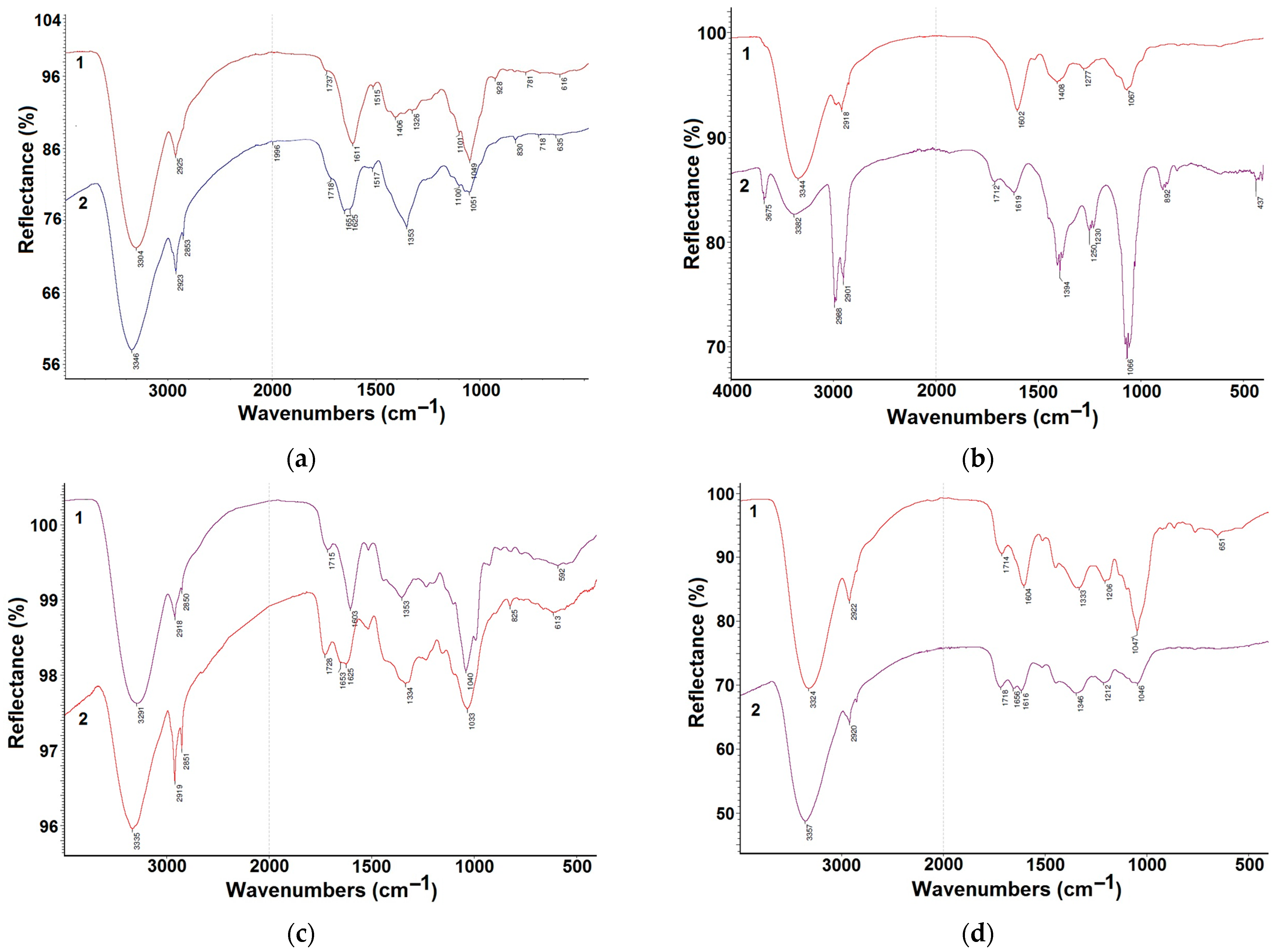
3.1. Fourier-Transform Infrared Spectroscopy (FTIR)

Figure 1 displays FTIR spectra of extracts of various plants and silver nanoparticles synthesized with their use. In the FTIR spectra of the extracts under study (dried directly on the crystal) (Figure 1a–d, spectrum 1), high-intensity absorption with maximum values in the range of 3290–3350 cm−1 is caused by the stretching of bonds between O–H alcohol groups and water (that is present in bound form as remnants after drying), as well as N–H bonds. The stretching of C–H bonds occurs in the range of 2850–2930 cm−1. The medium-intensity band with two peaks is a superposition of vibration bands of the carbonyl groups of ketones, amino- and carboxylic acids (1715–1730 cm−1), as well as bending vibrations of NH2 groups, water, double bonds and aromatic rings (1600–1650 cm−1). C–O–H bending vibrations and stretching of C–O alcohol groups and ether C–O–C bonds appear as the 1000–1100 cm−1 band, which, within the spectra of the four extracts, varies greatly in shape and intensity relative to other bands in each spectrum. These groups also absorb in the range of 1200–1500 cm−1, but the bands are weak and not characteristic.
Figure 1a shows the FTIR spectra of dried gooseberry extracts and the corresponding silver nanoparticles. Noteworthy is an increase in the frequency of stretching of O–H bonds at 40 cm−1 and a significant decrease in the intensity of the band of C–O–H bending vibrations. A change in the complex band with the peaks at 1745 cm−1 and 1611 cm−1 is also observed: it changes its shape due to the shift of the first peak from the low-frequency side to 1725 cm−1 and the splitting of the second (1651 cm−1 and 1610 cm−1). All this may indicate the involvement of hydroxyl and amino groups in the reduction of silver ions (formation of silver nanoparticles).
FTIR spectra of dried blue honeysuckle extracts and corresponding silver nanoparticles are presented in Figure 1b. They differ, first of all, by a significant decrease in the intensity of the band of O–H bond stretching and an increase in its frequency from 3344 cm−1 to 3383 cm−1. In addition, a band of free hydroxyl groups appears at 3680, 3670 cm−1. The absorption peak of the carbonyl group (1713 cm−1) appears more clearly, and the band at 1602 cm−1 shifts to 1619 cm−1 and becomes weak. At the same time, the intensity of the band in the range of 1000–1120 cm−1 increases. It is probable that the formation of nanoparticles occurs with the participation of –OH and –NH2 groups and the formation of C–O–C bonds.
During the formation of silver nanoparticles in the presence of strawberry extract (Figure 1c), as in cases shown in Figure 1a,b, the frequency of O–H bond stretching increases by more than 40 cm−1. The 1040 cm−1 band shifts to 1033 cm−1, and its intensity decreases. A shift in the peak of the carbonyl group band from 1715 cm−1 to 1728 cm−1 and an increase in its intensity are observed. Simultaneously, in the range of the –NH2 group vibration, the band becomes weaker. It broadens, and two peaks—1653 cm−1 and 1625 cm−1—appear instead of one peak at 1603 cm−1.
When using the sea buckthorn extract in phytosynthesis (Figure 1d), similar changes are observed, but they are more significant: the intensity of the band at 1447 cm−1 strongly decreases, and the band of –NH2 groups splits (1650 cm−1 and 1616 cm−1).
It should be noted that in the range of 1300–1400 cm−1 all samples demonstrate an increase in intensity due to the overlap with the nitrate ion band. Thus, significant changes in the IR spectra of plant extracts and synthesized silver sols indicate the presence of interaction between the extracts and silver ions during phytosynthesis, resulting from the involvement of –OH and –NH2 groups in the process.

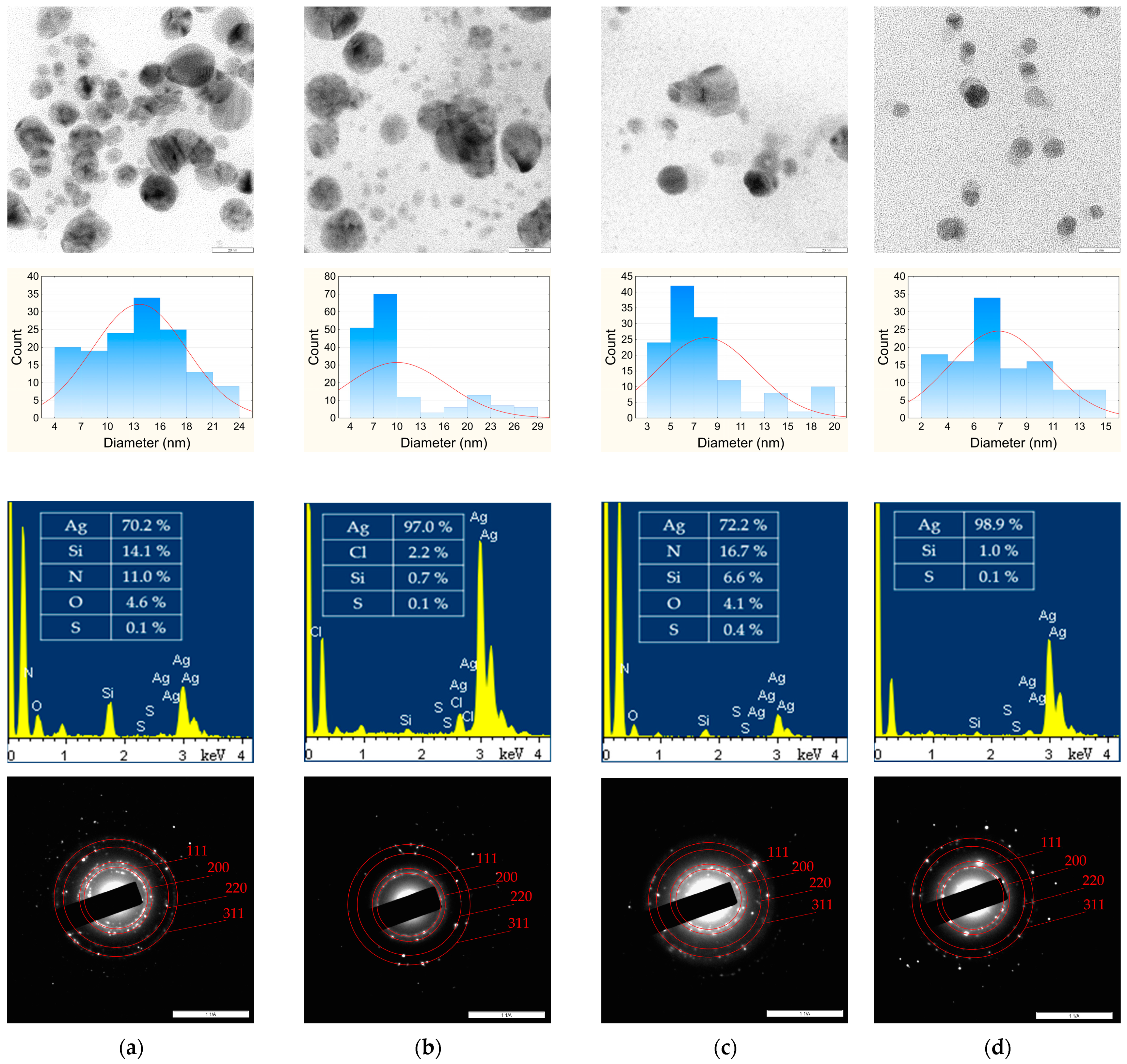
3.2. Transmission Electron Microscopy (TEM)

Figure 2 presents the TEM images of phyto-AgNPs synthesized with aqueous extracts of plant leaves. The resulting phyto-AgNPs are predominantly spherical in shape, and their sizes vary from 4–24 nm (gooseberry extract) and 4–29 nm (blue honeysuckle extract) to 3–20 nm (strawberry extract) and 2–15 nm (sea buckthorn extract). The micrographs show both well-dispersed phyto-AgNPs and a small number of their agglomerates. The agglomeration of phyto-AgNPs may be due to their sedimentation at the last stage of synthesis [40,41] resulting in the formation of larger and non-spherical particles. These were not taken into account when estimating the average size of phyto-AgNPs. Figure 2 also shows the particle size distribution histograms. The use of gooseberry, blue honeysuckle, strawberry and sea buckthorn extracts resulted in phyto-AgNPs with average diameters of 13, 10, 8 and 7 nm, respectively. A decrease in the average diameter of phyto-AgNPs is due to a decrease in the proportion of large particles and an increase in the proportion of small particles. All phyto-AgNPs are polydisperse (PI > 0.1), which may be due to a large number of phytocomponents in plant extracts that have various reducing and stabilizing properties. The results of TEM analysis are summarized in Table 2. It can be observed that an increase in the plant extract AOA (Table 1) is accompanied by a decrease in the average diameter of phyto-AgNPs, but it does not affect their polydispersity. The results obtained are consistent with the findings in [35], which reported that an increase in the plant extract AOA is accompanied by a decrease in the average diameter of phytosynthesized gold nanoparticles.
Semi-quantitative analysis of phyto-AgNP samples was performed by energy-dispersive X-ray spectroscopy (EDS) using a copper substrate, the contribution of which was not taken into account in the calculations (Figure 2). The obtained EDX spectra show peaks of silver with a characteristic dispersion energy of about 3 keV. Low intensities of nitrogen (N), silicon (Si), oxygen (O), chlorine (Cl) and sulfur (S) indicate the existence of an organic shell around the silver nucleus [10,11,13,14,15].
The selected area electron diffraction (SAED) crystallographic technique was also used in the characterization of phyto-AgNP samples. SAED patterns of phyto-AgNPs are represented as a set of point reflections that form a pattern of concentric rings (Figure 2) characteristic of a polycrystalline sample [42]. Indexing the diffraction patterns of phyto-AgNPs showed that a number of Bragg reflections correspond to sets of lattice planes (111), (200), (220), (311), which indicate a cubic face-centered structure (Fm3m). The calculated values of the phyto-AgNP lattice constant lie in the range of 0.4123–0.4216 nm (Table 2) and are approximate to the reference value of the silver crystal lattice parameter, equal to 0.4086 nm at 20 °C [43]. The error in determining the phyto-AgNP lattice parameters according to the TEM data did not exceed 5%.

3.3. UV–Vis Spectroscopy

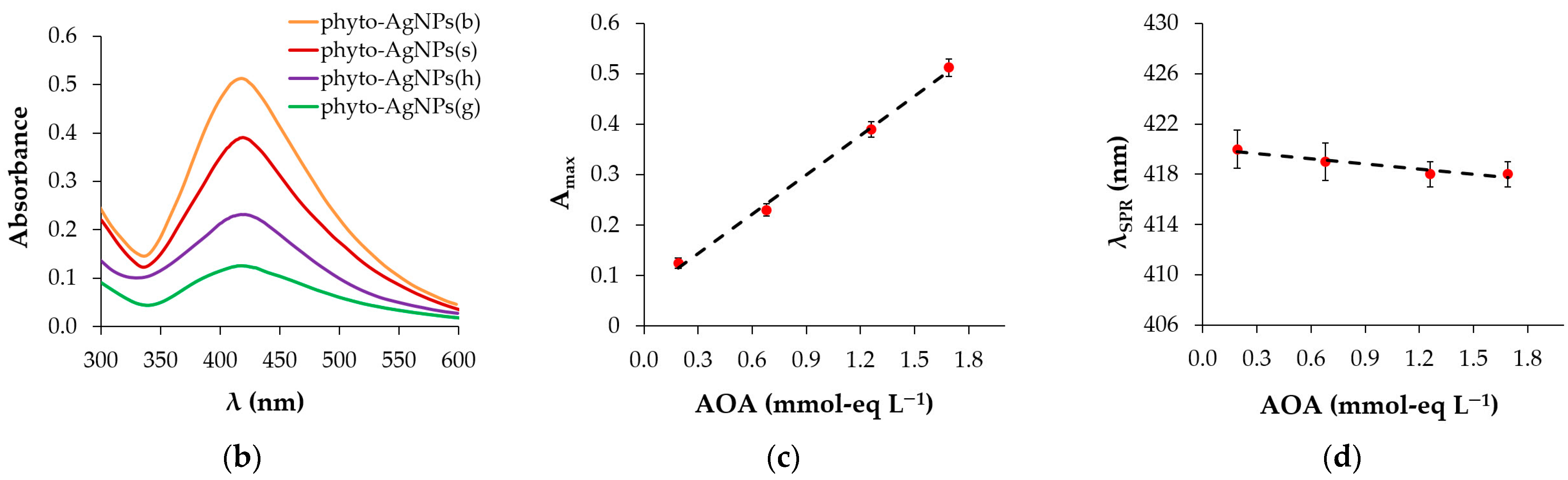
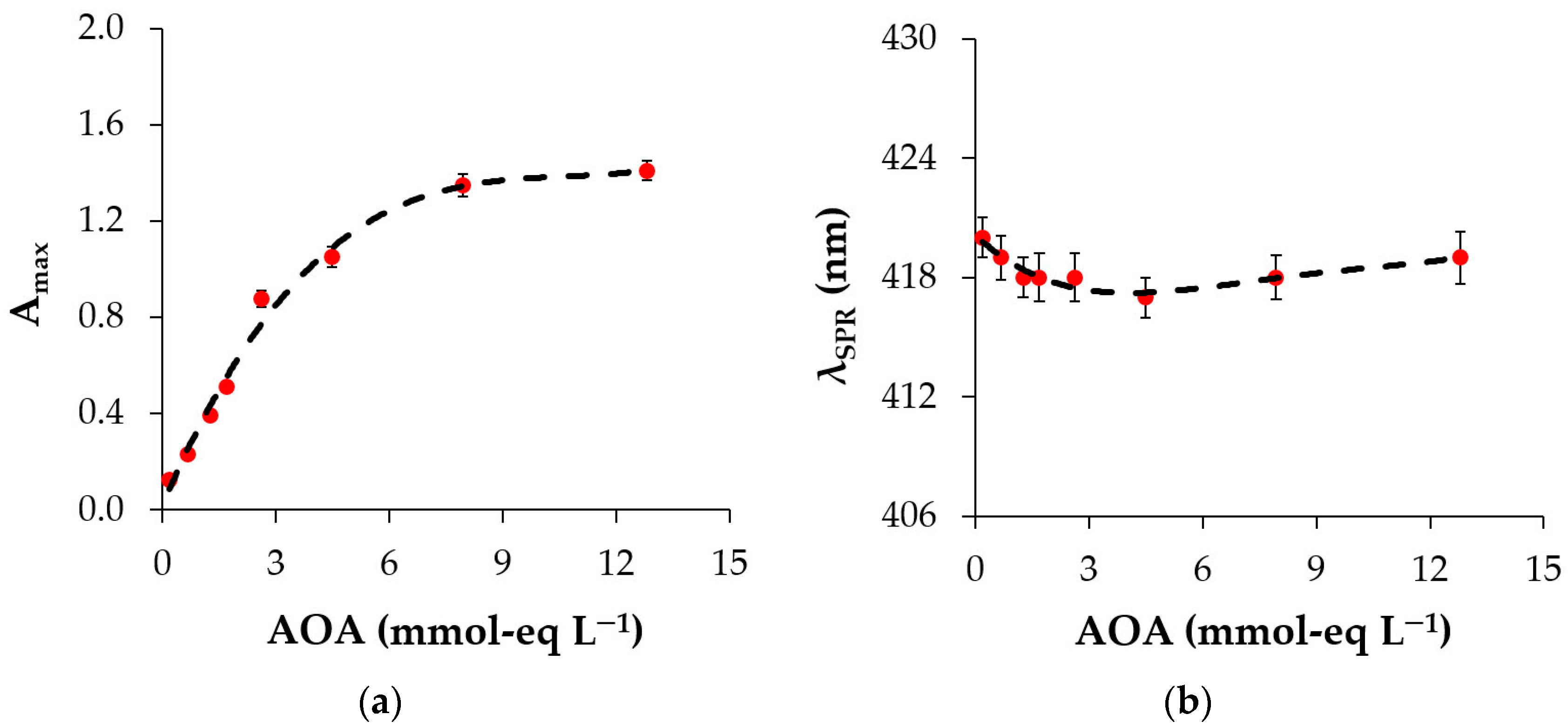
3.3.1. Effect of Plant Extract AOA

To examine the effect of plant AOA on the optical properties of phyto-AgNPs, silver sols were synthesized using extracts of different plants as well as extracts of the same plant. In the first case, different AOA of the extract was determined by the type of plant, thus the weights of the selected samples of cut leaves and the aliquots used for synthesis were constant. In the second case, AOA of the extract varied by the weight of the leaf sample and the extract aliquot during synthesis.
Figure 3a presents photographs of silver sols obtained with the same extract aliquot of different plants (gooseberry, blue honeysuckle, strawberry and sea buckthorn). As can be seen from the photographs, silver sols have different colors depending on the type of plant used for synthesis. In the series phyto-AgNPs(g)—phyto-AgNPs(h)—phyto-AgNPs(s)—phyto-AgNPs(b), the color of silver sols varies from light brown to dark brown. Changes in the color of silver sols reflect their absorption spectra (Figure 3b). The presence of surface plasmon resonance (SPR) bands of phyto-AgNPs in the UV–Vis spectra makes it possible to assess the effect of plant extract on the optical properties of phyto-AgNPs. The lowest SPR peak intensity at a wavelength of 408–420 nm is observed for the silver sol obtained using gooseberry extract, and the highest intensity is observed with the use of the sea buckthorn extract. Considering the trend towards an increase in the extract AOA in the order gooseberry < blue honeysuckle < strawberry < sea buckthorn (Table 1), it can be concluded that higher plant extract AOA results in higher intensity of the SPR of silver sols (Figure 3c). It shifts to the short-wave region, the so-called “blue shift” (Figure 3d).
To test whether such a pattern would be observed when using an extract of the same plant, different aliquots of the sea buckthorn extract were used for phytosynthesis of silver sols. Aliquots of 0.6, 1.0 and 2.0 mL of sea buckthorn extract with AOA = 15.7 mmol-eq L−1 and 1.2 and 2.0 mL of extract with AOA = 41 mmol-eq L−1, prepared using a larger portion of the plant powder in order to minimize dilution of the reaction mixture, were taken. Thus, on a reaction volume basis, the extract AOA was 1.7, 2.6, 4.5, 7.9 and 13 mmol-eq L−1, respectively. As a result of phytosynthesis, five silver sols were obtained, whose optical properties were studied by the spectrophotometric method. Figure 4a shows the spectra of the obtained sols. With an increase in AOA of sea buckthorn extracts, Amax of silver sols increases (Figure 4b). Their λSPR is practically independent of AOA (Figure 4c).
After combining the results of optical studies for different plants and a single plant into one set, the dependences Amax = f(AOA) and λSPR = f(AOA) were obtained (Figure 5). An increase in the plant extract AOA to 5 mmol-eq L−1 leads to a sharp increase in Amax (Figure 5a) and a slight decrease in λSPR (Figure 5b). At AOA > 5 mmol-eq L−1, a smoother increase in Amax and a subtle increase in λSPR occur. Since the optical density is directly proportional to the number of particles in the system [44] and the wavelength λSPR is a reflection of the particle size [45], it can be argued that with an increasing extract AOA, the number of smaller silver nanoparticles in the sol grows, which is consistent with the results of microscopic studies. Most likely, at AOA > 5 mmol-eq L−1, the concentration of small nanoparticles in the sol becomes so high that they begin to aggregate, causing a slight shift in λSPR towards longer wavelengths. Thus, the AOA value of the extract causes a size-dependent change in the magnitude and position of the nanoparticle plasmon resonance peak.

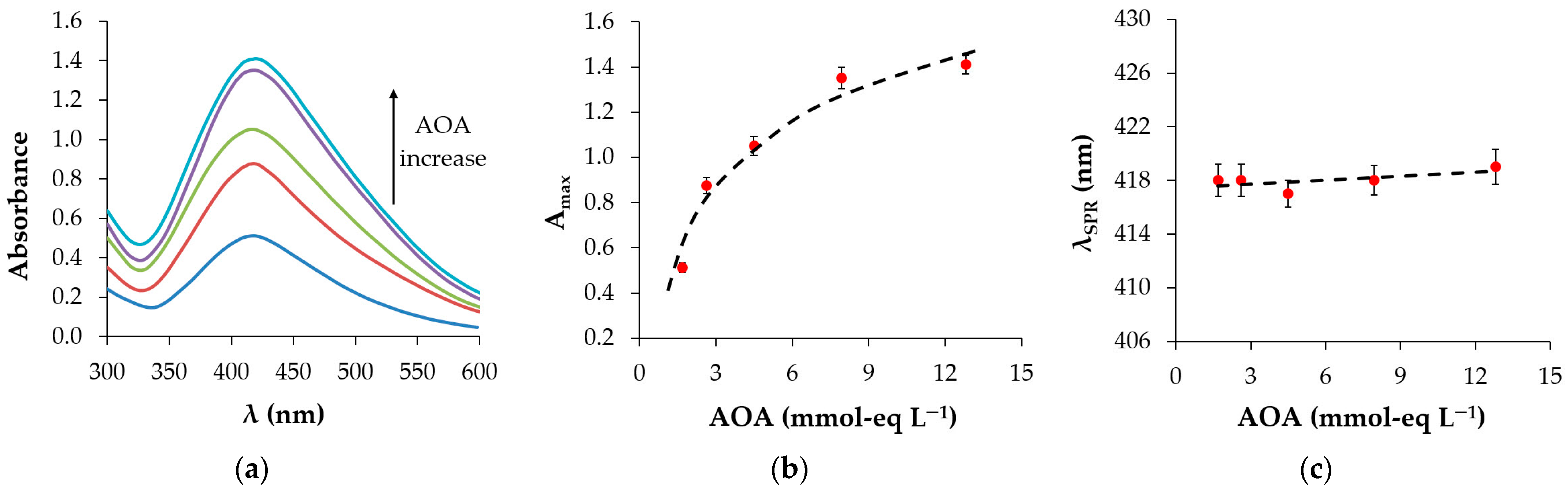
3.3.2. Maturation of Silver Sols

To assess the completeness of hydrothermal phytosynthesis of silver sols, maturation of phyto-AgNPs was studied. The process was carried out by standing phyto-AgNPs for 16 h in the solution they were formed in. At the end of this time, the absorption spectra of the silver sols were recorded. From Figure 6a it is evident that after 16 h of maturation, Amax of all silver sols increases, but to varying degrees. Thus, for phyto-AgNPs(g) the optical density increases by 2.6 times; phyto-AgNPs(h)—1.6 times; phyto-AgNPs(s)—1.3 times; and phyto-AgNPs(b)—1.2 times relative to the optical density measured immediately after the end of phytosynthesis. λSPR is slightly higher for silver sols formed using extracts of gooseberry and blue honeysuckle than using strawberry and sea buckthorn extracts (Figure 6b).
These results show that after 16 h of maturation, fewer nanoparticles are formed in silver sols obtained using plant extracts with higher AOA compared to extracts with lower AOA. This is due to the fact that extracts with higher AOA have a greater reducing ability and ensure a higher rate of nanoparticle formation during hydrothermal synthesis. Therefore, the main part of silver nanoparticles manages to form within 5 min of temperature synthesis while using strawberry and sea buckthorn extracts with high AOA. For plant extracts with lower AOA (gooseberry, particularly), much more time is required. Thus, the rate of silver ion reduction using extracts with higher AOA is higher than using extracts with lower AOA.

3.4. Electrochemical Investigations

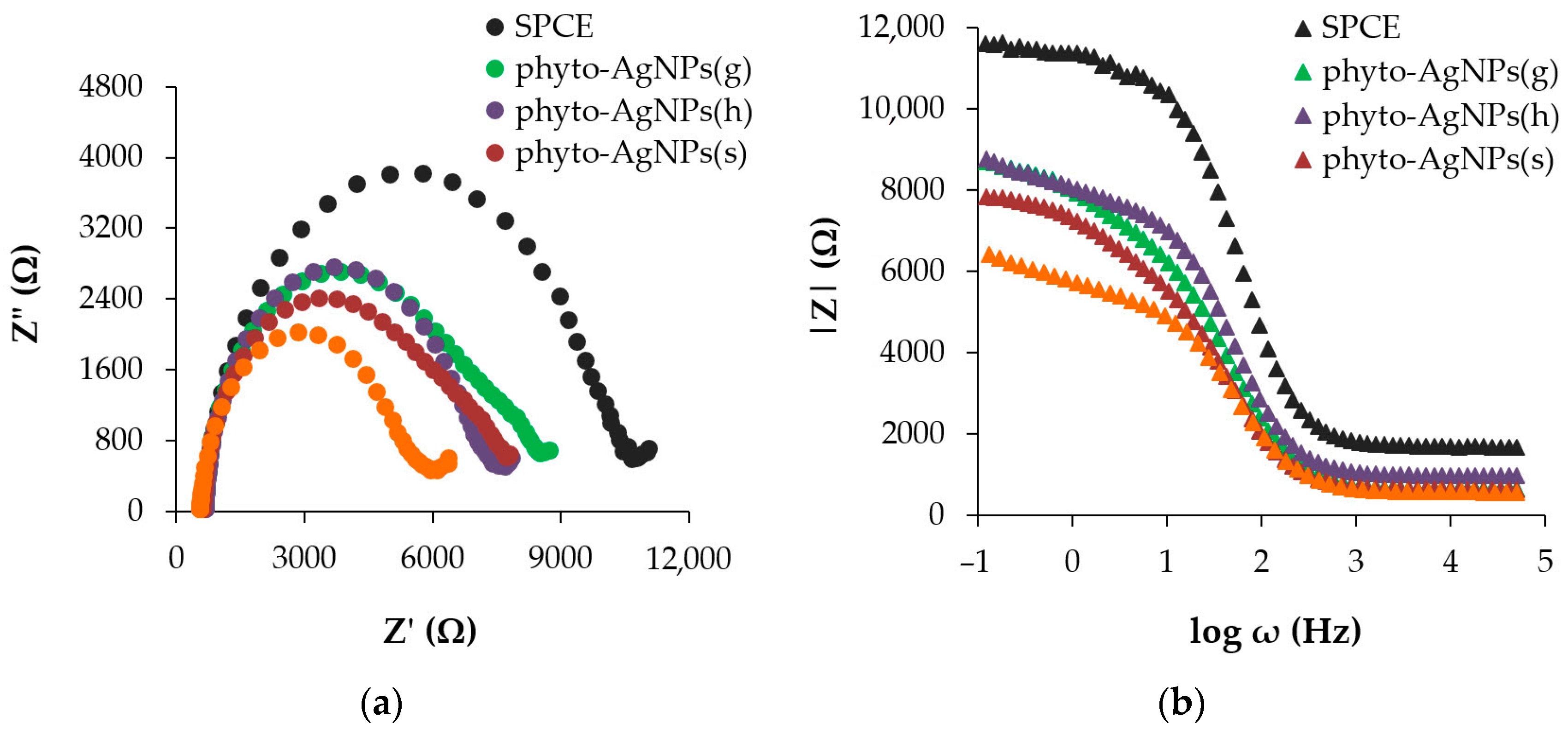
3.4.1. Electrochemical Impedance Spectroscopy (EIS)

Figure 7a shows Nyquist plots for an unmodified SPCE and SPCE modified with silver nanoparticles synthesized using plant extracts with different AOA. The value of charge transfer resistance (Rct), equal to the diameter of the spectrum semi-circle, characterizes the kinetics of electron transfer of a standard redox probe at the interface of the electrode surface. The Rct value for the unmodified SPCE is 9603.7 Ohm. For the SPCE modified with phytosynthesized AgNPs, Rct value varies from 5199 Ohm (for sea buckthorn extract with the highest AOA) to 7194 Ohm (for gooseberry extract with the lowest AOA). A decrease in resistance by 1.33–1.85 times indicates an increase in conductivity in the presence of phyto-AgNPs.
From the Bode plot (Figure 7b), it is evident that at frequencies from 0.1 Hz to ∼10 Hz, the electrodes modified with phyto-AgNPs exhibit significantly lower impedance values compared to the unmodified SPCE. Moreover, the higher the AOA of the plant extract used for the synthesis of phyto-AgNPs, the lower the values of |Z| in the frequency range from 0.1 Hz to ~10 Hz and the height of the vertical section of the dependence |Z| = f(log ω) in the range from 10 Hz to ~500 Hz. Based on the Nyquist and Bode plots, it can be concluded that there is a trend towards a decrease in charge transfer resistance in the series phyto-AgNPs(g) > phyto-AgNPs(h) > phyto-AgNPs(s) > phyto-AgNPs(b) with increasing AOA of the plant extract used for synthesis.

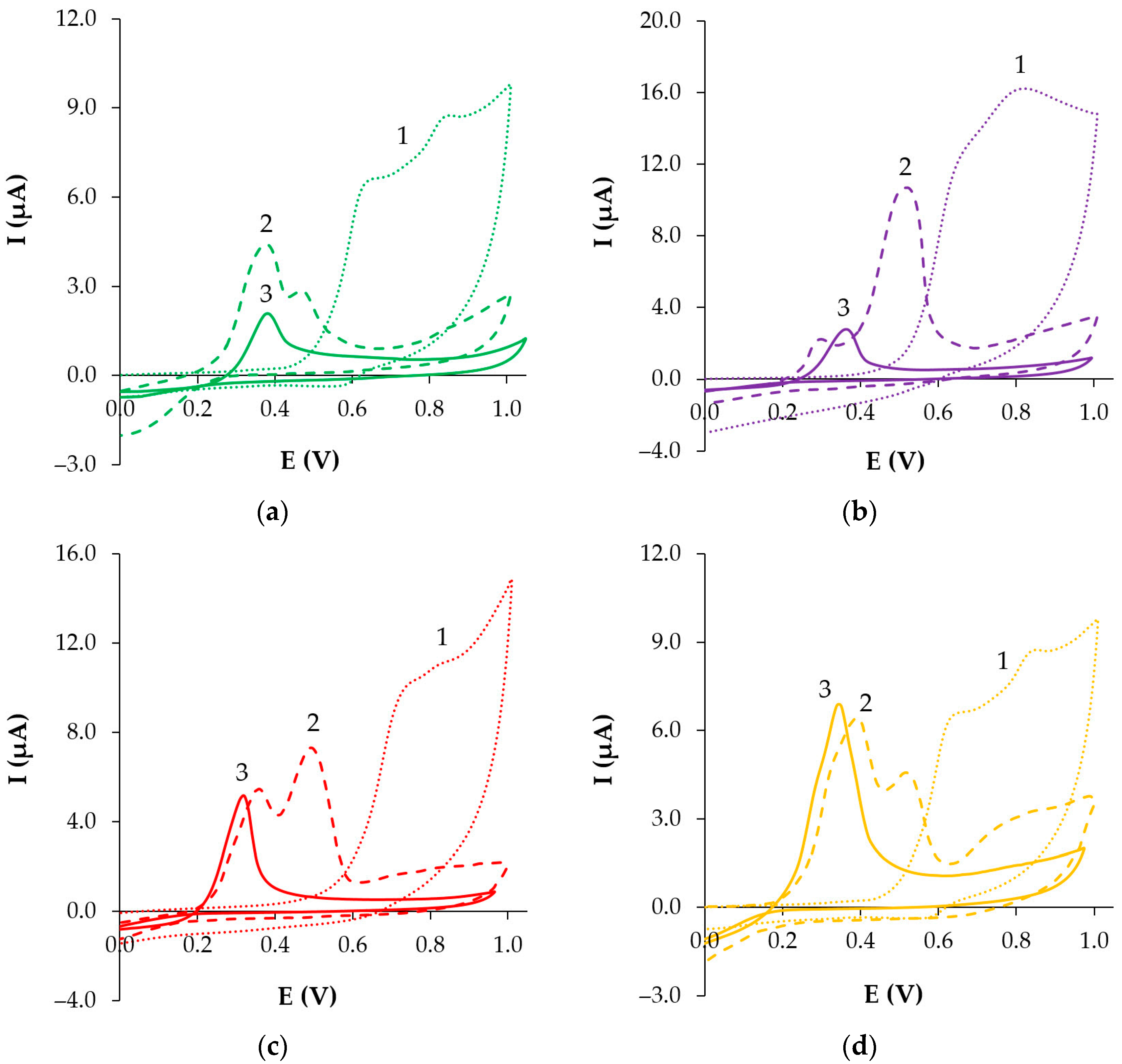
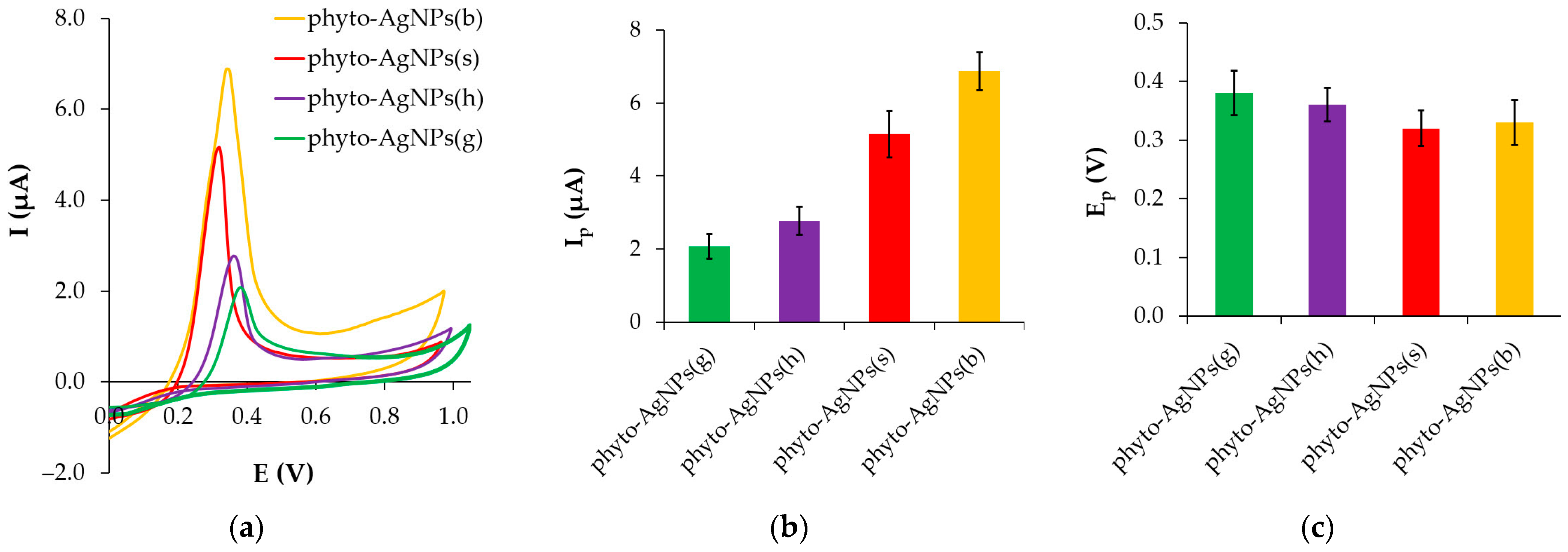
3.4.2. Cyclic Voltammetry (CV)

Figure 8 illustrates cyclic voltammograms of the plant extracts, as well as native and “washed” silver nanoparticles synthesized using these extracts. The components of gooseberry, blue honeysuckle, strawberry and sea buckthorn extracts are irreversibly oxidized on the SPCE in the potential range of 0.5–1.0 V and two waves are formed, quite clearly distinguishable for gooseberry and sea buckthorn and poorly distinguishable for blue honeysuckle and strawberry. In the cyclic voltammograms of native phytosynthesized silver nanoparticles, two anodic signals are observed at (0.35 ± 0.07) V and (0.51 ± 0.05) V, and a weakly pronounced cathodic wave at E < 0.1 V. For washed silver nanoparticles, only one signal is recorded in the potential range of 0.33–0.38 V, depending on the AOA of the plant extract used (Figure 9a). The disappearance of the second anodic signal is observed upon repeated registration of the electrooxidation cycle of native phytosynthesized silver nanoparticles, which indicates the departure of the compound that forms it from the electrode surface. It can be assumed that this signal is caused by some remnants of plant extracts or products of their interaction with silver ions, which are removed from the electrode surface as a result of oxidation or washing of the silver sol. Hereinafter, only washed silver nanoparticles were used in electrochemical studies.
There is a clear relationship between the magnitude of the silver nanoparticle oxidation current and the AOA of the plant extract used for particle synthesis (Figure 9). Cyclic voltammograms of silver nanoparticles phytosynthesized using plant extracts with different AOA are presented in Figure 9a. An increase in AOA in the plant series gooseberry < blue honeysuckle < strawberry < sea buckthorn is accompanied by an increase in the peak current (Ip) of silver nanoparticle oxidation (Figure 9b). Thus, the peak current of phyto-AgNPs(b) obtained using the sea buckthorn extract with the maximum AOA is 3.3 times higher than the peak oxidation current of phyto-AgNPs(g) synthesized using the gooseberry extract with the lowest AOA. As can be seen from Figure 9c, with an increase in the plant extract AOA, the peak potential (Ep) of the silver nanoparticle oxidation shifts by approximately 30–60 mV to the cathode side, which proves an increase in the electroactivity of nanoparticles synthesized using extracts with higher AOA (strawberry and sea buckthorn) compared to extracts with lower AOA (gooseberry and blue honeysuckle).

3.5. Correlation Analysis

Correlation analysis was performed between the plant extract AOA and the following parameters characterizing phyto-AgNP properties: average diameter ( d ¯ ), polydispersity index (PI), charge transfer resistance (Rct), light absorption (Amax) and surface plasmon resonance wavelength (λSPR), the magnitude of peak current (Ip) and peak potential (Ep). The results obtained are summarized in Table 3. The plant extract AOA negatively correlated with the following parameters: d ¯ (r = −0.98, p = 0.019), λSPR (r = −0.96, p = 0.041), Rct (r = −0.95, p = 0.053) and Ep (r = −0.92, p = 0.084). At the same time, the plant extract AOA positively correlated with these parameters: Amax (r = 0.99, p = 0.003) and Ip (r = 0.98, p = 0.018). The lack of correlation with PI (r = −0.13, p = 0.869) indicates that the polydispersity of phyto-AgNPs is determined by plant extract metabolites with reducing properties. However, the composition and amount of the latter may vary for different plants.
With the increase in the extract AOA, the number of molecules with reducing properties increases, which contributes to the formation of a greater number of crystallization centers of silver nanoparticles. The higher concentration of antioxidants in the plant extract also ensures improved capping of nanoparticles and the production of finer and more stable nanosuspensions. Sols of phyto-AgNPs formed by using extracts with higher AOA demonstrate an increase in the intensity of the plasmon resonance band and its shift to the “blue” region, as well as a decrease in charge transfer resistance, an increase in the peak oxidation current of nanosilver and a decrease in its potential compared to phyto-AgNPs formed in extracts with lower AOA. Thus, a cause-and-effect relationship is observed in the form of the “AOA–size–properties” triad. These data indicate that the plant extract AOA is one of the key parameters of phytosynthesis. By changing plant extract AOA, it is possible to obtain phyto-AgNPs of different sizes which affect their optical and electrochemical properties. Thus, a change in the wave width and length of plasma resonance was reported in [45], along with a shift in the potential of the silver oxidation peak and a change in the surface energy properties of nanoparticles [46] depending on their diameter.

4. Conclusions

The synthesis of silver nanoparticles using extracts from plant leaves is a simple, effective, economical, fast, affordable and eco-friendly way to obtain biocompatible nanoparticles that are widely used in various fields, namely, medicine, food industry, cosmetology, agriculture and (bio)sensorics. The article presents the results of a comprehensive study of the optical and electrochemical properties of silver nanoparticles synthesized using four plant extracts with different antioxidant activity (AOA). These properties are shown to be characterized by size-dependent behavior and correlate with AOA. In particular, with an increase in the AOA of plant extracts, the kinetics of phytosynthesis increases, the size of silver nanoparticles decreases, the intensity of the plasmon resonance band increases and its “blue shift” occurs. An increase in electrochemical activity and a decrease in the resistance to charge transfer of nanoparticles are also observed. The established patterns allow us to consider AOA as a key parameter of phytosynthesis and the “AOA–size–properties” triad as a practical guide for obtaining nanoparticles of certain sizes and properties. The results of this work can make a significant contribution to the development of phytosynthesis methodology and to producing silver nanoparticles with prescribed properties.

Author Contributions

Conceptualization, N.S.; methodology, N.S.; validation, M.B.; formal analysis, A.T.; investigation, V.T., E.K. and V.K.; writing—original draft preparation, N.S., A.T. and M.B.; writing—review and editing, N.S. and A.T.; visualization, A.T. and M.B.; funding acquisition, M.B. All authors have read and agreed to the published version of the manuscript.

Funding

This research was funded by Russian Science Foundation (RSF), grant number 23-23-00353. URL: https://rscf.ru/project/23-23-00353/ (accessed on 14 October 2024).

Data Availability Statement

Data are contained within the article.

Acknowledgments

The authors express their gratitude to Skrylnik M.Yu. (Institute of Metallurgy, Ural Branch of the Russian Academy of Sciences) for assistance in conducting impedimetric studies. The authors would like to thank Koryakova O.V. (Laboratory of Structural Research Methods, Institute of Organic Synthesis, Ural Branch of the Russian Academy of Sciences) for assistance in conducting FTIR studies, using the facilities of Resource Sharing Center “Spectroscopy and Analysis of Organic Compounds”.

Conflicts of Interest

The authors declare no conflicts of interest.

References

  1. Magdy, G.; Aboelkassim, E.; Elhaleem, S.M.A.; Belal, F. A comprehensive review on silver nanoparticles: Synthesis approaches, characterization techniques, and recent pharmaceutical, environmental, and antimicrobial applications. Microchem. J. 2024, 196, 109615. [Google Scholar] [CrossRef]
  2. Faris, V.M.; Barzinjy, A.A.; Hamad, S.M. The Applications of Green Synthesized Silver Nanoparticles: A Review. Eurasian J. Sci. Eng. 2022, 8, 16–34. [Google Scholar] [CrossRef]
  3. Dhaka, A.; Mali, S.C.; Sharma, S.; Trivedi, R. A review on biological synthesis of silver nanoparticles and their potential applications. Results Chem. 2023, 6, 101108. [Google Scholar] [CrossRef]
  4. Ong, W.T.J.; Nyam, K.L. Evaluation of silver nanoparticles in cosmeceutical and potential biosafety complications. Saudi J. Biol. Sci. 2022, 29, 2085–2094. [Google Scholar] [CrossRef]
  5. Angelopoulou, P.; Giaouris, E.; Gardikis, K. Applications and Prospects of Nanotechnology in Food and Cosmetics Preservation. Nanomaterials 2022, 12, 1196. [Google Scholar] [CrossRef]
  6. Satsangi, N. Synthesis and Characterization of Biocompatible Silver Nanoparticles for Anticancer Application. J. Inorg. Organomet. Polym. Mater. 2020, 30, 1907–1914. [Google Scholar] [CrossRef]
  7. Basova, T.V.; Vikulova, E.S.; Dorovskikh, S.I.; Hassan, A.; Morozova, N.B. The use of noble metal coatings and nanoparticles for the modification of medical implant materials. Mater. Des. 2021, 204, 109672. [Google Scholar] [CrossRef]
  8. Arroyo, G.V.; Madrid, A.T.; Gavilanes, A.F.; Naranjo, B.; Debut, A.; Arias, M.T.; Angulo, Y. Green synthesis of silver nanoparticles for application in cosmetics. J. Environ. Sci. Health Part A Toxic/Hazard. Subst. Environ. Eng. 2020, 55, 1304–1320. [Google Scholar] [CrossRef]
  9. Wani, I.A. Review—Recent Advances in Biogenic Silver Nanoparticles & NanoComposite Based Plasmonic-Colorimetric and Electrochemical Sensors. ECS J. Solid State Sci. Technol. 2021, 10, 047003. [Google Scholar] [CrossRef]
  10. Desalegn, T.; Ravikumar, C.R.; Murthy, H.C.A. Eco-friendly synthesis of silver nanostructures using medicinal plant Vernonia amygdalina Del. leaf extract for multifunctional applications. Appl. Nanosci. 2021, 11, 535–551. [Google Scholar] [CrossRef]
  11. Umair Raza, M.; Abasi, F.; Shahbaz, M.; Ehsan, M.; Seerat, W.; Akram, A.; Raja, N.I.; Mashwani, Z.u.-R.; Hassan, H.U.; Proćków, J. Phytomediated Silver Nanoparticles (AgNPs) Embellish Antioxidant Defense System, Ameliorating HLB-Diseased ‘Kinnow’ Mandarin Plants. Molecules 2023, 28, 2044. [Google Scholar] [CrossRef]
  12. Singh, R.; Hano, C.; Nath, G.; Sharma, B. Green Biosynthesis of Silver Nanoparticles Using Leaf Extract of Carissa carandas L. and Their Antioxidant and Antimicrobial Activity against Human Pathogenic Bacteria. Biomolecules 2021, 11, 299. [Google Scholar] [CrossRef] [PubMed]
  13. Nguyen, L.A.T.; Van Mai, B.; Van Nguyen, D.; Nguyen, N.Q.T.; Van Pham, V.; Pham, T.L.M.; Le, H.T. Green synthesis of silver nanoparticles using Callisia fragrans leaf extract and its anticancer activity against MCF-7, HepG2, KB, LU-1, and MKN-7 cell lines. Green Process. Synth. 2023, 12, 20230024. [Google Scholar] [CrossRef]
  14. Javed, B.; Nadhman, A.; Mashwani, Z.R. Phytosynthesis of Ag nanoparticles from Mentha longifolia: Their structural evaluation and therapeutic potential against HCT116 colon cancer, Leishmanial and bacterial cells. Appl. Nanosci. 2020, 10, 3503–3515. [Google Scholar] [CrossRef]
  15. Murthy, H.C.A.; Zeleke, T.D.; Ravikumar, C.R.; Kumar, M.R.A.; Nagaswarupa, H.P. Electrochemical properties of biogenic silver nanoparticles synthesized using Hagenia abyssinica (Brace) JF. Gmel. medicinal plant leaf extract. Mater. Res. Express 2020, 7, 055016. [Google Scholar] [CrossRef]
  16. Kumar, V.; Yadav, S.C.; Yadav, S.K. Syzygium cumini leaf and seed extract mediated biosynthesis of silver nanoparticles and their characterization. J. Chem. Technol. Biotechnol. 2010, 85, 1301–1309. [Google Scholar] [CrossRef]
  17. Obaid, A.Y.; Al-Thabaiti, S.A.; El-Mossalamy, E.H.; Al-Harbi, L.M.; Khan, Z. Extracellular bio-synthesis of silver nanoparticles. Arab. J. Chem. 2017, 10, 226–231. [Google Scholar] [CrossRef]
  18. Elemike, E.E.; Fayemi, O.E.; Ekennia, A.C.; Onwudiwe, D.C.; Ebenso, E.E. Silver Nanoparticles Mediated by Costus afer Leaf Extract: Synthesis, Antibacterial, Antioxidant and Electrochemical Properties. Molecules 2017, 22, 701. [Google Scholar] [CrossRef]
  19. Ajaykumar, A.P.; Mathew, A.; Chandni, A.P.; Varma, S.R.; Jayaraj, K.N.; Sabira, O.; Rasheed, V.A.; Binitha, V.S.; Swaminathan, T.R.; Basheer, V.S.; et al. Green Synthesis of Silver Nanoparticles Using the Leaf Extract of the Medicinal Plant, Uvaria narum and Its Antibacterial, Antiangiogenic, Anticancer and Catalytic Properties. Antibiotics 2023, 12, 564. [Google Scholar] [CrossRef]
  20. Giri, A.K.; Jena, B.; Biswal, B.; Pradhan, A.K.; Arakha, M.; Acharya, S.; Acharya, L. Green synthesis and characterization of silver nanoparticles using Eugenia roxburghii DC. extract and activity against biofilm-producing bacteria. Sci. Rep. 2022, 12, 8383. [Google Scholar] [CrossRef]
  21. Farnad, N.; Farhadi, K. Introducing potato starch-ecofriendly silver nanoparticles as a novel binary system for nanoencapsulation of riboflavin. Food Chem. 2023, 398, 133910. [Google Scholar] [CrossRef] [PubMed]
  22. Ahmad, B.; Chang, L.; Satti, U.Q.; Rehman, S.u.; Arshad, H.; Mustafa, G.; Shaukat, U.; Wang, F.; Tong, C. Phyto-Synthesis, Characterization, and In Vitro Antibacterial Activity of Silver Nanoparticles Using Various Plant Extracts. Bioengineering 2022, 9, 779. [Google Scholar] [CrossRef] [PubMed]
  23. Rashmi, B.N.; Harlapur, S.F.; Avinash, B.; Ravikumar, C.R.; Nagaswarupa, H.P.; Anil Kumar, M.R.; Gurushantha, K.; Santosh, M.S. Facile green synthesis of silver oxide nanoparticles and their electrochemical, photocatalytic and biological studies. Inorg. Chem. Commun. 2020, 111, 107580. [Google Scholar] [CrossRef]
  24. Yadi, M.; Mostafavi, E.; Saleh, B.; Davaran, S.; Aliyeva, I.; Khalilov, R.; Nikzamir, M.; Nikzamir, N.; Akbarzadeh, A.; Panahi, Y.; et al. Current developments in green synthesis of metallic nanoparticles using plant extracts: A review. Artif. Cells Nanomed. Biotechnol. 2018, 46, S336–S343. [Google Scholar] [CrossRef]
  25. Waiezi, S.; Nik Malek, N.A.N.; Asraf, M.H.; Sani, N.S. Preparation, Characterization, and Antibacterial Activity of Green-Biosynthesised Silver Nanoparticles using Clinacanthus nutans Extract. Biointerface Res. Appl. Chem. 2023, 13, 171. [Google Scholar] [CrossRef]
  26. Elemike, E.E.; Onwudiwe, D.C.; Fayemi, O.E.; Botha, T.L. Green synthesis and electrochemistry of Ag, Au, and Ag–Au bimetallic nanoparticles using golden rod (Solidago canadensis) leaf extract. Appl. Phys. A 2019, 125, 42. [Google Scholar] [CrossRef]
  27. Phuong, T.N.M.; Thanh Hai, N.T.; Thu Thuy, N.T.; Phu, N.V.; Huong, N.T.; Hang Nga, D.T.; Quoc Huy, H.X.; Hoa, T.T. Factors affecting synthesis of silver-nanoparticles and antimicrobial applications. Hue Univ. J. Sci. Nat. Sci. 2020, 129, 25–31. [Google Scholar] [CrossRef]
  28. Liu, H.; Zhang, H.; Wang, J.; Wei, J. Effect of temperature on the size of biosynthesized silver nanoparticle: Deep insight into microscopic kinetics analysis. Arab. J. Chem. 2020, 13, 1011–1019. [Google Scholar] [CrossRef]
  29. Philip, D. Biosynthesis of Au, Ag and Au–Ag nanoparticles using edible mushroom extract. Spectrochim. Acta Part A 2009, 73, 374–381. [Google Scholar] [CrossRef]
  30. Ahmed, S.; Ahmad, M.; Swami, B.L.; Ikram, S. A review on plants extract mediated synthesis of silver nanoparticles for antimicrobial applications: A green expertise. J. Adv. Res. 2016, 7, 17–28. [Google Scholar] [CrossRef]
  31. Vanlalveni, C.; Lallianrawna, S.; Biswas, A.; Selvaraj, M.; Changmai, B.; Rokhum, S.L. Green synthesis of silver nanoparticles using plant extracts and their antimicrobial activities: A review of recent literature. RSC Adv. 2021, 11, 2804–2837. [Google Scholar] [CrossRef] [PubMed]
  32. Abada, E.; Mashraqi, A.; Modafer, Y.; Al Abboud, M.A.; El-Shabasy, A. Review green synthesis of silver nanoparticles by using plant extracts and their antimicrobial activity. Saudi J. Biol. Sci. 2024, 31, 103877. [Google Scholar] [CrossRef] [PubMed]
  33. Akintelu, S.A.; Olugbeko, S.C.; Folorunso, A.S.; Oyebamiji, A.K.; Folorunso, F.A. Potentials of phytosynthesized silver nanoparticles in biomedical fields: A review. Int. Nano Lett. 2021, 11, 273–293. [Google Scholar] [CrossRef]
  34. Shaikh, W.A.; Chakraborty, S.; Owens, G.; Islam, R.U. A review of the phytochemical mediated synthesis of AgNP (silver nanoparticle): The wonder particle of the past decade. Appl. Nanosci. 2021, 11, 2625–2660. [Google Scholar] [CrossRef] [PubMed]
  35. Stozhko, N.Y.; Bukharinova, M.A.; Khamzina, E.I.; Tarasov, A.V.; Vidrevich, M.B.; Brainina, K.Z. The Effect of the Antioxidant Activity of Plant Extracts on the Properties of Gold Nanoparticles. Nanomaterials 2019, 9, 1655. [Google Scholar] [CrossRef]
  36. Brainina, K.; Stozhko, N.; Bukharinova, M.; Khamzina, E.; Vidrevich, M. Potentiometric method of plant microsuspensions antioxidant activity determination. Food Chem. 2019, 278, 653–658. [Google Scholar] [CrossRef]
  37. Stozhko, N.Y.; Bukharinova, M.A.; Khamzina, E.I.; Tarasov, A.V. Electrochemical Properties of Phytosynthesized Gold Nanoparticles for Electrosensing. Sensors 2022, 22, 311. [Google Scholar] [CrossRef]
  38. Brainina, K.; Tarasov, A.; Khamzina, E.; Kazakov, Y.; Stozhko, N. Disposable Potentiometric Sensory System for Skin Antioxidant Activity Evaluation. Sensors 2019, 19, 2586. [Google Scholar] [CrossRef]
  39. Mohanaparameswari, S.; Balachandramohan, M.; Sasikumar, P.; Rajeevgandhi, C.; Vimalan, M.; Pugazhendhi, S.; Ganesh Kumar, K.; Albukhaty, S.; Sulaiman, G.M.; Abomughaid, M.M.; et al. Investigation of structural properties and antibacterial activity of AgO nanoparticle extract from Solanum nigrum/Mentha leaf extracts by green synthesis method. Green Process. Synth. 2023, 12, 20230080. [Google Scholar] [CrossRef]
  40. Lateef, A.; Akande, M.A.; Azeez, M.A.; Ojo, S.A.; Folarin, B.I.; Gueguim-Kana, E.B.; Beukes, L.S. Phytosynthesis of silver nanoparticles (AgNPs) using miracle fruit plant (Synsepalum dulcificum) for antimicrobial, catalytic, anticoagulant, and thrombolytic applications. Nanotechnol. Rev. 2016, 5, 507–520. [Google Scholar] [CrossRef]
  41. Ahmad, T.; Wani, I.A.; Manzoor, N.; Ahmed, J.; Asiri, A.M. Biosynthesis, structural characterization and antimicrobial activity of gold and silver nanoparticles. Colloids Surf. B Biointerfaces 2013, 107, 227–234. [Google Scholar] [CrossRef] [PubMed]
  42. Hammond, C. The Basics of Crystallography and Diffraction, 3rd ed.; Oxford University Press: New York, NY, USA, 2009; pp. 281–282. [Google Scholar]
  43. Arblaster, J.W. Selected Values of the Crystallographic Properties of the Elements; ASM International: Materials Park, OH, USA, 2018; p. 363. [Google Scholar]
  44. Hung, N.D.; Nam, V.N.; ThiNhan, T.; Dung, T.T.N. Quantitative concentration determination of silver nanoparticles prepared by DC high voltage electrochemical method. Vietnam J. Chem. 2018, 56, 553–558. [Google Scholar] [CrossRef]
  45. Mekuye, B. The Impact of Size on the Optical Properties of Silver Nanoparticles Based on Dielectric Function. In Nanotechnology and Nanomaterials-Annual; Jiménez-Suárez, A., Seisdedos, G., Eds.; IntechOpen: London, UK, 2024; Volume 2024, pp. 1–20. [Google Scholar] [CrossRef]
  46. Brainina, K.Z.; Galperin, L.G.; Kiryuhina, T.Y.; Galperin, A.L.; Stozhko, N.Y.; Murzakaev, A.M.; Timoshenkova, O.R. Silver nanoparticles electrooxidation: Theory and experiment. J. Solid State Electrochem. 2012, 16, 2365–2372. [Google Scholar] [CrossRef]
Figure 1. FTIR spectra for gooseberry (a), blue honeysuckle (b), strawberry (c) and sea buckthorn (d). Spectra 1 and 2 correspond to the plant extract and phyto-AgNP sol, respectively.
Physchem 04 00028 g001
Figure 2. TEM images, histograms of particle distribution by size, EDS spectra and SAED patterns of phyto-AgNPs synthesized using extracts of gooseberry (a), blue honeysuckle (b), strawberry (c) and sea buckthorn (d).
Physchem 04 00028 g002
Figure 3. Photographs (a) and absorption spectra (b) of phyto-AgNP sols synthesized using extracts of various plants. Amax (c) and λSPR (d) of phyto-AgNP sols depending on the plant extract AOA.
Physchem 04 00028 g003aPhyschem 04 00028 g003b
Figure 4. Absorption spectra of phyto-AgNPs(b) sols obtained as a result of synthesis using sea buckthorn extracts with different AOA (a). Dependence of Amax (b) and λSPR (c) of phyto-AgNPs(b) sols on AOA of sea buckthorn extracts.
Physchem 04 00028 g004
Figure 5. Dependence of Amax (a) and λSPR (b) of phyto-AgNP sols on the plant extract AOA.
Physchem 04 00028 g005
Figure 6. Comparison of Amax (a) and λSPR (b) of phyto-AgNP sols obtained immediately after synthesis at pH 5 and 16 h after it.
Physchem 04 00028 g006
Figure 7. Nyquist plot (a) and Bode plot (b) for bare and phyto-AgNP-modified SPCE in 0.1 mol L−1 KCl solution containing 5 mmol L−1 K3[Fe(CN)6].
Physchem 04 00028 g007
Figure 8. Cyclic voltammograms of plant extracts (1), native (2) and “washed” (3) phytoAgNPs synthesized using gooseberry (a), blue honeysuckle (b), strawberry (c) and sea buckthorn (d). Background 0.1 mol L–1 H2SO4. Potential scan rate 50 mV s−1.
Physchem 04 00028 g008
Figure 9. Cyclic voltammograms of phyto-AgNPs synthesized using extracts of different plants at pH 5 (a). Dependences of Ip (b) and Ep (c) of phyto-AgNP oxidation on plant extract AOA.
Physchem 04 00028 g009
Table 1. AOA of aqueous extracts of plant leaves (n = 5).
Plant ExtractAOA, mmol-eq L−1
Gooseberry (Ribes uva-crispa)1.8 ± 0.3
Blue honeysuckle (Lonicera caerulea)6.4 ± 1.1
Strawberry (Fragaria vesca)11.8 ± 0.9
Sea buckthorn (Hippophae rhamnoides)15.7 ± 1.2
Table 2. Characterization of phyto-AgNPs by TEM.
AgNPsDiameter Range (nm)Diameter Value (nm)PILattice Parameter (nm)
phyto-AgNPs(g)4–2413 ± 50.150.4216
phyto-AgNPs(h)4–2910 ± 70.490.4201
phyto-AgNPs(s)3–208 ± 40.250.4195
phyto-AgNPs(b)2–157 ± 30.180.4123
PI, polydispersity index.
Table 3. Correlation matrix.
ParametersAOA d ¯ PIAmaxλSPRRctIpEp
AOA1−0.98 *−0.130.99 *−0.96 *−0.950.98 *−0.92
d ¯ 1−0.04−0.96 *0.99 *0.88−0.930.94
PI 1−0.20−0.030.26−0.310.08
Amax 1−0.94−0.96 *0.99 *−0.90
λSPR 10.82−0.900.98 *
Rct 1−0.97 *0.75
Ip 1−0.88
Ep 1
AOA, antioxidant activity; d ¯ , average diameter; PI, polydispersity index; Amax, absorption at surface plasmon resonance wavelength; λSPR, surface plasmon resonance wavelength; Rct, charge transfer resistance; Ip, peak current; Ep, peak potential. The asterisk (*) indicates the Pearson linear correlation coefficients with a significance level of p < 0.05.
Disclaimer/Publisher’s Note: The statements, opinions and data contained in all publications are solely those of the individual author(s) and contributor(s) and not of MDPI and/or the editor(s). MDPI and/or the editor(s) disclaim responsibility for any injury to people or property resulting from any ideas, methods, instructions or products referred to in the content.

