Article

Sex and Stature Estimation from Scapular Measurements: Development and Independent Validation in Northeastern Thai Population

by
Suthat Duangchit
1,2,
Naphatchaya Imkrajang
3,
Worrawit Boonthai
4,5,
Nareelak Tangsrisakda
6,
Sararat Innoi
6,
Sitthichai Iamsaard
6 and
Chanasorn Poodendaen
2,3,*
1
Department of Physiology, Faculty of Medical Science, Naresuan University, Phitsanulok 65000, Thailand
2
Centre of Excellence in Medical Biotechnology, Naresuan University, Phitsanulok 65000, Thailand
3
Department of Anatomy, Faculty of Medical Science, Naresuan University, Phitsanulok 65000, Thailand
4
Department of Anthropology, Faculty of Sociology and Anthropology, Thammasat University, Pathum Thani 12120, Thailand
5
Thammasat University Research Unit in Physical Anthropology and Health Science, Thammasat University, Pathum Thani 12120, Thailand
6
Department of Anatomy, Faculty of Medicine, Khon Kaen University, Khon Kaen 40002, Thailand
*
Author to whom correspondence should be addressed.
Forensic Sci. 2025, 5(4), 66; https://doi.org/10.3390/forensicsci5040066 (registering DOI)
Submission received: 30 September 2025 / Revised: 28 October 2025 / Accepted: 21 November 2025 / Published: 23 November 2025

Abstract

Background/Objectives: Determination of the biological profile, particularly sex and stature, constitutes an essential component for individual identification in forensic and archaeological anthropology; however, validation of anthropometric equations remains inadequately implemented in contemporary research. This study addresses two limitations: the isolated development of sex and stature estimation methods and the lack of rigorous validation using independent samples. Methods: In this research, we analyzed 400 well-preserved scapulae from a Northeastern Thai population divided into training (n = 300: 150 male scapulae, 150 female scapulae) and validation (n = 100: 50 male scapulae, 50 female scapulae) groups. Eight standardized measurements were used for both sex and stature estimation. Results: All measurements demonstrated significant sexual dimorphism, with males exhibiting larger dimensions. For sex estimation, a multivariate model incorporating the maximum scapular height, maximum length of the spine, and scapula weight achieved 96.3% accuracy in the training samples and maintained 95.0% accuracy in independent validation. For stature estimation, a three-variable equation combining scapula weight, longitudinal scapular length, and maximum scapular breadth was strongly correlated (R = 0.769, SEE = 5.32 cm) with consistent performance across validation samples. Conclusions: Validation testing confirmed the high accuracy, reliability, and stability of both equations when applied to independent samples, with no significant differences in performance metrics between training and validation groups. These validated equations provide reliable standards for forensic practitioners analyzing scapular remains in practical applications within the northeastern Thai population.

1. Introduction

Skeletal examination provides crucial biological information supporting forensic identification and archaeological investigations [1,2]. Sex and stature assessments represent fundamental requirements for identification procedures. Although pelvic and cranial elements are traditionally preferred for sex determination and long bones for height estimation, these osseous structures commonly experience damage or destruction from environmental influences, trauma, or intentional dismemberment during forensic scenarios [3]. When preferred skeletal elements are unavailable, forensic practitioners require validated alternative approaches for sex and stature estimation [4,5,6]. Among alternative options, the scapula has gained recognition as a valuable skeletal element due to its anatomical position and distinctive morphological features [7,8,9]. Scapular sexual dimorphism originates during fetal development [10] and intensifies throughout maturation. The scapula presents multiple clearly defined anatomical reference points enabling consistent measurement protocols [11], exhibiting robust reliability for sex determination across various populations [12,13]. Furthermore, investigations have confirmed scapular effectiveness for stature estimation with precision matching conventional long bone methods [14,15,16].
Researchers have developed population-specific standards for biological profiling utilizing different skeletal components [17,18,19,20]; however, extensive scapular morphology research remains insufficient. Most investigations have focused on northern [21] and central Thai groups, generating a significant knowledge gap concerning the northeastern territory. This limitation is important since northeastern Thailand accounts for approximately 32% of the country’s population (based on 2022 census) [22] and encompasses unique ethnic composition, including Thai-Lao, Thai-Khmer, and Thai-Korat communities [23,24]. Missing region-specific standards may reduce forensic analysis precision in this densely populated area.
Previous studies across diverse populations have demonstrated successful sex determination from scapular measurements with high accuracy rates ranging from 77–97% [7,11,12,13,21]. Similarly, stature estimation from scapular dimensions has shown strong correlations at R = 0.65–0.85 in various populations [14,16,25]. However, earlier investigations have identified two substantial methodological constraints in scapular studies. First, most research examined sex determination or stature estimation separately rather than creating integrated methodologies from single skeletal elements [9,25,26,27]. Second, numerous anthropometric equations lack appropriate validation through independent testing samples [6,8,10,12,13] representing a critical methodological issue in forensic anthropology. Studies have shown that equations created exclusively from training datasets often exhibit decreased precision when implemented on different populations [28,29,30]. While the present study employs similar standardized osteometric measurements as previous research [13,21,27], it addresses both methodological gaps by simultaneously developing integrated sex and stature estimation equations from identical scapular measurements and validating both through independent testing in a previously unstudied northeastern Thai population.
To address these methodological and geographical limitations, this investigation had two primary objectives. First, comprehensive equations for both sex and stature estimation were developed using eight standardized scapular measurements in a northeastern Thai population. Second, these equations were validated using an independent sample from the same population to assess their reliability and practical applicability in forensic contexts. By establishing population-specific standards for northeastern Thailand and demonstrating rigorous validation methodology, this research contributes to enhanced forensic and archaeological anthropological practice.

2. Materials and Methods

2.1. Samples and Ethical Considerations

This cross-sectional study analyzed both left and right scapulae from 200 individuals (100 males, 100 females) who met the inclusion criteria, yielding a total of 400 scapulae from skeletal remains of body donors at the Unit of Human Bone Warehouse for Research (UHBWR), Department of Anatomy, Faculty of Medicine, Khon Kaen University, Thailand. The age at death ranged from 20 to 80 years, with males averaging 66.69 ± 13.02 years and females 64.15 ± 11.57 years. Inclusion criteria were anatomically complete scapulae with documented sex, ethnicity, age at death, and ante-mortem stature. Exclusion criteria included pathological conditions such as bone tumors, fractures, erosive lesions, or skeletal deformities affecting morphometric measurements. The sample was randomly divided into training (n = 300: 150 male scapulae, 150 female scapulae) and validation (n = 100: 50 male scapulae, 50 female scapulae) groups using computer-generated random numbers. The two groups were completely independent, with no individuals appearing in both groups. This research received ethical approval from the Center for Ethics in Human Research, Khon Kaen University (approval code: HE671710) and followed institutional guidelines for human remains research.

2.2. Measurements and Data Collection

These seven measurements (Figure 1) were selected based on validated protocols from previous scapular research [13,21,27]. Scapular weight was added as a novel parameter representing overall bone mass, and defined as below:
  • Maximum scapular height (MSH): Maximum linear distance between the superior and inferior angles.
  • Maximum scapular breadth (MSB): Transverse distance from the glenoid margin midpoint to the most projecting point of the medial border.
  • Maximum length of the spine (MLS): The maximum linear distance from the lateral point of the acromion to the medial end of the spine at the medial border.
  • Length of the axillary border (LAB): Distance from the inferior glenoid cavity to the inferior angle.
  • Longitudinal scapular length (LSL): Linear distance from the acromion process lateral point to the inferior angle.
  • Glenoid cavity breadth (GCB): Maximum transverse diameter of the glenoid cavity.
  • Glenoid cavity height (GCH): Maximum vertical diameter between the glenoid cavity’s superior and inferior margins.
  • Scapula weight (SW): Total mass of completely dried scapula measured in grams.
Linear measurements were obtained using a digital Vernier caliper with 0.01 mm precision (Mitutoyo, Kawasaki, Japan), and scapular weight was measured using a digital scale with 0.01 g precision. All measurements were performed independently by two trained observers. Documented ante-mortem stature measurements in centimeters were extracted from body donation records.

2.3. Statistical Analyses

All statistical analyses, including model development and validation using stepwise multivariate logistic regression for sex estimation and stepwise multiple linear regression for stature estimation, were conducted using IBM SPSS Statistics version 23.0 (IBM Corp., Armonk, NY, USA). Interobserver reliability was assessed using intraclass correlation coefficient (ICC) model (2,1). Data distribution normality was evaluated using the Kolmogorov–Smirnov test. Sexual dimorphism was examined using independent t-tests for normally distributed variables and Mann–Whitney U tests for non-normally distributed variables, with p < 0.05 considered statistically significant.
Sex estimation utilized stepwise multivariate logistic regression with classification accuracy assessment. Stature estimation employed stepwise multiple linear regression analyzing relationships between scapular measurements and height using Pearson’s correlation coefficients. Models were developed for pooled and sex-specific samples, with accuracy evaluated using standard error of estimate (SEE). Validation metrics for sex estimation included accuracy rates, positive and negative predictive values, kappa coefficient, area under the curve, and likelihood ratios. Stature estimation validation utilized mean absolute error (MAE), mean absolute percentage error (MAPE), coefficient of determination (R2), and Bland–Altman plots for assessing agreement between estimated and actual stature.

3. Results

3.1. Measurement Reliability

Interobserver reliability was assessed using intraclass correlation coefficient (ICC) model (2,1) for all osteometric measurements. All measurements demonstrated excellent reliability, with ICC values ranging from 0.978 to 0.997. The highest reliability was observed for maximum scapular height (ICC = 0.997), while glenoid cavity breadth showed the lowest but still excellent reliability (ICC = 0.978). Scapular weight demonstrated high measurement consistency using calibrated digital scales.

3.2. Sexual Dimorphism

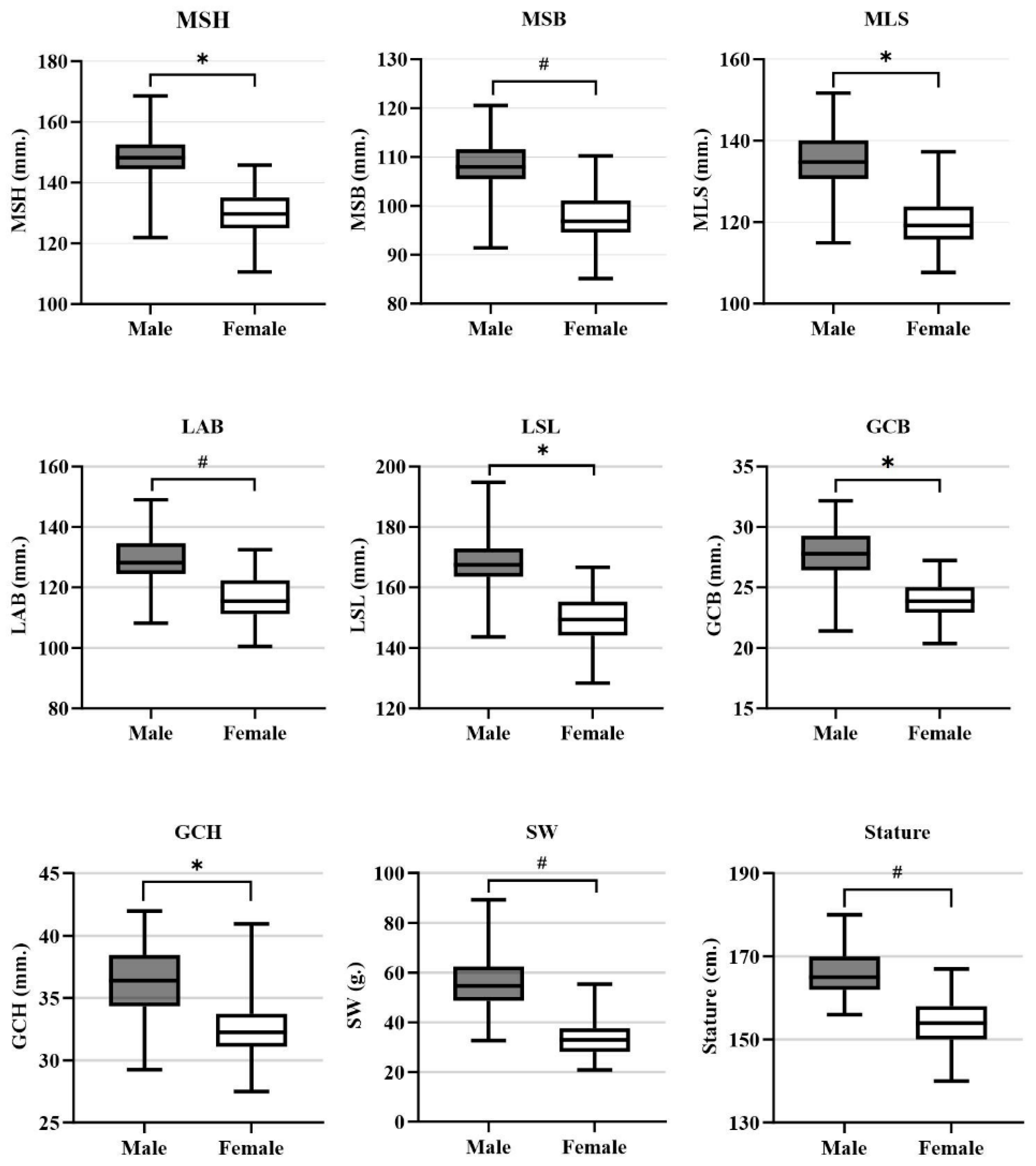
Five parameters (MSH, MLS, LSL, GCB, and GCH) showed normal distribution and were analyzed using independent t-tests, while four parameters (MSB, LAB, SW, and Stature) showed non-normal distribution and were analyzed using Mann–Whitney U tests (Figure 2). All measurements demonstrated significant sexual dimorphism (p < 0.01), with males consistently showing larger dimensions than females. The greatest differences were observed in scapular weight and longitudinal scapular length, while glenoid cavity measurements also revealed significant sex-based variations.

3.3. Logistic Regression Models for Sex Determination

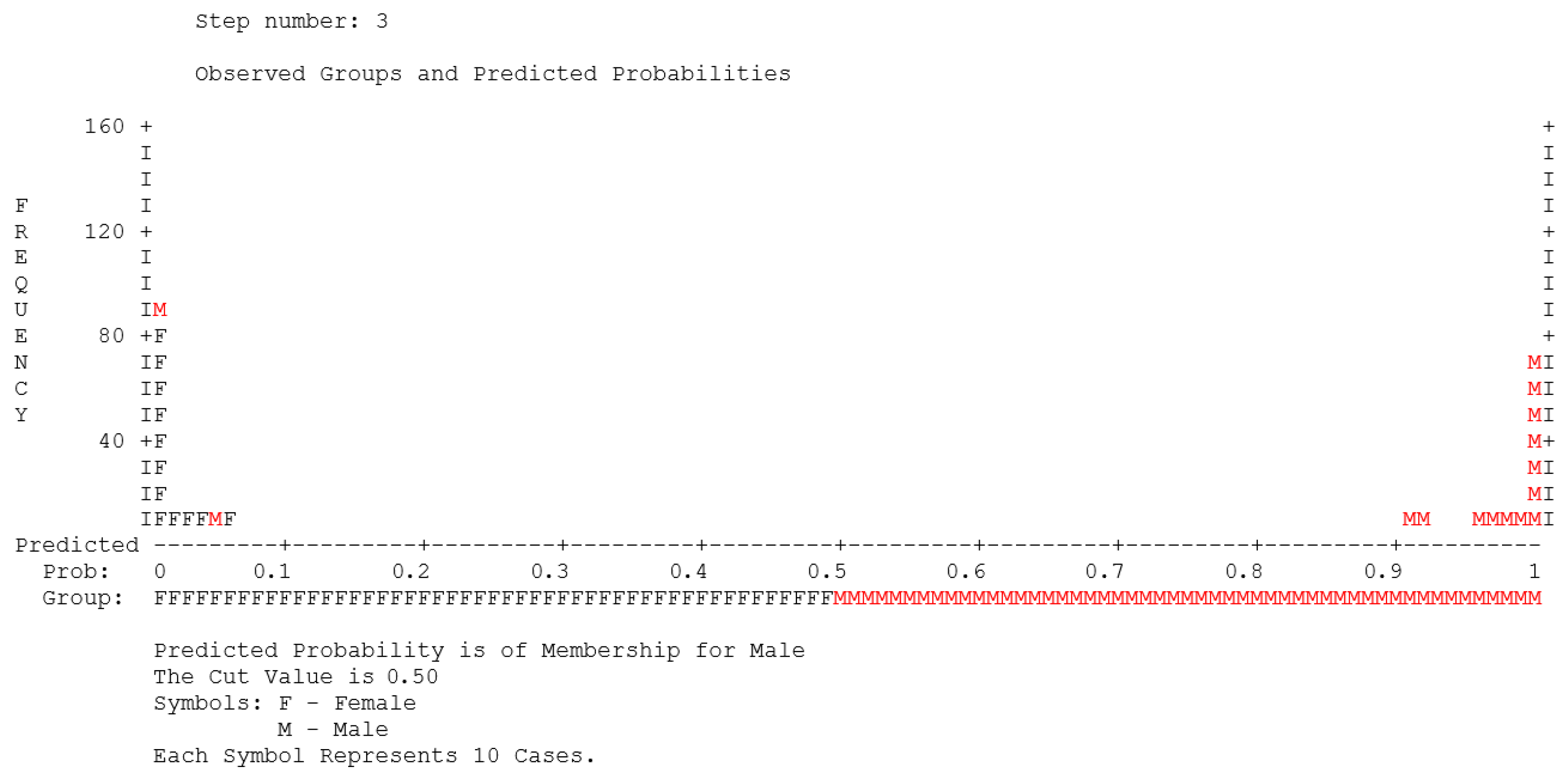
Stepwise multiple logistic regression analysis (forward likelihood ratio) was performed to develop sex estimation equations using scapular measurements (Table 1). The final model incorporated three significant predictors: MSH, MLS, and SW, achieving overall predictive accuracy of 96.3% (Nagelkerke R2 = 0.886). The stepwise process selected MSH first, followed by SW, then MLS, with Nagelkerke R2 values increasing from 0.784 to 0.886. Subsequently, the final model demonstrated excellent performance, with 95.3% accuracy for females and 97.3% accuracy for males (Table 2). All variables contributed significantly (p < 0.01), with odds ratios ranging from 1.171–1.202. To apply this equation, substitute the measured values into the formula. If the calculated result is greater than 0, the scapula is classified as male; if less than 0, it is classified as female. Additionally, the classification plot (Figure 3) showed distinct bimodal distribution with minimal overlap between sex groups, confirming strong discriminatory ability.

3.4. Stature Estimation from Scapular Measurements

Stepwise multiple regression analysis was used to develop stature estimation equations (Table 3). The optimal pooled-sample model incorporated three variables (SW, LSL, and MSB), achieving strong correlation (R = 0.769) with SEE of 5.32 cm. Sex-specific analysis revealed different patterns: for males, only MSB remained in the final equation, whereas females utilized a four-variable model (LSL, SW, GCH, GCB) with improved correlation and precision. Despite sex-specific equations offering marginally lower SEE values, the pooled-sample equation provides robust stature estimation suitable for diverse forensic contexts where sex determination may be uncertain or unavailable.

3.5. Independent Validation of Sex and Stature Estimation Equations

3.5.1. Validation Sample Characteristics

The validation sample consisted of 100 subjects (50 males, 50 females) randomly selected using simple random sampling from an independent northeastern Thai population. The mean age was 64.91 ± 11.91 years, with no significant age difference between sexes (p = 0.58). All scapular measurements showed significant sexual dimorphism (p < 0.01), with males consistently showing larger dimensions (Table 4). This pattern was consistent with the training sample, confirming sample comparability for validation testing.

3.5.2. Validation of the Sex Determination Equation

The three-variable logistic regression model was validated using the independent sample. The sex estimation equation is as follows:
Sex = 0.158 (MSH) + 0.162 (MLS) + 0.184 (SW) − 50.817
The equation demonstrated consistent performance between training and validation samples, with no significant differences (p > 0.05) across all diagnostic metrics (Table 5). The validation sample showed excellent discriminatory ability with kappa coefficient (0.900), AUC value (0.970), positive likelihood ratio (16.0), and negative likelihood ratio (0.04), confirming the equation’s reliability and practical applicability in forensic contexts.

3.5.3. Validation of the Stature Estimation Equation

The three-variable stature estimation equation was validated using the independent sample. The stature estimation formula is as follows:
Stature = 98.296 + 0.233 (SW) + 0.163 (LSL) + 0.246 (MSB)
The equation demonstrated consistent performance between training and validation samples, with no significant differences (p > 0.05) in accuracy metrics (Table 6). The coefficient of determination remained stable, and intraclass correlation coefficient confirmed good reliability. Bland–Altman analysis revealed acceptable agreement between predicted and actual stature measurements in both samples (Figure 4), with mean differences close to zero and similar limits of agreement, confirming the equation’s robustness and practical applicability.

4. Discussion

This study demonstrated significant sexual dimorphism in all scapular parameters (p < 0.01), with males consistently exhibiting larger dimensions than females. The multivariate logistic regression analysis identified MSH, MLS, and SW as the most significant discriminators, collectively achieving 96.3% classification accuracy. Scapular weight and longitudinal scapular length showed the most pronounced sex-based differences, consistent with previous findings [7,12,13]. These dimorphic patterns result from both biological and environmental factors. Sexual differences originate during fetal development [10] and intensify throughout maturation due to hormonal influences, particularly testosterone-driven bone mineralization and increased cortical thickness in males [31], and estrogen-mediated regulation of osteoblast activity and calcium homeostasis in females [32]. Environmental factors also contribute to these differences, as occupational activities in northeastern Thailand often involve differential physical demands between sexes [33]. Additionally, the scapula serves as an attachment site for upper limb muscles and experiences differential mechanical loading between sexes, stimulating bone remodeling through mechanical stress adaptation [34,35].
The multivariate logistic regression model incorporating MSH, MLS, and SW achieved high classification accuracy (96.3%), demonstrating the effectiveness of combining multiple scapular dimensions that reflect distinct developmental and functional pathways. MSH captures sexual dimorphism arising from skeletal scaling influenced by growth hormone and sex steroid interactions, while MLS reflects differential mechanical loading from muscle attachments where males experience greater stress from trapezius and deltoid development [36,37]. Notably, SW emerged as a powerful discriminator because it represents overall bone mass, reflecting integrated effects of cortical and trabecular bone development influenced by the testosterone–estrogen balance that capture sexual dimorphism in bone microarchitecture beyond linear measurements. However, stepwise regression risks producing artificially favorable results by overfitting to sample-specific patterns rather than capturing true population relationships, demanding rigorous validation procedures [38]. The maintained accuracy (95.0%) in independent validation samples demonstrates genuine biological differences rather than statistical artifacts, confirming the equations’ reliability for practical forensic applications.
Comparative analysis revealed that the current findings align with global patterns while exhibiting population-specific characteristics (Table 7). The northeastern Thai accuracy (96.3%) exceeded previous northern Thai results (78.0–88.0%) [21], suggesting regional morphological variation within Thailand potentially related to genetic ancestry and environmental factors. This performance compared favorably with dry bone studies in South African [39], American [7], Greek [11], and Spanish [40] populations, while surpassing CT-based studies in Italian [41], Chinese [16], and Iranian [9] populations. The discriminatory variables identified (MSH, MLS, SW) align with findings from American populations [7], whereas studies in Japanese [42] and Egyptian [43] populations emphasized glenoid cavity dimensions. These variations reflect population-specific morphological patterns and highlight the necessity for region-specific standards in forensic anthropological practice.
Multiple regression analysis demonstrated significant correlations between stature and scapular measurements, with notable sex-specific patterns. Females showed stronger correlations with stature than males, consistent with previous scapular studies [25,46] but contrasting with long bone research where male correlations typically predominate [47,48]. These differences reflect biological factors where females maintain more conservative body proportions [49], while males exhibit greater variability in body size ratios [50] and experience testosterone-driven muscular development [51]. Environmental factors such as gender-specific occupational activities may further contribute to these patterns [33]. Despite these underlying sex-based differences, multiple regression models incorporating various scapular dimensions demonstrated statistical advantages through capturing complex interrelationships between variables [52], minimizing error variance and providing greater robustness against measurement errors [53,54]. From a practical standpoint, pooled-sex equations were adopted due to comparable error values and enhanced applicability in forensic contexts where sex determination may be uncertain. This approach aligns with findings from northern Thai [21], Japanese [46], and Indian [55] populations, demonstrating that pooled-sex models provide acceptable accuracy while maintaining methodological flexibility.
This study highlights the critical importance of independent validation in forensic anthropological research, a methodological step often overlooked despite its fundamental significance for legal admissibility and scientific rigor. The consistent performance between training and validation samples demonstrates that the developed equations possess genuine discriminatory power rather than statistical artifacts arising from sample-specific variance or methodological overfitting—a distinction crucial for court proceedings where misidentification carries severe consequences. The maintained high kappa coefficients, AUC values, and likelihood ratios exceeding established forensic identification thresholds confirm the equations’ reliability within the northeastern Thai population and provide the statistical foundation necessary for expert testimony under scientific evidence standards. However, these findings are specific to the northeastern Thai population. Application to other populations may result in reduced performance, as documented in previous cross-population studies [28,38]. This limitation highlights the ongoing need for population-specific validation in forensic anthropological practice.
The stature estimation equation demonstrated consistent performance across training and validation samples. Bland–Altman analysis revealed acceptable agreement between predicted and actual stature measurements, with mean differences close to zero and comparable limits of agreement, confirming the equation’s robustness. The results demonstrate absence of systematic bias and stable precision, essential prerequisites for forensic applications [56].
This study demonstrated high success in sex and stature estimation from scapular measurements, providing validated population-specific standards for northeastern Thailand and an integrated approach valuable when traditional skeletal elements are unavailable or damaged. Practical implementation requires trained personnel, standardized protocols, and careful preservation assessment. Additionally, these population-specific equations require validation for other groups. Nevertheless, the methods offer practical solutions for forensic casework. Although developed using complete specimens, future research exploring fragmentary remains [57] and innovative technologies like deep learning and artificial intelligence technologies could expand applicability [58]. Most significantly, the rigorous independent validation framework employed here, assessing accuracy, reliability, and stability across separate samples, demonstrates these equations are not merely statistically significant but possess the requisite performance characteristics for dependable field application, ensuring practical applicability for real-world forensic identification where accurate biological profiling carries profound legal and humanitarian consequences.

5. Conclusions

This study developed and validated sex and stature estimation equations from scapular measurements in northeastern Thais. All the parameters showed significant sexual dimorphism (p < 0.01), with a three-variable model achieving 96.3% sex classification accuracy. Stature estimation via combined scapular weight, length, and breadth yielded strong correlations (R = 0.769, SEE = 5.32 cm). Independent validation confirmed the reliability across samples. This research addresses methodological gaps by providing comprehensive validated equations from a single skeletal element, offering forensic practitioners reliable alternatives when traditional elements are unavailable.
Limitations of the study: Several limitations require acknowledgment. Age-group stratification was not conducted, and the sample predominantly consisted of older adults (mean age 65.42 years), potentially overlooking age-related morphological changes in the scapula. Caution is warranted when applying these equations to younger individuals. Occupational patterns that may influence scapular development and sexual dimorphism were not addressed. These equations require prior knowledge of northeastern Thai origin for accurate application. Determining population affinity in unidentified cases can be challenging, limiting practical use. Practitioners should apply these methods cautiously when origin is uncertain, and external validation across other Thai populations is needed. External validation using different regional populations was not performed, limiting broader applicability assessment. Additionally, the focus on complete scapulae restricts their utility because fragmentation remains common in forensic contexts. Finally, ante-mortem stature measurements were extracted from body donation records provided by the hospital, and these measurements could not be independently verified or re-measured, potentially introducing measurement variability.

Author Contributions

Conceptualization, S.D., W.B., S.I. (Sitthichai Iamsaard) and C.P.; methodology, S.D., N.I., S.I. (Sararat Innoi) and C.P.; formal analysis, S.D. and C.P.; resources, N.T. and S.I. (Sitthichai Iamsaard); data curation, N.T., N.I., S.I. (Sararat Innoi) and S.I. (Sitthichai Iamsaard); writing—original draft preparation, S.D.; writing—review and editing, all authors.; project administration, C.P.; funding acquisition, S.D. and C.P. All authors have read and agreed to the published version of the manuscript.

Funding

This research was supported by an undergraduate student thesis grant from the Faculty of Medical Science, Naresuan University, Thailand.

Institutional Review Board Statement

This investigation received ethical approval from the Center for Ethics in Human Research, Khon Kaen University (HE671710), 11 December 2024.

Informed Consent Statement

Our study utilized skeletal specimens from the osteological collection at Khon Kaen University, Thailand, derived from voluntary body donors. We have obtained a Research Exemption Determination from the Khon Kaen University Ethics Committee for Human Research (KKUEC) under exemption category 6.7.3: Research studying bones, skeletons, extracted teeth, and cadavers.

Data Availability Statement

During the preparation of this work the authors used Claude Sonnet 4 in order to sentence refinement, grammatical verification, and minor translation. All conceptual frameworks, analytical approaches, research interpretations, scholarly discussions, and conclusions herein remain the exclusive intellectual contribution of the authors. After using this tool/service, the authors reviewed and edited the content as needed and take full responsibility for the content of the published article.

Acknowledgments

The authors gratefully acknowledge the Unit of Human Bone Warehouse for Research (UHBWR) at the Faculty of Medicine, Khon Kaen University, for providing access to the scapular specimens that were crucial for this study. Their institutional support was instrumental in the successful completion of this research.

Conflicts of Interest

The authors declare no conflict of interest.

References

  1. Franklin, D.; Swift, L.; Flavel, A. ‘Virtual anthropology’ and radiographic imaging in the Forensic Medical Sciences. Egypt J. Forensic Sci. 2016, 6, 31–43. [Google Scholar] [CrossRef]
  2. Franklin, D.; Flavel, A.; Obertova, Z.; Souter, C.; Paterson, A. Bioarchaeological analysis of human skeletal remains associated with the wrecking of the retourschip “Batavia”, 1629: Burials BIB 11–14. Aust. Archaeol. 2024, 90, 329–346. [Google Scholar] [CrossRef]
  3. Krishan, K.; Chatterjee, P.M.; Kanchan, T.; Kaur, S.; Baryah, N.; Singh, R.K. A review of sex estimation techniques during examination of skeletal remains in forensic anthropology casework. Forensic Sci. Int. 2016, 261, 165.e1–165.e8. [Google Scholar] [CrossRef]
  4. Hlad, M.; Veselka, B.; Steadman, D.W.; Herregods, B.; Elskens, M.; Annaert, R.; Boudin, M.; Capuzzo, G.; Dalle, S.; De Mulder, G.; et al. Revisiting metric sex estimation of burnt human remains via supervised learning using a reference collection of modern identified cremated individuals (Knoxville, USA). Am. J. Phys. Anthropol. 2021, 175, 777–793. [Google Scholar] [CrossRef]
  5. Jerković, I.; Bašić, Ž.; Anđelinović, Š.; Kružić, I. Adjusting posterior probabilities to meet predefined accuracy criteria: A proposal for a novel approach to osteometric sex estimation. Forensic Sci. Int. 2020, 311, 110273. [Google Scholar] [CrossRef]
  6. Mažuranić, A.; Bubalo, P.; Baković, M. Osteometric sex estimation using the humerus, the radius and the ulna in the contemporary population of Croatia. J. Forensic Leg. Med. 2025, 111, 102852. [Google Scholar] [CrossRef]
  7. Dabbs, G.R.; Moore-Jansen, P.H. A Method for Estimating Sex Using Metric Analysis of the Scapula. J. Forensic Sci. 2010, 55, 149–152. [Google Scholar] [CrossRef]
  8. Vassallo, S.; Davies, C.; Biehler-Gomez, L. Sex estimation using scapular measurements: Discriminant function analysis in a modern Italian population. Aust. J. Forensic Sci. 2021, 54, 785–798. [Google Scholar] [CrossRef]
  9. Ghasemi, B.; Ramezani, R.; Katourani, N.; Babahajian, A.; Yousefinejad, V. Anthropometric characteristics of scapula for sex determination using CT scans images in Iranian population. Forensic Imaging 2020, 23, 200408. [Google Scholar] [CrossRef]
  10. Oliveira Costa, A.C.; Feitosa de Albuquerque, P.P.; de Albuquerque, P.V.; Ribeiro de Oliveira, B.D.; Lima de Albuquerque, Y.M.; Caiaffo, V. Morphometric Analysis of the Scapula and Their Differences between Females and Males. Int. J. Morphol. 2016, 34, 1164–1168. [Google Scholar] [CrossRef]
  11. Papaioannou, V.A.; Kranioti, E.F.; Joveneaux, P.; Nathena, D.; Michalodimitrakis, M. Sexual dimorphism of the scapula and the clavicle in a contemporary Greek population: Applications in forensic identification. Forensic Sci. Int. 2012, 217, 231.e1–231.e7. [Google Scholar] [CrossRef]
  12. Hudson, A.; Peckmann, T.R.; Logar, C.J.; Meek, S. Sex determination in a contemporary Mexican population using the scapula. J. Forensic Leg. Med. 2016, 37, 91–96. [Google Scholar] [CrossRef]
  13. Curate, F.; Alves, I.; Rodrigues, T.; Garcia, S.J. Assigned sex estimation with the clavicle and scapula: A study in a Portuguese reference sample. Med. Sci. Law 2024, 64, 15–22. [Google Scholar] [CrossRef]
  14. Campobasso, C.P.; Di Vella, G.; Introna, F., Jr. Using scapular measurements in regression formulae for the estimation of stature. Boll. Soc. Ital. Biol. Sper. 1998, 74, 75–82. [Google Scholar]
  15. Giurazza, F.; Del Vescovo, R.; Schena, E.; Cazzato, R.L.; D’Agostino, F.; Grasso, R.F.; Silvestri, S.; Beomonte Zobel, B. Stature estimation from scapular measurements by CT scan evaluation in an Italian population. Legal Med. 2013, 15, 202–208. [Google Scholar] [CrossRef] [PubMed]
  16. Zhang, K.; Cui, J.H.; Luo, Y.Z.; Fan, F.; Yang, M.; Li, X.H.; Zhang, W.; Deng, Z.H. Estimation of stature and sex from scapular measurements by three-dimensional volume-rendering technique using in Chinese. Legal Med. 2016, 21, 58–63. [Google Scholar] [CrossRef]
  17. Monum, T.; Prasitwattanseree, S.; Das, S.; Siriphimolwat, P.; Mahakkanukrauh, P. Sex estimation by femur in modern Thai population. Clin. Ter. 2017, 168, e203–e207. [Google Scholar]
  18. Srinak, N.; Sukvitchai, P. Sex estimation from patella using discriminant analysis in Central Thai population. Can. Soc. Forensic Sci. J. 2023, 56, 231–247. [Google Scholar] [CrossRef]
  19. Jongmuenwai, W.; Boonpim, M.; Monum, T.; Sintubua, A.; Prasitwattanaseree, S.; Mahakkanukrauh, P. Sex estimation using radius in a Thai population. Anat. Cell Biol. 2021, 54, 321–331. [Google Scholar] [CrossRef] [PubMed]
  20. Duangto, P.; Mahakkanukrauh, P. Sex estimation from upper limb bones in a Thai population. Anat. Cell Biol. 2020, 53, 36–43. [Google Scholar] [CrossRef] [PubMed]
  21. Peckmann, T.R.; Scott, S.; Meek, S.; Mahakkanukrauh, P. Sex estimation from the scapula in a contemporary Thai population: Applications for forensic anthropology. Sci. Justice 2017, 57, 270–275. [Google Scholar] [CrossRef]
  22. National Statistical Office of Thailand. Number of Populations from Registration by Male, Female and Household, by Province, 2021–2024. 2022. Available online: https://www.nso.go.th/nsoweb/downloadFile/stat_impt/ie/file_xls_th (accessed on 1 April 2025). (In English).
  23. Kutanan, W.; Srithawong, S.; Kamlao, A.; Kampuansai, J. Mitochondrial DNA-HVR1 Variation Reveals Genetic Heterogeneity in Thai-Isan Peoples from the Lower Region of Northeastern Thailand. Adv. Anthropol. 2014, 4, 7–12. [Google Scholar] [CrossRef]
  24. Kutanan, W.; Kampuansai, J.; Srikummool, M.; Kangwanpong, D.; Ghirotto, S.; Brunelli, A.; Tassi, F.; Worawongkae, S.; Boattini, A.; Damasco, E.; et al. Complete mitochondrial genomes of Thai and Lao populations indicate an ancient origin of Austroasiatic groups and demic diffusion in the spread of Tai-Kadai languages. Hum. Genet. 2017, 136, 85–98. [Google Scholar] [CrossRef]
  25. Monum, T.; Opaburanakul, T.; Samai, W. Stature estimation using scapula measurements by postmortem computed tomography in the southern Thai population. Egypt J. Forensic Sci. 2024, 14, 39. [Google Scholar] [CrossRef]
  26. Ülkir, M.; Farımaz, M.; Ataç, G.K.; Kırıcı, Y.; Karaağaoğlu, E.; Aldur, M. Sex determination from scapula by volume rendering technique using in Turkish population. Int. J. Morphol. 2023, 41, 569–576. [Google Scholar]
  27. Ali, Z.; Cox, C.; Stock, M.K.; Zandee vanRilland, E.E.; Rubio, A.; Fowler, D.R. Estimating Sex Using Metric Analysis of the Scapula by Postmortem Computed Tomography. J. Forensic Sci. 2018, 63, 1346–1349. [Google Scholar] [CrossRef] [PubMed]
  28. Patterson, M.M.; Tallman, S.D. Cranial and Postcranial Metric Sex Estimation in Modern Thai and Ancient Native American Individuals. Forensic Anthropol. 2019, 2, 233–252. [Google Scholar] [CrossRef]
  29. Bidmos, M.A.; Mazengenya, P. Accuracies of discriminant function equations for sex estimation using long bones of upper extremities. Int. J. Legal Med. 2021, 135, 1095–1102. [Google Scholar] [CrossRef]
  30. Mittino, G.; Langstaff, H.; García-Donas, J.G. Sex and stature estimation on the tibia: A virtual pilot study on a contemporary Hispanic population. J. R. Anthropol. Inst. 2024, 30, 743–761. [Google Scholar] [CrossRef]
  31. Alswat, K. Gender Disparities in Osteoporosis. J. Clin. Med. Res. 2017, 9, 382–387. [Google Scholar] [CrossRef]
  32. Emmanuelle, N.E.; Marie-Cécile, V.; Florence, T.; Jean-François, A.; Françoise, L.; Coralie, F.; Alexia, V. Critical Role of Estrogens on Bone Homeostasis in Both Male and Female: From Physiology to Medical Implications. Int. J. Mol. Sci. 2021, 22, 1568. [Google Scholar] [CrossRef]
  33. Setboonsarng, S. Gender division of labour in integrated agriculture/aquaculture of Northeast Thailand. In Rural Aquaculture; Edwards, P., Little, D.C., Demaine, H., Eds.; CABI Publishing: Oxfordshire, UK, 2002; pp. 253–274. [Google Scholar]
  34. Mroz, K.H.; Sterczala, A.J.; Sekel, N.M.; Lovalekar, M.; Fazeli, P.K.; Cauley, J.A.; O’Leary, T.J.; Greeves, J.P.; Nindl, B.C.; Koltun, K.J. Differences in Body Composition, Bone Density, and Tibial Microarchitecture in Division I Female Athletes Participating in Different Impact Loading Sports. Calcif. Tissue Int. 2025, 116, 35. [Google Scholar] [CrossRef]
  35. Stock, J.T.; Macintosh, A.A. Lower limb biomechanics and habitual mobility among mid-Holocene populations of the Cis-Baikal. Quat. Int. 2016, 405, 200–209. [Google Scholar] [CrossRef]
  36. Emmert, M.E.; Emmert, A.S.; Goh, Q.; Cornwall, R. Sexual dimorphisms in skeletal muscle: Current concepts and research horizons. J. Appl. Physiol. 2024, 137, 274–299. [Google Scholar] [CrossRef] [PubMed]
  37. Oikonomopoulou, E.K.; Valakos, E.; Nikita, E. Population-specificity of sexual dimorphism in cranial and pelvic traits: Evaluation of existing and proposal of new functions for sex assessment in a Greek assemblage. Int. J. Legal Med. 2017, 131, 1731–1738. [Google Scholar] [CrossRef]
  38. Rose, S.; McGuire, T.G. Limitations of P-Values and R-squared for Stepwise Regression Building: A Fairness Demonstration in Health Policy Risk Adjustment. Am. Stat. 2019, 73, 152–156. [Google Scholar] [CrossRef]
  39. Scholtz, Y.; Steyn, M.; Pretorius, E. A geometric morphometric study into the sexual dimorphism of the human scapula. HOMO 2010, 61, 253–270. [Google Scholar] [CrossRef]
  40. Garzón-Alfaro, A.; Botella, M.; Rus Carlborg, G.; Prados Olleta, N.; González-Ramírez, A.R. Anthropometric study of the scapula in a contemporary population from granada. Sex estimation and glenohumeral osteoarthritis prevalence. PLoS ONE 2024, 19, e0305410. [Google Scholar] [CrossRef]
  41. Giurazza, F.; Schena, E.; Del Vescovo, R.; Cazzato, R.L.; Mortato, L.; Saccomandi, P.; Paternostro, F.; Onofri, L.; Zobel, B.B. Sex determination from scapular length measurements by CT scans images in a Caucasian population. In Proceedings of the 2013 35th Annual International Conference of the IEEE Engineering in Medicine and Biology Society (EMBC), Osaka, Japan, 3–7 July 2013; pp. 1632–1635. [Google Scholar]
  42. Torimitsu, S.; Makino, Y.; Saitoh, H.; Sakuma, A.; Ishii, N.; Yajima, D.; Inokuchi, G.; Motomura, A.; Chiba, F.; Yamaguchi, R.; et al. Sex estimation based on scapula analysis in a Japanese population using multidetector computed tomography. Forensic Sci. Int. 2016, 262, 285.e1–285.e5. [Google Scholar] [CrossRef] [PubMed]
  43. Paulis, M.G.; Abu Samra, M.F. Estimation of sex from scapular measurements using chest CT in Egyptian population sample. J. Forensic Radiol. Imaging 2015, 3, 153–157. [Google Scholar] [CrossRef]
  44. Omar, N.; Mohd Ali, S.H.; Shafie, M.S.; Nik Ismail, N.A.; Hadi, H.; Ismail, R.; Mohd Nor, F. Sex estimation from reconstructed scapula models using discriminant function analysis in the Malaysian population. Aust. J. Forensic Sci. 2019, 53, 199–210. [Google Scholar] [CrossRef]
  45. Maranho, R.; Ferreira, M.T.; Curate, F. Sexual Dimorphism of the Human Scapula: A Geometric Morphometrics Study in Two Portuguese Reference Skeletal Samples. Forensic Sci. 2022, 2, 780–794. [Google Scholar] [CrossRef]
  46. Torimitsu, S.; Makino, Y.; Saitoh, H.; Sakuma, A.; Ishii, N.; Yajima, D.; Inokuchi, G.; Motomura, A.; Chiba, F.; Yamaguchi, R.; et al. Stature estimation in Japanese cadavers based on scapular measurements using multidetector computed tomography. Int. J. Legal Med. 2015, 129, 211–218. [Google Scholar] [CrossRef]
  47. Chiba, F.; Makino, Y.; Torimitsu, S.; Motomura, A.; Inokuchi, G.; Ishii, N.; Hoshioka, Y.; Abe, H.; Yamaguchi, R.; Sakuma, A.; et al. Stature estimation based on femoral measurements in the modern Japanese population: A cadaveric study using multidetector computed tomography. Int. J. Legal Med. 2018, 132, 1485–1491. [Google Scholar] [CrossRef]
  48. Menéndez Garmendia, A.; Sánchez-Mejorada, G.; Gómez-Valdés, J.A. Stature estimation formulae for Mexican contemporary population: A sample based study of long bones. J. Forensic Leg. Med. 2018, 54, 87–90. [Google Scholar] [CrossRef]
  49. Dowthwaite, J.N.; Kliethermes, S.A.; Scerpella, T.A. Biological Benchmarks for Adult Bone Mass Proportions in Young Females: A Prospective Longitudinal Analysis. Am. J. Hum. Biol. 2025, 37, e70118. [Google Scholar] [CrossRef]
  50. Zajitschek, S.R.; Zajitschek, F.; Bonduriansky, R.; Brooks, R.C.; Cornwell, W.; Falster, D.S.; Lagisz, M.; Mason, J.; Senior, A.M.; Noble, D.W.; et al. Sexual dimorphism in trait variability and its eco-evolutionary and statistical implications. eLife 2020, 9, e63170. [Google Scholar] [CrossRef]
  51. Handelsman, D.J.; Hirschberg, A.L.; Bermon, S. Circulating Testosterone as the Hormonal Basis of Sex Differences in Athletic Performance. Endocr. Rev. 2018, 39, 803–829. [Google Scholar] [CrossRef]
  52. Ingvoldstad, M.E.; Walter, B.S. The impact of antimeric lower limb length asymmetry on adult stature estimation. J. Forensic Sci. 2022, 67, 2401–2408. [Google Scholar] [CrossRef] [PubMed]
  53. Murrah, W.M. Compound Bias due to Measurement Error When Comparing Regression Coefficients. Educ. Psychol. Meas. 2020, 80, 548–577. [Google Scholar] [CrossRef] [PubMed]
  54. Jeong, Y.; Jantz, L.M. Developing Korean-specific equations of stature estimation. Forensic Sci. Int. 2016, 260, 105.e1–105.e11. [Google Scholar] [CrossRef]
  55. Raj, K.; Gokul, K.V.; Yadav, G.; Gupta, S.K.; Tyagi, S.; Srinivasamurthy, A. Stature estimation from the scapula measurements using 3D-volume rendering technique by regression equations in the Northern Indian population. Med. Sci. Law. 2023, 64, 182–189. [Google Scholar] [CrossRef]
  56. Giavarina, D. Understanding Bland Altman analysis. Biochem. Med. 2015, 25, 141–151. [Google Scholar] [CrossRef] [PubMed]
  57. Ubelaker, D.H.; Wu, Y. Fragment analysis in forensic anthropology. Forensic Sci Res. 2020, 5, 260–265. [Google Scholar] [CrossRef] [PubMed]
  58. Chomean, S.; Chatthai, N.; Sangchay, N.; Kaset, C. Enhancing forensic sex identification through AI-based analysis of the foramen magnum. Forensic Sci. Int. Rep. 2025, 11, 100411. [Google Scholar] [CrossRef]
Figure 1. Osteometric parameters measured on the dry scapula: (a) posterior aspect showing MSH (maximum scapular height), MLS (maximum length of the spine), MSB (maximum scapular breadth), and LAB (length of the axillary border); (b) medial aspect showing LSL (longitudinal scapular length), GCH (glenoid cavity height), and GCB (glenoid cavity breadth). Scapular weight (SW) was measured separately using a digital scale.
Forensicsci 05 00066 g001
Figure 2. Sexual dimorphism in scapular parameters and stature in northeastern Thai population. All parameters showed significant differences between sexes (p < 0.01). Parameters marked with * were analyzed using independent t-tests; parameters marked with # were analyzed using Mann–Whitney U tests.
Forensicsci 05 00066 g002
Figure 3. Classification plot of predicted probabilities from the final multivariable logistic regression model for sex estimation.
Forensicsci 05 00066 g003
Figure 4. Bland–Altman plots comparing predicted and actual stature for (a) training sample (n = 300) and (b) validation sample (n = 100). Horizontal lines represent mean differences and 95% limits of agreement.
Forensicsci 05 00066 g004
Table 1. Multivariable logistic regression analysis with stepwise selection for sex estimation.
StepParameterBWaldp ValueExp (B)95% CI for Exp (B)Nagelkerke R2
1MSH0.32578.463<0.011.3831.288–1.4860.784
Constant−45.14678.247<0.01
2MSH0.22329.173<0.011.2501.153–1.3550.868
SW0.20529.682<0.011.2281.141–1.322
Constant−40.11345.188<0.01
3MSH0.15811.465<0.011.1711.069–1.2820.886
MLS0.1629.760<0.011.1761.062–1.302
SW0.18421.232<0.011.2021.111–1.299
Constant−50.81743.400<0.01
Table 2. Classification accuracy of stepwise multivariable logistic regression models for sex estimation.
StepSex Estimation EquationClassification Accuracy Rate (%)
FemaleMaleOverall
1Sex = 0.325 (MSH) − 45.14693.389.391.3
2Sex = 0.223 (MSH) + 0.205 (SW) − 40.11392.094.093.0
3Sex = 0.158 (MSH) + 0.162 (MLS) + 0.184 (SW) − 50.81795.397.396.3
Table 3. Stepwise multiple regression equations for stature estimation: pooled and sex-specific models.
SampleModelRegression EquationSEERp-Value
Overall1Stature = 140.886 + 0.429 (SW)5.810.737<0.01
2Stature = 105.651 + 0.258 (SW) + 0.270 (LSL)5.380.763<0.01
3Stature = 98.296 + 0.233 (SW) + 0.163 (LSL) + 0.246 (MSB)5.320.769<0.01
Male1Stature = 133.984 + 0.296 (MSB)5.140.329<0.01
Female1Stature = 101.548 + 0.349 (LSL)5.130.457<0.01
2Stature = 106.611 + 0.266 (LSL) + 0.221 (SW)4.930.534<0.01
3Stature = 96.662 + 0.240 (LSL) + 0.207 (SW) + 0.439 (GCH)4.850.558<0.01
4Stature = 102.323 + 0.276 (LSL) + 0.222 (SW) + 0.640 (GCH) − 0.756 (GCB)4.770.582<0.01
Table 4. Comparison of scapular measurements between sexes in the validation sample.
ParameterOverall (n = 100)Male (n = 50)Female (n = 50)p Value
Age (year)64.91 ± 11.9165.58 ± 11.8664.24 ± 12.040.58
Stature (cm)160.43 ± 8.23166.78 ± 5.03154.08 ± 5.41<0.01
MSH (mm)139.25 ± 12.12148.96 ± 7.63129.54 ± 6.80<0.01
MSB (mm)103.60 ± 7.56109.46 ± 4.9597.73 ± 4.58<0.01
MLS (mm)128.34 ± 10.35135.79 ± 7.64120.89 ± 6.70<0.01
LAB (mm)124.07 ± 9.12129.88 ± 7.42118.26 ± 6.63<0.01
LSL (mm)159.74 ± 11.83168.58 ± 7.84150.90 ± 7.86<0.01
GCB (mm)25.94 ± 2.9928.21 ± 2.3223.68 ± 1.50<0.01
GCH (mm)34.75 ± 3.4436.90 ± 3.0132.60 ± 2.33<0.01
SW (g)44.68 ± 14.0155.91 ± 9.8033.45 ± 6.58<0.01
Table 5. Validation of sex estimation equation: comparison of diagnostic accuracy between training and validation samples.
ParametersTraining Sample (n = 300)Validation Sample (n = 100)p Value
Overall accuracy (%)96.395.00.56 a
Male accuracy (Sensitivity) (%)97.396.00.64 a
Female accuracy (Specificity) (%)95.394.00.71 a
Positive Predictive Value (PPV) (%)95.494.10.64 a
Negative Predictive Value (NPV) (%)97.395.90.73 a
Positive Likelihood Ratio (LR+)20.716.0
Negative Likelihood Ratio (LR−)0.030.04
Kappa coefficient0.9270.900-
AUC (SE), (95% CI)0.985 (0.007)0.970 (0.018)-
(0.972–0.998)(0.934–1.000)
a Fisher’s exact test was performed because the expected frequency was less than 5.
Table 6. Validation of stature estimation equation: comparison of accuracy metrics between training and validation samples.
ParametersTraining Sample
(n = 300)
Validation Sample
(n = 100)
p-Value
MAE ± SD4.14 ± 2.983.65 ± 2.970.07 a
MAPE ± SD2.59 ± 1.852.28 ± 1.860.08 a
R20.6230.626-
ICC (95% CI)0.74 (00.69–0.79)0.74 (0.64–0.82)-
Note: a Independent t test.
Table 7. Sex estimation accuracy from scapular analysis across diverse populations.
AuthorPopulationMethodAccuracy Rate (%)
Scholtz et al., (2010) [39]South AfricanDry bone91.1–95.6
Dabbs & Moore-Jansen, (2010) [7]AmericanDry bone92.5–95.8
Papaioannou et al., (2012) [11]GreekDry bone77.8–97.0
Giurazza et al., (2013) [41]ItalianCT scan84.0–89.0
Paulis & Abu Samra, (2015) [43]EgyptianCT scan87.0–95.0
Zhang, (2016) [16]ChineseCT scan79.0–88.4
Torimitsu et al., (2016) [42]JapaneseCT scan75.7–94.5
Oliveira Costa, (2016) [10]BrazilianDry boneNA
Hudson et al., (2016) [12]MexicanDry bone82.9–91.1
Peckmann, et al., (2017) [21]Northern ThaiDry bone78.0–88.0
Ali et al., (2018) [27]Maryland (USA)CT scan94.5 a
Omar et al., (2019) [44]MalaysianCT scan82.5–95.0
Vassallo et al., (2021) [8]ItalianDry bone65.0–96.0
Maranho et al., (2022) [45]PortugueseDry bone80.1 a
Ghasemi et al., (2022) [9]IranianCT scan76.0–93.0
Garzón-Alfaro et al., (2024) [40]SpanishDry bone92.1–98.3
Curate et al., (2024) [13]PortugueseDry bone85.3–91.2
Duangchit et al. (This study)Northeastern ThaiDry bone95.3–97.3
Note: a reported in only the highest value, NA: not assessed.
Disclaimer/Publisher’s Note: The statements, opinions and data contained in all publications are solely those of the individual author(s) and contributor(s) and not of MDPI and/or the editor(s). MDPI and/or the editor(s) disclaim responsibility for any injury to people or property resulting from any ideas, methods, instructions or products referred to in the content.