Article

Feasibility and Reliability of Ammer–Coelho Computational Tool for Sex Estimation: A Pilot Study on an Elderly Scottish Sample

by
Mackenzie S. Todd
1,2 and
Julieta G. García-Donas
2,*
1
Department of Genetics, Cell Biology and Anatomy, College of Medicine, University of Nebraska Medical Center, Omaha, NE 68198, USA
2
Centre for Anatomy and Human Identification, School of Science and Engineering, University of Dundee, Nethergate, Dundee DD1 4HN, UK
*
Author to whom correspondence should be addressed.
Forensic Sci. 2025, 5(4), 49; https://doi.org/10.3390/forensicsci5040049
Submission received: 11 August 2025 / Revised: 14 October 2025 / Accepted: 15 October 2025 / Published: 18 October 2025

Abstract

Background/Objectives: Estimating the sex from unknown individuals is a critical step when constructing their biological profile. The distal humerus is a useful sex discriminator as shown through metric, morphoscopic, and geometric morphometric approaches. A recently developed web application using geometric morphometric techniques has provided an accessible tool for estimating sex from the shape of the olecranon fossa. The aims of this study were to examine the accuracy of the Ammer–Coelho web application on Scottish individuals, as well as test its repeatability and reproducibility among seven different observers. Methods: The right humerus was obtained from 52 Scottish individuals, and the Ammer–Coelho web application was used to estimate sex. Total accuracy rates and sex-specific rates were calculated, and an analysis of Cohen’s and Fleiss’ kappa was performed. Results: The results demonstrate an overall accuracy of 69.23% with a sex bias of −5.33%, with 55.56% of the sample being accurately estimated with probabilities equal to or higher than 0.95. Substantial agreement was reported for intra-observer error, and an overall low agreement was reported for inter-observer error Conclusions: This is the first study that evaluates the Ammer–Coelho web application. A tendency to perceive more triangular shapes (male appearance) rather than oval shapes (female appearance) resulted in a high level of observer errors, with only 6% of females correctly estimated across the seven observers. The low accuracy rates obtained could also indicate inter-population variation, as shown by other studies. Due to the results obtained, research considering different levels of observers’ experience and diverse population samples is needed to confirm our findings.

1. Introduction

Forensic anthropologists are often tasked with developing the biological profile of unknown human remains. This process entails estimating population affinity, sex, age-at-death, and stature from unknown skeletal remains, as well as assessing any signs of pathology, trauma, and other potential identifying traits [1,2,3]. As further developments occur in the field aiming to improve the means for human identification, scholars are creating new methods, such as software applications, for estimating the biological profile [4,5,6,7].
Biological sex is a crucial piece of information that excludes half of the population if accurately estimated, and sex estimation methods have been developed based on the principles of sexual dimorphism in humans [8,9,10,11,12]. Two main approaches are traditionally available for estimating sex: metric and morphological techniques [3,13]. Additionally, there is a third and relatively more advanced methodology called geometric morphometrics (GMM) [3,14]. GM methods have been extensively used in biology for the assessment of morphological differences by using landmarks quantified as Cartesian coordinates that represent the shape of biological organisms [15,16,17], with studies also exploring human variation, e.g., [18,19]. Morphological and metric methods can present limitations due to population variation in relation to size; additionally, size can also affect the perception of shape and form [15,20]. The use of GMM might mitigate these issues due to its ability to assemble the shape of an object independently of size [3]. It has been demonstrated, however, that GMM can produce both similar and less accurate results than traditional metric methods when using certain skeletal elements such as the GMM analysis of cranial outlines for sex estimation [21].
In addition to the pelvis and skull [9,22,23,24], a variety of bones and approaches have been explored for sex estimation, e.g., [1,25,26,27]. Among those, the distal end of the humerus has been investigated for sex estimation on the basis that it develops differently due to sexually dimorphic characteristics and because it is commonly preserved [28], with recent studies also highlighting the importance of correlation and robust statistical approaches [29]. Regarding the distal humerus, the carrying angle refers to the forearm’s lateral deviation from the axis of the humerus in natomical position, with the forearm deviating from the humeral axis by approximately 20 to 25 degrees in females, as opposed to a deviation of 10 to 15 degrees in males [28,30]. Differential body proportion in relation to pelvis and shoulder size between males and females will potentially affect the forearm position while walking, with shorter forearms possibly leading to greater carrying angles [28,30,31]. Accordingly, sex estimation methods have used different aspects of the distal humerus, including features such as the olecranon fossa. The first developed technique consisting of a morphological sex estimation method for the distal humerus was developed from a European sample with accuracies for combined traits ranging from 70% to 92% [31]. Subsequent research aimed to explore other reference population samples further utilizing the principles of GMM to capture the olecranon fossa shape, with classification accuracies ranging from 57 to 93% for males and 61–95% in females being reported [32,33,34].
Following the emergence of web-based online applications for anthropological analysis, e.g., [35,36], Ammer et al. [34] released a computational tool applying GM approaches for sex estimation using the olecranon fossa of the distal humerus on a contemporary Portuguese sample (n = 151; 71 males, 80 females; ages unspecified; from the 21st Century Identified Skeletal Collection at the University of Coimbra (CEI/XXI)). The authors tested 11 different principal components (PCs), with the optimal model including two PCs (PC3 and PC5) and an overall accuracy of 88.10%. As a web-based application, it has the benefit of reducing the reliance on the user’s previous experience by eliminating the difficulty of choosing between scores and phases commonly used in traditional morphoscopic methods, e.g., [37,38]. Additionally, the technique is potentially applicable to use on different target samples, as GM shape analysis is unaffected by size [34]. Thus, the aim of the present study is to test the feasibility and accuracy of the Ammer–Coelho sex estimation web application, exploring both the correct classification power of the method on an unrelated reference sample of elderly Scottish individuals, as well as testing the repeatability and reproducibility of the technique by assessing intra- and inter-observer error rates.

2. Materials and Methods

The materials used for this study were collected from Thiel embalmed bodies at the Centre for Anatomy and Human Identification (CAHID, School of Science and Engineering, University of Dundee). Ethical permission for the use of this skeletal material for research is covered by the Anatomy Act (1984) and the Human Tissue (Scotland) Act 2006 [39], with the research project being further approved by the CAHID Thiel Advisory Team [40]. The right humeri from 52 Scottish individuals (27 male and 25 female) were included in this study; the selection of the right side and the slightly uneven sex distribution were due to the limited availability of eligible donors, and issues with bilateral asymmetry were not considered. The sample has a mean age for males and females of 84.37 and 85.64, respectively (Table 1). Each bone was dissected, with any remaining soft tissue being gently cleaned using tools such as a periosteal elevator, dressing tweezers, and Adson tweezers to reveal the shape of the olecranon fossa. The selection criteria consisted of humeri with no obvious pathologies or trauma that would compromise the posterior-distal end of the bone.
The morphology of the olecranon fossa of each humerus was assessed using the web application, Ammer–Coelho (https://osteomics.com/Ammer-Coelho/ (accessed on 15 April 2025)). Ammer–Coelho’s method provides a simple model consisting of two PCs obtained by the GM analysis. The user is allowed to select right or left humerus depending on the side available for analysis. Regarding the sexually dimorphic expression of the olecranon fossa shape, PC3 corresponds to triangularity and roundness, while PC5 relates to convexity and concavity [34]. For each humerus, PC3 and PC5 are represented by an adjustable scale that can be manipulated. The software provides a sex estimation based on the combined values selected by both PCs, being supported by metrics such as Mahalanobis distance, probabilistic results for males and females, and a linear discriminant score (LDS). The probabilistic results represent the probability of the individual belonging to the allocated group, and the LDSs are calculated based on the analysis performed for the specific individual, with an overall reported accuracy of 88.1%. The output always allocates a sex classification, even when probabilities are close to 0.50 [34]. Observers were tasked with adjusting the PC3 and PC5 sliders in the Ammer–Coelho interface until the simulated olecranon fossa shape best represented the specific shape of the specimen under examination. Once satisfied, they reported the PC values provided by the software, along with the associated probabilities and LDS values.

Statistical Analysis

Statistical tests were run on SPSS (v.28.0) and Microsoft Excel. To test the repeatability and reproducibility of the web application, 10 humeri were randomly selected to assess observer error (5 male and 5 female). Intra-observer error was assessed by one of the authors (MST) after two weeks of data collection. Inter-observer error was assessed by the participation of seven observers. All observers were blind to the sex distribution or the sex of the specimens. The inter-observer pool consisted of postgraduate students and staff within CAHID, with all participants having either completed or nearly completed an MSc or holding a BSc in the field of anatomy, forensic, or biological anthropology. Participants were familiar with the traditional sex estimation method on the distal end of the humerus [31] but not necessarily with the Ammer–Coehlo web application. Sex was estimated by the observers based on the ten humeri using the Ammer–Coelho web application, and a printed copy of Ammer et al.’s [34] publication was provided to the participants for consultation. Intra- and inter-observer errors were assessed using Cohen’s and Fleiss’ kappa (κ) coefficients, respectively, with 95% confidence interval (CI) calculations, and the levels of agreement were determined as slight (<0.20), fair (0.20–0.40), moderate (0.41–0.60), substantial (0.61–0.80), and almost perfect agreement (>0.80) [41,42]. In the case of inter-observer error, Cohen’s kappa coefficient was used to compare each observer’s data against the main observer’s estimation, while Fleiss’ kappa coefficient was used to provide an overall level of agreement (kappa score) among the seven observers and the main observer [43]. Moreover, overall accuracy rates, as well as male and female classification errors and sex bias (male classification–female classification), were recorded for intra- and inter-observer errors.
In terms of the reliability of Ammer–Coelho’s method to estimate sex on the elderly Scottish sample, different values were calculated. The positive predictive value (PPV) is the likelihood that correct estimations are, in fact, correct. The negative predictive value (NPV) is thus the likelihood that incorrect estimations are incorrect. Sensitivity represents the method’s ability to detect a true positive, and specificity is its ability to detect a true negative [44]. Overall accuracy rates (total number of correctly estimated individuals divided by total number of individuals multiplied by 100) were calculated. The probabilistic results for males and females provided by the Ammer–Coelho web application were recorded and used to assess the reliability of the estimation, with probability thresholds being considered as equal or over 0.50, 0.80, and 0.95, representing different levels of confidence in the sex assigned by the method and following recommendations from previous studies [45,46,47,48,49]. Moreover, each recorded LDS was plotted against the obtained probability for each individual to graphically represent the level of confidence of the estimated sex. Finally, PC3 and PC5 values collected were organized in a morphospace scatter plot similar to the plot shown on the Ammer–Coelho web application for the Portuguese reference sample [34]. The PCs obtained for each Scottish individual were plotted to show the variability of the current dataset, producing shape simulator scatter dots representing the estimated sex and the known sex, separately.

3. Results

3.1. Observer Error

The intra-observer agreement, as indicated by Cohen’s kappa Coefficient, suggested an overall substantial agreement with 95% CI indicating moderate-to-perfect agreement (κ = 0.80 (95% CI, 0.45–1), p = 0.01). The correct classification accuracy rate obtained for both males and females was 60%, resulting in no sex bias, with an overall intra-observer sex estimation accuracy rate of 60%.
Inter-observer error results are reported in Table 2. The overall accuracy rates of individual observers ranged from 20% to 60%, with an average correct classification of 40%. Inter-observer accuracy rates were higher for male individuals and provided a wider range (40–100%), while female individuals showed lower accuracy rates and a smaller percentage range (0–20%). The average observer accuracy rate was 74% for male individuals and 6% for female individuals, resulting in a sex bias of 68%. The results were heavily skewed towards observers estimating the individuals as male. The reproducibility of the method between the author and each observer was assessed with Cohen’s kappa, displaying coefficients ranging from −0.190 to 0.348 and p-values over 0.05, with five observers obtaining fair agreement overall and the remaining two obtaining poor agreement with values lower than 0.20 (Table 2). The negative kappa coefficient indicated that for one observer, there was less agreement than if the judgment had been random. The 95% CI suggested that agreement ranged from moderate to perfect for three observers, from slight to substantial for two observers, and from slight to moderate for one of the observers.
To gauge the overall agreement between all observers involved in the study, agreement was assessed with Fleiss’ kappa coefficient, indicating an overall slight agreement and a 95% CI with the lower and upper bounds ranging from slight-to-fair agreement (κ = 0.174, 95% CI = 0.06–0.29, p-value = 0.004).

3.2. Ammer–Coelho Performance on the Scottish Sample

The Ammer–Coelho web application, when applied to the 52 Scottish individuals, produced an overall accuracy of 69.23%, resulting in a sex bias of −5.33%. Using males as a positive result for sex diagnosis, a PPV of 72% and NPV of 66.67% were calculated, with sensitivity and specificity for male individuals being 66.67% and 72%, respectively. These values indicated the application’s slightly increased ability to correctly identify female individuals over males.
Regarding the estimations as indicated by the probabilistic values, 83.33% and 55.56% of the individuals that were accurately estimated reported probabilities greater than or equal to 0.80 and 0.95, respectively (Table 3).
To extract more information from the sex estimation performance provided by the Ammer–Coelho web application, the Scottish sample’s LDSs were plotted against the obtained probabilities, including both the correctly and incorrectly sex estimated individuals (Figure 1). The LDSs and probabilities obtained for the Scottish sample suggested that the minimum LDS required for the highest probability of 95% was found at approximately −1.07 for females, while the minimum LDS for this probability threshold was found at approximately 1.32 for males.
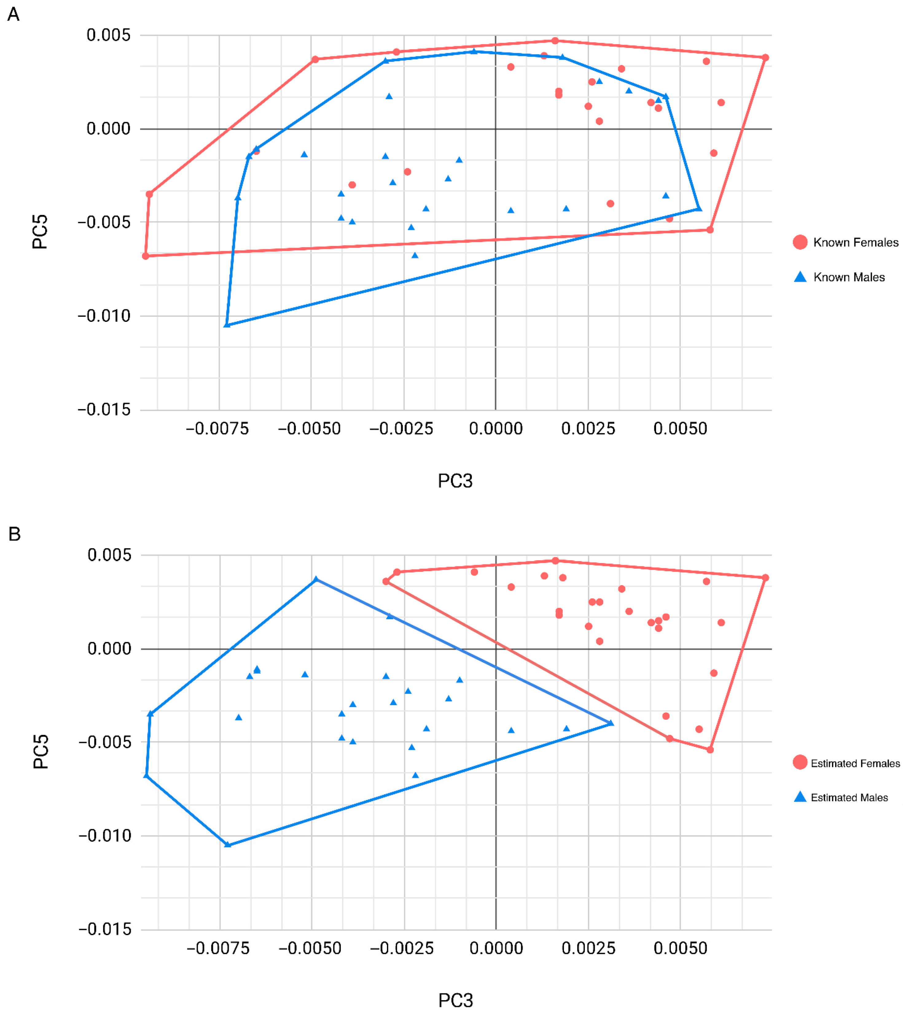
Moreover, the PC3 and PC5 values obtained for the Scottish individuals through the shape simulator were plotted to replicate the morphospace found on the Ammer–Coelho PCA playground webpage, representing the estimated sexes and the known sex, separately. As seen in Figure 2A, which represents known sex according to the entered PCs values, all four quadrants of the morphospace seemed to be occupied by the data points, with many individuals falling on either negative or positive PC values. When the data points were categorized by the sex estimated by the Ammer–Coelho Shape Simulator software (Figure 2B), there was a clear line of discrimination with no overlap between the clusters, with individuals estimated as male primarily located in the negative regions of both PCs, while those estimated as female falling primarily in the positive regions of both PCs. A well-functioning application would produce two similar plots rather than the differing examples seen in this study. The observed discrepancy between Figure 2A,B demonstrates the limitations of the program’s discriminative power when applied to the Scottish sample.

4. Discussion

The development of skeletal methods for forensic identification has been increasing recently, with research being performed using different skeletal elements and diverse populations, as well as traditional and more advanced statistical approaches and technological aids [35,48,50,51]. Among the information required to construct the biological profile, accurate biological sex estimation is crucial for identification purposes as it excludes half of the population and could be needed for subsequent assessment if applying sex-specific methods [3,11,52,53]. As novel sex estimation methods are being developed, testing their reliability and accuracy is essential to avoid jeopardizing investigations [54,55]. Thus, the aim of the present study is two-fold. Firstly, it focuses on evaluating the accuracy of a recently developed software-based technique [34] that uses GMM properties on the olecranon fossa to estimate sex in a Scottish elderly sample. Moreover, the repeatability and reproducibility of the online tool were tested through intra- and inter-observer errors to assess the performance of different observers with similar levels of experience. The overall accuracy rates obtained for the Scottish sample in comparison to the original method classification rates and the level of inter-observer agreement could be related to issues regarding observers’ experience, software application technicalities, or inter-population differences, among others. Limitations in relation to the sample size and the specific nature of the sample population (elderly individuals) should be taken into consideration when interpreting our findings. Moreover, reduced reproducibility raises concerns about courtroom admissibility and reliability [56,57], as a method that cannot be consistently replicated across observers might require refinement before use in forensic contexts [58].

4.1. Intra- and Inter-Observer Error

Macroscopic observation of traits is traditionally used as a quick and easy methodological approach for sex estimation using skeletal elements like the pelvis or the skull [59]. However, subjectivity has been reported as playing an impact on the techniques’ performances with problems arising from, for example, difficulty in assessing specific features, inherent technical and methodological issues, and the observer’s experience [47,60,61]. In the present study, inter-observer average accuracy rates for sex estimation were high, with errors being 29.23% below the analysis performed by the main observer. The poor inter-observer agreement observed here might suggest that adjusting PC3 and PC5 sliders could require more standardized training, with this subjectivity potentially making the method less appropriate for legal settings [56,62]. Macroscopic traits for population affinity estimation, accounting for similar morphological approaches as the one required for the olecranon fossa, reported variable inter-observer errors, with disagreement between experienced and inexperienced observers having an impact on the method’s reliability [63,64]. Moreover, inter-observer differences in scoring cranial non-metric traits compromised sex classification accuracies, as shown in another study [60]. In this study, all observers have similar background knowledge, and thus, their previous experience in the field and its impact on their performance were mitigated [60]. The difference in the observations and accuracy rates reported between the main observer and the other participants could be explained by the fact that the former was exposed to and engaged with a larger sample for a longer time. This could have contributed to a better understanding of the morphological inter-individual variation in the sample under study, explaining also the lower intra-observer error reported. Familiarity with the traits under observation has been highlighted as a factor having an impact on the performance of observers applying methods for sex estimation, and improved traits descriptions have been suggested as a solution [65]. Perhaps adding more detailed written explanations about the specific characteristics to be assessed could further guide the observers into constructing the appropriate shape on the simulator as seen on the specimen under consideration.
Web applications for morphological and metric skeletal assessment have been produced for sex and population affinity estimations, e.g., [66,67,68]. Issues associated with the observation of specific traits using software-based data collection protocols have been noted, with virtual learning initiatives aiming to support the observation of the structures in a three-dimensional (3D) plane [61,69]. Methodologically speaking, the use of the Ammer–Coelho web application translates the olecranon fossa into a 2D structure representing shape through when represented in GM analysis [34]. Additionally, it provides a GM simulator that assists the user with matching the olecranon fossa’s outline shape of the target specimen to the online reference data, as opposed to the traditional approach that compares the unknown individual with the images provided [31]. In the present study, the poor agreement reported for inter-observer error, along with the high sex bias (68.0%), might suggest a tendency of the observers to consistently observe traits associated with male morphology, perceiving triangularity and convexity to a greater degree than roundness and concavity. The original method developed by Rogers (1999) [31] and followed by Ammer et al. (2019) [34] proposed guidelines for the assessment of the olecranon fossa shape, with males having a roughly triangular outline shape and uniformly shallow fossa. The female olecranon fossa description presents two key features, a deep oval shape and a shallow proximal extension, with the latter only seen in some females. The proximal extension of the female olecranon fossa was also noted by other authors when revising Rogers’ (1999) [31] methods [70,71]. Revisiting inter-observer error, it is possible that the observers in this study interpreted the proximal extension as the border of the olecranon fossa, thus explaining the high levels of triangularity reported for known female individuals and decreasing the overall accuracy rates. The difficulties of assessing specific traits for sex estimation methods have been reported previously, e.g., [60]. Overlapping between the sexes’ morphologies for the olecranon fossa might indicate low sexual dimorphism for the traits observed [32] but could be related to the observers not having a full understanding of the features being assessed. As mentioned, more detailed descriptions may help observers grasp not only the traits, but also the potentially diverse range of morphological variation presented on the current population. The incorporation of a training interface within the online tool showing the user the different settings and examples of the olecranon fossa morphology of male, female, and ambiguous individuals could reduce the impact that the traits and the web application might potentially have on the performance of the observers, as well as reducing sex bias.
In the future, the possibility of introducing the assessment of the olecranon fossa in the web application through 3D visualization might help the user to better understand the features under study [72], especially when dealing with characteristics related to the perception of depth, such as concavity and convexity. The combination of the level of experience of the observers in relation to the method, along with both the methodological approach and the online tool being used could have potentially resulted in the discrepancies and bias reported by the inter-observer error analysis.

4.2. Ammer–Coelho for Sex Estimation on the Scottish Sample

Regarding the reliability of the Ammer–Coelho web application, this validation study provided an overall accuracy of 69.23%, which is lower than the original method (88.10%) [34]. Our results indicate slightly higher correct classification rates for female individuals, resulting in a sex bias of −5.33%, with similar bias trends observed by the original method when using 11 PCs [34]. Sex bias greater than 5% is considered large and having an impact on the reliability of the methods [26,65], and thus, our findings suggest a positive outcome for the method under scrutiny. However, it is important to consider that other morphological studies on sex estimation on the distal humerus have reported higher sex bias, showing a lower discriminatory power of the selected features and a larger overlap between males and females [28].
The use of probabilities can assist the expert on decision making [48,73]. In the Scottish sample, more than 80% and 63% of the individuals were estimated with probabilities equal to or greater than 0.80 and 0.95 thresholds, respectively. Considering that sex estimation traditionally accounts for a binary outcome, a cut-off value of more than 0.50 for classification into one of the two groups could be applied. Nonetheless, caution must be taken when classifying close to the zone of uncertainty, with reliable classification being considered those with a posterior probability of at least 0.95 [74,75]. The probabilistic results for the Ammer–Coelho web application provide a sex estimation following the 0.50 threshold. In the current study, a higher percentage of accurately estimated males and females obtained posterior probabilities of at least 0.80 compared to inaccurate male and female estimates. However, some cases were misclassified with high probabilities, as seen in Figure 1. As noted elsewhere, prioritizing high probabilities might minimize the error, but the trade-off will be a decrease in the overall accuracy of the method [76]. Further validation studies with diverse users and different samples will corroborate the accuracy and performance of the Ammer–Coelho web application, as our findings are limited to the observers and the sample size and demographic characteristics of the Scottish elderly sample under investigation.
Sex estimation studies performed on the olecranon fossa shape on different populations have reported variable accuracy rates ranging from 37% to 100% [28,31,32,71,77]. Discrepancies in the classification rates might be due to inter-population differences, as demonstrated by the lower accuracies obtained by Tallman and Blanton (2020) [28] on a target sample from Thailand proving that statistical approaches such as binary regression performed the best with accuracies from 74 to 100%. A South African sample was tested by Vance et al. (2011) [33], who incorporated a scoring method and reported overall accuracies of 77%, indicating lower discriminating power than other papers [34] but demonstrating a degree of sexual dimorphism in the individuals under study. Thus, potential inter-population variability related to differing degrees of expression and morphology patterns of the olecranon fossa between the Scottish target sample and the Portuguese reference sample should be further considered when discussing the performance of the online tool, as the reference database may differ in age and sex distribution in relation to our sample [13,78]. Perhaps adding new samples to the web application will allow for the possibility of refining the algorithm to adjust to different populations, as well as conducting cross-validation studies with independent datasets. Furthermore, although age has been proven not to affect the sex traits associated with the olecranon fossa and other skeletal areas [28,31,79,80], the accumulation of biomechanical loads, age-related pathologies, and physiological adjustments such as hormonal changes can alter bone mass and skeletal structures, thus affecting the feasibility and accuracy of the methods [81,82]. Therefore, the elderly nature of the Scottish sample could have had an impact on our findings, and a wider age range should be included in future research.

4.3. Limitations of the Study

This is the first piece of research that addresses the accuracy and reliability of the Ammer–Coelho web application [34] on a not-related target sample. Although some trends were reported in this validation study, further research is required to confirm our findings. This study focused on the Ammer–Coelho web application [34] which applied GM analysis on a dataset of Portuguese individuals, providing a tool for online users to estimate sex. To draw further conclusions, it will be necessary to run an independent GM analysis using the target sample to fully assess morphological changes for discriminating between the sexes in the Scottish individuals, through data visualization as well as the optimal PCs that represent the current sample. The reliance on right humeri only prevented assessment of bilateral asymmetry, and the elderly nature of the sample may have obscured sexually dimorphic features. The relatively small sample size further reduces statistical power and may affect sex bias, with all confounding factors limiting the conclusion’s generalizability [83].

5. Conclusions

The potential of the distal humerus morphology for sex estimation, along with its high likelihood to survive damage, imply that this is a promising area for investigation. The present study aimed to test the performance and feasibility of the recently developed Ammer–Coelho sex estimation method on an elderly Scottish sample and explore the impact of intra- and inter-observer errors on the web application.
Lower accuracy rates than those of the original method are reported, suggesting that other factors, such as inter-population variation and statistical differences, could have an impact on the performance of the method on an unrelated sample. While the size of our sample was adequate for the purpose of this study, a larger sample would cover a wider range of feature expression variability which would be beneficial to confirm our findings. The possibility of including the left and right side for comparing potential bilateral asymmetry could also be investigated. Furthermore, incorporating individuals representing other age ranges and other population origins will be useful to explore the potential impact of age on the sexual dimorphic features under consideration, as well as the impact of interpopulation variability.
In terms of the observer errors reported, repeatability is confirmed, but reproducibility will need further investigation. Several factors such as the observers’ level of experience, exposure to the material, and methodological issues in relation to the presentation of the olecranon fossa outline, as well as technical matters related to the use of the online tool, could all be considered confounding factors contributing to the low accuracy rates and to the low agreement obtained by the observers.
While the Ammer–Coelho web application performed less effectively on the elderly Scottish sample than on the Portuguese individuals, it offers clear advantages by being an accessible, free web-based application with a standardized interface broadening the applicability of GM in forensic anthropology. Further research is required to confirm the reliability and reproducibility of the online tool, and future practical steps could include algorithm refinement and integration of larger datasets along with the development of a GM population-specific model for the Scottish sample.

Author Contributions

Conceptualization, M.S.T. and J.G.G.-D.; methodology, M.S.T. and J.G.G.-D.; validation, M.S.T.; formal analysis, M.S.T.; investigation, M.S.T. and J.G.G.-D.; resources, J.G.G.-D.; data curation, M.S.T.; writing—original draft preparation, M.S.T. and J.G.G.-D.; writing—review and editing, M.S.T. and J.G.G.-D.; visualization, M.S.T. and J.G.G.-D.; supervision, J.G.G.-D.; project administration, J.G.G.-D. All authors have read and agreed to the published version of the manuscript.

Funding

This research received no external funding.

Institutional Review Board Statement

Ethical permission for the use of skeletal material for research is covered by the Anatomy Act (1984) and the Human Tissue (Scotland) Act 2006, allowing for human material to be used for anatomical examination, education, and training, as well as research purposes (https://www.gov.scot/publications/body-donation-in-scotland-guidance/ (accessed 1 May 2025). Additionally, further approval for this project was granted by the University of Dundee Thiel Advisory group who reviewed the research and granted access to the skeletal sample. All work was authorized and overseen by a Licensed Teacher of Anatomy (https://www.gov.scot/publications/body-donation-in-scotland-guidance/) (accessed on 14 October 2025). The ethical approval for the inter-observer error study using participants was granted by the CAHID ethics committee (UOD_SSEREC_CAHID_MSc_2023_10). More detail link to the University of Dundee ethics procedures-https://www.dundee.ac.uk/research-governance-policy/ethics/non-clinical-research-ethics (accessed on 14 October 2025).

Informed Consent Statement

Not Applicable. Ethical permission for the use of skeletal material for research is covered by the Anatomy Act (1984) and the Human Tissue (Scotland) Act 2006, allowing for human material to be used for anatomical examination, education and training as well as research purposes (https://www.gov.scot/publications/body-donation-in-scotland-guidance/) (accessed on 14 October 2025).

Data Availability Statement

The raw data supporting the conclusions of this article will be made available by the authors on reasonable request.

Acknowledgments

We would like to thank all the participants that volunteered for the inter-observer error. A huge thank to the technical team in CAHID for all the support throughout the collection of the material and assisting with the project. Finally, thanks to the donors, as without them, this project would not have been possible.

Conflicts of Interest

The authors declare no conflicts of interest.

References

  1. Chandrakanth, H.V.; Kanchan, T.; Krishan, K. Osteometric Analysis for Sexing of Modern Sternum—An Autopsy Study from South India. Leg. Med. 2014, 16, 350–356. [Google Scholar] [CrossRef]
  2. Kemkes-Grottenthaler, A. The Reliability of Forensic Osteology—A Case in Point. Forensic Sci. Int. 2001, 117, 65–72. [Google Scholar] [CrossRef]
  3. Krishan, K.; Chatterjee, P.M.; Kanchan, T.; Kaur, S.; Baryah, N.; Singh, R.K. A Review of Sex Estimation Techniques during Examination of Skeletal Remains in Forensic Anthropology Casework. Forensic Sci. Int. 2016, 261, 165.e1–165.e8. [Google Scholar] [CrossRef]
  4. Hefner, J.T. Cranial Nonmetric Variation and Estimating Ancestry. J. Forensic Sci. 2009, 54, 985–995. [Google Scholar] [CrossRef]
  5. Klales, A.R. MorphoPASSE: Morphological Pelvis and Skull Sex Estimation Program. In Sex Estimation of the Human Skeleton; Elsevier: Amsterdam, The Netherlands, 2020; pp. 271–278. [Google Scholar] [CrossRef]
  6. Curate, F.; d’Oliveira Coelho, J.; Silva, A.M. CalcTalus: An Online Decision Support System for the Estimation of Sex with the Calcaneus and Talus. Archaeol. Anthropol. Sci. 2021, 13, 74. [Google Scholar] [CrossRef]
  7. Curate, F.; Navega, D.; Cunha, E.; d’Oliveira Coelho, J. DXAGE 2.0—Adult Age at Death Estimation Using Bone Loss in the Proximal Femur and the Second Metacarpal. Int. J. Leg. Med. 2022, 136, 1483–1494. [Google Scholar] [CrossRef] [PubMed]
  8. Moore, M.K. Sex Estimation and Assessment. In Research Methods in Human Skeletal Biology; DiGangi, E., Moore, M.K., Eds.; Elsevier: Amsterdam, The Netherlands, 2012; pp. 91–114. [Google Scholar]
  9. Giles, E.; Elliot, O. Sex Determination by Discriminant Function Analysis of Crania. Am. J. Phys. Anthropol. 1963, 21, 53–68. [Google Scholar] [CrossRef] [PubMed]
  10. Nikitovic, D. Sexual Dimorphism (Humans). In The International Encyclopedia of Biological Anthropology; John Wiley & Sons, Ltd.: Hoboken, NJ, USA, 2018; pp. 1–4. [Google Scholar] [CrossRef]
  11. Bonczarowska, J.H.; Bonicelli, A.; Papadomanolakis, A.; Kranioti, E.F. The Posterior Portion of the Ilium as a Sex Indicator: A Validation Study. Forensic Sci. Int. 2019, 294, 216.e1–216.e6. [Google Scholar] [CrossRef]
  12. Frayer, D.W.; Wolpoff, M.H. Sexual Dimorphism. Annu. Rev. Anthropol. 1985, 14, 429–473. [Google Scholar] [CrossRef]
  13. Ubelaker, D.H.; DeGaglia, C.M. Population Variation in Skeletal Sexual Dimorphism. Forensic Sci. Int. 2017, 278, 407.e1–407.e7. [Google Scholar] [CrossRef]
  14. Gonzalez, P.N.; Bernal, V.; Perez, S.I. Geometric Morphometric Approach to Sex Estimation of Human Pelvis. Forensic Sci. Int. 2009, 189, 68–74. [Google Scholar] [CrossRef]
  15. Bookstein, F.L. Size and Shape Spaces for Landmark Data in Two Dimensions. Stat. Sci. 1986, 1, 181–222. [Google Scholar] [CrossRef]
  16. Bookstein, F.L. Morphometric Tools for Landmark Data; Cambridge University Press: Cambridge, UK, 1997. [Google Scholar]
  17. Mitteroecker, P.; Gunz, P.; Bernhard, M.; Schaefer, K.; Bookstein, F.L. Comparison of Cranial Ontogenetic Trajectories among Great Apes and Humans. J. Hum. Evol. 2004, 46, 679–698. [Google Scholar] [CrossRef]
  18. Steyn, M.; Pretorius, E.; Hutten, L. Geometric Morphometric Analysis of the Greater Sciatic Notch in South Africans. HOMO 2004, 54, 197–206. [Google Scholar] [CrossRef] [PubMed]
  19. Kimmerle, E.H.; Ross, A.; Slice, D. Sexual Dimorphism in America: Geometric Morphometric Analysis of the Craniofacial Region. J. Forensic Sci. 2008, 53, 54–57. [Google Scholar] [CrossRef] [PubMed]
  20. Kalbouneh, H.; Mubarak, N.; Daradkeh, S.; Ashour, O.; Alkhatib, A.M.; Suboh, L.; Nofal, A.; Mahafzah, W.; Alsalem, M. Estimation of Sex Based on Metrics of the Sternum in a Contemporary Jordanian Population: A Computed Tomographic Study. Medicine 2021, 100, e28169. [Google Scholar] [CrossRef] [PubMed]
  21. Murphy, R.E.; Garvin, H.M. A Morphometric Outline Analysis of Ancestry and Sex Differences in Cranial Shape. J. Forensic Sci. 2018, 63, 1001–1009. [Google Scholar] [CrossRef]
  22. Phenice, T.W. A Newly Developed Visual Method of Sexing the Os Pubis. Am. J. Phys. Anthropol. 1969, 30, 297–301. [Google Scholar] [CrossRef]
  23. Walker, P.L. Sexing Skulls Using Discriminant Function Analysis of Visually Assessed Traits. Am. J. Phys. Anthropol. 2008, 136, 39–50. [Google Scholar] [CrossRef]
  24. Washburn, S.L. Sex Differences in the Pubic Bone. Am. J. Phys. Anthropol. 1948, 6, 199–208. [Google Scholar] [CrossRef]
  25. Mostafa, E.M.; El-Elemi, A.H.; El-Beblawy, M.A.; Dawood, A.E.-W.A. Adult Sex Identification Using Digital Radiographs of the Proximal Epiphysis of the Femur at Suez Canal University Hospital in Ismailia, Egypt. Egypt. J. Forensic Sci. 2012, 2, 81–88. [Google Scholar] [CrossRef]
  26. Hemy, N.; Flavel, A.; Ishak, N.-I.; Franklin, D. Sex Estimation Using Anthropometry of Feet and Footprints in a Western Australian Population. Forensic Sci. Int. 2013, 231, 402.e1–402.e6. [Google Scholar] [CrossRef]
  27. Kotěrová, A.; Velemínská, J.; Dupej, J.; Brzobohatá, H.; Pilný, A.; Brůžek, J. Disregarding Population Specificity: Its Influence on the Sex Assessment Methods from the Tibia. Int. J. Leg. Med. 2017, 131, 251–261. [Google Scholar] [CrossRef] [PubMed]
  28. Tallman, S.D.; Blanton, A.I. Distal Humerus Morphological Variation and Sex Estimation in Modern Thai Individuals. J. Forensic Sci. 2020, 65, 361–371. [Google Scholar] [CrossRef]
  29. Abdelghani, N.; Barut, C.; Ogut, E. The Investigation of Cranial Fossae in the Intracranial Cavity of Fixed Cadaveric Skull Bases: Associations with Sex, Laterality, and Clinical Significance. Surg. Radiol. Anat. 2024, 46, 1305–1329. [Google Scholar] [CrossRef]
  30. Grabiner, M. The Elbow and Radioulnar Joints. In Kinesiology and Applied Anatomy; Lea & Febiger: London, UK, 1989; pp. 136–150. [Google Scholar]
  31. Rogers, T. A Visual Method of Determining the Sex of Skeletal Remains Using the Distal Humerus. J. Forensic Sci. 1999, 44, 57–60. [Google Scholar] [CrossRef]
  32. Vance, V.L.; Steyn, M.; L’Abbé, E.N. Nonmetric Sex Determination from the Distal and Posterior Humerus in Black and White South Africans: Nonmetric Sex Estimation from Distal Humerus. J. Forensic Sci. 2011, 56, 710–714. [Google Scholar] [CrossRef]
  33. Vance, V.L.; Steyn, M. Geometric Morphometric Assessment of Sexually Dimorphic Characteristics of the Distal Humerus. HOMO 2013, 64, 329–340. [Google Scholar] [CrossRef]
  34. Ammer, S.; d’Oliveira Coelho, J.; Cunha, E.M. Outline Shape Analysis on the Trochlear Constriction and Olecranon Fossa of the Humerus: Insights for Sex Estimation and a New Computational Tool. J. Forensic Sci. 2019, 64, 1788–1795. [Google Scholar] [CrossRef] [PubMed]
  35. Navega, D.; Coelho, C.; Vicente, R.; Ferreira, M.T.; Cunha, E. AncesTrees: Ancestry Estimation with Randomized Decision Trees. Int. J. Leg. Med. 2015, 129, 1145–1153. [Google Scholar] [CrossRef] [PubMed]
  36. Scott, G.R.; Pilloud, M.A.; Navega, D.; d’Oliveira Coelho, J.; Cunha, E.; Irish, J.D. rASUDAS: A New Web-Based Application for Estimating Ancestry from Tooth Morphology. Forensic Anthropol. 2018, 1, 18–31. [Google Scholar] [CrossRef]
  37. İşcan, M.Y.; Loth, S.R.; Wright, R.K. Metamorphosis at the Sternal Rib End: A New Method to Estimate Age at Death in White Males. Am. J. Phys. Anthropol. 1984, 65, 147–156. [Google Scholar] [CrossRef]
  38. Haas, J.; Buikstra, J.E.; Ubelaker, D.H. Standards for Data Collection from Human Skeletal Remains: Proceedings of a Seminar at the Field Museum of Natural History Organized by Jonathan Haas, Réimpression; Arkansas Archeological Survey Research Series; Arkansas Archeological Survey: Fayetteville, AR, USA, 1994. [Google Scholar]
  39. Human Tissue (Scotland) Act 2006 (asp 4). An Act of the Scottish Parliament to Make Provision in Relation to Activities Involving Human Tissue. 2006. Available online: https://www.legislation.gov.uk/asp/2006/4/contents (accessed on 29 September 2025).
  40. Scottish Government. Body Donation in Scotland: Guidance. 2023. Available online: https://www.gov.scot/publications/body-donation-in-scotland-guidance/ (accessed on 29 September 2025).
  41. Landis, J.R.; Koch, G.G. The Measurement of Observer Agreement for Categorical Data. Biometrics 1977, 33, 159. [Google Scholar] [CrossRef]
  42. Tang, W.; Hu, J.; Zhang, H.; Wu, P.; He, H. Kappa Coefficient: A Popular Measure of Rater Agreement. Shanghai Arch. Psychiatry 2015, 27, 62–67. [Google Scholar] [CrossRef] [PubMed]
  43. Fleiss, J.L.; Levin, B.A.; Paik, M.C. Statistical Methods for Rates and Proportions, 3rd ed.; Wiley Series in Probability and Statistics; Wiley-Interscience: Hoboken, NJ, USA, 2003. [Google Scholar] [CrossRef]
  44. Trevethan, R. Sensitivity, Specificity, and Predictive Values: Foundations, Pliabilities, and Pitfalls in Research and Practice. Front. Public Health 2017, 5, 307. [Google Scholar] [CrossRef] [PubMed]
  45. Bartholdy, B.P.; Sandoval, E.; Hoogland, M.L.P.; Schrader, S.A. Getting Rid of Dichotomous Sex Estimations: Why Logistic Regression Should Be Preferred Over Discriminant Function Analysis. J. Forensic Sci. 2020, 65, 1685–1691. [Google Scholar] [CrossRef] [PubMed]
  46. Jerković, I.; Bašić, Ž.; Anđelinović, Š.; Kružić, I. Adjusting Posterior Probabilities to Meet Predefined Accuracy Criteria: A Proposal for a Novel Approach to Osteometric Sex Estimation. Forensic Sci. Int. 2020, 311, 110273. [Google Scholar] [CrossRef]
  47. Lesciotto, K.M.; Klales, A.R. Sex Estimation Using Metrics of the Innominate: A Test of the DSP2 Method. J. Forensic Sci. 2025, 70, 249–257. [Google Scholar] [CrossRef]
  48. Kranioti, E.F.; Nathena, D.; Michalodimitrakis, M. Sex Estimation of the Cretan Humerus: A Digital Radiometric Study. Int. J. Leg. Med. 2011, 125, 659–667. [Google Scholar] [CrossRef]
  49. Sieber, K.S.; García-Donas, J.G. Population Affinity Estimation on a Spanish Sample: Testing the Validity and Accuracy of Cranium and Mandible Online Software Methods. Leg. Med. 2023, 60, 102180. [Google Scholar] [CrossRef]
  50. Reynolds, M.S.; MacGregor, D.M.; Alston-Knox, C.L.; Meredith, M.; Barry, M.D.; Schmutz, B.; Gregory, L.S. Bayesian Modeling Predicts Age and Sex Are Not Required for Accurate Stature Estimation from Femoral Length. Forensic Sci. Int. 2018, 289, 452.e1–452.e14. [Google Scholar] [CrossRef]
  51. Krüger, G.C.; L’Abbé, E.N.; Stull, K.E. Sex Estimation from the Long Bones of Modern South Africans. Int. J. Leg. Med. 2017, 131, 275–285. [Google Scholar] [CrossRef]
  52. İşcan, M.Y.; Loth, S.R.; Wright, R.K. Age Estimation from the Rib by Phase Analysis: White Males. J. Forensic Sci. 1984, 29, 1094–1104. [Google Scholar] [CrossRef] [PubMed]
  53. İşcan, M.Y.; Loth, S.R.; Wright, R.K. Age Estimation from the Rib by Phase Analysis: White Females. J. Forensic Sci. 1985, 30, 853–863. [Google Scholar] [CrossRef]
  54. Steyn, M.; İşcan, M.Y. Osteometric Variation in the Humerus: Sexual Dimorphism in South Africans. Forensic Sci. Int. 1999, 106, 77–85. [Google Scholar] [CrossRef] [PubMed]
  55. Liebenberg, L.; Krüger, G.C.; L’Abbé, E.N.; Stull, K.E. Postcraniometric Sex and Ancestry Estimation in South Africa: A Validation Study. Int. J. Leg. Med. 2019, 133, 289–296. [Google Scholar] [CrossRef]
  56. Lesciotto, K.M. Daubert and the Effect on Biological Profile Research. Forensic Anthropol. 2024, 7. Available online: https://journals.upress.ufl.edu/fa/article/view/2368 (accessed on 15 July 2025). [CrossRef]
  57. Christensen, A.M.; Crowder, C.M.; Ousley, S.D.; Houck, M.M. Error and Its Meaning in Forensic Science. J. Forensic Sci. 2014, 59, 123–126. [Google Scholar] [CrossRef] [PubMed]
  58. Klemm, M.M. Inter- and Intra-Observer Error in Forensic Anthropology. Neb. Anthropol. 2024, 35, 93–114. Available online: https://digitalcommons.unl.edu/nebanthro/12 (accessed on 1 July 2025).
  59. İşcan, M.Y. Human Skeleton in Forensic Medicine, 3rd ed.; Charles, C., Ed. Thomas Publisher, Limited: Springfield, IL, USA, 2013. [Google Scholar]
  60. Lewis, C.J.; Garvin, H.M. Reliability of the Walker Cranial Nonmetric Method and Implications for Sex Estimation. J. Forensic Sci. 2016, 61, 743–751. [Google Scholar] [CrossRef]
  61. Kamnikar, K.R.; Plemons, A.M.; Hefner, J.T. Intraobserver Error in Macromorphoscopic Trait Data. J. Forensic Sci. 2018, 63, 361–370. [Google Scholar] [CrossRef]
  62. Nakhaeizadeh, S.; Dror, I.E.; Morgan, R.M. Cognitive Bias in Forensic Anthropology: Visual Assessment of Skeletal Remains Is Susceptible to Confirmation Bias. Sci. Justice 2014, 54, 208–214. [Google Scholar] [CrossRef]
  63. Klales, A.R.; Kenyhercz, M.W. Non-Metric Assessment of Ancestry Through Cranial Macromorphoscopics: A Validation of the Hefner (2009) Method; American Academy of Forensic Sciences: Atlanta, GA, USA; Colorado Springs, CO, USA, 2012. [Google Scholar]
  64. Klales, A.R.; Kenyhercz, M.W. Morphological Assessment of Ancestry Using Cranial Macromorphoscopics. J. Forensic Sci. 2015, 60, 13–20. [Google Scholar] [CrossRef]
  65. Lesciotto, K.M.; Doershuk, L.J. Accuracy and Reliability of the Klales et al. (2012) Morphoscopic Pelvic Sexing Method. J. Forensic Sci. 2018, 63, 214–220. [Google Scholar] [CrossRef] [PubMed]
  66. d’Oliveira Coelho, J.; Navega, D. Cranial Nonmetric Traits Ancestry Estimation. Available online: https://osteomics.com/hefneR/ (accessed on 14 October 2025).
  67. Constantinou, C.; Nikita, E. SexEst: An Open Access Web Application for Metric Skeletal Sex Estimation. Int. J. Osteoarchaeol. 2022, 32, 832–844. [Google Scholar] [CrossRef]
  68. Hefner, J.; Ousley, S.; Richardson, R. MaMD Analytical. 2024. Available online: https://mamdanalytical.com/ (accessed on 15 June 2025).
  69. Spiros, M. 3D MMS Initiative: Cranial & Postcranial Three-Dimensional Interactive Learning Instrument. Available online: https://www.3dmms-initiative.com/ (accessed on 15 June 2025).
  70. Wanek, V.L. A Qualitative Analysis for Sex Determination in Humans Utilizing Posterior and Medial Aspects of the Distal Humerus. Master’s Thesis, Portland State University, Portland, OR, USA, 2002. [Google Scholar]
  71. Falys, C.; Schutkowski, H.; Weston, D. The Distal Humerus-A Blind Test of Rogers’ Sexing Technique Using a Documented Skeletal Collection. J. Forensic Sci. 2005, 50, 1289–1293. [Google Scholar] [CrossRef]
  72. Craik, K.; Collings, A.J. A Preliminary Study into the Impact of Using Three-Dimensional Models in Forensic Anthropology Learning and Teaching. Sci. Justice 2022, 62, 814–821. [Google Scholar] [CrossRef]
  73. Obertová, Z.; Stewart, A.; Cattaneo, C. Statistics and Probability in Forensic Anthropology; Academic Press: Cambridge, MA, USA, 2020. [Google Scholar] [CrossRef]
  74. Morrison, G.S.; Weber, P.; Basu, N.; Puch-Solis, R.; Randolph-Quinney, P.S. Calculation of Likelihood Ratios for Inference of Biological Sex from Human Skeletal Remains. Forensic Sci. Int. Synerg. 2021, 3, 100202. [Google Scholar] [CrossRef]
  75. Brůžek, J.; Santos, F.; Dutailly, B.; Murail, P.; Cunha, E. Validation and Reliability of the Sex Estimation of the Human Os Coxae Using Freely Available DSP2 Software for Bioarchaeology and Forensic Anthropology. Am. J. Phys. Anthropol. 2017, 164, 440–449. [Google Scholar] [CrossRef]
  76. Pilmann Kotěrová, A.; Santos, F.; Bejdová, Š.; Rmoutilová, R.; Attia, M.H.; Habiba, A.; Velemínská, J.; Brůžek, J. Prioritizing a High Posterior Probability Threshold Leading to Low Error Rate over High Classification Accuracy: The Validity of MorphoPASSE Software for Cranial Morphological Sex Estimation in a Contemporary Population. Int. J. Leg. Med. 2024, 138, 1759–1768. [Google Scholar] [CrossRef]
  77. López-Lázaro, S.; Pérez-Fernández, A.; Alemán, I.; Viciano, J. Sex Estimation of the Humerus: A Geometric Morphometric Analysis in an Adult Sample. Leg. Med. 2020, 47, 101773. [Google Scholar] [CrossRef] [PubMed]
  78. Seldin, M.F.; Shigeta, R.; Villoslada, P.; Selmi, C.; Tuomilehto, J.; Silva, G.; Belmont, J.W.; Klareskog, L.; Gregersen, P.K. European Population Substructure: Clustering of Northern and Southern Populations. PLoS Genet. 2006, 2, e143. [Google Scholar] [CrossRef]
  79. Suazo Galdames, I.; Zavando, D. Age Effect in the Morphological Traits Performance for Sex Determination in Human Skulls and Mandibles. Int. J. Morphol. 2012, 30, 296–301. [Google Scholar] [CrossRef]
  80. Selliah, P.; Martino, F.; Cummaudo, M.; Indra, L.; Biehler-Gomez, L.; Campobasso, C.P.; Cattaneo, C. Sex Estimation of Skeletons in Middle and Late Adulthood: Reliability of Pelvic Morphological Traits and Long Bone Metrics on an Italian Skeletal Collection. Int. J. Leg. Med. 2020, 134, 1683–1690. [Google Scholar] [CrossRef]
  81. Ahlborg, H.G.; Johnell, O.; Turner, C.H.; Rannevik, G.; Karlsson, M.K. Bone Loss and Bone Size After Menopause: Obstet. Gynecol. Surv. 2004, 59, 103–105. [Google Scholar] [CrossRef]
  82. Velemínská, J.; Fleischmannová, N.; Suchá, B.; Dupej, J.; Bejdová, Š.; Kotěrová, A.; Brůžek, J. Age-Related Differences in Cranial Sexual Dimorphism in Contemporary Europe. Int. J. Leg. Med. 2021, 135, 2033–2044. [Google Scholar] [CrossRef] [PubMed]
  83. Carvallo, D.; Retamal, R. Sex estimation using the proximal end of the femur on a modern Chilean sample. Forensic Sci. Int. Rep. 2020, 2, 100077. [Google Scholar] [CrossRef]
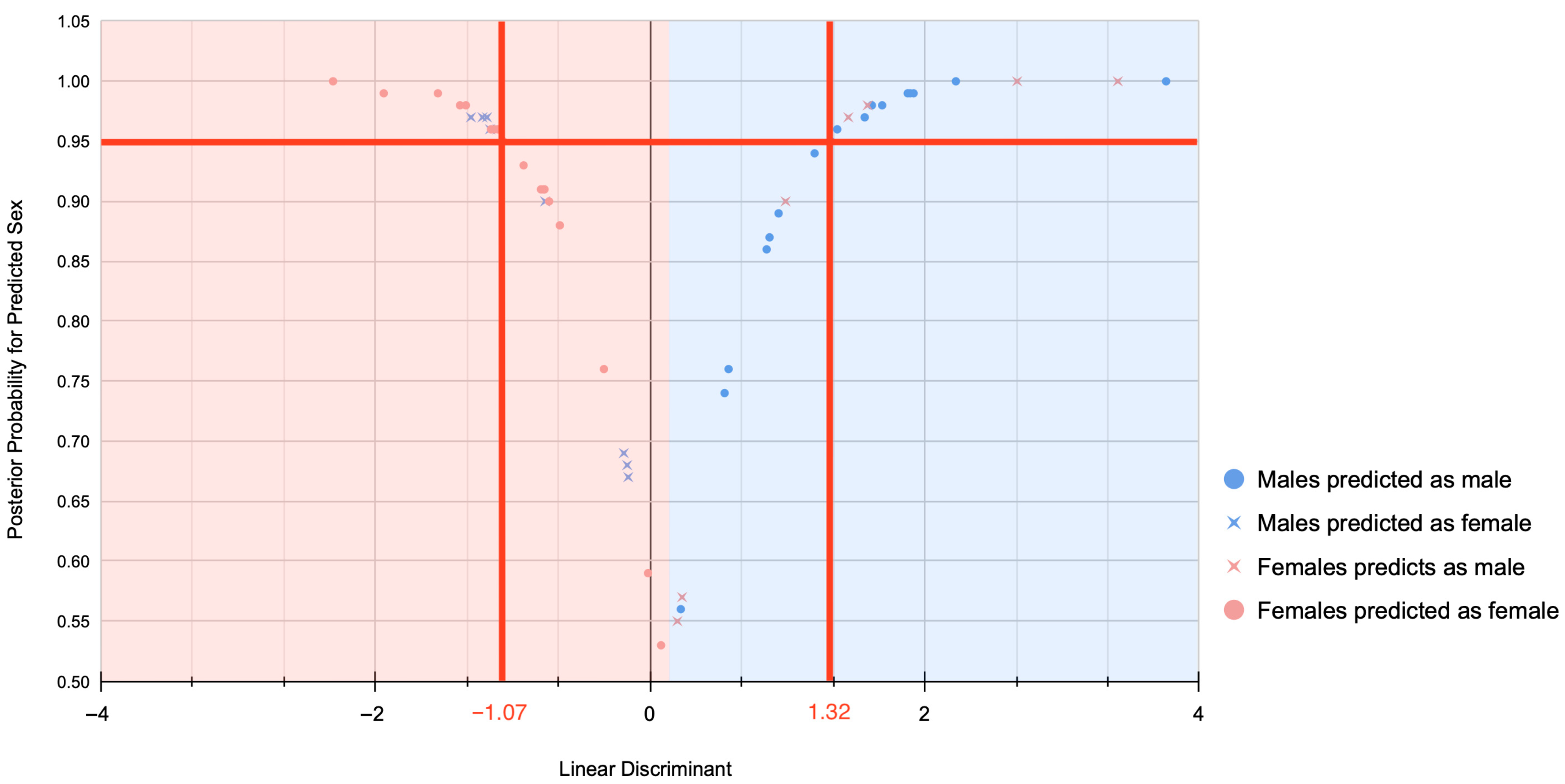
Figure 1. Probability plot demonstrating the relationship between the linear discriminant scores and probability (above 0.50) results for estimated sex on the Scottish sample.
Forensicsci 05 00049 g001
Figure 2. Morphospace scatter plots of PC3 and PC5 values of the olecranon fossae obtained for the Scottish sample representing known sex (A) and estimated sex (B), separately. The lines represent approximate boundaries of the morphospace area for females (red) and males (blue).
Forensicsci 05 00049 g002
Table 1. Demographics of the Scottish sample under study.
Number of
Individuals
Age RangeMean AgeSD
Males2770–9884.376.68
Females2577–9585.645.69
Total5270–9884.986.19
SD = Standard deviation.
Table 2. Accuracy rates for inter-observer error and Cohen’s kappa coefficient for agreement between participants and main observer.
ObserverMale
Accuracy%
Female
Accuracy%
Overall
Accuracy%
Sex Bias%Cohen’s Kappa Coefficient and
Confidence Intervals (CI)
A40020400.348 (95% CI, 0.24 to 0.94), p = 0.26
B1000501000.00
C1002060800.286 (95% CI, 0.18–0.76), p = 0.19
D602040400.348 (95% CI, 0.24 to 0.94), p = 0.26
E8004080−0.190 (95% CI, −0.51 to 0.139), p = 0.38
F60030600.348 (95% CI, 0.24 to 0.94), p = 0.26
G80040800.286 (95% CI, 0.18 to 0.76), p = 0.19
Average7464068.57
Table 3. Accuracy rates considering different probability thresholds for the Ammer–Coelho web application on the sample under study.
Probability Threshold
>0.50 ≥0.80 ≥0.95
Male (n/%)18/10015/83.3311/61.11
Female (n/%)18/10015/83.339/50
Overall (N/%)36/10030/83.3320/55.56
Disclaimer/Publisher’s Note: The statements, opinions and data contained in all publications are solely those of the individual author(s) and contributor(s) and not of MDPI and/or the editor(s). MDPI and/or the editor(s) disclaim responsibility for any injury to people or property resulting from any ideas, methods, instructions or products referred to in the content.