Abstract
Fat embolism syndrome (FES) can be challenging to diagnose by forensic pathologists. For the diagnosis of FES, there is no benchmark test. Postmortem diagnosis requires a full autopsy and specific ancillary examination. However, the high variability in the clinical presentation of FES represents a relevant issue, and there is no consensus on the postmortem assessment. This is the case of a 33-year-old man who died of FES one week after a car accident. He suffered multiple fractures, but was hemodynamically stable and showed no neurological changes. The patient died a few days after hospital discharge. Additionally, he had osteogenesis imperfecta type III, a genetic disorder associated with bone fragility. To the best of our knowledge, no study has assessed whether and how osteogenesis imperfecta contributes to the onset of FES. Despite the heterogeneous manifestations of FES, the present case met many of the proposed clinical and histological diagnostic criteria. Therefore, we briefly review FES diagnostic criteria, show the postmortem diagnostic workup, and discuss the hypothetical link between osteogenesis imperfecta and FES.
1. Introduction
Fat embolism is defined as the presence of fat globules in the pulmonary or peripheral circulation, and fat embolism syndrome (FES) refers to the clinical symptoms that follow an identifiable insult [1]. Since Zenker first described it in 1861, FES has posed a serious challenge because of the wide range of possible clinical presentations [1]. The most common clinical presentation of FES is a multisystem disorder affecting the lungs and brain. Other commonly presented signs and symptoms include disorders of the cardiovascular system, retinal manifestations, and cutaneous disorders [2]. Zenker reported the presence of fat droplets in the lung capillaries of a railroad worker who died from a crush injury [3]. Frequently, fat embolism occurs after trauma and during orthopedic procedures and is most commonly related to intramedullary orthopedic surgery and lower extremity trauma [2]. It can also occur after other forms of trauma, such as severe burns, liver injury, closed-chest cardiac massage, bone marrow transplantation, liposuction, parental lipid infusion, decompression sickness, extracorporeal circulation, acute hemorrhagic pancreatitis, prolonged corticosteroid therapy, sickle cell disease, and carbon tetrachloride poisoning [3,4]. However, the pathogenesis of FES is still debated.
FES comprises respiratory, neurological, cutaneous, and hematologic manifestations related to the intravascular embolization of fat arising from within the fractured bone marrow space. Respiratory distress, neurological changes, and a petechial rash represent the classic triad of symptoms. Symptoms usually appear several days after a significant trauma involving the pelvis or lower extremities and may rapidly lead to death. Many authors have proposed different diagnostic criteria for FES owing to its unpredictable clinical presentation [2]. In fact, one of the challenges in the study and identification of FES is that there is no benchmark test [1]. Many cases of FES are misdiagnosed, and postmortem examination should provide sufficient means for appropriate diagnosis [5].
Here, we present a case of post-traumatic FES in an adult man with osteogenesis imperfecta (OI) type III, a rare collagen disease that frequently leads to post-traumatic complications. OI is a phenotypically heterogeneous, rare connective tissue disorder. It is known as brittle bone disease because it causes bone fragility [6]. The severity varies extensively, ranging from intrauterine bone fractures and perinatal lethality to very mild forms without fractures [7]. The clinical presentation of OI also includes a wide range of extra-skeletal manifestations. Typical extra-skeletal manifestations are blue sclera, dentinogenesis imperfecta, hyperlaxity of ligaments and skin, hearing impairment, and the presence of Wormian bones on skull radiographs [8]. The initial diagnosis is mostly based on clinical and radiographic findings. OI patients’ hallmark characteristics are fractures after mild trauma, bowing deformities of long bones, and growth deficiency. Other skeletal features include macrocephaly, flat midface and triangular facies, chest wall deformities like pectus excavatum or pectus carinatum, barrel chest, and scoliosis or kyphosis [9]. Radiological findings comprise generalized osteopenia, different combinations of long-bone bowing, under-tubulation and metaphyseal flaring, gracile ribs, narrow thoracic apex, and vertebral compressions [8].
The type III variant of OI is progressive and severe and is included in the progressively deforming subgroup of OI [10]. The present case showed all of the described signs and symptoms of FES and a complete postmortem diagnostic workup. The study aims to briefly review clinical and postmortem criteria for FES diagnosis and present the untested hypothesis of the association between FES and OI.
2. Case
A 33-year-old man with osteogenesis imperfecta (OI) type III had a car accident and was immediately admitted to the emergency department.
At the admission to the emergency department, the man’s blood pressure was 140/90 mmHg, and the saturation of oxygen was 100%. An arterial blood gas (ABG) test yielded results within the normal range. The patient was hemodynamically stable and did not show alterations in mental status or focal neurological signs. He breathed spontaneously with a Venti-Mask with FiO2 of 0.4. Radiographs revealed a right supracondylar femoral fracture and a distal fracture of the fourth metacarpal finger bone. A few hours after hospital admission, the patient required intensive care because of the sudden onset of severe dyspnea.
The next day, blood test results showed that the patient’s red blood cell (RBC) count decreased from 4.84 × 10⁶/microL to 3.35 × 10⁶/microL; hemoglobin (Hb) decreased from 14.3 to 9.8 g/dL; hematocrit (Hct) decreased from 43.5% to 30.3%; and platelet count decreased from 319 × 103/microL to 230 × 103/microL. After a few hours, an ABG test showed that pH was 7.44, pCo2 was 35 mmHg, pO2 was 25 mmHg, Hct was 30%, and Hb was 9.9 g/dL.
Cranial computed tomography (CT) revealed occipital and parietal bone fractures. Chest and abdominal CT showed no pathological signs, so pulmonary embolism and abdominal hemorrhages were excluded. The O2 therapy (with Venti-Mask with FiO2 30% at 7 L/min) stabilized pH (7.37), pcO2 (50 mmHg), pO2 (105 mmHg), and Hb (9.3 g/dL) values. Blood tests revealed lower Hct (22.7%) and Hb (7.3 g/dL) three days after admission. Hence, the patient was transfused with two RBC units. The following day, the patient was discharged from the hospital, with Hb values of 9.4 g/dL and an oxygen therapy prescription when needed. The patient died four days after hospital discharge.
A postmortem examination of the male corpse revealed that the body was 124 cm tall and showed severe kyphoscoliosis, which is a characteristic joint deformation in OI type III.
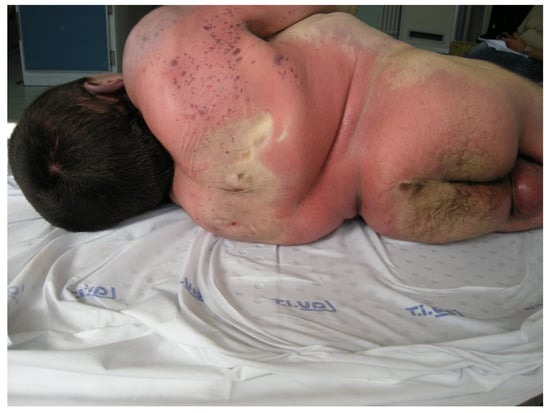
Both the upper limbs and chest showed multiple irregular abrasions and contusions due to trauma. Purple petechiae of different sizes (0.2–2 cm) were discernible on the back surface of the right arm and right side of the thorax [Figure 1].

Figure 1.
Scapular petechiae on the back.
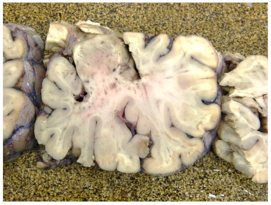
The autopsy confirmed fractures detected with a previous CT scan. The brain weighed 1575 g. The macroscopic evaluation of the brain revealed intense congestion of the meningeal vessels and several widespread petechiae in the temporal lobe. No other pathological findings were found [Figure 2].

Figure 2.
Temporal lobe section of the formaldehyde-fixated brain.
Macroscopic evaluation of the lungs revealed acute edema and congestion of the vessels. Both were non-specific findings of lung damage. No other organ was damaged by the trauma.
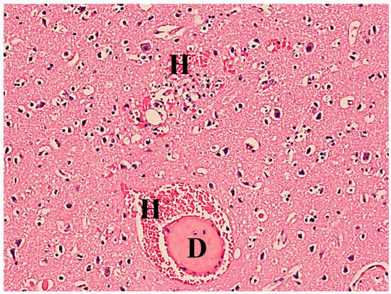
Histopathological examination revealed fat droplets in the alveolar capillaries of the lungs and cerebral blood vessels, and small hemorrhages in the brain tissue (Figure 3).

Figure 3.
Brain tissue with signs of acute stasis, fat droplets (D), and small hemorrhages (H). Hematoxylin–eosin stain, 40× magnification.
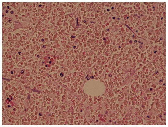
Moreover, lung samples were frozen and stained with Sudan III. The Sudan III staining of the frozen tissue colored the drops orange, revealing their fatty nature [11]. The frozen lung samples showed 20–40 emboli per field (Figure 4).

Figure 4.
Lung tissue stained with Sudan III showing the multiple fat drops in orange color.
Hence, according to the clinical presentation, the autopsy findings, and the ancillary examination results, the cause of death was pulmonary fat embolism due to FES.
3. Discussion
Fat embolism syndrome (FES) originates from long bone and pelvic fractures due to traumatized bone fat and bone marrow that travel to the lungs and block pulmonary circulation [12]. FES encompasses a spectrum of symptoms and signs of respiratory, neurological, cutaneous, and hematological manifestations caused by fat emboli. Talbot et al. demonstrated that 82% of patients with fractures had fat emboli, whereas only 33% had FES [13]. Fukumoto et al. reported that FES occurred in 19% of major trauma cases, and 67% of trauma patients without evidence of FES had circulating fat globules [3,14]. As there is still no consensus regarding the diagnosis of FES in clinical practice, its exact incidence remains unknown. Nevertheless, a retrospective study of over 3000 long bone fractures found an incidence of 0.9% [3], while Fabian et al. prospectively demonstrated an FES incidence of at least 11% by studying an increased pulmonary shunt fraction [15]. Although the pathophysiology of FES remains unclear, mechanical and biochemical factors have been proposed. The “mechanical” theory states that fat particles originating from disrupted adipose tissue or bone marrow are forced into torn venues in areas of trauma. The embolization of fat particles may cause the direct obstruction of pulmonary capillaries, leading to ventilation–perfusion mismatch, low oxygen saturation, and dyspnea. According to this theory, symptom severity is proportional to the number of vessels closed by fat emboli [16]. The mechanical theory is supported by the funding of “echogenic material” passing into the right heart during orthopedic surgery. Nevertheless, it does not sufficiently explain the 24–72 h delay often seen between surgery and FES manifestation [17]. Other educated guesses about FES pathogenesis involve multiple biochemical mechanisms that may play a role in the formation of fat emboli, which are part of the biochemical theory. The “biochemical” theory proposes biochemical changes including fat globules entering the pulmonary vasculature where they are broken down to free fatty acids, leading to a microvascular inflammatory response with the development of acute lung injury. The fat-hydrolyzed products (including free fatty acids) have been shown to cause ARDS and cardiac contractile dysfunction. The biochemical theory is supported by the slight increase in free fatty acids levels in acutely ill patients’ blood. Also, it partly accounts for the timescale required for the development of the symptoms and the delayed onset of them [12,17].
The clinical diagnosis of FES requires the characteristic signs and symptoms in the context of supportive imaging and predisposing insult. Chest radiography frequently reveals no lung alterations. Brain MRI sometimes reveals a star-field pattern of diffuse, punctate, and hyperintense lesions on diffusion-weighted imaging [18]. FES has autopsy features that are unique and can aid in diagnosis. The macroscopic pulmonary findings may include increased lung weight and volume, with a marbled appearance of the visceral pleura due to alternating areas of hemorrhage. Moreover, petechiae or ecchymoses may be present beneath the visceral pleura [19].
Many authors have proposed diagnostic criteria; the most used are Gurd’s criteria. The diagnosis of FES can be ruled out using two primary criteria or one major criterion with four minor criteria. The major signs include hypoxia, mental state changes, and petechiae, referred to as the classic triad of symptoms (pulmonary, neurological, and dermatological). Rather, the minor signs include tachycardia, thrombocytopenia, anemia, hyperpyrexia, retinal changes, hematuria, and a high erythrocyte sedimentation rate [20] [Table 1].

Table 1.
Gurd’s criteria for diagnosis of FES.
Schonfeld proposed a diagnostic score based on the clinical findings; the diagnosis of FES requires a five-point score. The most important signs, which give a higher score, are petechiae, chest X-ray changes, and hypoxemia. The presence of only petechiae can lead to the diagnosis of FES because it gives five points [21] [Table 2].

Table 2.
Schonfeld’s criteria for diagnosis of FES.
According to Lindeque’s criteria, FES is confirmed if a patient with tibial or femoral fracture shows one of the red flags indicative of respiratory distress [22] [Table 3].

Table 3.
Lindeque’s criteria for diagnosis of FES.
Even though FES often has a fatal outcome, there is no consensus on postmortem diagnosis [23]. In 1958, Emson proposed a postmortem grading system for the severity of fat emboli by counting the fat emboli (>10 µm in diameter) in frozen lung sections stained with Oil Red O. This allows three gravity classes to be distinguished [19] [Table 4].

Table 4.
Emson’s FES grading system.
Neri et al. confirmed the diagnosis of FES by Sudan III staining for lipids and immunohistochemistry with anti-CD 61 and anti-fibrinogen antibodies. Using this stain, fat emboli appeared as orange intravascular globules surrounded by platelet aggregation and fibrinogen [24].
In the present case, the patient showed two significant signs, hypoxia and petechiae, and one minor sign of unexplained anemia. Thus, Gurd’s criteria were satisfied. The presence of petechiae and hypoxemia is worth eight points according to Schonfeld’s criteria, confirming the FES diagnosis. Since the patient reported a femoral fracture and presented with respiratory fatigue and low PO2 values, even the Lindeque criteria were fulfilled. Finally, the patient’s presentation was consistent with grade 2 of the Sevitt classification [25].
The postmortem examination of frozen lung samples showed 20–40 emboli per field, which satisfied Emson’s criteria of a moderate degree of fat embolism. When we applied volumetric analysis of fat emboli using Neri’s grading system, the samples showed moderate embolism. Hence, the cause of death was ruled as FES.
The patient had osteogenesis imperfecta type III (OI). OI is a genetically determined disorder of connective tissue characterized by bone fragility. The disease state encompasses a phenotypically and genotypically heterogeneous group of inherited disorders resulting from more than 150 observed mutations in genes that code for type I collagen. OI type III (progressive deformation) is the most severe non-lethal form [8]. OI patients may sustain hundreds of fractures owing to traumatic or pathological causes, one of which is the most frequent cause of morbidity [8]. As FES usually develops after a long bone fracture, it is reasonable to expect that suffering a higher number of fractures would lead to an intrinsically augmented risk of developing FES. Moreover, osteogenesis imperfecta leads to alterations in bone material proprieties and may hypothetically affect the tendency to form fat embolies. Despite this, no direct association was observed and described between osteogenesis imperfecta and FES. On the other hand, a study based in the United States speculated that patients with bone loss or lower bone density had a lower relative risk of FES because they are likely to suffer minimal trauma fractures in specific sites like the neck of the femur. In contrast, FES risk is mainly related to fractures in sites rich in yellow marrow [26,27]. Nevertheless, differently from other conditions of bone fragility, fractures of the neck of the femur are rare given the high prevalence of long bone fractures in osteogenesis imperfecta [28].
In patients with OI presenting sudden death, FES should be suspected. Even if there is no consensus about postmortem diagnosis, based on the results of this study, it is possible to propose a diagnostic algorithm for forensic pathologists. First, it is necessary to conduct a careful analysis of the clinical history with special attention being paid to the signs and symptoms included in Gurd’s, Schonfeld’s, and Lindeque’s criteria. If the clinical presentation is suggestive of FES, the pathologist should collect multiple samples of lungs in both peripheric and peri-ileal sections. Different methods were proposed for sample preparation. Turillazzi et al. suggested the use of confocal laser scanning in order to identify the emboli [28]. Alternatively, it is possible to stain fat emboli using Oil Red O in frozen lung sections or to use Sudan III staining for lipids and immunohistochemistry with anti-CD 61 and anti-fibrinogen antibodies, as suggested by Neri et al. [24]. Once the fat emboli have been stained, the diagnosis of FES should include a quantitative assessment of their size and number. The method of quantitative evaluation varies according to the staining employed. Turillazzi proposed determining the ratio between embolized tissue areas and the total tissue area of all samples to obtain a reliable parameter for diagnosing FES [28]. Emson’s criteria for fat emboli grading are applicable if the samples are stained with Oil Red O or Sudan III. Further studies are needed to establish a reliable parameter for fat emboli evaluation.
In summary, the diagnosis of FES relies on the deep analysis of case history, and the demonstration of fat embolism.
4. Conclusions
FES diagnosis requires a complex workup and can be challenging for pathologists. Due to its heterogeneous manifestations, there are many different clinical and histological diagnostic criteria of FES; each one includes a deep case history assessment and specific histological examinations. There is no consensus about the postmortem diagnosis of FES; however, the demonstration of fat embolism requires more than the routine ancillary examination. Hence, clinical suspicion should drive the postmortem assessment.
To the best of our knowledge, no direct association has been observed between OI and FES. There are no studies dealing with this, and the present case’s findings shed light on the potential pathological correlation between OI and FES. Due to the pathogenesis of FES related to orthopedic trauma and the frequency of fractures suffered by OI patients, the possible association between FES and OI deserves further evaluation. Pathologists should suspect FES in patients with OI who died suddenly.
Author Contributions
F.C., C.C., and M.B.: Writing—Original Draft; L.A. and D.F.: Writing—Review and Editing; C.C. and S.N.: Methodology and Investigation; B.S.: Supervision. All authors have read and agreed to the published version of the manuscript.
Funding
This research received no external funding.
Institutional Review Board Statement
The study did not require ethical approval as it did not involve living humans. As stated in Recital 27 of the GDPR, “This Regulation does not apply to the personal data of deceased persons” (https://gdpr-info.eu/recitals/no-27/, accessed on 17 April 2024).
Informed Consent Statement
Not applicable.
Data Availability Statement
Supplementary data are not available due to privacy restrictions.
Acknowledgments
The authors acknowledge Francesco Merlanti (University of Bari) for his contribution to the preparation of the histological slides.
Conflicts of Interest
The authors declare no conflicts of interest.
References
- Rothberg, D.L.; Makarewich, C.A. Fat Embolism and Fat Embolism Syndrome. J. Am. Acad. Orthop. Surg. 2019, 27, E346–E355. [Google Scholar] [CrossRef] [PubMed]
- Taviloglu, K.; Yanar, H. Fat embolism syndrome. Surg. Today 2007, 37, 5–8. [Google Scholar] [CrossRef] [PubMed]
- Bulger, E.M.; Smith, D.G.; Maier, R.V.; Jurkovich, G.J. Fat Embolism Syndrome. A 10-Year Review. Arch. Surg. 1997, 132, 435–439. [Google Scholar] [CrossRef] [PubMed]
- Jenkins, K.; Chung, F.; Wennberg, R.; Etchells, E.E.; Davey, R. Fat Embolism Syndrome and Elective Knee Arthroplasty. Can. J. Anaesth. 2002, 49, 19–24. [Google Scholar] [CrossRef]
- Cantu, C.A.; Pavlisko, E.N. Liposuction-Induced Fat Embolism Syndrome: A Brief Review and Postmortem Diagnostic Approach. Arch. Pathol. Lab. Med. 2018, 142, 871–875. [Google Scholar] [CrossRef] [PubMed]
- Palomo, T.; Vilacą, T.; Lazaretti-Castro, M. Osteogenesis imperfecta: Diagnosis and treatment. Curr. Opin. Endocrinol. Diabetes Obes. 2017, 24, 381–388. [Google Scholar] [CrossRef] [PubMed]
- Shapiro, J.R.; Stover, M.L.; Burn, V.E.; McKinstry, M.B.; Burshell, A.L.; Chipman, S.D.; Rowe, D.W. An Osteopenic Nonfracture Syndrome with Features of Mild Osteogenesis Imperfecta Associated with the Substitution of a Cysteine for Glycine at Triple Helix Position 43 in the pro Alpha 1 (I) Chain of Type I Collagen. J. Clin. Invest. 1992, 89, 567–573. [Google Scholar] [CrossRef][Green Version]
- Forlino, A.; Marini, J.C. Osteogenesis imperfecta. Lancet 2016, 387, 1657–1671. [Google Scholar] [CrossRef]
- Forlino, A.; Cabral, W.A.; Barnes, A.M.; Marini, J.C. New Perspectives on Osteogenesis Imperfecta. Nat. Rev. Endocrinol. 2011, 7, 540–557. [Google Scholar] [CrossRef] [PubMed]
- Folkestad, L. Mortality and morbidity in patients with osteogenesis imperfecta in Denmark. Dan. Med. J. 2018, 65, B5454. [Google Scholar]
- Kao, S.J.; Yeh, D.Y.-W.; Chen, H.I. Clinical and pathological features of fat embolism with acute respiratory distress syndrome. Clin. Sci. 2007, 113, 279–285. [Google Scholar] [CrossRef] [PubMed]
- Milroy, C.M.; Parai, J.L. Fat Embolism, Fat Embolism Syndrome and the Autopsy. Acad. Forensic. Pathol. 2019, 9, 136–154. [Google Scholar] [CrossRef] [PubMed]
- Talbot, M.; Schemitsch, E.H. Fat embolism syndrome: History, definition, epidemiology. Injury 2006, 37 (Suppl. S4), S3–S7. [Google Scholar] [CrossRef] [PubMed]
- Fukumoto, L.E.; Fukumoto, K.D. Fat Embolism Syndrome. Nurs. Clin. N. Am. 2018, 53, 335–347. [Google Scholar] [CrossRef] [PubMed]
- Fabian, T.C.; Hoots, A.V.; Stanford, D.S.; Patterson, C.R.; Mangiante, E.C. Fat embolism syndrome: Prospective evaluation in 92 fracture patients. Crit. Care Med. 1990, 18, 42–46. [Google Scholar] [CrossRef] [PubMed]
- Zilg, B.; Råsten-Almqvist, P. Fatal Fat Embolism After Penis Enlargement by Autologous Fat Transfer: A Case Report and Review of the Literature. J. Forensic. Sci. 2017, 62, 1383–1385. [Google Scholar] [CrossRef] [PubMed]
- Gupta, A.; Reilly, C.S. Fat embolism. Contin. Educ. Anaesth. Crit. Care Pain 2007, 7, 148–151. [Google Scholar] [CrossRef]
- Kosova, E.; Bergmark, B.; Piazza, G. Fat embolism syndrome. Circulation 2015, 131, 317–320. [Google Scholar] [CrossRef]
- Emson, H.E. Fat Embolism Studied in 100 Patients Dying after Injury. J. Clin. Path. 1958, 11, 28–35. [Google Scholar] [CrossRef]
- Gurd, A.R. Fat embolism: An aid to diagnosis. J. Bone Joint Surg. Br. 1970, 52, 732–737. [Google Scholar] [CrossRef]
- Schonfeld, S.A.; Ploysongsang, Y.; Dilisio, R.; Crissman, J.D.; Miller, E.; Hammerschmidt, D.E.; Jacob, H.S. Fat embolism prophylaxis with corticosteroids. A prospective study in high-risk patients. Ann. Intern. Med. 1983, 99, 438–443. [Google Scholar] [CrossRef] [PubMed]
- Uransilp, N.; Muengtaweepongsa, S.; Chanalithichai, N.; Tammachote, N. Fat Embolism Syndrome: A Case Report and Review Literature. Case Rep. Med. 2018, 2018, 1479850. [Google Scholar] [CrossRef] [PubMed]
- Miller, P.; Prahlow, J.A. Autopsy diagnosis of fat embolism syndrome. Am. J. Forensic. Med. Pathol. 2011, 32, 291–299. [Google Scholar] [CrossRef] [PubMed]
- Neri, M.; Riezzo, I.; Dambrosio, M.; Pomara, C.; Turillazzi, E.; Fineschi, V. CD61 and fibrinogen immunohistochemical study to improve the post-mortem diagnosis in a fat embolism syndrome clinically demonstrated by transesophageal echocardiography. Forensic. Sci. Int. 2010, 202, e13–e17. [Google Scholar] [CrossRef] [PubMed]
- Sevitt, S. The Significance and Classification of Fat-Embolism. Lancet 1960, 276, 825–828. [Google Scholar] [CrossRef] [PubMed]
- Stein, P.D.; Yaekoub, A.Y.; Matta, F.; Kleerekoper, M. Fat embolism syndrome. Am. J. Med. Sci. 2008, 336, 472–477. [Google Scholar] [CrossRef] [PubMed]
- Chow, W.; Negandhi, R.; Kuong, E.; To, M. Management pitfalls of fractured neck of femur in osteogenesis imperfecta. J. Child. Orthop. 2013, 7, 195–203. [Google Scholar] [CrossRef]
- Turillazzi, E.; Riezzo, I.; Neri, M.; Pomara, C.; Cecchi, R.; Fineschi, V. The diagnosis of fatal pulmonary fat embolism using quantitative morphometry and confocal laser scanning microscopy. Pathol. Res. Pract. 2008, 204, 259–266. [Google Scholar] [CrossRef]
Disclaimer/Publisher’s Note: The statements, opinions and data contained in all publications are solely those of the individual author(s) and contributor(s) and not of MDPI and/or the editor(s). MDPI and/or the editor(s) disclaim responsibility for any injury to people or property resulting from any ideas, methods, instructions or products referred to in the content. |
© 2024 by the authors. Licensee MDPI, Basel, Switzerland. This article is an open access article distributed under the terms and conditions of the Creative Commons Attribution (CC BY) license (https://creativecommons.org/licenses/by/4.0/).