Review

Challenges and (Un)Certainties for DNAm Age Estimation in Future

by
Helena Correia Dias
1,2,3,*,
Eugénia Cunha
2,3,
Francisco Corte Real
3,4 and
Licínio Manco
1
1
University of Coimbra, Research Centre for Anthropology and Health (CIAS), Department of Life Sciences, 3000-456 Coimbra, Portugal
2
University of Coimbra, Centre for Functional Ecology (CEF), Laboratory of Forensic Anthropology, Department of Life Sciences, 3000-456 Coimbra, Portugal
3
National Institute of Legal Medicine and Forensic Sciences, 3000-548 Coimbra, Portugal
4
Faculty of Medicine, University of Coimbra, 3000-370 Coimbra, Portugal
*
Author to whom correspondence should be addressed.
Forensic Sci. 2022, 2(3), 601-614; https://doi.org/10.3390/forensicsci2030044
Submission received: 27 July 2022 / Revised: 3 September 2022 / Accepted: 7 September 2022 / Published: 15 September 2022
(This article belongs to the Special Issue Feature Papers in Forensic Sciences in 2022)

Abstract

:
Age estimation is a paramount issue in criminal, anthropological, and forensic research. Because of this, several areas of research have focused on the establishment of new approaches for age prediction, including bimolecular and anthropological methods. In recent years, DNA methylation (DNAm) has arisen as one of the hottest topics in the field. Many studies have developed age-prediction models (APMs) based on evaluation of DNAm levels of many genes in different tissue types and using different methodological approaches. However, several challenges and confounder factors should be considered before using methylation levels for age estimation in forensic contexts. To provide in-depth knowledge about DNAm age estimation (DNAm age) and to understand why it is not yet a current tool in forensic laboratories, this review encompasses the literature for the most relevant scientific works published from 2015 to 2021 to address the challenges and future directions in the field. More than 60 papers were considered focusing essentially on studies that developed models for age prediction in several sample types.

1. Introduction

Age estimation is a paramount issue in forensic science, being required for the identification process of deceased and living individuals. For deceased ones, including human skeletonized remains, the estimative of age can lead to an exclusion; for living individuals, age estimation is important to solve cases of immigration, cases of minors, or for determination of criminal responsibility, for instance [1,2,3,4].
Aging, being a complex biological process, is difficult to predict and the use of a multidisciplinary approach can improve the age estimation [5,6,7]. For instance, Shi et al. [6] demonstrated the power of the combination of the anthropological approach (dental and skeletal ages) with the epigenetic approach using DNA methylation (DNAm) levels. During recent years, several areas of knowledge such as anthropology, odontology, chemistry, genetics, and more recently epigenetics, have been continually focused on the improvement of age estimation research. As a result, new methods in several fields have been proposed. In any case, there is not an elected method or approach for age estimation that can be applied to all the forensic cases (living and deceased individuals), with the same accuracy in all age ranges [1,8]. In the past, the evaluation of changes in DNAm levels in age-associated genes has been explored as the epigenetic modification with the strongest potential for age prediction in forensic contexts [3,9,10,11,12,13,14,15,16,17,18,19]. As a result, DNAm levels of many age-correlated genes have been evaluated in different tissue types, proposing the development of many highly accurate age prediction models (APMs) [20]. Despite the increase in research on epigenetic age or DNAm age estimation (DNAm age) in recent years, this tool for age estimation was not implemented until now in forensic laboratories, essentially because of several challenges and confounding factors that need to be considered in future research.
The main aim of this review is to provide in-depth knowledge about DNAm as a tool for age estimation in forensic contexts. For this main purpose, in the first step, we discuss the most promising epigenetic mechanism (DNAm) associated with aging research and the underlying mechanisms of DNAm changes with age that allowed the use of DNAm levels as a tool for age estimation (points 2 and 3). In the second step, we explain the basis of some of the most currently used methodologies for DNAm evaluation and quantification, and review some of the most important works in the literature to date, focusing on the evaluation of the correlation between DNAm and age in many different tissue types (points 4 and 5). Lastly, we discuss future directions and challenges for the application of DNAm as a practicable tool for age estimation in forensic laboratories, considering the implementation of DNAm age in forensic cases (points 6 and 7).

2. DNA Methylation (DNAm): An Epigenetic Mechanism

Epigenetics is a large area of research nowadays. The main epigenetic features (histone modifications, regulation by non-coding RNAs and DNAm) have been associated with several clinical conditions, such as cancer and Alzheimer’s disease, and forensic issues, including age estimation [21,22,23]. One of the most important bases of aging research is DNAm, which has arisen in recent years as one of the most promising and investigated epigenetic features associated with aging [3,12,15,17,18,19]. DNAm is characterized by the addition of a methyl group (CH3) to the fifth carbon (5C) position of cytosines in the DNA molecule [24,25]. Commonly, the methylation of DNA occurs in dinucleotide CpGs (5′-CpG-3′ cytosine–phosphate–guanine) across the genome. However, there are some CpG sites located at clusters named CpG islands, mainly in the promoter regions of the active genes, in which there is no methylation. During aging, there is a change in the human genome methylation levels: most CpGs across the genome lose methylation, and CpG islands gain methylation [10,26,27,28,29,30]. These changes in the pattern of DNAm can be consistent across the individuals or result from stochastic factors. Based on these consistent alterations in DNAm levels of some genes (age-correlated genes), the first generation of “epigenetic clocks” has arisen in different tissue types [9,10,31,32,33].

3. Underling Mechanisms of DNAm Changes with Age

As mentioned previously, with aging several changes in DNAm have been observed: consistent alterations across the individuals (epigenetic clock), due to nonstochastic events [29,34]; or non-consistent DNAm changes that lead to a DNAm divergence across individuals due to stochastic or environmental factors, such as smoking habits, alcohol consumption, or physical activity (epigenetic drift) [16,29,35,36]. Indeed, the level of methylation of the CpG sites across the genome can be influenced by several intrinsic factors, such as sex, age and ancestry, or external factors, such as diet, nutrition, stress, toxin exposure, and lifestyles, being useful to predict many individual epigenomic variations also referred to as the epigenetic fingerprint. These epigenomic marks can be related to many phenotypic aspects such as individual lifestyle, health status, physical appearance and individual age estimation [37]. Despite these two levels of influence, several studies have been conducted in recent years to investigate the relationship between the chronological age of individuals and DNAm levels of some CpGs located at many genes, such as ELOVL2, FHL2, EDARADD, PDE4C, PENK, CCDC102B, C1orf132, TRIM59, and KLF14 [16,19,20,38,39,40]. Some of these genes showed a positive correlation with age, meaning that DNAm levels increase with the increase in chronological age; others revealed a negative age correlation, in which DNAm patterns decrease with the increase in age (Figure 1).
It should be noted that the same genes or CpGs reveal different patterns of age correlation in different tissues, for instance blood or bone samples, or considering the state of life of the individual in the same tissue type. As shown in Figure 1, using the same methodology (SNaPshot) for assessing the DNAm levels of the same CpGs from ELOVL2, FHL2, C1orf132, KLF14, and TRIM59, higher age correlation values were obtained for blood samples from living individuals in comparison to the values obtained in bone samples for the same CpGs. This can be related to the tissue specificity of DNAm levels. Several studies have shown different age correlation values for the same genes in different tissues [9,42,43,44]. This points to the necessity of evaluating the most promising gene in each tissue or building multi-tissue APMs with genes that revealed similar values of correlation among several tissues such as the case of the ELOVL2 gene [32,42]. In addition, the state of life of the individual can also influence the correlation with chronological age. In a study developed by our group [40] it has been shown that age correlation values obtained for ELOVL2, FHL2, C1orf132, KLF14, and TRIM59 genes, captured using the SNaPshot method, were higher in blood samples from living individuals in comparison to age-correlated values captured in blood samples from deceased individuals (Figure 1). This can be related to postmortem DNAm differences although this issue has not been clarified to date [40,45].

4. Methodologies for DNAm Evaluation

Based on the highest age-correlated markers, several authors built many tissue-specific APMs in recent years [16,19,20,38,39,46,47,48]. In these studies, the evaluation of DNAm levels is undertaken essentially after sodium bisulfite conversion of genomic DNA. Bisulfite conversion is a chemical modification that allows the easy identification of methylated cytosine and non-methylated cytosine. This treatment with sodium bisulfite leads to the conversion of non-methylated cytosine to uracil, while the methylated cytosine remains as cytosine. Thus, after conversion with sodium bisulfite, followed by amplification using polymerase chain reaction (PCR) and sequencing methods such as SNaPshot, pyrosequencing, Sanger sequencing, or massively parallel sequencing (MPS), the level of methylation in the chromatogram or electropherogram can be quantified [42,49,50].
Of note, it seems that the basics of each methodology can influence DNAm quantification. A recent study by Freire-Aradas and collaborators [48] showed that depending on the age-correlated gene, the measurement of DNAm levels can be influenced if different methodologies were used. For instance, the DNAm level of the C1orf132 gene is not independent of the methodology used to access to DNAm levels. However, ELOVL2 and FHL2 genes revealed similar patterns of DNAm in EpiTYPER, pyrosequencing, and MiSeq methodologies. Meanwhile, for DNAm levels of ELOVL2, different values were reported between SNaPshot and EpiTYPER or MiSeq methodologies, which can be explained by the use of dyes with different signals intensities in the SNaPshot method, which can require caution in the interpretation of DNAm quantification [48].
Pyrosequencing has been a widely used method for DNAm evaluation in the past [5,10,43,51,52,53,54,55,56,57,58,59]. Pyrosequencing is easy to use and reveals quantitative data, however, demands high costs [37]. In recent years, SNaPshot, despite being a semi-quantitative method, has shown promising results in DNAm assessment due to multiplexing analysis [42,60,61,62]. In addition, the droplet digital PCR (ddPCR) method has also revealed promising results in age estimation allowing to improve age prediction compared to other methodologies [63]. However, until now only three studies have developed APMs based on this method [6,63,64].
In the near future, it is expected that massively parallel sequencing (MPS), a powerful technology that shows large multiplexing capacities, high sensitivity, and single base resolution, could become a currently used method in forensic contexts for DNAm evaluation. Meanwhile, MPS has been used in a few studies in recent years [44,65,66,67,68] showing high model accuracy.
Lastly, considering the methodological aspects of assessment of DNAm levels and the current use of many methodologies for quantification of methylation information, it seems that, in the future, it will be necessary to evaluate the differences between laboratories using the same methodology [58] and differences between different methodologies [48] or propose one elected method with standard guidelines to access DNAm levels.

5. Epigenetic Models for Age Estimation Based on DNAm Changes

5.1. Tissue-Specific APMs

Several models for age estimation have been proposed in the literature. To the best of our knowledge, the first study that evaluated the correlation between DNAm levels and chronological age has been proposed by Bocklandt et al. [33]. The authors developed a powerful APM with only two genes in saliva samples, obtaining an accuracy of 5.2 years (Table 1). The authors used the Illumina Infinium 27K platform to access to DNAm levels of EDARADD, TOM1L1, and NPTX2 genes. After that, several other APMs have been built, essentially for blood samples [5,10,11,40,42,44,51,53,54,55,56,57,58,59,64,68,69,70,71,72,73]. With the growth of DNAm age research, many authors have investigated DNAm levels in many tissue types such as buccal swabs [42,43,44,52,59], sperm [60,74], bloodstains [11,53,56], teeth [51,75,76,77,78], bones [44,77,79,80], and hair samples [81,82]. These studies included different genes or CpGs, different methodologies for evaluation of methylation levels, and used many statistical approaches. One of the most relevant works published in 2015 was proposed by Bekaert et al. [51]. The authors were the first to consider blood samples from deceased individuals, despite observing similar DNAm changes in blood from living and deceased individuals. Moreover, they were also the first to investigate DNAm levels of some genes in dentin samples. In their model for age prediction using blood samples developed with the pyrosequencing methodology, an accuracy of 3.75 years has been obtained using methylation information of four CpGs located at ELOVL2, PDE4C, EDARADD, and ASPA genes. In the same study, using dentin samples, an accurate APM with 4.86 years has been built using 7 CpGs located at three genes (PDE4C, ELOVL2 and EDARADD). In the same year, Zbieć-Piekarska and collaborators [53] and Huang and collaborators [11], investigating DNAm levels through pyrosequencing on blood and bloodstains, have shown that DNAm levels show stability in bloodstains. In addition, during 2015, Zbieć-Piekarska and collaborators [54], evaluated DNAm levels of 41 CpG sites from ELOVL2, C1orf132, TRIM59, KLF14, and FHL2 genes using pyrosequencing methodology on blood samples of Polish individuals, developing an online age predictive calculator with five CpGs (ELOVL2, Chr6:11044634; C1orf132, Chr1:207823681; TRIM59, Chr3:160450199; KLF14, Chr7:130734355; FHL2, Chr2:105399288) (www.agecalculator.ies.krakow.pl, accessed on 31 August 2022) with an MAD of 3.4 years. One year later, in 2016, Eipel and collaborators [43], using the same methodology, showed the importance of selecting the best genes or CpGs in each tissue type to obtain better age prediction accuracies. This is an important factor in forensic contexts and leads to the concern about the development of multi-tissue APMs. In 2017, Cho et al. [5] and Thong et al. [56] revealed the importance of evaluating the population-specific differences in DNAm patterns testing a previously developed age-predictive equation in different populations [54]. Similar to Zbieć-Piekarska et al. [54], they used the pyrosequencing method for evaluation of DNAm levels of several CpG sites located at ELOVL2, FHL2, TRIM59, KLF14, and C1orf132 genes, and observed several differences in age correlation values for the same CpG sites between populations: Polish [54], Koreans [5], and Singaporeans [56]; however, the model accuracy of the developed population-specific APMs remains relatively consistent. Of note, Cho et al. [5] and Zbieć-Piekarska et al. [54] developed a population-specific model with the five CpGs, while Thong et al. [56] developed an APM with three CpGs. Despite this, these studies also revealed an improvement in the model accuracy when a population-specific model is developed. For instance, Cho et al. [5] with the five best age-correlated sites (ELOVL2, Chr6:11044628; C1orf132 Chr1:207823681; TRIM59, Chr3:160450189; KLF14, Chr7:130734355; FHL2, Chr2:105399282) in Koreans achieved an MAD of 3.34 years, while an MAD of 4.18 years was obtained when applying the model of Zbieć-Piekarska et al. [54] to the Korean population sample. In addition, Thong et al. [56] developed a specific model for Singaporeans with three CpGs (ELOVL2, Chr6:11044642; TRIM59, Chr3:160450189; KLF14, Chr7:130734357) obtaining an MAD of 3.3 years (test set) and an MAD of 5.0 years (validation set). Using the validation set and testing the predictive equation of Zbieć-Piekarska et al. [54] with five CpGs, an MAD value of 4.8 years was obtained. In concordance, some years later, in 2020, our group showed that the value of age correlation obtained for CpGs located at ELOVL2, FHL2, KLF14, TRIM59, and C1orf132 genes captured using the SNaPshot methodology are higher in blood samples from Portuguese individuals [40] compared to blood samples from Koreans captured using the same methodology [42]. Despite this, strong (R between 0.70 and 0.9) or very strong (R between 0.9 and 1) age correlation values were obtained for the Portuguese and Korean individuals for CpGs located at all genes, except for the CpG at the C1orf132 gene in Korean people (R = −0.637) [42]. One powerful study in 2018 was the contribution from Naue and collaborators [44], in which the authors evaluated through MPS the correlation of DNAm levels from some genes in several samples from deceased individuals, including bone samples. To the best of our knowledge, this was the first attempt to evaluate the correlation between chronological age and DNAm levels of some genes in bone samples. In this study, the ELOVL2 gene was revealed to be one of the most promising genes to be used in bone samples following its power of prediction in several other tissues [20]. One year later, Gopalan and colleagues [79] using Illumina Infinium 450 K and Illumina EPIC arrays, developed the first bone clock “37 bone clock CpGs” based on DNAm levels of CpGs located, among other genes, at TRIM59, ELOVL2, and KLF14. In 2020, Correia Dias et al. [71], using the Sanger sequencing methodology, developed a specific model for blood samples from deceased individuals, the first model that used only blood from deceased individuals collected during autopsies. This APM developed for Portuguese individuals revealed an accuracy of 6.08 years. The authors observed some differences in DNAm levels between blood samples from living and deceased individuals. These DNAm differences between blood of living and deceased individuals have also been corroborated by another study in 2020 using the SNaPshot methodology [40]. Still, in the year of 2020, Márquez-Ruiz and collaborators [76] tested DNAm levels of CpGs located at ELOVL2, ASPA, and PDE4C genes in tooth samples. The authors developed an APM using DNAm levels captured using pyrosequencing with a MAE (mean absolute error) of 5.08 years. In the year of 2021, an important study was undertaken by Koop et al. [83] evaluating methylation levels of the PDE4C gene through the same methodology in buccal swabs from living and deceased individuals. The developed model with only one CpG of the PDE4C gene in buccal swabs from living individuals revealed a high age correlation, and an MAD of 9.1 years when applied to buccal swabs from deceased individuals. Another important contribution in the field of DNAm age research in this year were the studies proposed by Hao et al. [81] and Naue et al. [82] that investigated DNAm levels in hair samples using the multiplex methylation SNaPshot assay and MPS, respectively. Naue and collaborators [82] observed that the most powerful genes to be included in a final assay for hair samples were ELOVL2, KLF14, RPA2, TRIM59, and ZYG11A. In accordance, the study of Hao et al. [81] after building four APMs, selected an APM with 10 CpGs as the best for age prediction. This model showed a prediction accuracy of 3.68 years and included CpGs from ELOVL2 and KLF14, among other genes. To the best of our knowledge, these are the only two models developed for hair samples to date. Still, in the year of 2021, Zapico and collaborators [78] developed the fifth study including tooth samples. They evaluated DNAm levels of 46 CpGs located at ELOVL2, KLF14, SCGN, NPTX2, and FHL2 genes, using pyrosequencing in 20 third molars, developing four accurate APMs for pulp samples. The best model includes 14 CpGs from the five genes, reveals a very strong age correlation value (R = 0.987), and allowed for predicting age with a MAE of 1.55 years. In 2022, Correia Dias et al. [77] developed two APMs for bone samples using Sanger sequencing methodology and a multiplex methylation SNaPshot assay. The model developed using Sanger sequencing revealed an accuracy of 2.56 years; through the SNaPshot method, the developed bone-APM showed a MAD of 7.18 years. The authors also investigated DNAm levels in tooth samples, obtaining the best results for the SNaPshot methodology, building a model with two CpGs located at ELOVL2 and KLF14 genes, with a MAD value of 7.07 years.
A brief review of some of these tissue-specific APMs is presented in Table 1.

5.2. Multi-Tissue APMs

In addition, during these recent years, the development of multi-tissue APMs applied to several sample types has arisen [9,42,45,84]. Horvath [9] was the first to propose an accurate multi-tissue APM (accuracy of 2.9 years) that included the information of methylation levels from many genes in different cellular tissues such as whole blood, cerebellar samples, occipital cortex, colon, peripheral blood mononuclear cells, liver, lung, saliva, buccal epithelium, uterine cervix, uterine endometrium, CD4 T cells and CD14 monocytes, among others, using about 8000 samples. However, the authors included a larger number of CpGs in the model (353 CpGs). This brought a disadvantage for its usage in forensic contexts. Four years later, Alsaleh et al. [84] proposed a multi-tissue model for whole blood, saliva, semen, menstrual blood, and vaginal secretions, using methylation information of only 10 CpGs and revealing a model accuracy of 3.8 years. Although the inclusion of a larger number of markers leads to the improvement of the age prediction, the authors defended that to be practicable in forensic contexts, the number of markers must not exceed 10 CpGs. Another relevant multi-tissue with less CpGs was developed by Jung and collaborators [42] in 2019. This multi-tissue included only five CpGs and has been developed for saliva, blood, and buccal swabs. The age prediction accuracy obtained using this model measured through the MAD value was 3.6 years. Despite these promising multi-tissue APMs developed for different samples and highly accurate for use in forensic contexts, one challenge remains to be clarified: what about teeth and bones, which are the most promising human remains, very often found in forensic cases? To answer this challenge and improve age prediction in forensic cases, at the end of 2021, Correia Dias et al. [45] developed a study presenting two multi-tissue APMs developed using Sanger sequencing and SNaPshot methodologies, and applied to blood, bones, and teeth. These multi-tissue APMs, namely BBT-APMs (Blood–Bone–Tooth APMs) can be implemented in forensic laboratories and revealed high accuracy in age predictions, MAD = 6.06 years using methylation information captured using Sanger sequencing and MAD = 6.49 years using the SNaPshot assay. These BBT-APMs are the only multi-tissue APMs in the literature that included bone and tooth samples.
Some of other relevant aspects of these five multi-tissue APMs developed until now can be observed in Table 2.

6. Future Direction in DNAm Age Research

Despite the growth in DNAm age research and the great development of many tissue-specific APMs and some multi-tissue APMs, there are several confusing factors that should be considered before the implementation of DNAm age as a new tool for forensic casework. DNAm levels can be affected by intrinsic influences (such as aging, sex, or ancestry) or environmental factors (including lifestyle, disease, alcohol consumption, or social environments) [12,85]. The future challenges in this field should be the correct evaluation of the effect of these factors on the accuracy of age predictions through DNAm analysis.

6.1. Intrinsic Influences

One of the most relevant factors that should be considered for current use of DNAm age in forensic laboratories is the difference between predicted and chronological ages with the increase in age. As demonstrated in many studies [9,40,51,54,56,59,68,69,72], younger individuals show lower MAD values, reflecting a high model accuracy, comparing to older ages. This can be explained by the increase in differences between biological and chronological ages with the increase in age as a result of the higher accumulation of specific alterations in DNAm patterns due to the stochastic or environmental factors (epigenetic drift) [16,29,36]. Consequently, APMs developed essentially for younger individuals should not be suitable for application to older ones. In the forensic field, this can be a challenge. If we do not know the age range of the individual, we do not know the most suitable model to be applied in the case. Another factor that could influence DNAm age is the putative effect of the sex. Despite not being reflected in the model accuracy, different studies [10,54,58] found some differences between males and females. However, some other studies observed no significant sex influence on DNAm levels [11,51,76,86,87,88]. In any case, considering these confusing results, the effect of sex on DNAm levels of age-correlated genes should always be explored and needs to be clarified in future research.
In addition, the possible influence of ancestry should be considered for the implementation of APMs. It is necessary to test the predictive equations that have already been developed for specific population groups in other ancestry groups to address potential DNAm differences at the same CpG sites. This was completed by Cho et al. [5] in Koreans and Thong et al. [56] in Singaporean individuals, applying the APM of Zbieć-Piekarska et al. [54] developed in Polish individuals (MAD = 3.4 years). Additionally, our group [40] replicated the model of Jung et al. [42] developed in Korean individuals (MAD = 3.17 years) and observed a higher MAD value (15.26 years) in a sample set of Portuguese individuals, suggesting some differences in DNAm patterns probably due to ancestry.

6.2. Environmental Factors

Some environmental factors can also influence DNAm patterns of many genes. For instance, certain diseases can alter the methylation information of some genes, and consequently the accuracy of the APMs built with these genes can be affected, as shown by Spólnicka et al. [57]. The authors suggest that each gene can show a specificity and/or sensibility to each disease. As a result, there are some genes more accurate for measurement of age [57,89]. Thus, it is necessary to test the effect of some common diseases in DNAm levels of some age-correlated genes in order to avoid the influence of external factors in age predictions made through DNAm analysis. Another slightly more radical suggestion was proposed by Bell and collaborators [90] suggesting the construction of disease-specific models for age prediction based on DNAm evaluation as a future direction in the forensic field.
Finally, despite the confusing data that has been reported until now [85], the impact of several socioeconomic factors (such as education) and lifestyle choices (such as tobacco and alcohol consumption) on DNAm clocks should also be evaluated [91]. The knowledge of all the possible factors that influence methylation level assessment, and consequently the accuracy of DNAm clocks previously developed, is one of the most important issues for good practice and future application of DNAm age in forensic contexts.

6.3. Technical Aspects of DNAm Evaluation

In addition, it has been observed that there are several technical and laboratorial aspects that should be considered for the current use of DNAm age. For example, DNAm levels captured using Sanger sequencing methodology are not the same as those captured using SNaPshot, even using the same sample types and the same CpGs [40,72,77]. In blood samples from living individuals, when evaluating CpG located at C1orf132 (Chr1:207823681), the correlation value is −0.788 using Sanger sequencing (data not published), and for SNaPshot the value is higher, R = −0.924 [40]. In agreement, for bones, DNAm levels of C1orf132 (Chr1:207823681) captured using Sanger sequencing revealed a correlation value of −0.834, while for the SNaPshot method the value is lower, R = −0.507 [77]. In concordance, the use of other methodologies for DNAm evaluation such as MPS, pyrosequencing, digital PCR, among others, can lead to differences in DNA methylation information. For instance, in the study of Freire-Aradas et al. [48], the methylation information of the same CpG sites located at ELOVL2, MIR29B2, and FHL2 captured using pyrosequencing, MiSeq and EpiTYPERR, and SNaPshot technologies was compared. The authors observed comparable DNAm levels and, consequently, a comparable accuracy in age estimation for the three first methodologies. However, they observed greater differences in DNAm levels for SNaPshot methodology in comparison with the others. This can be explained by the semi-quantitative nature of the SNaPshot methodology focused on the measurement of fluorescence using dyes with different signal intensities. As a result, a decrease in age prediction accuracy using the SNaPshot method has been observed when comparing with the other three technologies investigated. This can also be a relevant aspect to implement DNAm age in forensic laboratories. With so many models built using different methodologies, which ones should be implemented in forensic routine? This and other questions remain to be clarified.

7. Implementation of DNAm Age in Forensic Cases

Age estimation through DNAm level evaluation has revolutionized the field of forensics. As previously mentioned, there are several confusing factors that need to be clarified before use of DNAm as a tool for age predictions. DNAm age has a long way to go before the implementation in medico-legal laboratories. Meanwhile, with the increase in APMs developed with SNaPshot and MPS, the routine use of DNAm age in forensic laboratories can be possible in the near future.
New efforts have been continually made to improve the access of age through DNAm levels. An example of these efforts is the development of online age-prediction calculators that allow the expert to use developed APMs constructed previously by other researchers across the world, such as the online calculator (https://dnamage.genetics.ucla.edu/, accessed on 31 August 2022) proposed by Horvath [9] in 2013, the age tool for age estimation (www.agecalculator.ies.krakow.pl, accessed on 31 August 2022) proposed by Zbieć-Piekarska and collaborators [54] in 2015, and the age predictor available at http://mathgene.usc.es/cgi-bin/snps/age_tools/processmethylation-first.cgi (accessed on 31 August 2022), proposed by Freire-Aradas et al. [87] in 2016, among others. This year (2022), our group developed an epigenetic calculator, named DNAMethylAGE (https://osteomics.com/DNAMethylAGE/, accessed on 31 August 2022), comprising all the previously developed tissue-specific and multi-tissue APMs through SNaPshot methodology for Portuguese individuals [40,41,45,77]. These online calculators, besides using different methodologies, can be used in forensic cases, allowing to predict epigenetic age and improving age estimation in forensic contexts. It will be expected that the experts, using these online calculators to evaluate and compare differences between population groups, age ranges, tissue types, laboratories, methodologies, among other variables, would provide new insights for DNAm age assessment.

8. Concluding Remarks

DNAm age has arisen in the field of forensics as the hottest topic. In the last decade, many investigations on forensic age prediction have been reported using different methodological approaches to assess DNAm levels, which are considered the most promising age prediction tool. Several models for age prediction were developed in several specific tissue types, essentially in blood samples from living individuals. Other APMs were suggested across multiple tissues. These models showed similar age prediction accuracies, despite using different methodological approaches, several age-correlated genes, different tissue types, or different statistical methods. Although several models for age estimation based on the evaluation of methylation levels have been built, to the best of our knowledge, none of these models are currently used in forensic laboratories for identification purposes. At this point, it seems important to establish guidelines or requirements for implementation of some of the developed models in forensic contexts. It is also clear that new advances and new methodologies should be tested in several tissues such as MPS, that seems a promising technique for DNAm age evaluation in forensic contexts. This is an important step because some differences in DNAm information captured from different methodologies were reported, such as for Sanger sequencing, SNaPshot, MPS, or pyrosequencing, among others. As a consequence, the first step could be to propose the most advantageous methodology for the evaluation of methylation information for forensic purposes. In addition, we should also keep in mind that there are several intrinsic and extrinsic aspects that need to be clarified, such as the effect of diseases, ancestry, and sex, among others. Being an epigenetic feature, DNAm can be affected by several factors rather than the age of the individual, and this could possibly affect the age prediction made through the assessment of DNAm levels. These confounders need to be elucidated in future research because DNAm analysis could be in the future the best choice for estimating the age of an individual in forensic laboratories.

Author Contributions

Conceptualization, H.C.D., supervision, L.M., F.C.R. and E.C.; writing—original draft, H.C.D.; writing—review and editing, L.M., F.C.R. and E.C. All authors have read and agreed to the published version of the manuscript.

Funding

This research has funding by Fundação para a Ciência e a Tecnologia (FCT). H.C.D. had a PhD grant from FCT (SFRH/BD/117022/2016).

Institutional Review Board Statement

Not applicable.

Informed Consent Statement

Not applicable.

Data Availability Statement

Not applicable.

Conflicts of Interest

The authors declare no conflict of interest.

References

  1. Cunha, E.; Baccino, E.; Martrille, L.; Ramsthaler, F.; Prieto, J.; Schuliar, Y.; Lynnerup, N.; Cattaneo, C. The problem of aging human remains and living individuals: A review. Forensic Sci. Int. 2009, 193, 1–13. [Google Scholar] [CrossRef] [PubMed]
  2. Franklin, D.; Flavel, A.; Noble, J.; Swift, L.; Karkhanis, S. Forensic age estimation in living individuals: Methodological considerations in the context of medico-legal practice. Res. Rep. Forensic Med. Sci. 2015, 5, 53–66. [Google Scholar] [CrossRef]
  3. Parson, W. Age Estimation with DNA: From Forensic DNA Fingerprinting to Forensic (Epi) Genomics: A Mini-Review. Gerontology 2018, 64, 326–332. [Google Scholar] [CrossRef] [PubMed]
  4. Nuzzolese, E.; Di Vella, G. Legal background of age estimation for the dead and the living. In Age Estimation: A Multidisciplinary Approach; Adserias-Garriga, J., Ed.; Elsevier: Oxford, UK, 2019; pp. 17–24. [Google Scholar]
  5. Cho, S.; Jung, S.E.; Hong, S.R.; Lee, E.H.; Lee, J.H.; Lee, S.D.; Lee, H.Y. Independent validation of DNA-based approaches for age prediction in blood. Forensic Sci. Int. Genet. 2017, 29, 250–256. [Google Scholar] [CrossRef]
  6. Shi, L.; Jiang, F.; Ouyang, F.; Zhang, J.; Wang, Z.; Shen, X. DNA methylation markers in combination with skeletal and dental ages to improve age estimation in children. Forensic Sci. Int. Genet. 2018, 33, 1–9. [Google Scholar] [CrossRef] [PubMed]
  7. Becker, J.; Mahlke, N.S.; Reckert, A.; Eickhoff, S.B.; Ritz-Timme, S. Age estimation based on different molecular clocks in several tissues and a multivariate approach: An explorative study. Int. J. Leg. Med. 2020, 134, 721–733. [Google Scholar] [CrossRef]
  8. Baccino, E.; Cunha, E.; Cattaneo, C. Aging the Dead and the Living; Elsevier: Amsterdam, The Netherlands, 2013; pp. 42–48. [Google Scholar]
  9. Horvath, S. DNA methylation age of human tissues and cell types. Genome Biol. 2013, 14, R115. [Google Scholar] [CrossRef]
  10. Weidner, C.I.; Lin, Q.; Koch, C.M.; Eisele, L.; Beier, F.; Ziegler, P.; Bauerschlag, D.O.; Jöckel, K.H.; Erbel, R.; Mühleisen, T.W.; et al. Aging of blood can be tracked by DNA methylation changes at just three CpG sites. Genome Biol. 2014, 15, R24. [Google Scholar] [CrossRef]
  11. Huang, Y.; Yan, J.; Hou, J.; Fu, X.; Li, L.; Hou, Y. Developing a DNA methylation assay for human age prediction in blood and bloodstain. Forensic Sci. Int. Genet. 2015, 17, 129–136. [Google Scholar] [CrossRef]
  12. Kader, F.; Ghai, M. DNA methylation and application in forensic sciences. Forensic Sci. Int. 2015, 249, 255–265. [Google Scholar] [CrossRef]
  13. Lee, H.Y.; Lee, S.D.; Shin, K.-J. Forensic DNA methylation profiling from evidence material for investigative leads. BMB Rep. 2016, 49, 359–369. [Google Scholar] [CrossRef] [PubMed]
  14. Zubakov, D.; Liu, F.; Kokmeijer, I.; Choi, Y.; van Meurs, J.B.J.; van Ijcken, W.F.J.; Uitterlinden, A.G.; Hofman, A.; Broer, L.; van Duijn, C.M.; et al. Human age estimation from blood using mRNA, DNA methylation, DNA rearrangement, and telomere length. Forensic Sci. Int. Genet. 2016, 24, 33–43. [Google Scholar] [CrossRef] [PubMed]
  15. Jylhävä, J.; Pedersen, N.L.; Hägg, S. Biological age predictors. EBioMedicine 2017, 21, 29–36. [Google Scholar] [CrossRef] [PubMed]
  16. Freire-Aradas, A.; Phillips, C.; Lareu, M.V. Forensic individual age estimation with DNA: From initial approaches to methylation tests. Forensic Sci. Rev. 2017, 29, 121–144. [Google Scholar] [PubMed]
  17. Vidaki, A.; Kayser, M. Recent progress, methods and perspectives in forensic epigenetics. Forensic Sci. Int. Genet. 2018, 37, 180–195. [Google Scholar] [CrossRef]
  18. Williams, G. The emerging field of forensic epigenetics. Forensic Sci. Int. 2018, 290, e24–e25. [Google Scholar] [CrossRef]
  19. Zolotarenko, A.D.; Chekalin, E.V.; Bruskin, S.A. Modern Molecular Genetic Methods for Age Estimation in Forensics. Russ J Genet. 2019, 55, 1460–1471. [Google Scholar] [CrossRef]
  20. Hanafi, M.G.; Soedarsono, N.; Auerkari, E.I. Biological age estimation using DNA methylation analysis: A systematic review. Sci Dent. J. 2021, 5, 1–11. [Google Scholar]
  21. Roberti, A.; Valdes, A.F.; Torrecillas, R.; Fraga, M.F.; Fernandez, A.F. Epigenetics in cancer therapy and nanomedicine. Clin. Epigenetics 2019, 11, 81. [Google Scholar] [CrossRef]
  22. Liu, X.; Jiao, B.; Shen, L. The Epigenetics of Alzheimer’s Disease: Factors and Therapeutic Implications. Front. Genet. 2018, 9, 579. [Google Scholar] [CrossRef]
  23. Lardenoije, R.; Latrou, A.; Kenis, G.; Kompotis, K.; Steinbusch, H.W.M.; Mastroeni, D.; Coleman, P.; Lemere, C.A.; Hof, P.R.; van den Hove, D.L.A.; et al. The epigenetics of aging and neurodegeneration. Prog. Neurobiol. 2015, 131, 21–64. [Google Scholar] [CrossRef] [PubMed]
  24. Schübeler, D. Function and information content of DNA methylation. Nature 2015, 517, 321–326. [Google Scholar] [CrossRef]
  25. Jiang, S.; Guo, Y. Epigenetic Clock: DNA Methylation in Aging. Stem Cells Int. 2020, 2020, 1047896. [Google Scholar] [CrossRef]
  26. Heyn, H.; Li, N.; Ferreira, H.J.; Moran, S.; Pisano, D.G.; Gomez, A.; Diez, J.; Sanchez-Mut, J.V.; Setien, F.; Javier Carmona, F.; et al. Distinct DNA methylomes of newborns and centenarians. Proc. Natl. Acad. Sci. USA 2012, 109, 10522–10527. [Google Scholar] [CrossRef] [PubMed]
  27. Florath, I.; Butterbach, K.; Müller, H.; Bewerunge-Hudler, M.; Brenner, H. Cross-sectional and longitudinal changes in DNA methylation with age: An epigenome-wide analysis revealing over 60 novel age-associated CpG sites. Human Mol. Genet. 2014, 23, 1186–1201. [Google Scholar] [CrossRef]
  28. Zampieri, M.; Ciccarone, F.; Calabrese, R.; Franceschi, C.; Bürkled, A.; Caiafa, P. Reconfiguration of DNA methylation in aging. Mech. Ageing Dev. 2015, 151, 60–70. [Google Scholar] [CrossRef]
  29. Jones, M.J.; Goodman, S.J.; Kobor, M.S. DNA methylation and healthy human aging. Aging Cell 2015, 14, 924–932. [Google Scholar] [CrossRef] [PubMed]
  30. Xiao, F.H.; Kong, Q.P.; Perry, B.; He, Y.H. Progress on the role of DNA methylation in aging and longevity. Brief Funct. Genom. 2016, 15, 454–459. [Google Scholar] [CrossRef]
  31. Garagnani, P.; Bacalini, M.G.; Pirazzini, C.; Gori, D.; Giuliani, C.; Mari, D.; Di Blasio, A.M.D.; Gentilini, D.; Vitale, G.; Collino, S.; et al. Methylation of ELOVL2 gene as a new epigenetic marker of age. Aging Cell 2012, 11, 1132–1134. [Google Scholar] [CrossRef]
  32. Hannum, G.; Guinney, J.; Zhao, L.; Zhang, L.; Hughes, G.; Sadda, S.; Klotzle, B.; Bibikova, M.; Fan, J.-B.; Gao, Y.; et al. Genome-wide methylation profiles reveal quantitative views of human aging rates. Mol. Cell 2013, 49, 359–367. [Google Scholar] [CrossRef]
  33. Bocklandt, S.; Lin, W.; Sehl, M.E.; Sanchez, F.J.; Sinsheimer, J.S.; Horvath, S.; Vilain, E. Epigenetic predictor of age. PLoS ONE 2011, 6, e14821. [Google Scholar] [CrossRef] [PubMed] [Green Version]
  34. Pal, S.; Tyler, J.K. Epigenetics and aging. Sci. Adv. 2016, 2, e1600584. [Google Scholar] [CrossRef] [PubMed]
  35. Fraga, M.F.; Ballestar, E.; Paz, M.F.; Ropero, S.; Setien, F.; Ballestar, M.L.; Heine-Suner, D.; Cigudosa, J.C.; Urioste, M.; Benitez, J.; et al. Epigenetic differences arise during the lifetime of monozygotic twins. Proc. Natl. Acad. Sci. USA. 2005, 102, 10604–10609. [Google Scholar] [CrossRef]
  36. Tan, Q.; Heijmans, B.T.; Hjelmborg, J.V.; Soerensen, M.; Christensen, K.; Christiansen, L. Epigenetic drift in the aging genome: A ten-year follow-up in an elderly twin cohort. Int. J. Epidemiol. 2016, 45, 1146–1158. [Google Scholar] [CrossRef]
  37. Hasnain, S.E. Forensic Epigenetic Analysis: The Path Ahead. Med. Princ. Pract. 2019, 28, 301–308. [Google Scholar] [CrossRef]
  38. Goel, N.; Karira, P.; Garg, V.K. Role of DNA methylation in human age prediction. Mech. Ageing Dev. 2017, 166, 33–41. [Google Scholar] [CrossRef]
  39. Bergsma, T.; Rogaeva, E. DNA Methylation Clocks and Their Predictive Capacity for Aging Phenotypes and Healthspan. Neurosci. Insights 2020, 21, 2633105520942221. [Google Scholar] [CrossRef] [PubMed]
  40. Correia Dias, H.; Cordeiro, C.; Pereira, J.; Pinto, C.; Corte Real, F.; Cunha, E.; Manco, L. DNA methylation age estimation in blood samples of living and deceased individuals using a multiplex SNaPshot assay. Forensic Sci. Int. 2020, 311, 110267. [Google Scholar] [CrossRef]
  41. Dias, M.H.C. DNA methylation as an age predictor in living and deceased individuals. Ph.D. Thesis, Universidade de Coimbra, Coimbra, Portugal, 2021. [Google Scholar]
  42. Jung, S.E.; Lim, S.M.; Hong, S.R.; Lee, E.H.; Shin, K.J.; Lee, H.Y. DNA methylation of the ELOVL2, FHL2, KLF14, C1orf132/MIR29B2C, and TRIM59 genes for age prediction from blood, saliva, and buccal swab samples. Forensic Sci. Int. Genet. 2019, 38, 1–8. [Google Scholar] [CrossRef]
  43. Eipel, M.; Mayer, F.; Arent, T.; Ferreira, M.R.P.; Birkhofer, C.; Gerstenmaier, U.; Costa, I.G.; Ritz-Timme, S.; Wagner, W. Epigenetic age predictions based on buccal swabs are more precise in combination with cell type-specific DNA methylation signatures. Aging 2016, 8, 1034–1048. [Google Scholar] [CrossRef]
  44. Naue, J.; Sänger, T.; Hoefsloot, H.C.J.; Lutz-Bonengel, S.; Kloosterman, A.D.; Verschure, P.J. Proof of concept study of age-dependent DNA methylation markers across different tissues by massive parallel sequencing. Forensic Sci. Int. Genet. 2018, 36, 152–159. [Google Scholar] [CrossRef] [PubMed]
  45. Correia Dias, H.; Manco, L.; Corte Real, F.; Cunha, E. A Blood-Bone-Tooth Model for Age Prediction in Forensic Contexts. Biology 2021, 10, 1312. [Google Scholar] [CrossRef] [PubMed]
  46. Horvath, S.; Raj, K. DNA methylation-based biomarkers and the epigenetic clock theory of ageing. Nat. Rev. Genet. 2018, 19, 371–384. [Google Scholar] [CrossRef]
  47. Liu, Z.; Leung, D.; Levine, M. Comparative analysis of epigenetic aging clocks from CpG characteristics to functional Associations. BioRxiv 2019, 512483. [Google Scholar]
  48. Freire-Aradas, A.; Pośpiech, E.; Aliferi, A.; Girón-Santamaría, L.; Mosquera-Miguel, A.; Pisarek, A.; Ambroa-Conde, A.; Phillips, C.; Casares de Cal, M.A.; Gómez-Tato, A.; et al. A Comparison of Forensic Age Prediction Models Using Data From Four DNA Methylation Technologies. Front. Genet. 2020, 19, 932. [Google Scholar] [CrossRef] [PubMed]
  49. Jiang, M.; Zhang, Y.; Fei, J.; Chang, X.; Fan, W.; Qian, X.; Zhang, T.; Lu, D. Rapid quantification of DNA methylation by measuring relative peak heights in direct bisulfite-PCR sequencing traces. Lab. Investig. 2010, 90, 282–290. [Google Scholar] [CrossRef]
  50. Parrish, R.R.; Day, J.J.; Lubin, F.D. Direct bisulfite sequencing for examination of DNA methylation patterns with gene and nucleotide resolution from brain tissues. Curr. Protoc. Neurosci. 2012, 60, 7–24. [Google Scholar] [CrossRef] [PubMed]
  51. Bekaert, B.; Kamalandua, A.; Zapico, S.C.; Van de Voorde, W.; Decorte, R. Improved age determination of blood and teeth samples using a selected set of DNA methylation markers. Epigenetics 2015, 10, 922–930. [Google Scholar] [CrossRef] [PubMed]
  52. Bekaert, B.; Kamalandua, A.; Zapico, S.C.; Van de Voorde, W.; Decorte, R. A selective set of DNA-methylation markers for age determination of blood, teeth and buccal samples. Forensic Sci. Int. Genet. Suppl. Ser. 2015, 5, e144–e145. [Google Scholar] [CrossRef]
  53. Zbieć-Piekarska, R.; Spólnicka, M.; Kupiec, T.; Makowska, Ż.; Spas, A.; Parys-Proszek, A.; Kucharczyk, K.; Płoski, R.; Branicki, W. Examination of DNA methylation status of the ELOVL2 marker may be useful for human age prediction in forensic science. Forensic Sci. Int. Genet. 2015, 14, 161–167. [Google Scholar] [CrossRef]
  54. Zbieć-Piekarska, R.; Spólnicka, M.; Kupiec, T.; Parys-Proszek, A.; Makowska, Ż.; Pałeczka, A.; Kucharczyk, K.; Płoski, R.; Branicki, W. Development of a forensically useful age prediction method based on DNA methylation analysis. Forensic Sci. Int. Genet. 2015, 17, 173–179. [Google Scholar] [CrossRef] [PubMed]
  55. Park, J.L.; Kim, J.H.; Seo, E.; Bae, D.H.; Kim, S.Y.; Lee, H.C.; Woo, K.M.; Kim, Y.S. Identification and evaluation of age-correlated DNA methylation markers for forensic use. Forensic Sci. Int. Genet. 2016, 23, 64–70. [Google Scholar] [CrossRef] [PubMed]
  56. Thong, Z.; Chan, X.L.S.; Tan, J.Y.Y.; Loo, E.S.; Syn, C.K.C. Evaluation of DNA methylation-based age prediction on blood. Forensic Sci. Int. Genet. Suppl. Ser. 2017, 6, e249–e251. [Google Scholar] [CrossRef]
  57. Spólnicka, M.; Pośpiech, E.; Pepłońska, B.; Zbieć-Piekarska, R.; Makowska, Ż.; Pięta, A.; Karłowska-Pik, J.; Ziemkiewicz, B.; Wężyk, M.; Gasperowicz, P.; et al. DNA methylation in ELOVL2 and C1orf132 correctly predicted chronological age of individuals from three disease groups. Int. J. Leg. Med. 2017, 132, 1–11. [Google Scholar] [CrossRef] [PubMed]
  58. Daunay, A.; Baudrin, L.G.; Deleuze, J.F.; How-Kit, A. Evaluation of six blood-based age prediction models using DNA methylation analysis by pyrosequencing. Sci. Rep. 2019, 9, 8862. [Google Scholar] [CrossRef]
  59. Pfeifer, M.; Bajanowski, T.; Helmus, J.; Poetsch, M. Inter-laboratory adaption of age estimation models by DNA methylation analysis-problems and solutions. Int. J. Leg. Med. 2020, 134, 953–961. [Google Scholar] [CrossRef]
  60. Lee, H.Y.; Jung, S.-E.; Oh, Y.N.; Choi, A.; Yang, W.I.; Shin, K.-J. Epigenetic age signatures in the forensically relevant body fluid of semen: A preliminary study. Forensic Sci. Int. Genet. 2015, 19, 28–34. [Google Scholar] [CrossRef]
  61. Hong, S.R.; Jung, S.E.; Lee, E.H.; Shin, K.J.; Yang, W.I.; Lee, H.Y. DNA methylation based age prediction from saliva: High age predictability by combination of 7 CpG markers. Forensic Sci. Int. Genet. 2017, 29, 118–125. [Google Scholar] [CrossRef]
  62. Hong, S.R.; Shin, K.-J.; Jung, S.-E.; Lee, E.H.; Lee, H.Y. Platform-independent models for age prediction using DNA methylation data. Forensic Sci. Int. Genet. 2019, 38, 39–47. [Google Scholar] [CrossRef]
  63. Han, Y.; Franzen, J.; Stiehl, T.; Gobs, M.; Kuo, C.C.; Nikolić, M.; Hapala, J.; Koop, B.E.; Strathmann, K.; Ritz-Timme, S.; et al. New targeted approaches for epigenetic age predictions. BMC Biol. 2020, 18, 71. [Google Scholar] [CrossRef]
  64. Manco, L.; Dias, H.C. DNA methylation analysis of ELOVL2 gene using droplet digital PCR for age estimation purposes. Forensic Sci. Int. 2022, 333, 111206. [Google Scholar] [CrossRef] [PubMed]
  65. Woźniak, A.; Heidegger, A.; Piniewska-Róg, D.; Pośpiech, E.; Xavier, C.; Pisarek, A.; Kartasińska, E.; Boroń, M.; Freire-Aradas, A.; Wojtas, M.; et al. Development of the VISAGE enhanced tool and statistical models for epigenetic age estimation in blood, buccal cells and bones. Aging 2021, 11, 6459–6484. [Google Scholar] [CrossRef]
  66. Vidaki, A.; Ballard, D.; Aliferi, A.; Miller, T.H.; Barron, L.P.; Syndercombe Court, D. DNA methylation-based forensic age prediction using artificial neural networks and next generation sequencing. Forensic Sci. Int. Genet. 2017, 28, 225–236. [Google Scholar] [CrossRef] [PubMed]
  67. Aliferi, A.; Ballard, D.; Gallidabino, M.D.; Thurtle, H.; Barron, L.; Syndercombe Court, D. DNA methylation-based age prediction using massively parallel sequencing data and multiple machine learning models. Forensic Sci. Int. Genet. 2018, 37, 215–226. [Google Scholar] [CrossRef]
  68. Naue, J.; Hoefsloot, H.C.J.; Mook, O.R.F.; Rijlaarsdam-Hoekstra, L.; van der Zwalm, M.C.H.; Henneman, P.; Kloosterman, A.D.; Verschure, P.J. Chronological age prediction based on DNA methylation: Massive parallel sequencing and random forest regression. Forensic Sci. Int. Genet. 2017, 31, 19–28. [Google Scholar] [CrossRef] [PubMed]
  69. Hamano, Y.; Manabe, S.; Morimoto, C.; Fujimoto, S.; Ozeki, M.; Tamaki, K. Forensic age prediction for dead or living samples by use of methylation-sensitive high resolution melting. Leg. Med. 2016, 21, 5–10. [Google Scholar] [CrossRef]
  70. Alghanim, H.; Antunes, J.; Silva, D.S.B.S.; Alho, C.S.; Balamurugan, K.; McCord, B. Detection and evaluation of DNA methylation markers found at SCGN and KLF14 loci to estimate human age. Forensic Sci. Int. Genet. 2017, 31, 81–88. [Google Scholar] [CrossRef]
  71. Correia Dias, H.; Cordeiro, C.; Corte Real, F.; Cunha, E.; Manco, L. Age estimation based on DNA methylation using blood samples from deceased individuals. J. Forensic Sci. 2020, 65, 465–470. [Google Scholar] [CrossRef]
  72. Correia Dias, H.; Cunha, E.; Corte Real, F.; Manco, L. Age prediction in living: Forensic epigenetic age estimation based on blood samples. Leg. Med. 2020, 47, 101763. [Google Scholar] [CrossRef]
  73. Zhang, J.; Fu, H.; Xu, Y. Age Prediction of Human Based on DNA Methylation by Blood Tissues. Genes 2021, 12, 870. [Google Scholar] [CrossRef]
  74. Lee, J.W.; Choung, C.M.; Jung, J.Y.; Lee, H.Y.; Lim, S.K. A validation study of DNA methylation-based age prediction using semen in forensic casework samples. Leg. Med. 2018, 31, 74–77. [Google Scholar] [CrossRef]
  75. Giuliani, C.; Cilli, E.; Bacalini, M.G.; Pirazzini, C.; Sazzini, M.; Gruppioni, G.; Franceschi, C.; Garagnani, P.; Luiselli, D. Inferring chronological age from DNA methylation patterns of human teeth. Am. J. Phys. Anthropol. 2016, 159, 585–595. [Google Scholar] [CrossRef] [PubMed]
  76. Márquez-Ruiz, A.B.; González-Herrera, L.; Luna, J.D.; Valenzuela, A. DNA methylation levels and telomere length in human teeth: Usefulness for age estimation. Int. J. Leg. Med. 2020, 134, 451–459. [Google Scholar] [CrossRef] [PubMed]
  77. Correia Dias, H.; Corte Real, F.; Cunha, E.; Manco, L. DNA methylation age estimation from human bone and teeth. Aust. J. Forensic Sci. 2022, 54, 163–176. [Google Scholar] [CrossRef]
  78. Zapico, C.S.; Gauthier, Q.; Antevska, A.; McCord, B.R. Identifying Methylation Patterns in Dental Pulp Aging: Application to Age-at-Death Estimation in Forensic Anthropology. Int. J. Mol. Sci. 2021, 22, 3717. [Google Scholar] [CrossRef] [PubMed]
  79. Gopalan, S.; Gaige, J.; Henn, B.M. DNA methylation-based forensic age estimation in human bone. BioRxiv 2019, 801647. [Google Scholar] [CrossRef]
  80. Lee, H.Y.; Hong, S.R.; Lee, J.E.; Hwang, I.K.; Kim, N.Y.; Lee, J.M.; Fleckhaus, J.; Jung, S.E.; Lee, Y.H. Epigenetic age signatures in bones. Forensic Sci. Int. Genet. 2020, 46, 102261. [Google Scholar] [CrossRef]
  81. Hao, T.; Guo, J.; Liu, J.; Wang, J.; Liu, Z.; Cheng, X.; Li, J.; Ren, J.; Li, Z.; Yan, J.; et al. Predicting human age by detecting DNA methylation status in hair. Electrophoresis 2021, 42, 1255–1261. [Google Scholar] [CrossRef]
  82. Naue, J.; Winkelmann, J.; Schmidt, U.; Lutz-Bonengel, S. Analysis of age-dependent DNA methylation changes in plucked hair samples using massive parallel sequencing. Rechtsmedizin 2021, 31, 226–233. [Google Scholar] [CrossRef]
  83. Koop, B.E.; Mayer, F.; Gündüz, T.; Blum, J.; Becker, J.; Schaffrath, J.; Wagner, W.; Han, Y.; Boehme, P.; Ritz-Timme, S. Postmortem age estimation via DNA methylation analysis in buccal swabs from corpses in different stages of decomposition-a “proof of principle” study. Int. J. Leg. Med. 2020, 135, 167–173. [Google Scholar] [CrossRef]
  84. Alsaleh, H.; McCallum, N.A.; Halligan, D.L.; Haddrill, P.R. A multi-tissue age prediction model based on DNA methylation analysis. Forensic Sci. Int. Genet. Suppl. Ser. 2017, 6, e62–e64. [Google Scholar] [CrossRef]
  85. He, X.; Liu, J.; Liu, B.; Shi, J. The use of DNA methylation clock in aging research. Exp. Biol. Med. 2021, 246, 436–446. [Google Scholar] [CrossRef] [PubMed]
  86. Koch, M.C.; Wagner, W. Epigenetic-aging-signature to determine age in different tissues. Aging 2011, 3, 1018. [Google Scholar] [CrossRef]
  87. Freire-Aradas, A.; Phillips, C.; Mosquera-Miguel, A.; Girón-Santamaría, L.; Gómez-Tato, A.; Casares de Cal, M.; Álvarez-Dios, J.; Ansede-Bermejo, J.; Torres-Español, M.; Schneider, P.M.; et al. Development of a methylation marker set for forensic age estimation using analysis of public methylation data and the Agena Bioscience EpiTYPER system. Forensic Sci. Int. Genet. 2016, 24, 65–74. [Google Scholar] [CrossRef] [PubMed]
  88. Freire-Aradas, A.; Phillips, C.; Girón-Santamaría, L.; Mosquera- Miguel, A.; Gómez-Tato, A.; Casares de Cal, M.Á.; Álvarez-Dios, J.; Lareu, M.V. Tracking age-correlated DNA methylation markers in the young. Forensic Sci. Int. Genet. 2018, 36, 50–59. [Google Scholar] [CrossRef] [PubMed]
  89. Bacalini, M.G.; Deelen, J.; Pirazzini, C.; De Cecco, M.; Giuliani, C.; Lanzarini, C.; Ravaioli, F.; Marasco, E.; van Heemst, D.; Suchiman, H.E.D.; et al. Systemic Age-Associated DNA Hypermethylation of ELOVL2 Gene: In Vivo and In Vitro Evidences of a Cell Replication Process. J. Gerontol. A Biol. Sci. Med. Sci. 2017, 72, 1015–1023. [Google Scholar] [CrossRef]
  90. Bell, C.G.; Lowe, R.; Adams, P.D.; Baccarelli, A.A.; Beck, S.; Bell, J.T.; Christensen, B.C.; Gladyshev, V.N.; Heijmans, B.T.; Horvath, S.; et al. DNA methylation aging clocks: Challenges and recommendations. Genome Biol. 2019, 20, 249. [Google Scholar] [CrossRef]
  91. Fiorito, G.; McCrory, C.; Robinson, O.; Carmeli, C.; Rosales, C.O.; Zhang, Y.; Colicino, E.; Dugué, P.A.; Artaud, F.; McKay, G.J.; et al. BIOS Consortium; Lifepath consortium. Socioeconomic position, lifestyle habits and biomarkers of epigenetic aging: A multi-cohort analysis. Aging 2019, 11, 2045–2070. [Google Scholar] [CrossRef]
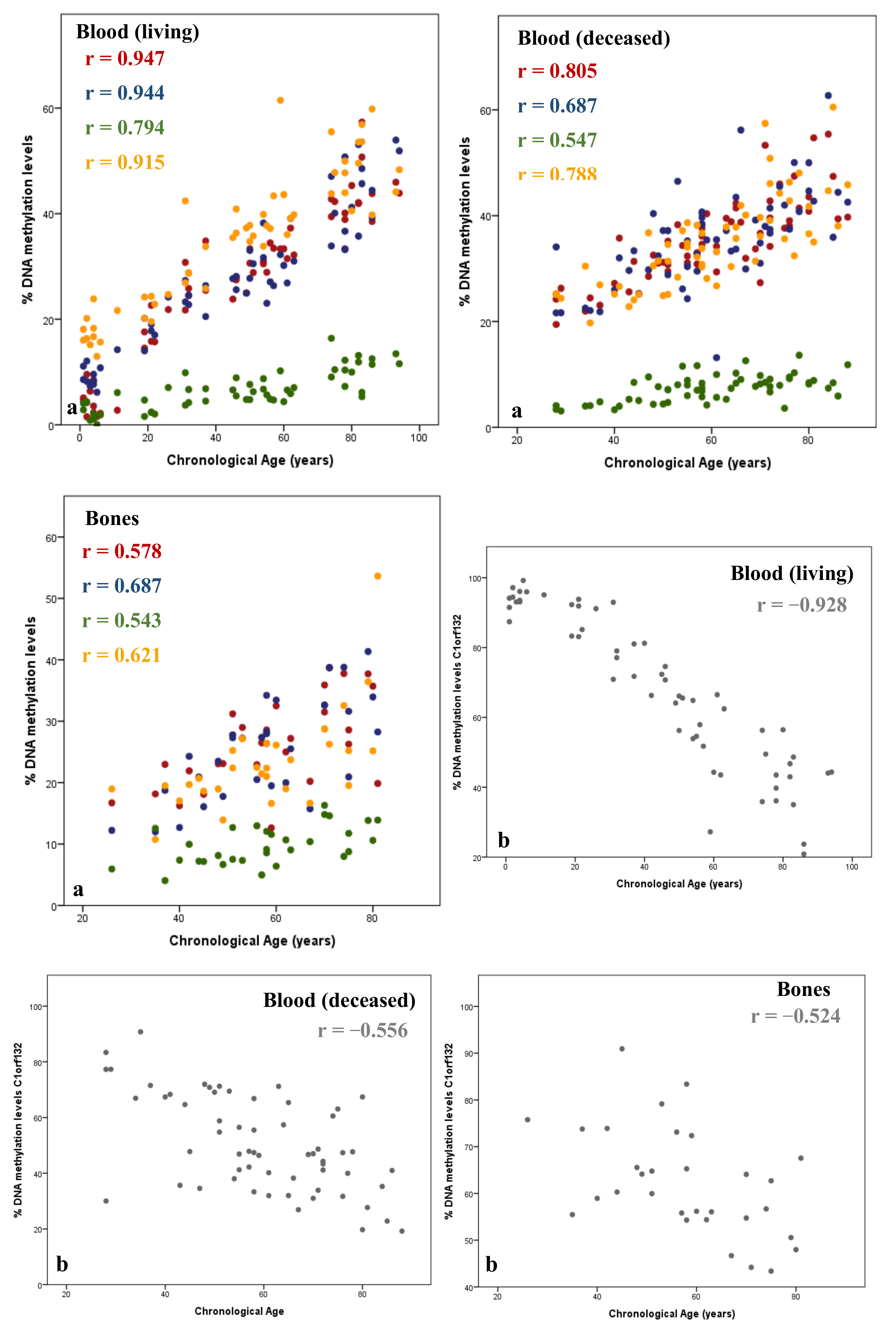
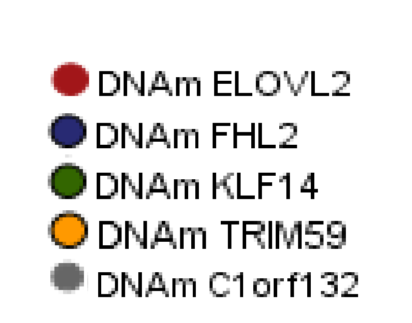
Figure 1. Positive (a) and negative (b) correlations between DNAm levels (%) and chronological age (years) in blood and bone samples. Methylation information is captured through SNaPshot assay in 59 blood samples of living individuals, 62 blood samples of deceased individuals, and 31 bone samples collected during routine autopsies. The corresponding Spearman correlation coefficients (r) are depicted inside each plot. Plots were adapted from ref. [41].
Forensicsci 02 00044 g001aForensicsci 02 00044 g001b
Table 1. Summary of some identified studies based on tissue-specific DNAm age estimation.
CpGs or GenesMain FindingsReference
NPTX2, EDARADD, TOM1L1The first study using DNAm levels for age prediction.
APM (2 CpGs) for saliva revealed an accuracy of 5.2 years.
[33]
ELOVL2, Clorf132, TRIM59, KLF14, FHL2The first age-prediction calculator available online for blood samples (www.agecalculator.ies.krakow.pl, accessed on 31 August 2022). Model with 5 CpGs revealed high accuracy with a MAD value of 3.4 years.[54]
ELOVL2High model accuracy using only 2 CpGs from ELOVL2: MAD = 5.03 years.
The first study that evaluated DNAm patterns in bloodstains, it has shown that the DNAm did not change after one-month storage as bloodstains.
[53]
ASPA, ELOVL2, PDE4C, EDARADDThe first study that investigated DNAm levels in blood samples from deceased individuals and dentin samples.
A MAD value of 3.75 years has been obtained evaluating 4 CpGs in blood from living and deceased individuals. An accurate APM with a 4.86 years of MAD value has been developed using 7 CpGs in dentin samples.
[51]
ELOVL2, FHL2, PENKThe first study that evaluated DNAm levels in different layers of tooth samples (cementum: 2.45 years; dentin: 7.07 years; dental pulp: 2.25 years). [75]
DDO, ELOVL2, F5, GRM2, HOXC4, KLF14, LDB2, MEIS1-AS3, NKIRAS2, RPA2, SAMD10, TRIM59, ZYG11A.The first study that evaluated the correlation between DNAm levels and age in bone samples.
The authors investigated the correlation between DNAm levels of 13 blood–age-correlated loci used in [44] and age in many samples from deceased individuals.
[44]
Total of 485.577 CpG sites investigated; CpGs selected are located at DDO, PRPH2, DHX8, ITGA2B and at one unknown gene with the Illumina ID number of 22398226Highly accurate models developed for young children (aged 6–15 years):
MAE = 0.47 years (boys); MAE = 0.33 years (girls).
The first study that combined anthropological and epigenetic approaches.
[6]
ELOVL2, FHL2, KLF14, C1orf132, TRIM59
Tissue-specific APMs for blood (MAD = 3.17 years), buccal swabs (MAD = 3.82 years), and saliva (MAD = 3.29 years).
A multi-tissue APM that is highly accurate (MAD = 3.55 years).
[42]
ELOVL2, PDE4C, FHL2, EDARADD, C1orf132The first study developed only for blood samples from deceased individuals.
MAD = 6.08 years.
[71]
CpGs located, among other genes, at TRIM59, ELOVL2 and KLF14The first model developed for bones namely the “37 bone clock CpGs”, revealing an accuracy of 4.9 years (RMSE).
DNAm levels of forensic samples have been evaluated, however, these were excluded.
[79]
ELOVL2, KLF14, C1orf132, FHL2, TRIM59Population-specific differences in DNAm levels. The authors applied the predictive equation developed by [37] in Korean to Portuguese living individuals obtaining a MAD value of 15.26 years.
APM for Portuguese people: MAD = 4.25 years (living); MAD =
5.36 years (deceased); MAD = 4.97 years (living and deceased individuals).
[40]
ELOVL2, KLF14, C1orf132, FHL2, TRIM59, PDE4C, EDARADDThe second APMs developed for bones in the literature (MAD = 7.18 years, using SNaPshot; MAD = 2.56 years, using Sanger sequencing).[77]
LAG3, SCGN, ELOVL2, KLF14, C1orf132, SLC12A5, GRIA2, PDE4CThe first study developed for hair samples.
Accuracy of 3.68 years using 10 CpGs.
[81]
Abbreviations: MAD, mean absolute deviation between predicted and chronological ages; RMSE, root mean square error; MAE, mean absolute error.
Table 2. Summary of some multi-tissue APMs based on DNAm levels.
YearCpGs Main FindingsReference
2013353 CpGsThe first multi-tissue model with different cellular tissues such as whole blood, occipital cortex, colon, peripheral blood mononuclear cells, liver, lung, saliva, buccal epithelium, among others, was developed using microarray hybridization technology, revealing an accuracy of 2.9 years.[9]
201710 CpGsA multi-tissue model developed for whole blood, saliva, semen, menstrual blood, and vaginal secretions with methylation data captured using the Illumina Infinium HM450 platform with an accuracy of 3.8 years. [84]
20195 CpGsAPM developed in Korean people for saliva, blood, and buccal swabs.
Multi-tissue with DNAm levels of ELOVL2, FHL2, KLF14, TRIM59, and C1orf132 genes developed using the SNaPshot method, revealing a MAD of 3.6 years.
[42]
2021The first multi-tissue APMs developed including bone and tooth samples.
Multi-tissue APMs developed for Portuguese individuals.
[45]
7 CpGsA Blood–Bone–Tooth APM (BBT-APM) with an MAD of 6.06 years developed with methylation information of CpGs located at EDARADD, FHL2,
ELOVL2, PDE4C, and C1orf132 genes using Sanger sequencing.
3 CpGsBBT-APM with a MAD of 6.49 years developed with DNAm levels of ELOVL2, KLF14, and C1orf132 genes, using the SNaPshot assay.
Publisher’s Note: MDPI stays neutral with regard to jurisdictional claims in published maps and institutional affiliations.