Article

The Andronowski Skeletal Collection for Histological Research: A Modern Anatomical Contribution

by
Janna M. Andronowski
* and
Joshua T. Taylor
Division of BioMedical Sciences, Faculty of Medicine, Memorial University of Newfoundland, 300 Prince Philip Drive, St. John’s, NL A1B 3V6, Canada
*
Author to whom correspondence should be addressed.
Forensic Sci. 2022, 2(1), 175-189; https://doi.org/10.3390/forensicsci2010014
Submission received: 4 February 2022 / Revised: 15 February 2022 / Accepted: 17 February 2022 / Published: 19 February 2022

Abstract

:
The Andronowski Skeletal Collection for Histological Research (ASCHR) comprises the fastest-growing documented modern human skeletal collection in the world developed specifically for histological and imaging research. Initiated in 2017 by Dr. Janna M. Andronowski, the ASCHR provides a resource for the study of skeletal microarchitectural variability with advancing age and between the sexes. The primary objective is to use this unique skeletal archive for histological and imaging research, with the goal of furthering knowledge of human bone biology. Bone procurement has focused on two sites commonly used in histological age-at-death estimation in anthropology: the mid-shaft sixth rib and femoral mid-shaft. The ASCHR consists of over 1200 bone samples from 621 individuals and thousands of imaging files, with age-at-death ranging from 15–105 years. Additional information collected about ASCHR donors includes occupational history; alcohol, tobacco, and drug use history; a health questionnaire; and cause and manner of death. The ASCHR offers a novel opportunity to devise regression formulae for histological age-at-death estimation and answer questions concerning age-related microarchitectural changes and biomechanical processes. It further serves as a skeletal reference database for researchers from various disciplines, including medicine, anthropology, and the biological sciences. Here, we describe the background of the collection, ethical considerations, bone procurement processes, demographic composition, and existing imaging and histological data available to researchers. Our primary aims are to (1) introduce the scientific community to ASCHR, (2) present descriptive and demographic information regarding the collection, and (3) encourage collaboration among national and international researchers interested in human skeletal biology.

1. Introduction

Human skeletal collections are essential for scientific research and teaching. The first recorded collections were initiated in the late 19th century with the Hamann–Todd Human Osteological Collection in 1893 [1] and the Robert J. Terry Anatomical Human Skeletal Collection in 1898 [2]. Such skeletal archives have served as the basis of numerous published journal articles, theses, and dissertations related to biological profiling and sex assessment (see [3] for a recent summary and analysis). Since their initiation, and at the time of this writing, a total of 153 skeletal collections have been reported worldwide [4]. It is critical to have well-documented collections representative of the living population since this is the demographic sample from which forensic cases are derived. There are inherent biases toward procuring specimens from both dissecting rooms and therapeutic, surgical procedures as they are most often from older individuals with pathological changes impacting their skeletal physiology. Bone procurement from individuals at medicolegal autopsy or at the time of organ donation, however, reflects a broader cross-section of a given community but requires research permissions, privacy contracts, and legal considerations. Thus, the formation of any postmortem bone collection is tied to essential ethical responsibilities and logistical considerations, with such factors likely contributing to the scarcity of modern anatomical collections. Recent events and subsequent public conversations have further shed light on the history of acquisition protocols for certain skeletal collections, calling their integrity and ethical soundness into question [5,6,7].
Since human skeletal collections are rare, invaluable, and irreplaceable, procuring samples for certain analyses (e.g., histology, isotopic analysis, nuclear and mitochondrial DNA testing) remains rarely permitted (e.g., Mann–Labrash Osteological Collection, William M. Bass Donated Skeletal Collection) [8,9]. Procurement techniques, including sectioning and coring, alter the macromorphology of intact elements but are required to visualize critical components of bone’s vascular and cellular networks. Traditional serial sectioning for bone histological analysis, for example, often involves the transverse sectioning of skeletal elements [10,11]. Further, bone procurement protocols for employing Synchrotron Radiation micro-Computed Tomography (SRμCT) experiments require the extraction of bone cores that fit into a determined field of view (FOV) [12,13]. Such minimally invasive techniques allow for more cosmetically acceptable sampling sites if traditionally used macroscopic skeletal indicators (e.g., for age and sex estimation) are avoided.

1.1. Skeletal Collections for Histological Research

Skeletal collections specifically for histological and imaging research remain scarce worldwide. Current well-documented collections include the Melbourne Femur Research Collection (MFRC; Australia), Ericksen collection (USA), and skeletal tissue collections at the National Museum of Health and Medicine (NMHM; USA). These collections have been instrumental in advancing scientific knowledge related to human growth and development, normal bone microarchitectural anatomy, histomorphometry, bone and joint pathology, and comparative anatomy [14,15,16,17,18,19,20,21,22,23]. The ASCHR collection (Canada), introduced and documented here, adds to this existing repertoire, and offers a unique modern anatomical archive specifically for histological and imaging analyses.

1.1.1. Melbourne Femur Research Collection

The Melbourne Femur Research Collection (MFRC) was established in 1991 for the histological assessment of age-at-death indicators from individuals of known age and sex. Initiated by Dr. John G. Clement [19], the collection is housed at the University of Melbourne in the Faculty of Medicine Dentistry and Health Sciences. The MFRC consists primarily of cadaveric specimens and surgical samples (e.g., femoral heads) obtained from local hospitals through the services of the Donor Tissue Bank of the Victorian Institute of Forensic Medicine. The MFRC includes approximately 500 femoral bone samples ranging from 1 cm transverse cross-sections of the mid-shaft to larger specimens (e.g., proximal five-eighths of femora) [19]. Donor age-at-death ranges from 1–95 years. Supplementary materials including biometric data, blood samples, clinical CT datasets, plain film or digital radiographs, and histological images are further available for a subset of MFRC specimens. The transfer of existing digital data files can be requested via a loan request made to MFRC curators. Research conducted on MFRC samples has yielded over 80 peer-reviewed journal articles and a repository of thousands of image files [14,16,17,19,20,24].

1.1.2. Ericksen Femur Collection

The Ericksen Femur Collection consists of human mid-shaft femoral cross-sections from 328 individuals (174 males, 154 females) [15]. Dr. Mary F. Ericksen developed the collection from femoral samples procured from cadavers at George Washington University Medical School, the cemetery remains from the Dominican Republic, and autopsy specimens from Chilé. The age-at-death ranges are 16–97 years for the male sub-sample and 14–94 years for females. The Ericksen collection is comprised primarily of femora from individuals of European ancestry (95%). Bone blocks and thin sections procured from the Ericksen femur collection are currently housed at The Ohio State University, Texas State University, and the Tarrant County Medical Examiner’s Office (Fort Worth, TX, USA). Since its initiation, research conducted using the Ericksen collection has yielded various academic contributions, including publications, grant technical reports, and conference presentations [15,21,22,23].

1.1.3. Skeletal Tissue Collections at the National Museum of Health and Medicine

The Anatomical Division of the National Museum of Health and Medicine (NMHM) maintains skeletal tissue collections that include extensive archives of whole-mount orthopedic pathology histological slides [18]. Various collections were spearheaded by Dr. Lent C. Johnson beginning in 1946 and continuously expanded during his tenure as chief of orthopedic pathology and later as a senior investigator and consultant. Currently, the collections include more than 10,000 slides of stained and undecalcified bone and joint specimens that illustrate bone and joint pathology, tumor pathology, human growth and development, normal musculoskeletal anatomy, histomorphometry, and comparative anatomy.
The Johnson–Sweet Whole Mount Collection of Orthopedic Pathology further includes a diverse catalog of bone diseases (e.g., osteosarcoma, osteomyelitis, chondrosarcoma, Paget’s disease, among others). The associated case materials include documentation of case history and patient treatment, radiographs, and follow-up notes. Additionally, the NMHM curates the historical Codman Registry of Bone Sarcoma, which is the oldest tumor registry in the United States. Materials include over 2600 cases of bone tumor pathology documented between 1920–1940 [18]. Associated documentation includes large-format histological slides, logbooks, card files, and correspondence. The museum additionally houses the original femoral bone histological thin sections produced by Dr. Ellis R. Kerley for his research on microscopic age changes in the bone structure of humans [25]. The original Kerley collection consisted of bone cross-sectional slides from the femur (n = 68), tibia (n = 33), and fibula (n = 25). Only a subset of these slides is available for study, given preservation issues with the mounting medium. Additional NMHM collections amassed by Kerley comprise histological slides from nonhuman primates including those from a malnutrition monkey study and a whole mount series from chimpanzees. The NMHM further maintains various uncatalogued histological collections including the Dallas Phemister Collection and Follis Nutritional Deficiency Research Archives. The former includes over 500 orthopedic pathology whole-mount sections of various sizes, curated between 1882–1951 (B. Spatola, personal communication, 28 January 2022). The Follis Archives contain several histological bone slide boxes from past nutritional studies conducted by Dr. Follis, with likely undiscovered materials associated with the larger Follis–Park study from Johns Hopkins Children’s Center, Harriet Lane Clinic. The Harriet Lane collection consists of approximately 1,260 cases of subadults from birth to eleven years of age. Materials primarily include sternal rib growth plates and epiphyses from various other skeletal elements.
Overall, the NMHM Anatomical Division curates one of the largest and most diverse bone slide collections (and associated subcollections) in the world. These materials are of great scientific value to the skeletal biology research community, particularly in the areas of orthopedic pathology, paleopathology, and biological and forensic anthropology.

1.1.4. Andronowski Skeletal Collection for Histological Research

In 2017, Dr. Janna M. Andronowski founded the Andronowski Skeletal Collection for Histological Research (ASCHR) to provide an ethically sourced, unique resource for studying the variability of skeletal microarchitecture with advancing age and between the sexes. The ASCHR consists of over 1200 bone samples from 621 individuals and thousands of imaging files, with age-at-death ranging from 15–105 years. Bone procurement has focused on two sites commonly used in histological age-at-death estimation in anthropology: the mid-shaft sixth rib and femoral mid-shaft. Several high-resolution imaging methods have been employed to analyze ASCHR specimens, including Synchrotron Radiation micro-Computed Tomography, laboratory micro-Computed Tomography, X-Ray Photoelectron Spectroscopy, Confocal Laser Scanning Microscopy, and fluorescence and brightfield light microscopy. A host of imaging data are further available (described below, Section 3.2). At present, the ASCHR is the newest and fastest-growing histological and imaging research-specific skeletal collection in the world.

2. Description of ASCHR Curatorial Processes and Ethical Considerations

2.1. Acquisition of Skeletal Material for ASCHR

Dr. Andronowski joined the Department of Biology at The University of Akron as academic faculty in 2017. During this appointment, she forged many fruitful collaborations with researchers, anatomists, organ donation non-profit organizations (OPOs), and medical examiners’ offices in the United States and Canada, with whom she continues to collaborate. Tissue procurement protocols and anatomical specimen requests were prepared for each institution/organization according to proprietary protocols and were reviewed and approved by Medical Advisory Boards and/or Ethics Panels. This subsequently allowed for the routine collection of cadaveric bone specimens in accordance with Medical Research guidelines. The study of the skeletal material was ethically cleared by The University of Akron Institutional Review Board for the Protection of Human Subjects and the Newfoundland and Labrador Health Research Ethics Board (Protocol Reference #2020.308). All cadaveric samples were collected with strict ethical oversight and explicit informed consent from the donor or next of kin. For organ donors, advanced procurement coordinators, who are responsible for securing authorizations to collect organs and tissues (e.g., heart valves, bone, corneas), specifically request permission to remove tissues for research. With approvals in place, questionnaires are then administered to the next of kin to inquire about past medical histories or medication regimens that may impact bone metabolism (e.g., osteoporosis, prolonged steroid use, etc.). Individuals with suspected bone turnover issues are identified and flagged in the ASCHR database.
Standard protocols were developed and implemented to ensure consistent procurement, maceration, curation, and storage of all ASCHR specimens. Samples include both embalmed and fresh bone tissues and primarily consist of two skeletal element types; the left femoral mid-shaft and left mid-shaft sixth rib. Cranial bones, including frontal, parietal, temporal, and occipital, have further been procured from roughly 100 donors.
Femoral and rib samples were collected by dissecting soft tissues (e.g., musculature, neurovasculature) away from the anterior mid-shafts and employing a bone saw (e.g., oscillating or reciprocating) or rib shears to extract three-inch bone blocks. An oscillating saw was used to procure cranial bones to obtain 2 × 2 cm specimens for imaging experiments.
Each sample is placed in an individual collection tube labeled with the donor identification number, sex, age, and cause of death. Upon return to the lab, specimens are wrapped in sterile saline-soaked gauze and stored in a sample tube/container in a −20 °C freezer until further processing. The collection is currently curated in the Division of BioMedical Sciences, Faculty of Medicine, Memorial University of Newfoundland in a well-protected, triple-locked, and secure environment that is continually monitored.
The cadaveric donor institutions provide additional demographic information such as population affinity/ancestry and primary and secondary cause of death. Organ donation non-profit organizations further provide a confidential organ chart and serology report for each donor that contains detailed information regarding demographic details, past medical history, drug use and rehabilitation history, next-of-kin questionnaire, toxicology report, physical assessment overview, and additional lab profiles (e.g., urinalysis, bacterial cultures, etc.). Please refer to the Supplementary Materials for examples of a fabricated organ chart (A) and serology report (B). Any identifiable donor information is protected and excluded from all subsequent data files.

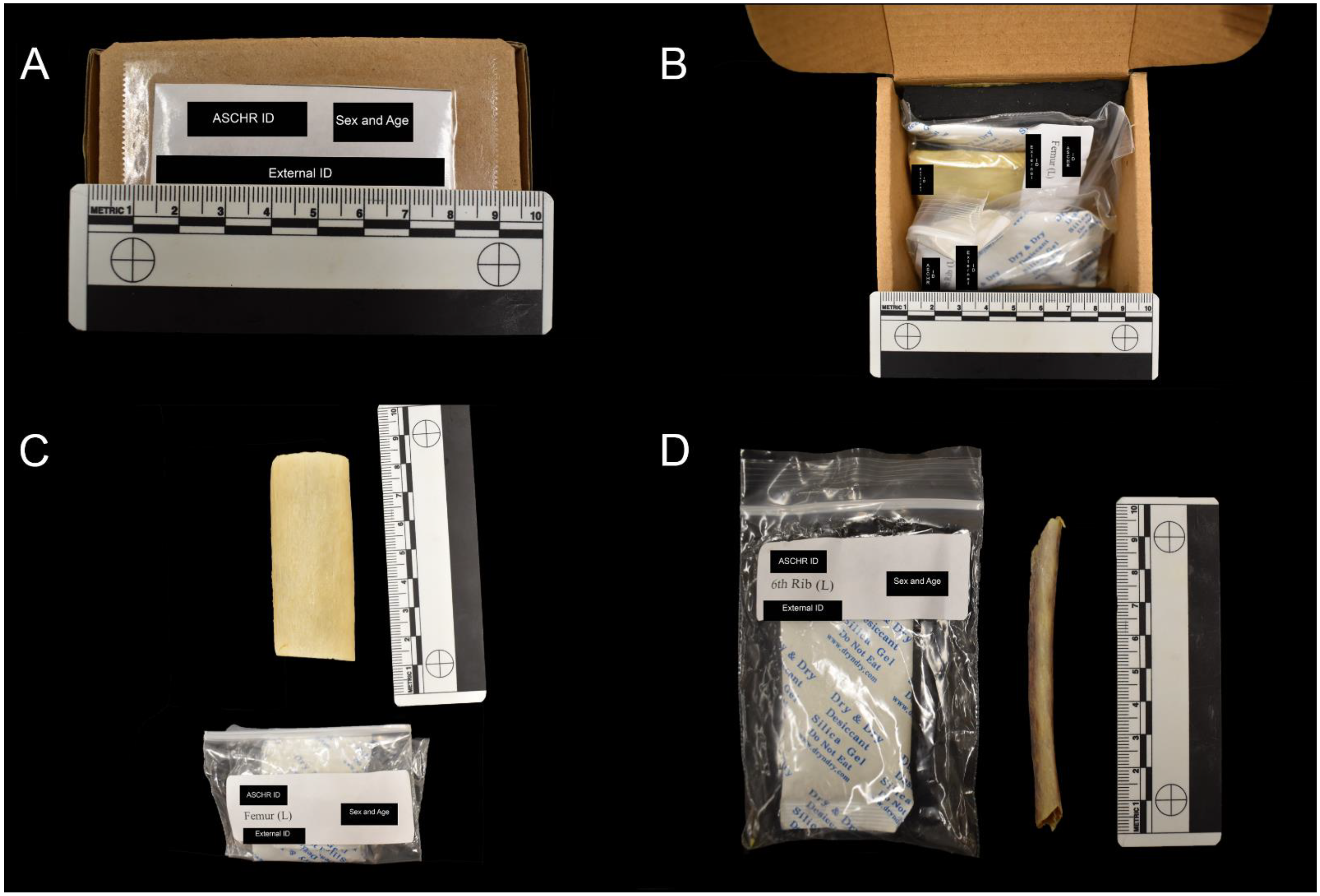
2.2. Processing and Curation

Samples are thawed and subsequently macerated using standard protocols [26], including incubation in a 45 °C water and protease solution (Tergazyme) for one to four hours. The time spent in the solution varies depending on skeletal element type, size, and the number of soft tissues present. Adhering soft tissues are removed using dental tools, and bone marrow is cleared from the medullary cavity with a handheld water flosser (e.g., WaterPik). Specimens are soaked in 70% ethanol at 4 °C for 24 h to remove lipids and air-dried for 24 h at ambient temperature.
Processed samples are placed in individually labeled bags with perforations to promote airflow. Specimen bags are housed in labeled corrugated cardboard boxes designated for each donor containing desiccant packets and foam to prevent moisture accumulation and to provide protection (Figure 1). All samples are labeled with an ASCHR-specific identification number using indelible ink and clear nail varnish. Andronowski Lab graduate student, Joshua T. Taylor, has processed, documented, and curated the vast majority of specimens in the ASCHR collection. He routinely checks each specimen to ensure the continued preservation and structural integrity of bone tissues and maintains thorough documentation, including a skeletal photo inventory.

3. ASCHR Demographic Information and Digital Data

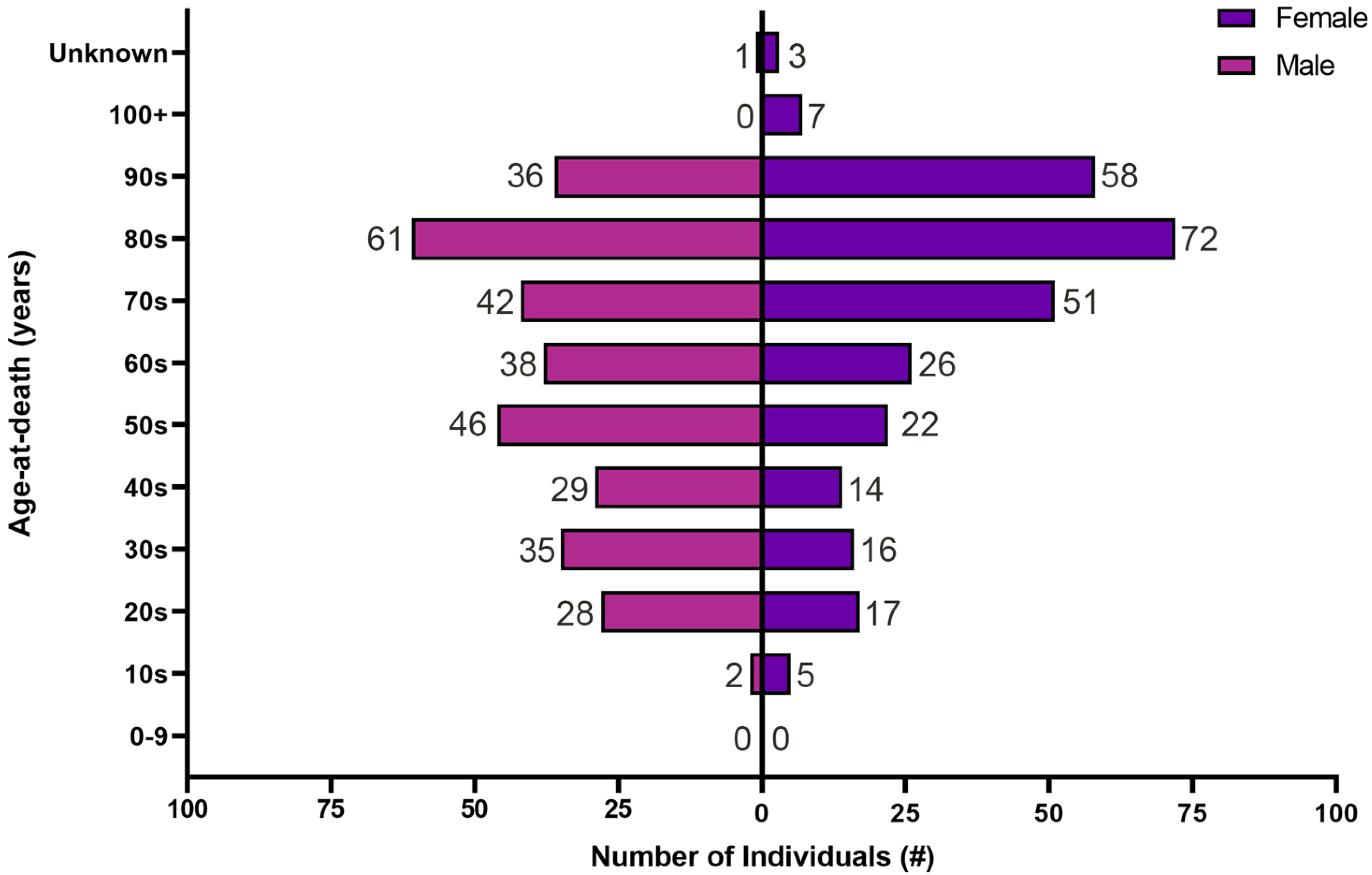
3.1. Demographic Information

The collection currently includes 1213 skeletal elements from 621 individual donors. The sex distribution is roughly equal with 318 (51%) male, 291 (47%) female, and 12 (2%) unidentified donors (Figure 2). Donor age-at-death ranges from 15–105 years. There are 214 (34%) donors between 15–59, 391 (63%) between 60–105 years old, and 16 (3%) individuals of undocumented age. The reported trend for sex and age is consistent when separated by skeletal elements (Figure 3 and Figure 4). The ASCHR continues to grow at an annual rate of approximately 200–300 skeletal elements per annum.
In addition to a comprehensive age-at-death range, the collection contains an extensive group of skeletal elements used in traditional histological analyses in biological anthropology. The majority of samples were procured from two sites commonly used in histological age-at-death estimation, left mid-shaft femoral diaphyses and left sixth mid-shaft ribs. There are currently 386 femoral blocks and 473 mid-shaft sixth rib segments. In addition, there are 106 individuals with samples available from the frontal, parietal, temporal, and occipital bones. Various other skeletal elements have been collected from a subset of ASCHR individuals, including clavicles, radii, ulnae, tibiae, metacarpals, and maxillae.
The individual donors are associated with comprehensive demographic information, including age-at-death, biological sex, population group affinity or ancestry, primary and secondary cause of death, organ charts, serology reports, and case-specific documentation concerning the individuals and/or the samples. Additional information for numerous ASCHR donors includes occupational history; alcohol, tobacco, and drug use history; a health questionnaire; and cause and manner of death. In total, there are 49 metadata fields with various subsections, including vitals, previous medical history, and lab profiles (e.g., bacterial cultures, chemistry, and toxicology). Forty-nine (7.9%) of the donors have portions of missing demographic information, however. Sex, age, and primary cause of death are unavailable for 12 (1.9%), 16 (3%), and 22 (3.5%) donors, respectively and are dramatically reduced from the missing ancestral data (49, 7.9%). The amount of unavailable demographic information is consistent with well-established skeletal collections. For example, the Hamann–Todd collection reports 0.6% of missing demographic information, and documentation associated with the Kirsten Skeletal Collection reports missing 10% of sex and population group affinity/ancestry [27].

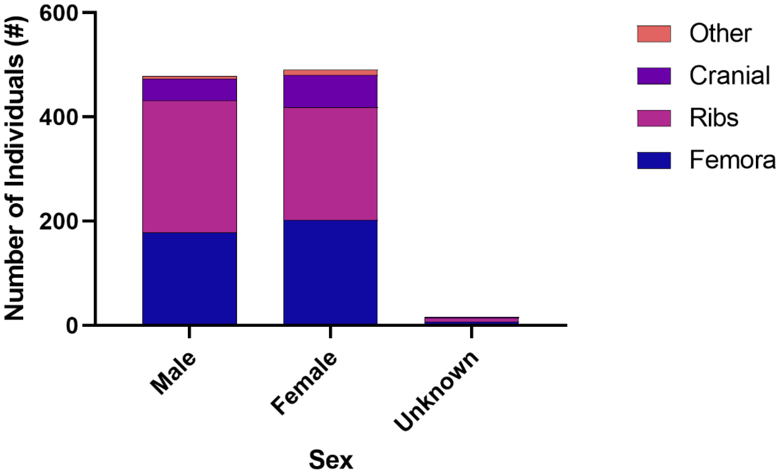
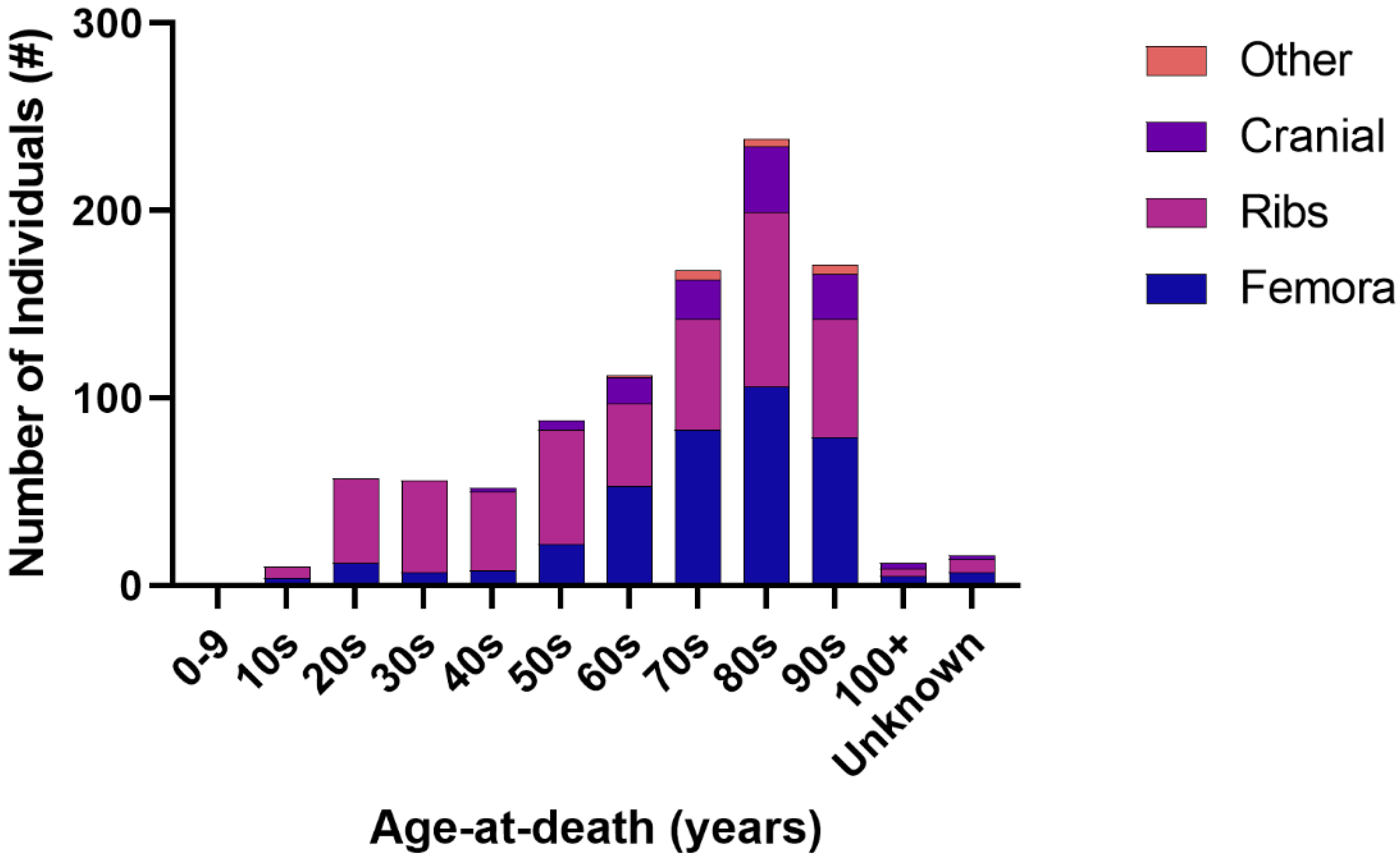
3.2. Histological and Imaging Data

Efforts to explore the dynamic process of bone remodeling have been aided by advances in high-resolution 3D imaging techniques. Imaging modalities such as micro-CT allow researchers to visualize and quantify bone in a spatial manner at higher resolutions than was previously obtainable. To that end, hundreds of ASCHR specimens have been visualized and analyzed using various 3D imaging modalities, resulting in over 30 terabytes of data. Such imaging data offer unique opportunities for remote analysis and collaboration. For example, big data sharing will provide researchers who may not have access to imaging infrastructure with opportunities to acquire existing imaging files.
The technology chosen for imaging depends on both the size of the region to be imaged and the resolution required to distinguish bone’s desired microstructural features. Increasing image resolution generally requires decreasing the FOV. A variety of imaging data are currently available for ASCHR and include datasets from brightfield, differential interference contrast (DIC), polarized, and fluorescence microscopy, micro-Computed Tomography (µCT), Synchrotron Radiation micro-Computed Tomography (SRµCT), X-ray Photoelectron Spectroscopy (XPS), and Confocal Laser Scanning Microscopy (CLSM) [10,11,12,13,28,29,30,31,32,33,34,35,36]. The availability of these imaging data allows for many benefits, including (1) opportunities to study ASCHR skeletal samples without having to secure expensive experimental imaging time, (2) providing a permanent record, often in 3D, of the bone collected, thus allowing specimens to be continuously studied nondestructively by researchers, and (3) the careful archiving of data that works to broaden distribution for users accessing the collection.

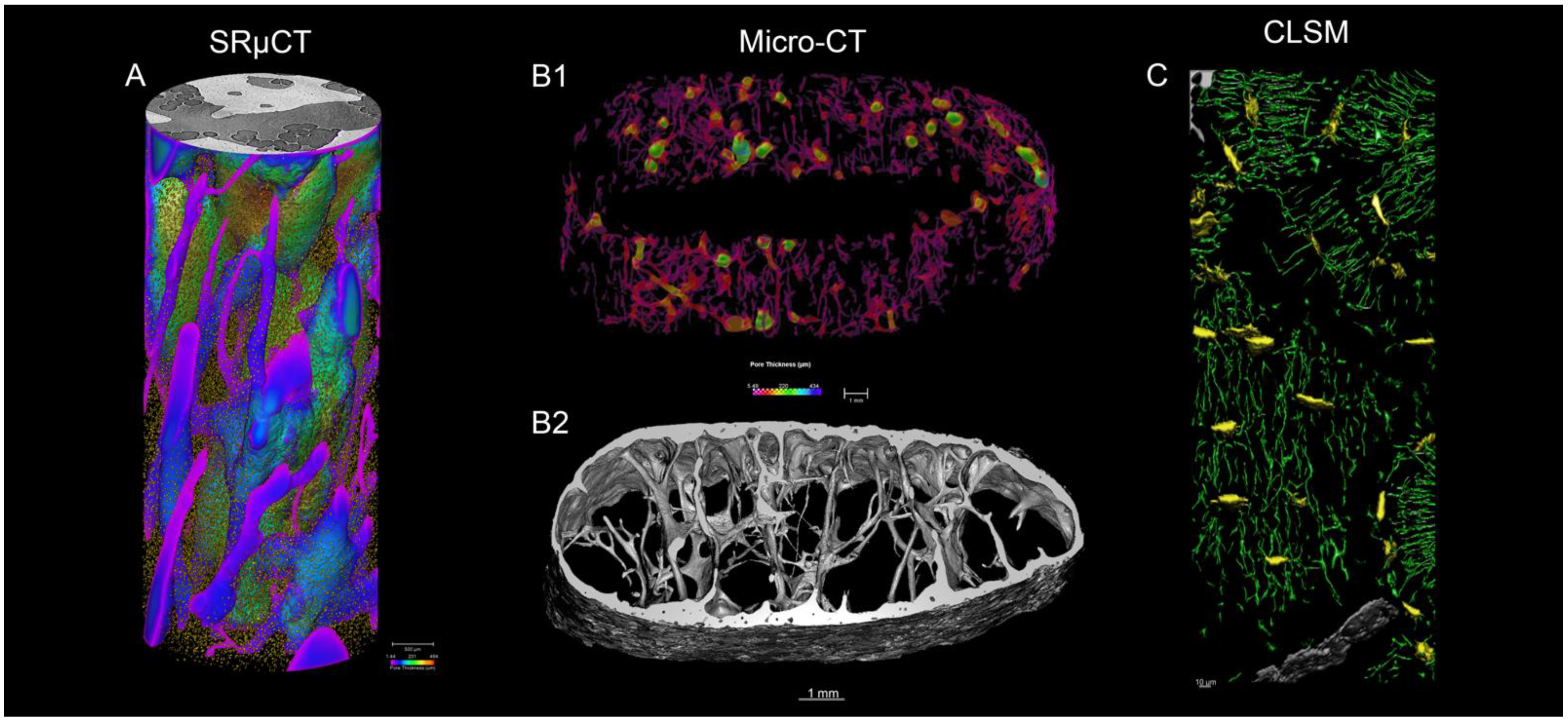
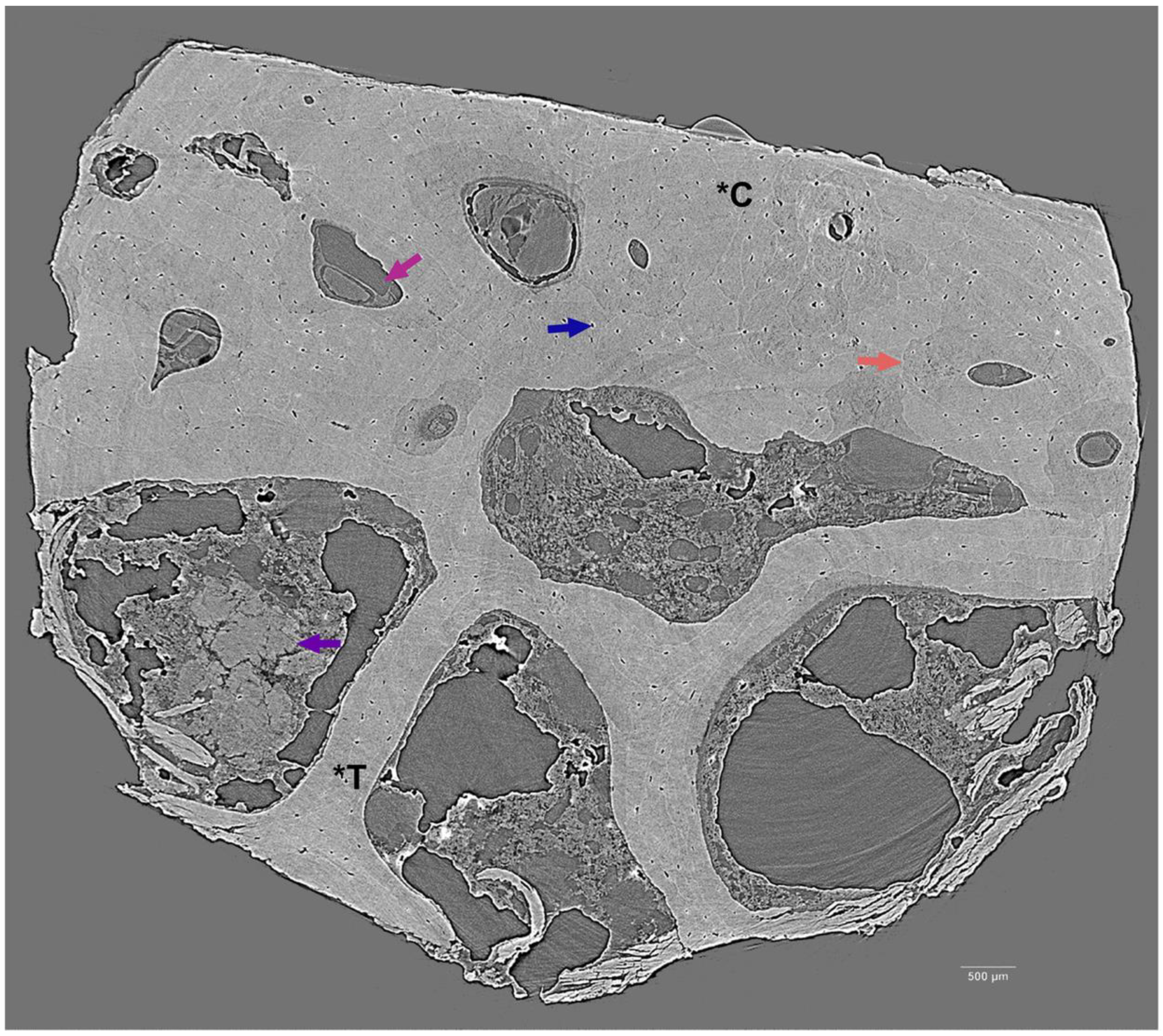
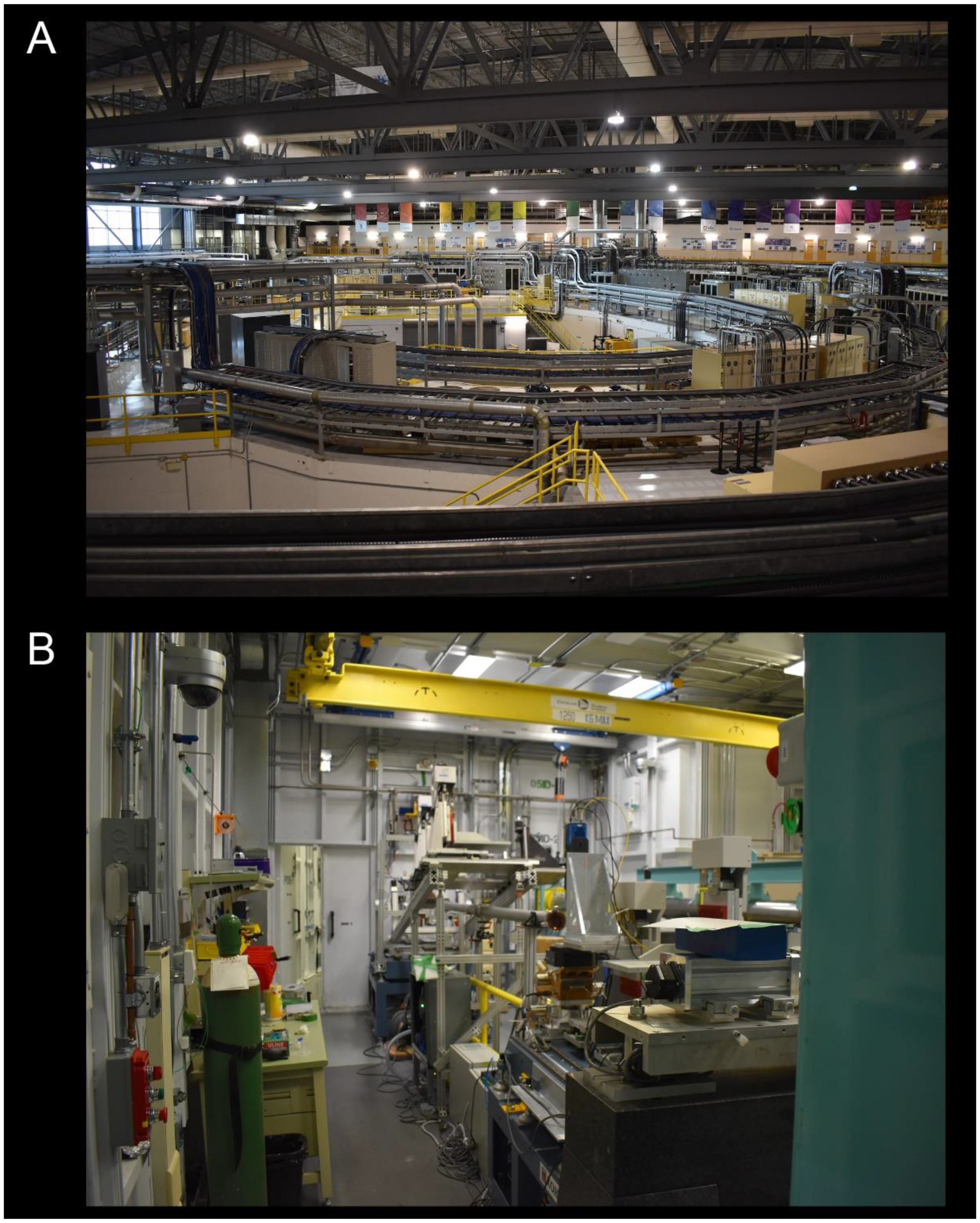
3.2.1. Synchrotron Radiation Micro-Computed Tomography

Synchrotron Radiation micro-Computed Tomography (SRµCT) uses brilliant breaking radiation produced by a synchrotron facility (Figure A1A). Recent experiments by our group at the Canadian Light Source (CLS) (Saskatoon, SK, Canada) have used ASCHR specimens to pioneer the application of SRµCT technology to forensic anthropological queries. Relevant projects have included (1) evaluating differences in 3D bone microarchitecture to shed light on differential nuclear DNA yield rates among bone tissue types [37], (2) documenting the presence and microstructural organization of osteon banding in the human cortical bone to aid in the differentiation of human from nonhuman bone [38], and (3) investigating the impact of opioid abuse on 2D microstructural parameters (e.g., OPD, cortical area) and 3D measures (e.g., intracortical porosity, osteocyte lacunar density) [33]. We are currently deploying a large ASCHR dataset to visualize cortical pores and osteocyte lacunae in cadaveric anterior femoral cores from across the lifespan (Figure 5A).
Synchrotron µCT experiments are routinely conducted by our group on the BioMedical Imaging and Therapy (BMIT) beamlines at CLS. The team has carried out experiments on both the Insertion Device (ID) and Bend Magnet (BM) beamlines. Bone cores for imaging are procured following the protocol outlined in Andronowski et al., 2020 [13]. Our most recent experimental set-up on the BM beamline includes a filtered white beam microscope (5× objective), photon energy of 30 keV, effective pixel size of 1.44 μm, and an object to detector distance of 5 cm (Figure A1B).
The ASCHR documentation currently contains complete SRµCT datasets for 265 donors from anterior femora and 127 from the cutaneous aspect of sixth ribs corresponding to many custom image processing and data analysis workflows. The datasets are reconstructed in 3D (Figure 6) using either NRecon (Bruker) or ufo-kit [39] and analyzed using a combination of Amira-Avizo (FEI, www.fei.com (accessed on 16 February 2022), CTAnalyser (Bruker), and ORS Dragonfly (www.theobjects.com/dragonfly/ (accessed on 16 February 2022)). Variables for vascular porosity typically measured include: % porosity, canal surface density, pore connectivity density, pore density, and mean and standard deviation of pore thickness and pore separation. Parameters pertaining to osteocyte lacunae include % lacunar volume, surface density, population density, and lacunar thickness.

3.2.2. Laboratory micro-Computed Tomography

Laboratory micro-Computed Tomography (µCT) allows for the visualization of 3D vascular pore networks in whole cross-sectional segments of small diameter bones. This macroscopic approach provides an increased FOV size and thus the analysis of structures, including tissue volume, cortical porosity, and vascular canal diameter. For example, the Andronowski Lab has visualized the porosity of the left mid-shaft six ribs (n = 25) from modern humans at a pixel size of 5.49 μm (Figure 5B). Double-wide scans were taken to ensure the entire rib was captured in the FOV. Each individual dataset contains 1070 cross-sectional images [29].
Our group has further employed µCT to analyze (1) a subset of frontal bone specimens for evaluation of Hyperostosis Frontalis Interna (HFI) progression, and (2) human/nonhuman comparative metacarpal specimens from ASCHR and black bear skeletons from the Cleveland Museum of Natural History, Division of Vertebrate Mammals [28].

3.2.3. Confocal Microscopy

Confocal Laser Scanning Microscopy (CLSM) employs a scanning laser to excite a fluorophore. Out-of-focused light is blocked with a spinning disc or pinhole, visualizing X, Y, and Z planes. CLSM offers the benefit of increased resolution that allows for the visualization of bone’s lacunar-canalicular network (LCN), which is not visible using high-resolution X-ray imaging modalities such as SRµCT. Our group has employed CLSM to image femoral specimens (n = 40) to visualize and quantify osteocyte lacunae and canaliculi in anterior intracortical regions of modern human cadaveric femora (Figure 5C). In our experiments, a Leica TCS SPE CLSM equipped with a motorized Z-Galvo stage (Leica Microsystems, Wetzlar, Germany) was employed with an immersion oil 63 × objective lens. A laser wavelength of 488 nm was set to 32.5% intensity with a spectral window ranging from 485 to 585 nm. Following imaging, adjacent regions were pairwise stitched in Fiji v.1.53c [40] and resampled to a pixel size of 0.3 micrometers in ORS Dragonfly software v.4.1 (Object Research Systems, Montréal, QC, Canada). Each image stack consisted of 101 consecutive slices to produce stacked 3D data. Lacunae were isolated by performing a 3D erosion in Dragonfly to separate their connections with associated canaliculi. Following isolation, a multiple region of interest (multi-ROI) function was applied to isolate each individual lacuna, producing a lacunar ROI. Morphometric data, including % volume, diameter, separation, connectivity density, and density, are acquired separately for lacunar and canalicular image stacks in CTAnalyser v.1.18.4.0 (Bruker, Kontich, Belgium).

4. ASCHR Scientific Contributions

4.1. Protocols for Research Requests

The ASCHR is open for use by researchers in biomedicine, anthropology, and related fields. For inquiries regarding collection access or existing imaging datasets, please contact Dr. Janna M. Andronowski and complete a Research Request Form (https://www.andronowskilab.com/andronowski-skeletal-collection; accessed on 12 January 2022). Each request will be considered based on scientific merit and the proposed broader impacts. Researchers are encouraged to attach additional supporting materials, including research grant proposals, ethical approval documents, equipment training documentation (if use is required for the proposed project), and other relevant documents (e.g., evidence of experience with experimental protocol). Student researchers must submit a signed letter from their primary supervisor to further support the research request form. Fees are not charged for collaborative research using ASCHR materials, but standard shipping and insurance costs apply.
Researchers may document their research endeavors using photography, X-ray imaging modalities, and microscopy but are required to provide the Andronowski Lab with copies of all images and associated data. The ASCHR and Dr. Andronowski should further be acknowledged in the resulting publications and presentations.

4.2. Significance of Collection

The skeletal collection is housed in the Division of BioMedical Sciences, Faculty of Medicine, Memorial University of Newfoundland. It currently boasts 1213 skeletal elements from 621 individuals with an approximately equal sex distribution and an age-at-death range of 15–105 years. The samples are associated with comprehensive demographic information, including population affinity/ancestry, primary and secondary cause of death, organ charts, serology reports, and case-specific documentation concerning the individuals and/or the samples. Available digital data, including SRµCT, µCT, and microscopic imaging files, offer unique opportunities for remote analysis and collaboration, particularly considering current COVID-19-related travel limitations and for departments and institutions where imaging infrastructure is limited. The collection is accessible to researchers both nationally and internationally, and research requests are evaluated based on the scientific merit of the research project and the proposed broader impacts. Our long-term goal is to provide a resource of international significance that attracts many requests from high-caliber scientists to explore its resources.
To date, the Andronowski Lab research team has studied ASCHR specimens for collaborative projects with colleagues of the (1) Departments of Anatomy, Physiology, and Pharmacology and Chemistry, the University of Saskatchewan, (2) Departments of Surgery and Mechanical Engineering, the University of Alberta, (3) Department of Anthropology, the University of Tennessee, Knoxville, (4) Forensic Anthropology Division, New York City Office of Chief Medical Examiner (NYC-OCME), (5) Divisions of Physical Anthropology and Vertebrate Mammals, Cleveland Museum of Natural History, (6) Lifebanc, and (7) the Cuyahoga County Medical Examiner’s Office. These collaborations are in addition to local collaborations with the Department of Biology and Division of BioMedical Sciences, Memorial University of Newfoundland, and the Office of the Chief Medical Examiner in St. John’s, NL.
The ASCHR has currently contributed modest advances of knowledge [12,13,29,30,31,32,33,34,35,36], though its potential significance offers promise to be expansive and lead to many novel and exciting discoveries.

5. Conclusions

The Andronowski Skeletal Collection for Histological Research (ASCHR) comprises the fastest-growing documented modern human skeletal collection developed specifically for histological and imaging research to date. It offers a unique, ethically sourced, and well-documented resource for the study of skeletal microarchitectural variability with advancing age and between the sexes. Dr. Andronowski began procuring skeletal samples in 2017 and has collected over 1200 bone specimens from 621 donors. She remains responsible for curating and maintaining ASCHR and overseeing the ethical framework and annual renewal process via the Newfoundland and Labrador Health Research Ethics Board (Protocol Reference #2020.308).
The collection continues to grow at an annual rate of approximately 200–300 skeletal elements per annum and offers an opportunity to devise novel regression formulae for histological age-at-death estimation and answer questions concerning age-related microarchitectural changes and biomechanical processes. It further serves as a skeletal reference database for researchers from various disciplines, including medicine, anthropology, and the biological sciences. Our hope is to encourage collaboration and data sharing among national and international researchers interested in human skeletal biology.

Supplementary Materials

The following supporting information can be downloaded at: https://www.mdpi.com/article/10.3390/forensicsci2010014/s1, Document S1: (A) Fabricated Organ Chart, (B) Serology Report.

Author Contributions

Conceptualization; Project Design; Methodology; Acquisition of Data; Data Visualization; Data Analysis/Interpretation; Data Curation; Figure Preparation; Project Administration; Supervision; Writing—original draft; Critical Revision of Manuscript; Writing—review and editing; Approving Final Version of Manuscript, J.M.A. Acquisition of Data; Data Visualization; Data Analysis/Interpretation; Data Curation; Figure Preparation; Writing—original draft; Critical Revision of Manuscript; Writing—review and editing; Approving Final Version of Manuscript, J.T.T. All authors have read and agreed to the published version of the manuscript.

Funding

Aspects of this work were supported by Award No. 2018-DU-BX-0188, awarded by the National Institute of Justice, Office of Justice Programs, U.S. Department of Justice.

Institutional Review Board Statement

The study of the skeletal material was ethically cleared by The University of Akron Institutional Review Board for the Protection of Human Subjects and the Newfoundland and Labrador Health Research Ethics Board (Protocol Reference #2020.308).

Informed Consent Statement

All bone tissue samples were collected with strict ethical oversight and explicit informed consent from the donor or next of kin.

Acknowledgments

The development of the ASCHR involved the efforts and support of many people and institutions. The authors thank Beth Dalzell from The University of Toledo College of Medicine and Life Sciences, the Department of Anatomy and Neurobiology at Northeast Ohio Medical University (NEOMED), Wright State University Boonshoft School of Medicine, and the SORC team of Lifebanc for access to cadaveric bone specimens. Certain specimen processing, data visualization, and collection curation (both skeletal and digital) would not have been possible without the support and assistance of current and past Andronowski Lab members from all training levels. J.M.A. sincerely thanks Reed Davis, Randi Depp, Hannah Stephen, Tyler Hicks, Gina Tubo, Logan Usher, Adam Schuller, Abigail LaMarca, Mary Beth Cole, Evin Hessel, Hannah Rutkowski, Jacob Haschak, Kelly Cooper, Linda Muakassa, Breanna White, Jason Davis, and Kassidy Wilson for their contributions ranging from assistance with cadaveric dissection to maceration, data management, and curatorial duties. The authors further wish to thank Brian Spatola of the National Museum of Health and Medicine, Anatomical Division, for providing current information regarding the skeletal collections curated within the department. Additionally, the research described in this paper was performed at the BMIT facility at the Canadian Light Source, which is supported by the Canada Foundation for Innovation, Natural Sciences and Engineering Research Council of Canada, the University of Saskatchewan, the Government of Saskatchewan, Western Economic Diversification Canada, the National Research Council Canada, and the Canadian Institutes of Health Research. The authors would like to thank the beamline scientists at the Canadian Light Source, particularly Adam Webb, Sergey Gasilov, Arash Panahifar, and Ning Zhu for the assistance in set-up and troubleshooting of the SkyScan SRµCT and white beam microscope systems. The authors further wish to thank Christine Dengler-Crish and Matthew Smith of NEOMED for access to their confocal laser scanning microscope, and Mike Marsh of Objects Research Systems Inc. for assistance with troubleshooting our CLSM Dragonfly workflow. Most importantly, we thank our selfless donors and their families. Without them, the ASCHR, and the novel, innovative research that it allows, would not be possible.

Conflicts of Interest

The authors declare no conflict of interest. The funders had no role in the design of the study; in the collection, analyses, or interpretation of data; in the writing of the manuscript, or in the decision to publish the results. The opinions, findings, and conclusions or recommendations expressed in this publication are those of the authors and do not necessarily reflect those of the Department of Justice.

Appendix A

Figure A1. Canada’s national synchrotron facility, the Canadian Light Source (CLS), as seen from the Mezzanine Level (A). The experimental X-ray Computed Tomography set-up employed by our team on the BioMedical Imaging and Therapy (BMIT) Bend Magnet (BM) beamline (B).
Forensicsci 02 00014 g0a1

References

  1. Işcan, M.A. Comparison of the Hamann–Todd and Terry Collections. Anthropologie 1992, 30, 35. [Google Scholar]
  2. Hunt, D.R.; Albanese, J. History and demographic composition of the Robert J. Terry anatomical collection. Am. J. Phys. Anthropol. 2005, 127, 406. [Google Scholar] [CrossRef] [PubMed]
  3. Alves-Cardoso, F.; Campanacho, V. The Scientific Profiles of Documented Collections via Publication Data: Past, Present, and Future Directions in Forensic Anthropology. Forensic Sci. 2022, 2, 37–56. [Google Scholar] [CrossRef]
  4. Petaros, A.; Caplova, Z.; Verna, E.; Adalian, P.; Baccino, E.; de Boer, H.H.; Cunha, E.; Ekizoglu, O.; Ferreira, M.T.; Fracasso, T.; et al. Technical Note: The Forensic Anthropology Society of Europe (FASE) Map of Identified Osteological Collections. Forensic Sci. Int. 2021, 11, 110995. [Google Scholar] [CrossRef]
  5. Imbler, S. Can Skeletons Have a Racial Identity? The New York Times, 19 October 2021. Available online: https://www.nytimes.com/2021/10/19/science/skeletons-racism.html(accessed on 16 February 2022).
  6. Schuessler, J. What Should Museums Do with the Bones of the Enslaved? The New York Times, 25 August 2021. Available online: https://www.nytimes.com/2021/04/20/arts/design/museums-bones-smithsonian.html(accessed on 16 February 2022).
  7. Williams, S.E.; Ross, A.H. Ethical dilemmas in skeletal collection utilization: Implications of the Black Lives Matter movement on the anatomical and anthropological sciences. Anat. Rec. 2021, 10, ar.24839. [Google Scholar] [CrossRef]
  8. Labrash, S.; Lozanoff, S. Standards and guidelines for willed body donations at the John A. Burn. Sch. Med. Hawaii Med. J. 2007, 66, 72. [Google Scholar]
  9. Mann, R.W.; Labrash, S.; Lozanoff, S. Medical School Hotline: A New Osteological Resource at the John A. Burns School of Medicine. Hawaii J. Health Soc. Welf 2020, 79, 202. [Google Scholar] [PubMed]
  10. Andronowski, J.M.; Crowder, C.; Martinez, M.S. Recent advancements in the analysis of bone microstructure: New dimensions in forensic anthropology. Forensic. Sci. Res. 2018, 3, 278. [Google Scholar] [CrossRef] [PubMed] [Green Version]
  11. Andronowski, J.M.; Cole, M.E. Current and emerging histomorphometric and imaging techniques for assessing age-at-death and cortical bone quality. WIREs Forensic Sci. 2021, 3, e1399. [Google Scholar] [CrossRef]
  12. Andronowski, J.M.; Davis, R.A.; Tubo, G.; Cooper, D.M.L. Application of Synchrotron Micro-Computed Tomography and Confocal Laser Scanning Microscopy to Evaluate Sex-Related Differences in the Human Osteocyte Lacunar-Canalicular Network across the Lifespan. Am. Assoc. Phys. Anthropol. 2019. Available online: https://www.researchgate.net/publication/335310785_Application_of_Synchrotron_micro-Computed_Tomography_and_Confocal_Laser_Scanning_Microscopy_to_Evaluate_Sex-Related_Differences_in_the_Human_Osteocyte_Lacunar-Canalicular_Network_Across_the_Lifespan (accessed on 16 February 2022).
  13. Andronowski, J.M.; Davis, R.A.; Holyoke, C.W. A Sectioning, Coring, and Image Processing Guide for High-Throughput Cortical Bone Sample Procurement and Analysis for Synchrotron Micro-CT. J. Vis. Exp. 2020, 10, e61081. [Google Scholar] [CrossRef] [PubMed]
  14. Carter, Y.; Suchorab, J.L.; Thomas, C.D.L.; Clement, J.G.; Cooper, D.M.L. Normal variation in cortical osteocyte lacunar parameters in healthy young males. J. Anat. 2014, 225, 328. [Google Scholar] [CrossRef]
  15. Ericksen, M.F. Histologic estimation of age at death using the anterior cortex of the femur. Am. J. Phys. Anthropol. 1991, 84, 171. [Google Scholar] [CrossRef] [PubMed]
  16. Fernandez, J.W.; Das, R.; Cleary, P.W.; Hunter, P.J.; Thomas, C.D.L.; Clement, J.G. Using smooth particle hydrodynamics to investigate femoral cortical bone remodelling at the Haversian level. Int. J. Numer. Methods Biomed. Eng. 2013, 29, 129. [Google Scholar] [CrossRef] [PubMed]
  17. Lerebours, C.; Thomas, C.D.L.; Clement, J.G.; Buenzli, P.R.; Pivonka, P. The relationship between porosity and specific surface in human cortical bone is subject specific. Bone 2015, 72, 109. [Google Scholar] [CrossRef] [PubMed]
  18. Spatola, B.F.; Damann, F.E.; Ragsdale, B.D. Bone Histology Collections of the National Museum of Health and Medicine. In Bone Histology; Crowder, C., Stout, S., Eds.; CRC Press: Boca Raton, FL, USA, 2012; Chapter 12; p. 313. [Google Scholar]
  19. Thomas, C.; Clement, J.G. The Melbourne Femur Collection: How a Forensic and Anthropological Collection Came to Have Broader Applications. In Bone Histology; Crowder, C., Stout, S., Eds.; CRC Press: Boca Raton, FL, USA, 2012; Chapter 13; p. 327. [Google Scholar]
  20. Wang, X.; Thomas, C.D.L.; Clement, J.G.; Das, R.; Davies, H.; Fernandez, J.W. A mechanostatistical approach to cortical bone remodelling: An equine model. Biomech. Model. Mechanobiol. 2016, 15, 29. [Google Scholar] [CrossRef]
  21. Crowder, C. Estimation of Age at Death Using Cortical Bone Histomorphometry; National Institute of Justice, U.S. Department of Justice: Washington, DC, USA, 2013. Available online: https://nij.ojp.gov/library/publications/estimation-age-death-using-cortical-bone-histomorphometry (accessed on 16 February 2022).
  22. Andronowski, J.M. Evaluating Age-related Bone Loss in the Ericksen Femur Collection. In Proceedings of the American Association of Physical Anthropologists 83rd Annual Meeting, Calgary, AB, Canada, 8–12 April 2014. [Google Scholar]
  23. Crowder, C.M.; Dominguez, V.M. A New Method for Histological Age Estimation of the Femur. In Proceedings of the American Academy of Forensic Sciences 64th Annual Scientific Meeting, Altanta, GA, USA, 20–25 February 2012. [Google Scholar]
  24. Kevey, D. Melbourne Femur Research Collection. 2017. Available online: https://dental.unimelb.edu.au/research/melbourne-femur-research-collection (accessed on 16 February 2022).
  25. Kerley, E.R. The Microscopic Determination of Age in Human-Bone. Am. J. Phys. Anthropol. 1965, 23, 149. [Google Scholar] [CrossRef] [PubMed]
  26. Crowder, C. Rib Histomorphometry for Adult Age Estimation. In Forensic Microscopy for Skeletal Tissues: Methods and Protocols; Bell, L.S., Ed.; Humana Press: Totowa, NJ, USA, 2012; Chapter 7; p. 109. [Google Scholar]
  27. Alblas, A.; Greyling, L.M. Geldenhuys, Composition of the Kirsten Skeletal Collection at Stellenbosch University. S. Afr. J. Sci. 2018, 114, 110. [Google Scholar]
  28. Andronowski, J.M.; Davis, R.A.; Stephen, H.E. Inferring bone attribution to species through micro-Computed Tomography: A comparison of third metapodials from Homo sapiens and Ursus americanus. J. Forensic. Radiol. Im. 2019, 18, 11. [Google Scholar] [CrossRef]
  29. Hicks, T.; Andronowski, J.M.; Davis, R. Age-Related Changes to Bone Microarchitecture in a Non-Weight Bearing Bone. Bachelor’s Thesis, The University of Akron, Akron, OH, USA, 2019; p. 871. Available online: https://ideaexchange.uakron.edu/honors_research_projects/871/ (accessed on 16 February 2022).
  30. Muakkassa, L. Assessment of Sex-Related Differences of the Osteocyte Lacunar-Canaliculaar Network across the Human Lifespan Using Synchrotron Micro-Computed Tomography. Bachelor’s Thesis, The University of Akron, Akron, OH, USA, 2019; p. 947. Available online: https://ideaexchange.uakron.edu/honors_research_projects/947/ (accessed on 16 February 2022).
  31. Tubo, G. Evaluating Sex Related Differences in the Osteocyte Lacunar Canalicular Network across the Lifespan: A Confocal Laser Scanning Microscopy Approach. Bachelor’s Thesis, The University of Akron, Akron, OH, USA, 2019; p. 1010. Available online: https://ideaexchange.uakron.edu/honors_research_projects/1010/ (accessed on 16 February 2022).
  32. Andronowski, J.M. The Andronowski Skeletal Collection for Histological Research. Mizzou Musculoskelet. Res. Symp. 2021. Available online: https://medicine.missouri.edu/centers-institutes-labs/thompson-laboratory-for-regenerative-orthopaedics/2021-mizzou-msk-research-symposium-abstracts (accessed on 16 February 2022).
  33. Andronowski, J.M.; Davis, R.A.; Cole, M.E. Investigating the Impact of Opioid Abuse on Intracortical Porosity and Bone Cellular Density: A Synchrotron-radiation micro-Computed Tomography Approach. In Proceedings of the American Academy of Forensic Sciences 72nd Annual Scientific Meeting, Anaheim, CA, USA, 17–22 February 2020. [Google Scholar]
  34. Cole, M.E.; Davis, R.A.; Taylor, J.T.; Andronowski, J.M. Automated Techniques for Cortical Bone Histological Variable Segmentation and Image Enhancement. In Proceedings of the American Academy of Forensic Sciences 73rd Annual Scientific Meeting, Virtual, 15–19 February 2021. [Google Scholar]
  35. Davis, R.A.; Stephen, H.; Andronowski, J.M. Inferring Species Origin Through Virtual Histology: A Comparison of Third Metapodials From From Homo sapiens and Ursus americanus Using Micro-Computed Tomography. In Proceedings of the American Academy of Forensic Sciences 71st Annual Scientific Meeting, Baltimore, MD, USA, 18–23 February 2019; 2019. [Google Scholar]
  36. Andronowski, J.M.; Mundorff, A.Z.; Davis, R.A.; Price, E.W. Application of X-ray photoelectron spectroscopy to examine surface chemistry of cancellous bone and medullary contents to refine bone sample selection for nuclear DNA analysis. J. Anal. Atom. Spectrom. 2019, 34, 2074. [Google Scholar] [CrossRef]
  37. Andronowski, J.M.; Mundorff, A.Z.; Pratt, I.V.; Davoren, J.M.; Cooper, D.M.L. Evaluating differential nuclear DNA yield rates and osteocyte numbers among human bone tissue types: A synchrotron radiation micro-CT approach. Forensic Sci. Int. Genet. 2017, 28, 211. [Google Scholar] [CrossRef] [PubMed]
  38. Andronowski, J.M.; Pratt, I.V.; Cooper, D.M.L. Occurrence of osteon banding in adult human cortical bone. Am. J. Phys. Anthropol. 2017, 164, 635. [Google Scholar] [CrossRef] [PubMed]
  39. Vogelgesang, M.; Farago, T.; Morgeneyer, T.F.; Helfen, L.; Rolo, T.d.S.; Myagotin, A.; Baumbach, T. Real-time image-content-based beamline control for smart 4D X-ray imaging. J. Synchrotron. Rad. 2016, 23, 1254. [Google Scholar] [CrossRef]
  40. Schindelin, J.; Arganda-Carreras, I.; Frise, E.; Kaynig, V.; Longair, M.; Pietzsch, T.; Preibisch, S.; Rueden, C.; Saalfeld, S.; Schmid, B.; et al. Fiji: An open-source platform for biological-image analysis. Nat. Methods 2012, 9, 676. [Google Scholar] [CrossRef] [PubMed] [Green Version]
Figure 1. Examples of ASCHR specimen labeling information and photo documentation (A,B) and procured bone specimens, including a mid-shaft femur (C) and mid-shaft sixth rib (D).
Forensicsci 02 00014 g001
Figure 2. Number of ASCHR donors per biological sex and decade of age-at-death.
Forensicsci 02 00014 g002
Figure 3. Number of ASCHR donors per biological sex and skeletal elements procured.
Forensicsci 02 00014 g003
Figure 4. Number of ASCHR donors per decade of age-at-death and skeletal elements procured.
Forensicsci 02 00014 g004
Figure 5. Examples of high-resolution datasets acquired, including Synchrotron Radiation micro-Computed Tomography (SRµCT) 3D render of a human mid-shaft femur (A). Vascular pores (blue/pink, yellow/green and osteocyte lacunae (gold) are segmented from dense cortical bone tissue. Scale bar = 500 µm; Pore Thickness ranges from 1.44–464 µm; Laboratory micro-Computed Tomography (µCT) 3D render of a transverse cross-section from a human mid-shaft sixth rib (middle). Vascular pores are visualized (B1) along with a 3D reconstruction of a full cross-section including both cortical and trabecular bone (B2). Scale bar = 1 mm; Pore Thickness ranges from 5.49–434 µm; Confocal Laser Scanning Microscopy (CLSM) image from a human mid-shaft femur featuring the osteocyte lacunar canalicular network (C). Osteocyte lacunae (gold) and canaliculi (green) are visualized. Scale bar = 10 µm.
Forensicsci 02 00014 g005
Figure 6. Example of a single Synchrotron Radiation micro-Computed Tomography (SRµCT) reconstructed slice from a mid-shaft sixth rib. High-resolution synchrotron imaging can visualize cortical (*C) and trabecular (*T) bone, soft tissues (purple arrow), resorption spaces (pink arrow), osteocyte lacunae (blue arrow), and cement lines (orange arrow). Scale bar = 500 µm.
Forensicsci 02 00014 g006
Publisher’s Note: MDPI stays neutral with regard to jurisdictional claims in published maps and institutional affiliations.

