Article

Description of Silvibacterium acidisoli sp. nov. and Edaphobacter albus sp. nov. and a Proposal for Taxonomic Rearrangements Within the Family Acidobacteriaceae Based on Comparative Genome Analysis

School of Life Sciences, Sun Yat-sen University, Guangzhou 510275, China
*
Author to whom correspondence should be addressed.
Taxonomy 2025, 5(3), 40; https://doi.org/10.3390/taxonomy5030040
Submission received: 27 May 2025 / Revised: 30 July 2025 / Accepted: 4 August 2025 / Published: 8 August 2025

Abstract

Acidobacteriota are difficult to cultivate but pervasively and copiously distributed across nearly all ecosystems, especially soils, such as agricultural, peat, arctic tundra and metal-contaminated soils. Most of the currently available isolates are affiliated with the family Acidobacteriaceae. However, the current taxonomic structure of Acidobacteriaceae was established based mainly on 16S rRNA gene phylogeny, and several described genera appear to be polyphyletic or taxonomically unresolved. To resolve these issues, genome sequence analyses (18 genomes sequenced in this study and 49 genomes obtained from the NCBI database) along with phenotypic data analysis were used in this study. Phylogenomic analysis and the overall genome relatedness indices (OGRIs)—average nucleotide identity (ANI), average amino acid identity (AAI), percentage of conserved proteins (POCP)—were performed on 67 Acidobacteriota genomes. As a result, proposals for 13 novel combinations are made. Firstly, to resolve the polyphyly of the genus Granulicella, it is suggested that G. aggregans TPB6028T, G. arctica MP5ACTX2T, G. pectinivorans DSM 21001T, G. rosea TPO1014T, G. sapmiensis S6CTX5AT, G. sibirica AF10T and G. tundricola MP5ACTX9T be reclassified to Edaphobacter genus. Secondly, Bryocella elongata is a deep phylogenetic branching pattern of Granulicella elongata comb. nov. Thirdly, due to their deeply phylogenetic branching and low ANI and AAI values, two novel genera, Alloterriglobus gen. nov. and Rhizacidiphilus gen. nov., are proposed, respectively, which encompass Alloterriglobus saanensis comb. nov., Rhizacidiphilus albidus comb. nov. and Rhizacidiphilus tenax comb. nov. Fourthly, Alloacidobacterium dinghuense 4Y35T is placed into genus Pseudacidobacterium. Lastly, based on the phenotypic and genomic data, merging the Terracidiphilus into Occallatibacter genus is proposed. In addition, we describe two novel isolates from forest soil designated ZG23-2T and 4G125T, which are phylogenetically located within this family.

1. Introduction

The phylum Acidobacteriota is one of the most ubiquitously distributed and phylogenetically diverse on the planet, even though it was defined as a phylum relatively recently [1]. This difficult-to-culture group was traced by molecular analyses in hot springs, agricultural soils, desert soils, forest soils, arctic tundra soils, acid mine drainage and high-altitude lake sediments [2,3].
The first described species of the phylum Acidobacteriota was Acidobacterium capsulatum obtained from acid mine drainage in Japan [4]. The second isolate, described as Holophaga foetida, was tentatively assigned to the phylum Proteobacteria in the course of phylogenetic analysis with δ-subclass of Proteobacteria in 1994 [5]. In 1996, in the process of phylogenetic analysis of dissimilatory Fe(III)-reducing bacteria, the species Geothrix fermentans was shown to be a close relative of H. foetida [6]. No relationship between the Acidobacterium capsulatum and the new lineage consisting of H. foetida and G. fermentans was built until the phylum Holophaga/Acidobacterium was established in 1997 [7]. Based on molecular surveys of the 16S and 23S rRNA genes, the acidobacterial diversity was expanded to 8 subgroups in 1998 [8], to 11 subdivisions in 2005 [9] and to 26 subdivisions in 2007 [10]. Subsequently, another closely related bacterium, Acanthopleuribacter pedis, isolated from a marine sample, was described [11]. Since these isolates were very distantly related to other subdivisions, the class Holophagae was proposed for subdivision 8. Acidobacteriae and Blastocatellia, the class ranks, also were defined for subdivision 1 and subdivision 4, respectively [12,13]. Recently, Dedysh and Yilmaz refined the taxonomic structure of the phylum Acidobacteriota based on 16S rRNA gene sequence analysis [14]. Twenty-six subdivisions were assigned to 15 class-level units and the classes Vicinamibacteria (subdivisions 6, 9 and 17) and Thermoanaerobaculia (subdivision 23) were described.
Currently, there are a total of 62 species belonging to 29 genera: twelve genera of subdivision 1 (Acidobacteriae), two of subdivision 3, eight of subdivision 4 (Blastocatellia), two of subdivision 6 (Vicinamibacteria), three of subdivision 8 (Holophagae), one of subdivision 10 and one of subdivision 23 (Thermoanaerobaculia) [14,15]. The great majority of isolates (40 out of 62 species) belong to the family Acidobacteriaceae. The family contains twelve genera: Acidicapsa, Acidisarcina, Acidipila, Acidobacterium, Bryocella, Edaphabacter, Granulicella, Occallatibacter, Silvibacterium, Telmatobacter, Terracidiphilus and Terriglobus. Cell morphology of described Acidobacteriaceae species is wider than it is written here. Most strains can grow optimally at low pH. Cells are rod-shaped and non-motile, but cells of Acidipila rosea AP8T are cocci [16]. The isolate Edaphobacter modestus Jbg-1T was reported with motile ability at ≤ pH 5.5 [17]. They are all heterotrophic, most species are aerobic, and some species (Acidobacterium capsulatum, Telmatobacter bradus) are facultative anaerobic bacteria [4,18]. The major fatty acid of Acidobacteriaceae is iso-C15:0, and the main type of quinoid is MK-8. The G + C contents of the genomic DNA range from 54.1% to 63.2%.
Despite the high abundance and diversity across terrestrial ecosystems worldwide of Acidobacteriota, our knowledge of their physiological characteristics and ecological roles remains rudimentary because they are recalcitrant and difficult to cultivate under laboratory conditions. Genome analysis by Ward et al. showed that Acidobacterium capsulatum, ‘Candidatus Koribacter versatilis’ and ‘Candidatus Solibacter usitatus’ all contain cellulase genes and α-glucuronidase genes, and the latter two also contain β-glucosidase genes. It is speculated that they can use more complex substrates, such as hemicellulose and cellulose. The three genes encoding transporters account for about 6% of the genome, and they all encode a low-specificity major facilitator superfamily transporter and a high-affinity ABC transporter for sugar, which is conducive to their growth under oligotrophic conditions [15,19,20,21]. A large-scale comparative genome analysis covering subdivisions 1, 3, 4, 6, 8 and 23 (n = 24) conducted by Eichorst et al. [22] revealed their ability to adapt to different oxygen gradients owing to the existence of low- and high-affinity respiratory oxygen reductases in multiple genomes. The ability to utilize a variety of carbohydrates, as well as inorganic and organic nitrogen sources, has also been detected in most genomes, and these two characteristics are beneficial to bacterial growth in environments with fluctuating nutrient environments.
In recent years, more described genera (Acidisarcina, Occallatibacter, Silvibacterium, Terracidiphilus) had been added to the Acidobacteriaceae [21,23,24,25]. Since these new strains were published around the same time, they were not included in each other’s phylogenetic analysis. According to our preliminary phylogenetic analysis based on single 16S rRNA gene, genera Acidipila, Acidisarcina and Silvibacterium clustered with the genus Acidobacterium, and genera Acidicapsa, Occallatibacter, Telmatobacter and Terracidiphilus formed a group distinct from Bryocella, Edaphobacter, Granulicella and Terriglobus. The Granulicella members were phylogenetically intermixed with the genus Edaphobacter. Thus, the objective of the present study is to improve the phylogenetic framework for classification of the family Acidobacteriaceae. Phylogenomic trees inferred from genome-sequenced strains and overall genome relatedness indices were provided as proofs of some taxa revised. Moreover, some phenotypes supporting reclassifications were collected or detected.

2. Materials and Methods

2.1. Bacterial Strains Collection

The strains ZG23-2T and 4G125T were isolated from soil samples collected from the Dinghushan Biosphere Reserve (DHSBR; 23°10′ N 112°31′ E) located in Guangdong Province, PR China, during a soil microbial survey. The DHSBR has an annual mean temperature of 20.9 °C and rainfall of 1956 mm, and the soil samples collected there were acidic lateritic red earth soils having a pH of 4.0–5.0. The following methods were used for the isolation: 10 g soil was suspended in 100 mL sterile saline (0.85% NaCl) and the resultant suspension was serially diluted with the same saline. The 10−3 to 10−5 diluted suspensions were plated on DSMZ 1671 acidicapsa medium (GYS medium) and DSMZ medium 1266 VL55 (Braunschweig, Germany) using leaching solution of forest soil from the DHSBR, instead of distilled water. Leaching solution: 500 g soil was added to 1000 mL water, stirred for 30 min and then centrifuged at 4000 rpm. The supernatant was filtered using 0.45 μm membrane to remove debris. The plates were aerobically incubated at 28 °C for 15 days. A single colony was purified by sub-culturing three times under the same conditions and then stored in 25% (v/v) glycerol at −80 °C.
Other strains used in this study (Table S1) were purchased or exchanged from: 1. KOREAN Agricultural Culture Collection (KACC); 2. Korean Collection for Type Cultures (KCTC); 3. NITE Biological Resource Center (NBRC); 4. German Collection of Microorganisms and Cell Cultures (DSMZ); 5. Laboratorium voor Microbiologie, Universiteit Gent (LMG). Strains were routinely maintained on GYS agar or 1:10-diluted HD agar at 28 °C.

2.2. Phenotypic Characterisation

The growth of strains ZG23-2T and 4G125T on GYS medium under the following conditions was examined: 4–42 °C (4, 12, 20, 28, 33, 37 and 42 °C), pH 2.0–8.0 (at 0.5 pH unit intervals, buffered with 0.2 M Na2HPO4 + 0.1 M citric acid for pH ≤ 5.5 and 0.2 M KH2PO4 + 0.2 M NaOH for pH ≥ 6) and NaCl concentrations of 0–5% (w/v, at 0.5% intervals). The growth of ZG23-2T and 4G125T was monitored by measuring OD600 of the cultures in liquid GYS medium under the conditions stated for 10 days, except for the tests on temperatures of 4 and 12 °C, which were tested after 4 weeks. For other tests carried out in this study, strains ZG23-2T, 4G125T and their reference strains were cultured on GYS medium at 28 °C for 10 days, except otherwise stated.
Gram staining was performed using the standard method. Motility was tested by piercing into a semisolid GYS medium (0.35% agar) in a test tube. Anaerobic growth of the above strains was verified using an AnaeroPouch (MGC) with the following method: 50 µL exponential growth inoculum was inoculated into 2 mL liquid GYS medium in a 24-well cell culture plate, and then incubated inside the anaerobic bag at 28 °C. The same cultures but incubated under aerobic conditions were also established as a control. The growth of the cultures was monitored by measuring OD600 of the cultures for 10 days.
Catalase activity was determined by observing bubble production in 3% (v/v) H2O2, and oxidase activity was determined using 1% (w/v) tetramethyl-p-phenylenediamine. Determination of carbon source utilization and other biochemical characteristics were performed using the API 50CH, API 20NE and API ZYM galleries according to the instructions of the manufacturer (bioMérieux, Shanghai, China).
The cellular fatty acids of strains ZG23-2T, 4G125T and their reference strains Silvibacterium bohemicum S15T, Silvibacterium dinghuense comb. nov. DHOF10T and Edaphobacter acidisoli 4G-K17T were analyzed using biomass collected from cultures grown on GYS agar at 28 °C for 7–14 days. Fatty acid methyl esters were obtained from 40 mg cells by saponification, methylation and extraction using previous methods [25]. The fatty acid methyl esters mixtures were separated using the Sherlock Microbial Identification System (midi, Microbial ID). Polar lipids of strains ZG23-2T, 4G125T, Silvibacterium bohemicum S15T, Silvibacterium dinghuense comb. nov. DHOF10T and Edaphobacter acidisoli 4G-K17T were extracted and analyzed according to previous methods [25]. The total polar lipids were separated on silica gel plates by two-dimensional thin-layer chromatography with a solvent system composed of chloroform–methanol–water (65:25:4, v/v/v) in the first direction and chloroform–methanol–acetic acid–water (80:15:12:4, v/v/v/v) in the second direction. Molybdophosphoric acid was used for detection of all lipids, ninhydrin reagent for lipids containing free amino groups, molybdenum blue reagent for phosphorus-containing lipids, and p-anisaldehyde and α-naphthol for sugar-containing lipids.

2.3. Genome Selection and Quality Assessment

Eighteen genomes were sequenced in this study. The genome data of the remaining 48 strains were obtained from the NCBI database (https://www.ncbi.nlm.nih.gov/ (accessed on 6 July 2024). DNA was extracted by using a DNeasy Blood and Tissue Kit (Qiagen, Hilden, Germany). Sequencing, and assembling and annotation were implemented by Genewiz Inc. (Suzhou, China), BioMarker Technology Co., Ltd. (Beijing, China) or Benagen Technology Services Co., Ltd. (Wuhan, China). For the draft genome, the raw data were obtained using with either an Illumina HiSeq 4000 instrument or an Illumina NovaSeq instrument and the Velvet (version 1.2.10) [26] software was used to assemble clean data after quality control. For the complete genome, the subreads after filtration, original from the Nanopore PromethION platform, were assembled by Unicycler (version 0.4.8) [27] software, and Pilon [28] software (version 1.1.4) was used to correct the assembled genome using the second-generation sequencing data (if there is no second-generation data, skip this step). Using SOAPnuke [29] to perform quality control on the original data, when either the N content exceeds 10% or the low quality (Q ≤ 5) base exceeds 50% in any sequencing read, it would be removed. All genomes meet the MIMAG standards for genome-based taxonomy. Coding genes were predicted through Prokka (version 1.1.2) [30] software. Prokka is a collection of genetic element prediction tools, which calls Prodigal [31] to predict coding genes, Aragorn [32] to predict tRNA, and rnammer [33] to predict rRNA. Those predicted genes were then annotated by BLAST+2.17.0 [34] against the COG [35], KEGG [36], Swiss-Port and TrEMBL [37] databases. Function annotation based on the Pfam [38] database was implemented using hmmer-3.3.2 [39] software. Table 1 lists detailed information about these genomes.

2.4. Phylogenetic Analyses Based on 16S rRNA Gene and Genome Sequences

For strains equipped with genome data, the 16S rRNA gene was extracted from the genome using RNAmmer 1.2 [33]. For others, the 16S rRNA gene sequences were downloaded from the Ezbiocloud database (version 2020.10.12, https://www.ezbiocloud.net/ (accessed on 6 September 2024)). Phylogenetic trees based on the 16S rRNA gene were reconstructed using MEGA (version 7.0) software [40] with the neighbor-joining [41], maximum-parsimony [42] and maximum-likelihood algorithms [43]. Kimura’s two-parameter model [44] was used to determine evolutionary distances with bootstrap analysis (1000 resamples) [45] to evaluate the robustness of the tree topology.
Since 16S rRNA gene-based phylogeny has been demonstrated not to accurately determine taxonomic relationships at genus and species levels, the phylogenomic approach based on the concatenated 92 up-to-date bacterial core gene (UBCG) set and 387 universal marker proteins (PhyloPhlAn) with a higher resolution was preferred in this study.
A phylogenomic tree was reconstructed based on the 92 up-to-date bacterial core gene (UBCG) set with the UBCG pipeline (www.ezbiocloud.net/tools/ubcg (accessed on 6 September 2024)) developed by Na et al. using the JAVA programming language [46]. Some external tools were installed for the UBCG pipeline: Prodigal [31] for 92 UBCG extraction from genome assemblies; hmmsearch [47] for marker gene identification; MAFFT [48] for multiple alignments of each gene; FastTree [49] for drawing an approximate maximum likelihood tree, respectively.
The second phylogenomic tree was also reconstructed based on amino acid sequences using the PhyloPhlAn 3.0 pipeline [50] with the phylophlan database (400 universal proteins). The Diamond [51] was used for marker protein identification from input proteomes. Trimal [52] was used for trimming the completed sequence. MAFFT and FastTree also were used for sequence alignment and constructing a phylogenetic tree of the connected sequences, respectively.

2.5. Genome Relatedness Indices Calculation

The genome similarities between each pair of strains were quantified by the average nucleotide identity (ANI) at the nucleotide level, as well as the average amino acid identity (AAI) and percentage of conserved proteins (POCP) at the amino acid level, which have been widely used in prokaryotic taxonomy. The ANI was assessed using the orthoANIu pipelines [53] integrated with orthoANI [54] and USEARCH [55]. Briefly, a pair of genomes were cut into 1020 bp fragments, and these were used to run reciprocal USEARCH searches and identify a pair of fragments with reciprocal best hits. The ANI values were determined as the average of nucelotide identity values for all reciprocal best hits.
The Prodigal software (version 2.6.3) output sequences were used to calculate the AAI and POCP values. The BLAST+2.17.0 program was used to search and define bidirectional best BLAST hits (parameters as: E-value, ≥30% sequence identity cut-off, and ≥70% alignment length) between genomes [22]. The AAI was determined by calculating the mean protein sequence similarity of the higher in each pair of bidirectional best BLAST hits. The percentage of conserved proteins (POCP) [56] was calculated using R script (https://github.com/fujch7/pocp (accessed on 08 September 2024)). The parameters were set as Qin et al. described (E-value, ≥40% sequence identity cut-off and ≥50% alignment length). The multiple alignment of the 16S rRNA gene sequence was carried out by the MAFFT program and then the sequence similarity value was produced by DNAstar (version 7.1) software from the alignment.

2.6. Species and Genus Assignments Based on Genotypic and Phenotypic Data

Phylogenetically, the essential requirement for a set of isolates to be considered as part of the same taxon is that they should form a monophyletic clade [57]. Therefore, the assignments of species and genus were first determined based on the monophyly in the phylogenomic tree and then were validated by the comparison of the ANI, AAI and POCP values and the phenotypic data [57]. Data on several phenotypic characteristics commonly tested between organisms were collected from others’ papers (Table 2). Fatty acid profiles were collected from others’ publications or obtained from this study (Table 3).

3. Results and Discussion

3.1. Phenotypic Characterisation

Strain ZG23-2T grew at 12–37 °C (optimal, 28 °C), pH 3.5–6.5 (optimal, pH 4.5–5.5) and NaCl concentrations of 0–1.0% (w/v), and strain 4G125T grew at 12–37 °C (optimal, 28 °C), pH 3.5–6.5 (optimal, pH 4.5–5.5) and NaCl concentrations of 0–2.0% (w/v). The colonies of ZG23-2T appeared round and convex; those of strain 4G125T were round, convex and white after incubation on GYS medium at 28 °C for 10 days. Both strains were Gram-stain-negative, non-spore-forming, and ovoid rods. Strain ZG23-2T did not form capsules and was non-motile; strain 4G125T formed capsules and showed motility in the GYS medium. Anaerobic growth testing using AnaeroPouch showed that neither strain ZG23-2T nor strain 4G125T could grow under anaerobic conditions. Strains ZG23-2T and 4G125T did not produce H2S and were oxidase negative. Strain ZG23-2T showed catalase activity, unlike strain 4G125T. Strain ZG23-2T metabolized 5-ketogluconate, unlike its closest relatives Silvibacterium bohemicum S15T and Silvibacterium dinghuense DHOF10T. Strain 4G125T was able to utilize d-ribose, potassium 2-ketogluconate and potassium 5-ketogluconate, unlike its close phylogenetic neighbor Edaphobacter acidisoli 4G-K17T. The enzymatic profile of strains 4G125T and ZG23-2T are listed in Table S2.
The major fatty acids (>5%) of strain ZG23-2T and the reference strains Silvibacterium bohemicum S15T and Silvibacterium dinghuense DHOF10T were iso-C15:0, C16:0, C16:1ω7c and C18:1ω9c, but the proportions of fatty acids were different (Table 3). The strains ZG23-2T and 4G125T contained phosphatidylethanolamine, unidentified phospholipids, aminolipids, and polar lipids (Figure S1). The proportion of iso-C15:0 in Silvibacterium bohemicum S15T is higher than those of other strains. The proportion of C18:1ω9c in Silvibacterium dinghuense DHOF10T is higher than those of other strains. Strain 4G125T contained similar fatty acid types to the reference strain Edaphobacter acidisoli 4G-K17T, but the proportions of iso-C15:0, C16:1ω7c and C16:0 were different. The polar lipid profiles of strains ZG23-2T and 4G125T were different from those of the reference strains Silvibacterium bohemicum S15T and Edaphobacter acidisoli 4G-K17T, respectively. So the strains ZG23-2T and 4G125T were different from the reference strains.

3.2. The 16S rRNA Gene-Based Analysis

The currently isolated acidobacterial strains of the family Acidobacteriaceae are separated into four major clades (MC, Figure 1). The two isolates ZG23-2T and 4G125T were inserted within the genera Silvibacterium and Edaphobacter (<95% ANI), which formed MC2 and MC4, respectively. However, inconsistent topology was observed in MC4 and MC1. The tree topology inferred via the neighbor-joining algorithm illustrated that the Telmatobacter, Terracidiphilus and Occallatibacter genera located in a tight cluster. The two genera Edaphobacter and Granulicella were mixed, although they should represent two independent monophyletic branches in the phylogenetic tree. Seven type strains of the genus Granulicella were claded into the genus Edaphobacter. G. acidiphila MCF40T and G. mallensis MP5ACTX8T were clustered with E. bradus 4MSH08T in another independent phylogeny. This conflicting tree structure implies the following: (i) the limited samples used for phylogeny analysis defined a novel taxon due to the difficult-to-cultivate acidobacterial species; (ii) there is a relatively low phylogenetic resolution and unstable tree topology at the genus level based on the single 16S rRNA gene [80]. Thus, the taxonomic status of several species should be revised based on the phylogenomic analysis.
According to the 16S rRNA gene-based pairwise comparisons (Table S1), the isolated strains ZG23-2T (1488 bp) and 4G125T (1490 bp) shared the highest similarities with S. dinghuense DHOF10T (98.5%) and E. flagellatus HZ411T (97.9%), respectively, lower than the 98.65% threshold of 16S rRNA gene sequence similarity [81]. Combining the 16S rRNA gene phylogenetic relationships, these two strains may be a novel species of the genera Silvibacterium and Edaphobacter, respectively. In addition, in the current Acidobacteriaceae taxonomic framework, different species shared 93.1–97.7% 16S rRNA gene identities at inter-genus levels and 93.9–99.9% at intra-genus levels. Overlapping of data ranges between inter-genus and intra-genus levels is clearly visible.

3.3. Genome-Based Phylogenetic Analysis

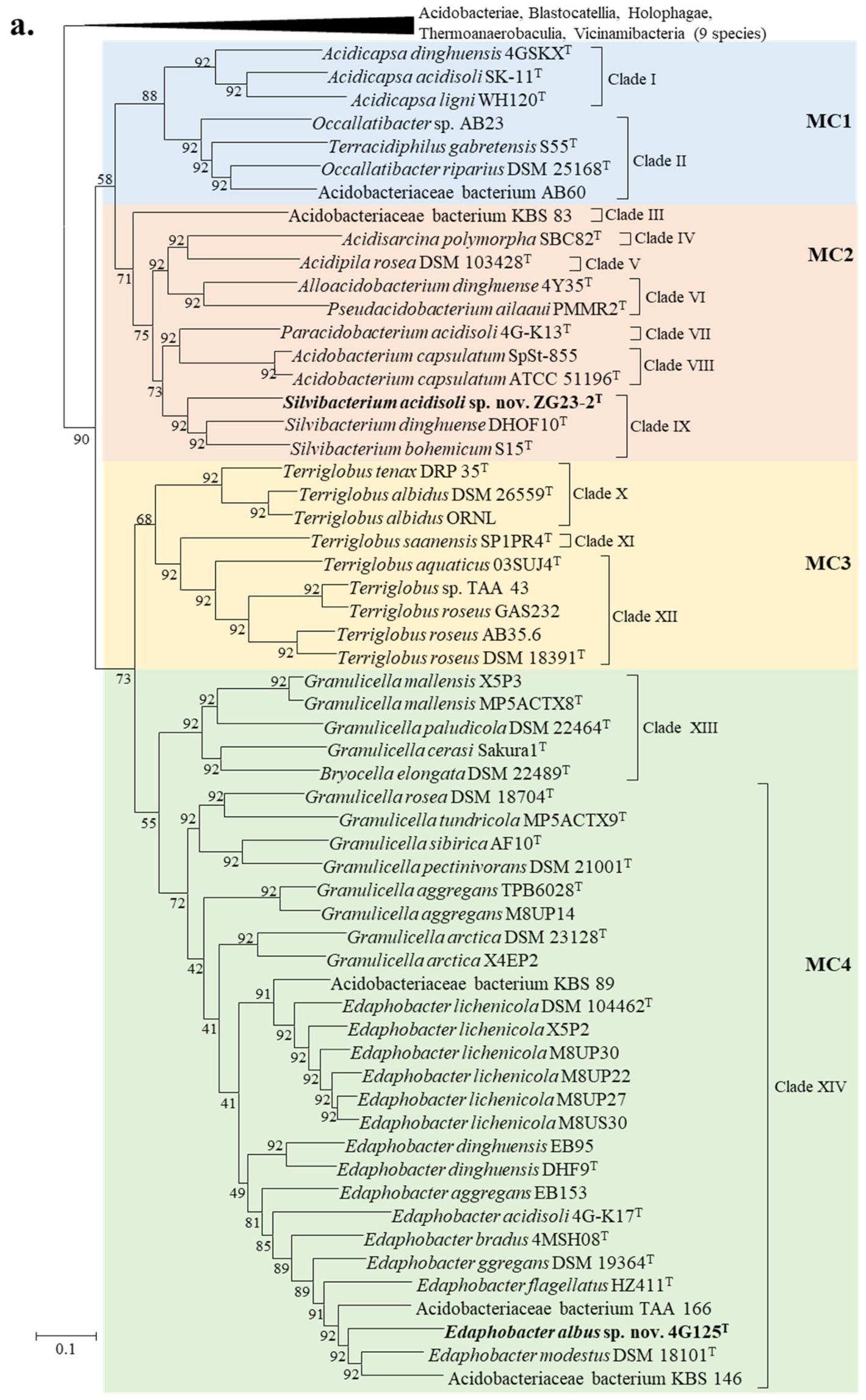
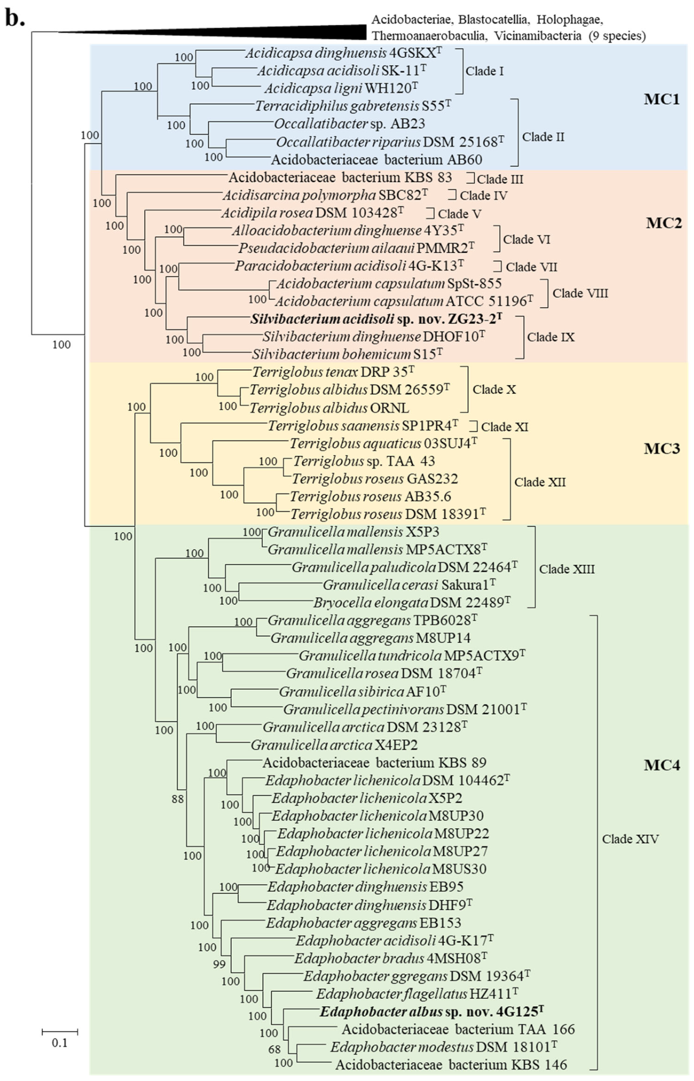
Compared to the 16S-rRNA-based approach, the UBCG- and phylophlan-based phylogenomic analysis provide a better resolution on classifying bacteria at the species and genus levels. Therefore, the 92 bacterial core genes-based (Figure 2a) and 387 universal marker proteins-based (Figure 2b) phylogenomic trees were reconstructed, containing 67 acidobacterial genomes.
These two phylogenomic trees shared a similar topology, but the phylophlan tree was equipped with high bootstrap values at branches. Strains ZG23-2T and 4G125T were consistently clustered with the genera Silvibacterium and Edaphobacter in the two phylogenomic trees, respectively. The tiny differences between the two phylogenomic trees appeared in the Granulicella genus. Granulicella species were distributed in three branches in the UBCG tree but in two branches in the phylophlan tree. Unlike the 16S rRNA tree, the intermixed positions between Granulicella and Edaphobacter genera were improved in the genome phylogenies. Edaphobacter species formed a cluster in both phylogenomic trees. Like the 16S rRNA tree, G. aggregans TPB6028T, G. aggregans M8UP14, G. arctica DSM 23128T, G. pectinivorans DSM 21001T, G. rosea DSM 18704T, G. sibirica AF10T and G. tundricola MP5ACTX9T were closer to Edaphobacter genus (Clade XIV). It is sufficiently proved that Granulicella was a non-monophyletic group, and reassignment of several Granulicella species stated above to Edaphobacter genus was highly recommended by the phylogenomic trees (Clade XIV). Bryocella elongata, currently the only species of the Bryocella genus, apparently nested within the clade consisting of the rest of Granulicella species—G. cerasi Sakura1T, G. mallensis MP5ACTX8T and G. paludicola DSM 22464T (Clade XIII)—indicating the need for reclassification. Deep branches appear within the Terriglobus genus, which implies that the current Terriglobus may not be a monophyletic lineage. However, it is difficult to define a genus just on the basis of evolutionary distance. Thus, the ambiguous boundary in Terriglobus genus is discussed below with more analyses. Acidobacteriaceae bacterium KBS 83 occupied a deeply independent branch ((Clade III)), indicating a potential genus. Terracidiphilus-Occallatibacter lineage (Clade II) always clustered well in the 16S rRNA gene phylogenetic and phylogenomic trees. Merging the two genera into one genus is recommended. The non-nominal Acidobacteriaceae bacteria AB60 and AB23 may represent two potential species of Clade II genus according to the phylogenetic positions.

3.4. Reclassification Supported by AAI, ANI and POCP Values

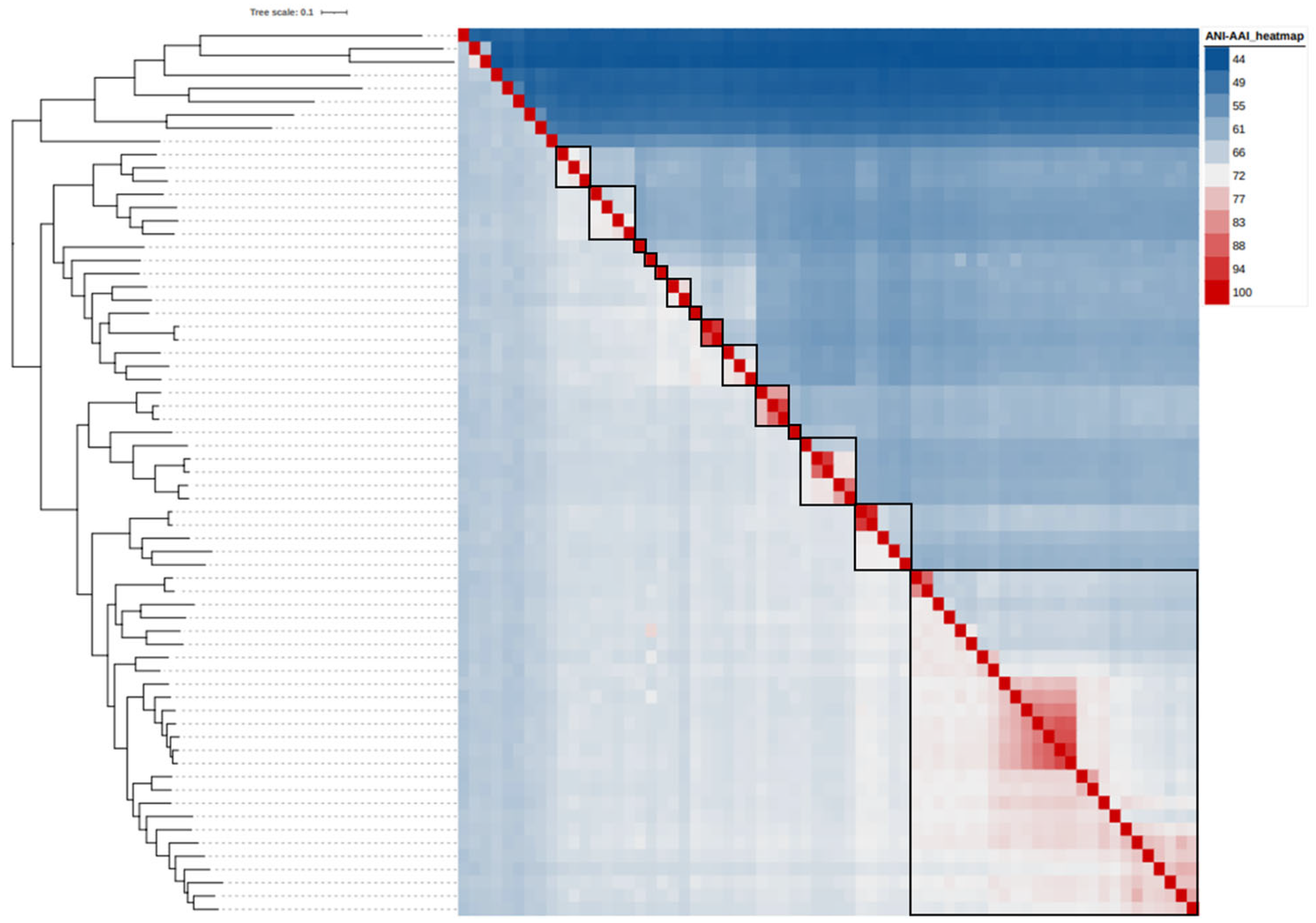
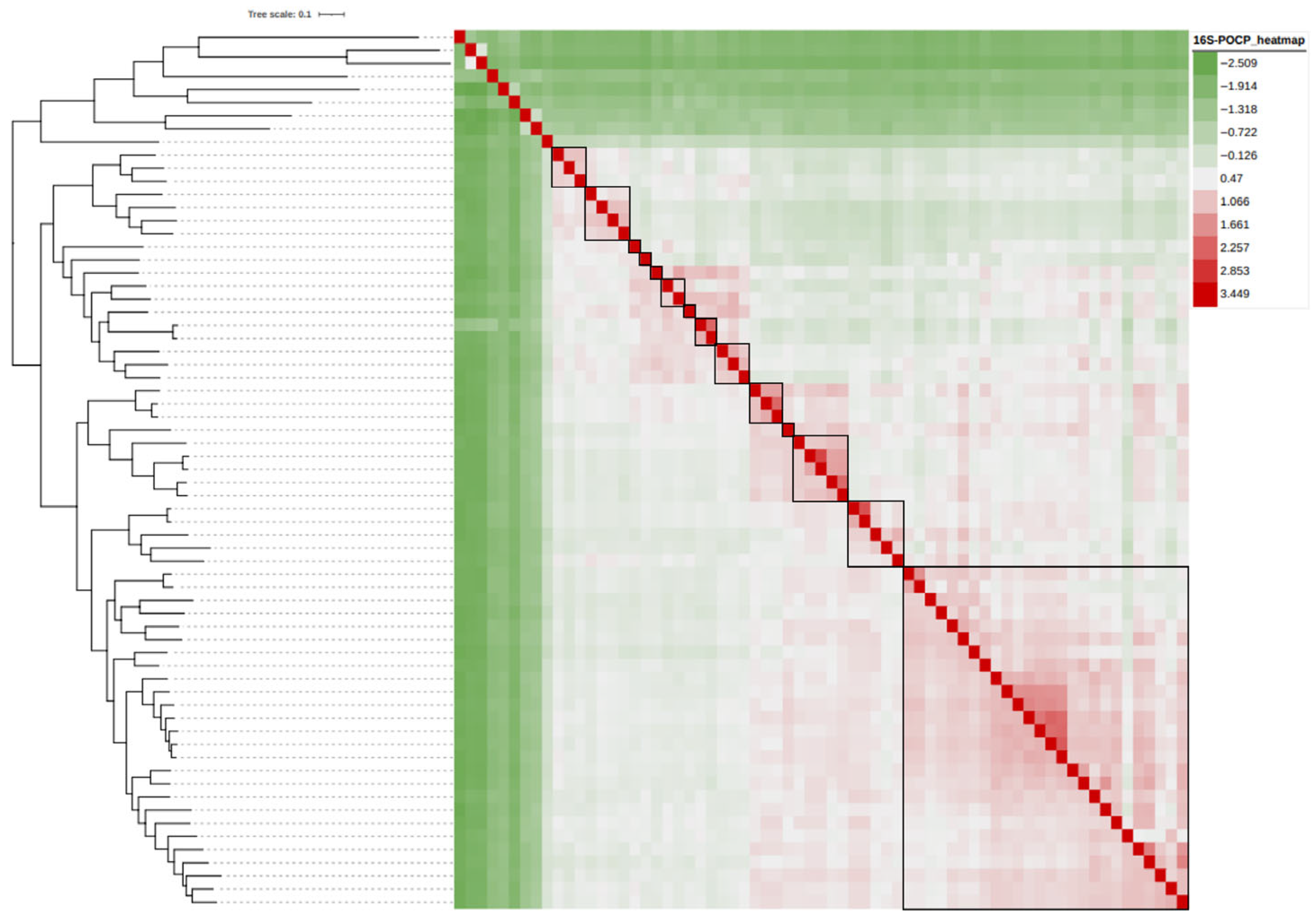
Taxonomic reorganization is required at the genus level because of polyphyletic (Granulicella) and paraphyletic (Occallatibacter, Terriglobus) genera. Reorganization requires setting the criteria for diversity allowed within genera, using a genome-based phylogeny as a primary guideline to identify clades that are monophyletic. Here, the viability of genome-based similarity indices—AAI, ANI and POCP—as supplements to genome phylogeny to infer genera was examined. Sixty-seven acidobacterial genomes were used in this study to produce 2211 ANI, AAI and POCP values. Heatmaps (Figure 3 and Figure 4) were depicted using iTOL (https://itol.embl.de (accessed on 8 September 2024)) [82] on the phylophlan tree.
In the current family Acidobacteriaceae framework, the different species shared 55.8–74.9% of AAI, 67.3–75.2% of ANI and 35.6–67.0% of POCP at the inter-genus levels and shared 60.9–95.9% of AAI, 69.6–93.7% of ANI and 45.7–86.6% of POCP at the intra-genus levels (Table S1). Overlap between the inter-genus and intra-genus levels is clearly visible, as can be seen in the phylogeny analyses. The lowest values at the intra-genus level appeared in genus Granulicella or Terriglobus. In a previous study, defined 73.98% (±0.64%, 73.34–74.62%) ANI and 50% POCP thresholds for the genus boundary were proposed based on massive genome sequence calculations across genera [56,83]. For the Acidobacteriaceae family, applying this boundary shows some pairwise comparisons of at least 50% POCP that would automatically violate the monophyly rule for taxon delineation. For example, the Terriglobus species had higher than 50% POCP with major Edaphobacter species, which would merge the two genera members into the same genus. The same could be said for the Acidicapsa-Silvibacterium genera. On the other hand, taking into consideration comparisons with at least 50% POCP that also exhibited monophyly in the genome phylogeny, an inferred genus-level cluster would require splitting previously described species from the same genus into different genera. For example, in the inferred Edaphobacter clade (Figure 2), several pairwise comparisons between Edaphobacter aggregans DSM 19364T and Edaphobacter dinghuensis EB95, Edaphobacter dinghuensis DHF9T and Edaphobacter acidisoli 4G-K17T displayed POCP values lower than 50%, which would separate the Edaphobacter aggregans DSM 19364T from Edaphobacter genus. Accordingly, the 50% POCP boundary is not an appropriate metric to delineate genera of Acidobacteriaceae family. With at least a 73.34% ANI threshold, many genomes in the Acidobacteriaceae would be split into different genera except for well-clustered Edaphobacter genus. Such a huge change suggests the previously proposed ANI genus boundary is not suitable for the current Acidobacteriaceae.
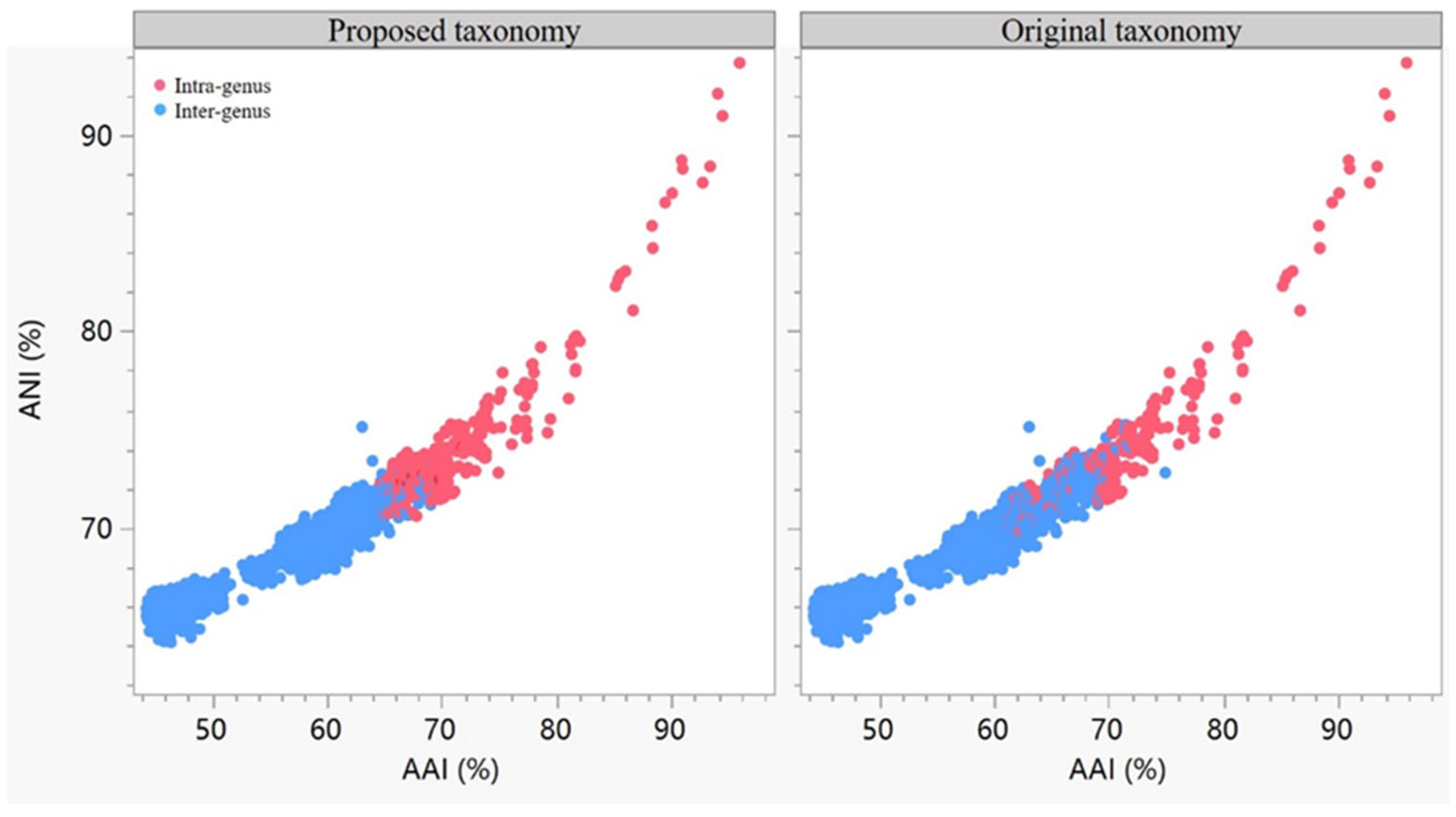
Although there is no generally recognized AAI genus boundary, calculations by Luo et al. suggested that prokaryotic taxa exhibit AAI comparisons typically ranging 60–80% at the genus level [84]. In the Acidobacteriaceae family, over 60% of comparisons had at least 60% AAI, which would result in aggregating the majority of genomes into a single genus, a consequence contradictory to that which ANI inferred. Thus, a higher AAI threshold needs to be determined for this group. Considering AAI, ANI and the phylogenomic tree structure together, 66% AAI and 72% ANI as the genus limits were proposed to distinguish different genera. As shown in Figure 5, the intra-genus and inter-genus dots in the proposed taxonomy of the family Acidobacteriaceae seemed to be more reasonable and compact against its original taxonomy. Applying the AAI and ANI thresholds resulted in separating Terriglobus into three different units (Clade X–Clade XII). Taking thresholds and monophyletic rules into consideration, the 14 inferred genera (Clade I–Clade XIV) maintained most of the current classifications for the identified genomes (Figure 2 and Figure 3). The variable AAI and ANI ranges among the inferred genus clades may be due to a lack of representation of some genera. But variations in AAI can be expected since different prokaryotic taxa, even those that are closely related, can evolve at different rates due to differences in responses to evolutionary and ecological processes.
According to species delineation standards, 95–96% ANI threshold [85], there are several misclassified species that need to be addressed. The type strains Terriglobus albidus DSM 26559T and Terriglobus albidus ORNL exhibit 87.6% ANI with each other, indicating that they do not represent the same species. Therefore, it is proposed to rename the ORNL genome as Terriglobus sp. ORNL. As such, Acidobacterium capsulatum SpSt-855, Edaphobacter lichenicola X5P2, E. lichenicola M8UP30, E. lichenicola M8UP22, E. lichenicola M8UP27, E. lichenicola M8US30, E. dinghuensis EB95, E. aggregans EB153, Granulicella aggregans M8UP14, G. arctica X4EP2, Terriglobus roseus GAS232 and T. roseus AB35.6 genomes can be distinguished from the closest type strains, respectively. Each represents a putative novel species.

3.5. Reclassification Supported by Phenotypic Characteristics and Fatty Acid Profiles

Most strains of Acidobacteriaceae are obviously acidophilic and capable of growth at low temperature. Pigment, motility and capsule are variable across genera.
High amounts of iso-C15:0 appear in all known Acidobacteriaceae species. Moreover, the distribution of other major fatty acid (>5%) components has obvious differences at the genus level (Table 3). In Clade I, the second most abundant fatty acid component contained in the strains is iso-C17:1 ω7c. Among them, Acidicapsa acidisoli SK-11T and A. dinghuensis 4GSKXT employed summed feature 9 (iso-C17:1 ω9c/C16:0 10-methyl) as the second highest. It is worth mentioning that iso-C17:1 ω7c is occasionally identified as iso-C17:1 ω9c type in Acidobacteriota group, and it is also identified as iso-C17:1 ω8c later [25,79]. It is not yet clear which component is correctly identified. For the convenience of comparison, in this context, summed feature 9 and iso-C17:1 ω7c are considered as the same. In Clade II, members also have iso-C17:1 ω7c as the second most abundant component, and unlike the first clade, all strains in the second clade also have more iso-C17:0. In Clade VI, the major fatty acids profile of Pseudacidobacterium ailaaui PMMR2T includes iso-C15:0, iso-C17:0 and summed feature 9. The main components of Alloacidobacterium dinghuense 4Y35T are iso-C15:0, C16:0, C16:1 ω7c, C18:1 ω9c and summed feature 9, which is inconsistent with P. ailaaui PMMR2T. However, the ANI and AAI values between the two strains are 72.8% and 74.9%, respectively. Both the genome relatedness and the phylogenomic relationship verified the feasibility of reclassifying strain 4Y35T into Pseudacidobacterium (Table S1 and Figure 2). The major fatty acids of the isolated strain ZG23-2T are composed of iso-C15:0, C16:0, C16:1 ω7c and C18:1 ω9c, which is in line with clustered Silvibacterium species, supporting assigning this novel strain to Silvibacterium genus. The ANI and AAI values between Terriglobus species are in the ranges of 69.6–88.4% and 61.4–93.3%, respectively. It is strongly recommended by the ANI and AAI thresholds to split this genus into three units (Figure 2, Clade X to Clade XII). Since T. roseus DSM 18391T is the type species of the genus, we propose to move Terriglobus tenax and Terriglobus albidus into Rhizacidiphilus gen. nov., and Terriglobus saanensis into Alloterriglobus gen. nov. Rhizacidiphilus genus employs iso-C15:0 and C16:1 ω7c as predominant fatty acids, whereas Alloterriglobus genus takes iso-C15:0, C16:1 ω7c, iso-C13:0 and C16:0 as predominant fatty acids. The remaining Terriglobus genus employs iso-C15:0, C16:0 and C16:1 ω7c as the main fatty acids.
Genome phylogeny shows the genus Granulicella to be paraphyletic (Figure 2, Clade XIII and Clade XIV). Lower ANI and AAI values between Granulicella species in the two clades appeared than that between Granulicella species of Clade XIV and Edaphobacter species (Table S1). Accordingly, we propose to transfer this part of Granulicella species into Edaphobacter. Granulicella sapmiensis S6CTX5AT genome is not sequenced, but this species exhibited the closest relationship with Granulicella pectinivorans TPB6011T (99.1% 16S rRNA gene sequence similarity and 31 ± 3% experimental DDH) [72]. Moreover, the second Granulicella clade members and Granulicella sapmiensis have the same major fatty acids as Edaphobacter species (Table 3). Outer-membrane vesicles, reported in G. rosea DSM 18704T, G. sibirica AF10T and G. tundricola MP5ACTX9T [71,72,74], also are observed in E. bradus 4MSH08T, E. flagellatus HZ411T and E. lichenicola DSM 104462T [76,78]. Based on the genome phylogeny, genomic comparisons and phenotypic features, we propose to transfer G. pectinivorans DSM 21001T, G. rosea DSM 18704T, G. aggregans TPB6028T, G. arctica MP5ACTX2T, G. sibirica AF10T, G. tundricola MP5ACTX9T and G. sapmiensis S6CTX5AT into Edaphobacter as E. pectinivorans comb. nov., E. rosea comb. nov., E. xylanilytica comb. nov., E. arctica comb. nov., E. sibirica comb. nov., E. tundricola comb. nov. and E. sapmiensis comb. nov., respectively. Acidobacteriaceae bacterium strains TAA 166, KBS 146 and KBS 89 are confirmed as three novel Edaphobacter species by genome relatedness, phylogenomic and chemotaxonomic analyses, and they should be renamed as Edaphobacter sp.

4. Conclusions

This work highlights the relevance of the use of genomics in prokaryotic taxonomy. We successfully established a genome-based phylogeny for the currently available Acidobacteriaceae genome sequences. In addition, pairwise genome comparisons were used to supplement the robust phylogeny to confidently allow the reclassification of several previously identified genomes, as well as provide identities for some unidentified genomes. This work serves as a foundation for the classification of current and future isolates within Acidobacteriaceae.

5. Description of Alloterriglobus gen. nov.

Al.lo.ter.ri.glo′bus (Gr. masc. adj. allos, another, other, different; N.L. masc. n. Terriglobus, a bacterial genus; N.L. fem. n. Alloterriglobus, a genus different from Terriglobus).
Characteristics for this genus were derived from the original paper [68]. The cells are Gram-negative, non-motile, aerobic rods. Catalase-positive and oxidase-negative. The major cellular fatty acids are iso-C15:0, C16:1 ω7c, iso-C13:0 and C16:0. The DNA G + C content is 57.3 mol%.
The type species of the genus is Alloterriglobus saanensis.

6. Description of Rhizacidiphilus gen. nov.

Rhi.za.ci.di.phi.lus (Gr. fem. n. rhiza, root; L. neut. adj. acidum, acid, sour; Gr. masc. adj. philos, loving; N.L. masc. n. Rhizacidiphilus, bacteria from rhizosphere soil and acid-loving).
Characteristics for this genus were derived from the literature [66,67]. The cells are Gram-staining-negative, non-motile, non-spore-forming, non-capsule-forming, aerobic and short rods. Divides by binary fission. Catalase and oxidase are positive or negative. Nitrate is not reduced to nitrite. The major fatty acids are iso-C15:0 and C16:1 ω7c. The major polar lipids are phosphatidylethanolamine. The predominant menaquinone is MK-8. The DNA G + C content as calculated from genome sequences is around 57.6–59.8 mol%, while the range provided in the literature is 58.5–62.1 mol%.
The type species of the genus is Rhizacidiphilus tenax.

7. Description of Edaphobacter albus sp. nov.

Edaphobacter albus (al′bus. L. masc. adj. albus, white-colored, referring to the color of the colonies of the type strain).
The cells are Gram-stain-negative, aerobic, rod-shaped (0.5–0.8 × 1.0–1.6 µm), capsulated and motile with a single polar flagellum. The colonies are round and white with entire edges on GYS medium. The temperature and pH for growth are in the ranges of 12–37 °C and pH 4.0–7.0. Optimal growth occurs at 28 °C and pH 5.0–6.5. NaCl inhibits growth at concentrations above 3.0% (w/v). H2S and indole production, glucose fermentation, arginine dihydrolase and gelatin hydrolysis are absent. Nitrate is reduced to nitrite. Catalase-positive and oxidase-negative. The following enzyme activities are present: urease, arginine hydrolase, alkaline phosphatase, esterase (C4), lipid esterase (C8), leucine arylamidase, valine arylamidase, cystine arylamidase, acid phosphatase, naphthol-AS-BI-phosphohydrolase, α-galactosidase, β-galactosidase, β-uronidase, α-glucosidase, β-glucosidase, N-Acetyl-glucosaminidase, α-mannosidase and α-fucosidase. The following substrates can be used as sole carbon sources for growth: arabinose, D-ribose, D-xylose, L-xylose, D-galactose, D-glucose, D-fructose, D-mannose, inositol, D-mannitol, Methyl-α-D-mannopyranoside, Methyl-α-D-glucopyranoside, N-Acetyl D-glucosamine, amygdalin, arbutin, esculin, salicin, D-cellobiose, D-maltose, lactose, α-D-melibiose, D-sucrose, D-trehalose, D-melezitose, D-raffinose, starch, glycogen, D-gentiobiose, D-turanose, D-(-)-lyxose, D-fucose, potassium gluconate, potassium 2-ketogluconate and potassium 5-ketogluconate. The following substrates cannot be used as carbon sources: glycerol, erythritol, D-adonitol, Methyl-β-D-xylopyranoside, L-sorbose, dulcitol, D-sorbitol, inulin, xylitol, D (-)-tagatose, L-fucose, arabitol, capric acid, adipic acid, malic acid, citric acid and phenylacetic acid. The main fatty acids are iso-C15:0, C16:0 and C16:1 ω7c. The polar lipid components include phosphatidylethanolamine (PE), unknown aminolipid (AL), unidentified phospholipids (PL) and unknown lipids (L).
The type strain of this species is 4G125T (=KACC 21729T=NBRC 114262T), which was isolated from forest soil collected at the DHSBR in Guangdong Province. The G + C content of the genome is 55.0 mol%. The 16S rRNA gene sequence and genome sequence have been uploaded to the GenBank database with accession numbers MT892923 and GCA_014274685.1, respectively.

8. Description of Silvibacterium acidisoli sp. nov.

Silvibacterium acidisoli (a.ci.di.so’li. L. masc. adj. acidus, acidic; L. neut. n. solum, soil; N.L. gen. neut. n. acidisoli, of acidic soil).
Gram-stain-negative, oval to short rod-shaped (0.3–0.8 × 0.5–1.5 µm), capsulated, non-motile and aerobic cells. Colonies are round, translucent, smooth and with entire edges in GYS medium. The temperature and pH ranges for growth are 12–33 °C and pH 3.5–6.0. Optimal growth occurs at 20–28 °C and pH 5.0–6.0. Tolerance to salinity is 2.5% (w/v). Does not produce indole or H2S. Glucose fermentation and nitrate reduction are negative. The activities of oxidase and catalase are negative. Positive for phenol-AS-BI-phosphohydrolase, alkaline phosphatase, α-glucosidase, esterase (C4), β-glucuronidase, lipase (C8) and lipase (C14) activities, but negative for urease, valine arylamidase, α-galactosidase, N-acetylglucosaminidase, and β-fucosidase activity. The following substrates can be used as sole carbon sources for growth: L-xylose, D-fructose, methyl-β-D-xylopyranoside, methyl-α-D-mannose, D-glucose, amygdalin, N-Acetyl D-glucosamine, arbutin, lactose, inulin, L-fucose, esculin, xylitol, D-maltose, glycogen, D-trehalose, D-gentiobiose, D-turanose, potassium 5-ketogluconate and capric acid, but the following substrates cannot: glycerol, L-rhamnose, D-arabinose, L- -sorbose, D-melezitose, L-arabinose, inositol, D-ribose, D-xylose, D-adonitol, D-fucose, D-galactose, D-arabinol, D-mannose, D-cellobiose, D(-)-tagatose, dulcitol, D-mannitol, D-raffinose, L-arabitol, erythritol, D-sorbitol, methyl-α-D-glucopyranoside, salicin, D-sucrose, starch, D-(-)-lyxose, potassium gluconate, D-melibiose and potassium 2-ketogluconate. The major fatty acids (> 5%) are iso-C15:0, C16:0, C16:1 ω7c and C18:1 ω9c. The major polar lipids comprise phosphatidylethanolamine (PE), unidentified aminolipids (AL), unknown phospholipids (PL) and a glycolipid (GL). The type strain of this species is ZG23-2T (=GDMCC 1.2710T=KCTC 49686T), which was isolated from forest soil collected at the Dinghushan Biosphere Reserve (DHSBR) in Guangdong Province. The G + C content of the genome is 58.4 mol%. The 16S rRNA gene sequence and genome sequence of the strain have been uploaded to the GenBank database with accession numbers OK445524 and CP085323, respectively.

9. Description of Alloterriglobus saanensis comb. nov.

Alloterriglobus saanensis (sa.a.nen′sis. N.L. masc. adj. saanensis, pertaining to Mount Saana, Finland).
Basonym: Terriglobus saanensis [68].
The species description is provided by Minna et al. [68]. The DNA G + C content of the type strain is 57.3%. The type strain is SP1PR4T (=DSM 23119T=ATCC BAA-1853T).

10. Description of Edaphobacter arcticus comb. nov.

Edaphobacter arcticus (arc′ti.cus. L. masc. adj. arcticus, northern, arctic, referring to the arctic ecosystem where the type strain was isolated).
Basonym: Granulicella arctica [72].
The description is as given for Granulicella arctica. Based on phylogenomic analyses, genome comparisons and phenotypic characteristics, the species is revised as an Edaphobacter species. The type strain is MP5ACTX2T (=ATCC BAA-1858T= DSM 23128T).

11. Description of Edaphobacter pectinivorans comb. nov.

Edaphobacter pectinivorans (pec.ti.ni.vor′ans. N.L. neut. n. pectinum, pectin; L. pres. part. vorans, devouring; N.L. part. adj. pectinivorans, pectin-devouring, referring to the ability to use pectin as a growth substrate).
Basonym: Granulicella pectinivorans [71].
The description is as given for Granulicella pectinivorans. The genomic G + C content of the type strain is 61.3%. The type strain is TPB6011T (=VKM B-2509T=DSM 21001T).

12. Description of Edaphobacter roseus comb. nov.

Edaphobacter roseus (ro.se′us. L. masc. adj. roseus, rose-colored).
Basonym: Granulicella rosea [71].
The description is as given for Granulicella rosea. The genomic G + C content of the type strain is 62.9%. The type strain is TPO1014T (=DSM 18704T=ATCC BAA-1396T).

13. Description of Edaphobacter sapmiensis comb. nov.

Edaphobacter sapmiensis (sap.mi.en′sis. N.L. masc./fem. adj. sapmiensis, of or pertaining to Sapmi Land, the area inhabited by the Sámi people (Sámi, Sápmi)).
Basonym: Granulicella sapmiensis [72].
The description is as given for Granulicella sapmiensis. Based on the DDH, 16S rRNA gene sequence similarity and phenotypic characteristics, the species is revised as an Edaphobacter species. The type strain is S6CTX5AT (=LMG 26174T=DSM 23136T).

14. Description of Edaphobacter sibiricus comb. nov.

Edaphobacter sibiricus (si.bi′ri.cus. N.L. masc. adj. sibiricus, originating from Siberia, referring to the site of isolation).
Basonym: Granulicella sibirica [74].
The description is as given for Granulicella sibirica. The genomic G + C content of the type strain is 61.3%. The type strain is AF10T (=DSM 104461T=VKM B-3276T).

15. Description of Edaphobacter tundricola comb. nov.

Edaphobacter tundricola (tun.dri.co′la. N.L. n. tundra, a cold treeless region; L. masc./fem. suff. -cola, dweller; N.L. masc./fem. n. incola, dweller; N.L. masc./fem. n. tundricola, tundra dweller).
Basonym: Granulicella tundricola [72].
The description is as given for Granulicella tundricola. The type strain is MP5ACTX9T (=ATCC BAA-1859T=DSM 23138T).

16. Description of Edaphobacter xylanilyticus comb. nov.

Edaphobacter xylanilyticus (xy.la.ni.ly′ti.cus. N.L. neut. n. xylanum, xylan; N.L. masc. adj. lyticus, able to loosen, able to dissolve; from Gr. masc. adj. lytikos, dissolving; N.L. masc. adj. xylanilyticus, xylan-dissolving).
Basonym: Granulicella aggregans [71].
The description is as given for Granulicella aggregans. Based on phylogenomic analyses, genome comparisons and phenotypic characteristics, the species is revised as an Edaphobacter species. The genomic G + C content of the type strain is 59.8%. The type strain is TPB6028T (=LMG 25274T=VKM B-2571T).

17. Description of Granulicella elongata comb. nov.

Granulicella elongata (e.lon.ga′ta. L. fem. part. adj. elongata, elongated, stretched out, pertaining to the elongated cell shape).
Basonym: Bryocella elongata [70].
The description is as given for Bryocella elongata. The genomic G + C content of the type strain is 62.0%. The type strain is SN10T (=LMG 25276T=DSM 22489T).

18. Description of Occallatibacter gabretensis comb. nov.

Occallatibacter gabretensis (ga.bret.en′sis N.L. masc. adj. gabretensis, pertaining to Gabreta, the Celtic name of the Bohemian Forest, the mountain range in central Europe, where the type strain was isolated).
Basonym: Terracidiphilus gabretensis [24].
The description is as given by Paula et al. [24]. The genomic G + C content of the type strain is 57.3%. The type strain is S55T (=NBRC 111238T=CECT 8791T).

19. Description of Pseudacidobacterium dinghuense comb. nov.

Pseudacidobacterium dinghuense (ding.hu.en’se. N.L. neut. adj. dinghuense, pertaining to Dinghu mountain, PR China, where the soil samples were collected).
Basonym: Alloacidobacterium dinghuense.
The description of the species is as given by Zhang et al. [64]. The type strain is 4Y35T (=KACC 21728T=NBRC 114261T).

20. Description of Rhizacidiphilus albidus comb. nov.

Rhizacidiphilus albidus (al′bi.dus. L. masc. adj. albidus, white, referring to the color of the colonies).
Basonym: Terriglobus albidus [66].
The species description is provided by Pascual et al. [66]. The genomic G + C content of the type strain is 57.6%. The type strain is Ac_26_B10T (=DSM 26559T=LMG 27984T).

21. Description of Rhizacidiphilus tenax comb. nov.

Rhizacidiphilus tenax (ten′ax. L. masc. adj. tenax, sticky, holding firm, referring to the organism’s viscous colonies).
Basonym: Terriglobus tenax [67].
The description is as given by Whang et al. [67]. The genomic G + C content of the type strain is 59.8%. The type strain of this species is DRP 35T (=KACC 16474T=NBRC 109677T).

22. Emended Description of Edaphobacter Koch et al. 2008

After the inclusion of Granulicella arctica, G. aggregans, G. rosea, G. tundricola, G. sibirica, G. pectinivorans and G. sapmiensis, the original description [17] needs to be modified as follows. The cells are short, ovoid to elongated rods. Several strains are motile or capsulated. The major fatty acids are iso-C15:0, C16:1 ω7c and C16:0. The G + C content is 56.5–62.9 mol%. The type species is Edaphobacter modestus.

23. Emended Description of Occallatibacter Foesel et al. 2016

The characteristics of genus Occallatibacter are as described by Foesel et al. [25], with the follow revision: the DNA G + C content ranges from 57.3 to 60.2 mol%. The type species is Occallatibacter riparius.

24. Emended Description of Pseudacidobacterium Myers et al. 2016, Zhang et al. 2022

The characteristics of genus Pseudacidobacterium are from the papers [60,64], with the following revisions: the cells are capsulated or not; the major fatty acids are iso-C15:0 and summed feature 9 (iso-C17:1 ω9c/C16:0 10-methyl); the G + C content is from 55.8 to 56.5 mol%. The type species is Pseudacidobacterium ailaaui.

Supplementary Materials

The following supporting information can be downloaded at https://www.mdpi.com/article/10.3390/taxonomy5030040/s1. Figure S1: Two dimensional thin-layer chromatogram showing the polar lipid profiles of strains ZG23-2T (a) and 4G125T (b). PE: phosphatidylethanolamine, PL: unidentified phospholipid, GL: unidentified glycolipid, AL: unidentified aminolipid, L: unidentified polar lipid. Table S1: Carbon source utilization and enzymic activities of ZG23-2T, 4G125T and closest relatives. Strains: 1, ZG23-2; 2, Silvibacterium dinghuense comb. nov. DHOF10T; 3, Silvibacterium bohemicum S15T; 4, 4Y35; 5, Pseudacidobacterium ailaaui comb. nov. PMMR2T; 6, Acidobacterium capsulatum ATCC 51196T; 7, 4G125; 8, Edaphobacter modestus Jbg-1T; 9, Edaphobacter flagellatus HZ411T; 10, Edaphobacter bradus 4MSH08T; 11, Edaphobacter acidisoli 4G-K17T; 12, Edaphobacter aggregans DSM 19364T; 13, Edaphobacter dinghuensis DHF9T.

Author Contributions

Conceptualization, software, data curation, L.Q.; writing—original draft preparation, L.Q. and L.C.; writing—review and editing, L.C. All authors have read and agreed to the published version of the manuscript.

Funding

This research received no external funding.

Institutional Review Board Statement

The experiments reported in this study did not involve human participants and/or animals.

Informed Consent Statement

The experiments reported in this study did not involve human participants and/or animals.

Data Availability Statement

The data are available from the corresponding author on reasonable request.

Conflicts of Interest

The authors declare no conflicts of interest.

References

  1. Kalam, S.; Basu, A.; Ahmad, I.; Sayyed, R.Z.; El-Enshasy, H.A.; Dailin, D.J.; Suriani, N.L. Recent Understanding of Soil Acidobacteria and Their Ecological Significance: A Critical Review. Front. Microbiol. 2020, 11, 580024. [Google Scholar] [CrossRef] [PubMed]
  2. Eichorst, S.A.; Kuske, C.R.; Schmidt, T.M. Influence of plant polymers on the distribution and cultivation of bacteria in the phylum Acidobacteria. Appl. Environ. Microbiol. 2011, 77, 586–596. [Google Scholar] [CrossRef] [PubMed]
  3. Aszalos, J.M.; Krett, G.; Anda, D.; Marialigeti, K.; Nagy, B.; Borsodi, A.K. Diversity of extremophilic bacteria in the sediment of high-altitude lakes located in the mountain desert of Ojos del Salado volcano, Dry-Andes. Extremophiles 2016, 20, 603–620. [Google Scholar] [CrossRef] [PubMed]
  4. Kishimoto, N.; Kosako, Y.; Tano, T. Acidobacterium capsulatum gen. nov., sp. nov.: An acidophilic chemoorganotrophic bacterium containing menaquinone from acidic mineral environment. Current Microbiol 1991, 22, 1–7. [Google Scholar] [CrossRef]
  5. Liesack, W.; Bak, F.; Kreft, J.U.; Stackebrandt, E. Holophaga foetida gen. nov., sp. nov., a new, homoacetogenic bacterium degrading methoxylated aromatic compounds. Arch. Microbiol. 1994, 162, 85–90. [Google Scholar] [CrossRef]
  6. Lonergan, D.J.; Jenter, H.L.; Coates, J.D.; Phillips, E.J.; Schmidt, T.M.; Lovley, D.R. Phylogenetic analysis of dissimilatory Fe(III)-reducing bacteria. J. Bacteriol. 1996, 178, 2402–2408. [Google Scholar] [CrossRef]
  7. Ludwig, W.; Bauer, S.H.; Bauer, M.; Held, I.; Kirchhof, G.; Schulze, R.; Huber, I.; Spring, S.; Hartmann, A.; Schleifer, K.H. Detection and in situ identification of representatives of a widely distributed new bacterial phylum. FEMS Microbiol. Lett. 1997, 153, 181–190. [Google Scholar] [CrossRef]
  8. Hugenholtz, P.; Goebel, B.M.; Pace, N.R. Impact of culture-independent studies on the emerging phylogenetic view of bacterial diversity. J. Bacteriol. 1998, 180, 4765–4774. [Google Scholar] [CrossRef]
  9. Zimmermann, J.; Gonzalez, J.M.; Saiz-Jimenez, C.; Ludwig, W. Detection and phylogenetic relationships of highly diverse uncultured acidobacterial communities in altamira cave using 23S rRNA sequence analyses. Geomicrobiol. J. 2005, 22, 379–388. [Google Scholar] [CrossRef]
  10. Barns, S.M.; Cain, E.C.; Sommerville, L.; Kuske, C.R. Acidobacteria phylum sequences in uranium-contaminated subsurface sediments greatly expand the known diversity within the phylum. Appl. Environ. Microbiol. 2007, 73, 3113–3116. [Google Scholar] [CrossRef]
  11. Fukunaga, Y.; Kurahashi, M.; Yanagi, K.; Yokota, A.; Harayama, S. Acanthopleuribacter pedis gen. nov., sp. nov., a marine bacterium isolated from a chiton, and description of Acanthopleuribacteraceae fam. nov., Acanthopleuribacterales ord. nov., Holophagaceae fam. nov., Holophagales ord. nov. and Holophagae classis nov. in the phylum ‘Acidobacteria’. Int. J. Syst. Evol. Microbiol. 2008, 58, 2597–2601. [Google Scholar] [CrossRef] [PubMed]
  12. Pascual, J.; Wust, P.K.; Geppert, A.; Foesel, B.U.; Huber, K.J.; Overmann, J. Novel isolates double the number of chemotrophic species and allow the first description of higher taxa in Acidobacteria subdivision 4. Syst. Appl. Microbiol. 2015, 38, 534–544. [Google Scholar] [CrossRef] [PubMed]
  13. Oren, A.; da Costa, M.S.; Garrity, G.M.; Rainey, F.A.; Rossello-Mora, R.; Schink, B.; Sutcliffe, I.; Trujillo, M.E.; Whitman, W.B. Proposal to include the rank of phylum in the International Code of Nomenclature of Prokaryotes. Int. J. Syst. Evol. Microbiol. 2015, 65, 4284–4287. [Google Scholar] [CrossRef] [PubMed]
  14. Dedysh, S.N.; Yilmaz, P. Refining the taxonomic structure of the phylum Acidobacteria. Int. J. Syst. Evol. Microbiol. 2018, 68, 3796–3806. [Google Scholar] [CrossRef]
  15. Kielak, A.M.; Barreto, C.C.; Kowalchuk, G.A.; van Veen, J.A.; Kuramae, E.E. The Ecology of Acidobacteria: Moving beyond Genes and Genomes. Front. Microbiol. 2016, 7, 744. [Google Scholar] [CrossRef]
  16. Okamura, K.; Kawai, A.; Yamada, T.; Hiraishi, A. Acidipila rosea gen. nov., sp. nov., an acidophilic chemoorganotrophic bacterium belonging to the phylum Acidobacteria. FEMS Microbiol. Lett. 2011, 317, 138–142. [Google Scholar] [CrossRef]
  17. Koch, I.H.; Gich, F.; Dunfield, P.F.; Overmann, J. Edaphobacter modestus gen. nov., sp. nov., and Edaphobacter aggregans sp. nov., acidobacteria isolated from alpine and forest soils. Int. J. Syst. Evol. Microbiol. 2008, 58, 1114–1122. [Google Scholar] [CrossRef]
  18. Pankratov, T.A.; Kirsanova, L.A.; Kaparullina, E.N.; Kevbrin, V.V.; Dedysh, S.N. Telmatobacter bradus gen. nov., sp. nov., a cellulolytic facultative anaerobe from subdivision 1 of the Acidobacteria, and emended description of Acidobacterium capsulatum Kishimoto et al. 1991. Int. J. Syst. Evol. Microbiol. 2012, 62, 430–437. [Google Scholar] [CrossRef]
  19. Ward, N.L.; Challacombe, J.F.; Janssen, P.H.; Henrissat, B.; Coutinho, P.M.; Wu, M.; Xie, G.; Haft, D.H.; Sait, M.; Badger, J.; et al. Three genomes from the phylum Acidobacteria provide insight into the lifestyles of these microorganisms in soils. Appl. Environ. Microbiol. 2009, 75, 2046–2056. [Google Scholar] [CrossRef]
  20. Rawat, S.R.; Mannisto, M.K.; Bromberg, Y.; Haggblom, M.M. Comparative genomic and physiological analysis provides insights into the role of Acidobacteria in organic carbon utilization in Arctic tundra soils. FEMS Microbiol. Ecol. 2012, 82, 341–355. [Google Scholar] [CrossRef]
  21. Belova, S.E.; Ravin, N.V.; Pankratov, T.A.; Rakitin, A.L.; Ivanova, A.A.; Beletsky, A.V.; Mardanov, A.V.; Sinninghe Damsté, J.S.; Dedysh, S.N. Hydrolytic Capabilities as a Key to Environmental Success: Chitinolytic and Cellulolytic Acidobacteria From Acidic Sub-arctic Soils and Boreal Peatlands. Front. Microbiol. 2018, 9, 2775. [Google Scholar] [CrossRef]
  22. Eichorst, S.A.; Trojan, D.; Roux, S.; Herbold, C.; Rattei, T.; Woebken, D. Genomic insights into the Acidobacteria reveal strategies for their success in terrestrial environments. Environ. Microbiol. 2018, 20, 1041–1063. [Google Scholar] [CrossRef] [PubMed]
  23. Llado, S.; Benada, O.; Cajthaml, T.; Baldrian, P.; Garcia-Fraile, P. Silvibacterium bohemicum gen. nov. sp. nov., an acidobacterium isolated from coniferous soil in the Bohemian Forest National Park. Syst. Appl. Microbiol. 2016, 39, 14–19. [Google Scholar] [CrossRef] [PubMed]
  24. Garcia-Fraile, P.; Benada, O.; Cajthaml, T.; Baldrian, P.; Llado, S. Terracidiphilus gabretensis gen. nov., sp. nov., an Abundant and Active Forest Soil Acidobacterium Important in Organic Matter Transformation. Appl. Environ. Microbiol. 2016, 82, 560–569. [Google Scholar] [CrossRef] [PubMed]
  25. Foesel, B.U.; Mayer, S.; Luckner, M.; Wanner, G.; Rohde, M.; Overmann, J. Occallatibacter riparius gen. nov., sp. nov. and Occallatibacter savannae sp. nov., acidobacteria isolated from Namibian soils, and emended description of the family Acidobacteriaceae. Int. J. Syst. Evol. Microbiol. 2016, 66, 219–229. [Google Scholar] [CrossRef]
  26. Zerbino, D.R.; Birney, E. Velvet: Algorithms for de novo short read assembly using de Bruijn graphs. Genome Res. 2008, 18, 821–829. [Google Scholar] [CrossRef]
  27. Wick, R.R.; Judd, L.M.; Gorrie, C.L.; Holt, K.E. Unicycler: Resolving bacterial genome assemblies from short and long sequencing reads. PLoS Comput. Biol. 2017, 13, e1005595. [Google Scholar] [CrossRef]
  28. Walker, B.J.; Abeel, T.; Shea, T.; Priest, M.; Abouelliel, A.; Sakthikumar, S.; Cuomo, C.A.; Zeng, Q.; Wortman, J.; Young, S.K.; et al. Pilon: An integrated tool for comprehensive microbial variant detection and genome assembly improvement. PLoS ONE 2014, 9, e112963. [Google Scholar] [CrossRef]
  29. Chen, Y.; Chen, Y.; Shi, C.; Huang, Z.; Zhang, Y.; Li, S.; Li, Y.; Ye, J.; Yu, C.; Li, Z.; et al. SOAPnuke: A MapReduce acceleration-supported software for integrated quality control and preprocessing of high-throughput sequencing data. Gigascience 2018, 7, gix120. [Google Scholar] [CrossRef]
  30. Seemann, T. Prokka: Rapid prokaryotic genome annotation. Bioinformatics 2014, 30, 2068–2069. [Google Scholar] [CrossRef]
  31. Hyatt, D.; Chen, G.L.; Locascio, P.F.; Land, M.L.; Larimer, F.W.; Hauser, L.J. Prodigal: Prokaryotic gene recognition and translation initiation site identification. BMC Bioinform. 2010, 11, 119. [Google Scholar] [CrossRef]
  32. Laslett, D.; Canback, B. ARAGORN, a program to detect tRNA genes and tmRNA genes in nucleotide sequences. Nucleic Acids Res. 2004, 32, 11–16. [Google Scholar] [CrossRef] [PubMed]
  33. Lagesen, K.; Hallin, P.; Rødland, E.A.; Staerfeldt, H.H.; Rognes, T.; Ussery, D.W. RNAmmer: Consistent and rapid annotation of ribosomal RNA genes. Nucleic Acids Res. 2007, 35, 3100–3108. [Google Scholar] [CrossRef] [PubMed]
  34. Altschul, S.F.; Madden, T.L.; Schaffer, A.A.; Zhang, J.; Zhang, Z.; Miller, W.; Lipman, D.J. Gapped BLAST and PSI-BLAST: A new generation of protein database search programs. Nucleic Acids Res. 1997, 25, 3389–3402. [Google Scholar] [CrossRef]
  35. Tatusov, R.L.; Galperin, M.Y.; Natale, D.A.; Koonin, E.V. The COG database: A tool for genome-scale analysis of protein functions and evolution. Nucleic Acids Res. 2000, 28, 33–36. [Google Scholar] [CrossRef] [PubMed]
  36. Kanehisa, M.; Goto, S.; Kawashima, S.; Okuno, Y.; Hattori, M. The KEGG resource for deciphering the genome. Nucleic Acids Res. 2004, 32, D277–D280. [Google Scholar] [CrossRef]
  37. Boeckmann, B.; Bairoch, A.; Apweiler, R.; Blatter, M.C.; Estreicher, A.; Gasteiger, E.; Martin, M.J.; Michoud, K.; O’Donovan, C.; Phan, I.; et al. The SWISS-PROT protein knowledgebase and its supplement TrEMBL in 2003. Nucleic Acids Res. 2003, 31, 365–370. [Google Scholar] [CrossRef]
  38. Finn, R.D.; Coggill, P.; Eberhardt, R.Y.; Eddy, S.R.; Mistry, J.; Mitchell, A.L.; Potter, S.C.; Punta, M.; Qureshi, M.; Sangrador-Vegas, A.; et al. The Pfam protein families database: Towards a more sustainable future. Nucleic Acids Res. 2016, 44, D279–D285. [Google Scholar] [CrossRef]
  39. Eddy, S.R. Profile hidden Markov models. Bioinformatics 1998, 14, 755–763. [Google Scholar] [CrossRef]
  40. Kumar, S.; Stecher, G.; Tamura, K. MEGA7: Molecular Evolutionary Genetics Analysis Version 7.0 for Bigger Datasets. Mol. Biol. Evol. 2016, 33, 1870–1874. [Google Scholar] [CrossRef]
  41. Saitou, N.; Nei, M. The neighbor-joining method: A new method for reconstructing phylogenetic trees. Mol. Biol. Evol. 1987, 4, 406–425. [Google Scholar] [CrossRef] [PubMed]
  42. Fitch, W.M. Toward Defining the Course of Evolution: Minimum Change for a Specific Tree Topology. Syst. Biol. 1971, 20, 406–416. [Google Scholar] [CrossRef]
  43. Felsenstein, J. Evolutionary trees from DNA sequences: A maximum likelihood approach. J. Mol. Evol. 1981, 17, 368–376. [Google Scholar] [CrossRef] [PubMed]
  44. Kimura, M. A simple method for estimating evolutionary rates of base substitutions through comparative studies of nucleotide sequences. J. Mol. Evol. 1980, 16, 111–120. [Google Scholar] [CrossRef] [PubMed]
  45. Felsenstein, J. Confidence limits on phylogenies: An approach using the bootstrap. Evolution 1985, 39, 783–791. [Google Scholar] [CrossRef]
  46. Na, S.I.; Kim, Y.O.; Yoon, S.H.; Ha, S.M.; Baek, I.; Chun, J. UBCG: Up-to-date bacterial core gene set and pipeline for phylogenomic tree reconstruction. J. Microbiol. 2018, 56, 280–285. [Google Scholar] [CrossRef]
  47. Eddy, S.R. Accelerated Profile HMM Searches. PLoS Comput. Biol. 2011, 7, e1002195. [Google Scholar] [CrossRef]
  48. Katoh, K.; Standley, D.M. MAFFT multiple sequence alignment software version 7: Improvements in performance and usability. Mol. Biol. Evol. 2013, 30, 772–780. [Google Scholar] [CrossRef]
  49. Price, M.N.; Dehal, P.S.; Arkin, A.P. FastTree 2—approximately maximum-likelihood trees for large alignments. PLoS ONE 2010, 5, e9490. [Google Scholar] [CrossRef]
  50. Asnicar, F.; Thomas, A.M.; Beghini, F.; Mengoni, C.; Manara, S.; Manghi, P.; Zhu, Q.; Bolzan, M.; Cumbo, F.; May, U.; et al. Precise phylogenetic analysis of microbial isolates and genomes from metagenomes using PhyloPhlAn 3.0. Nat. Commun. 2020, 11, 2500. [Google Scholar] [CrossRef]
  51. Buchfink, B.; Xie, C.; Huson, D.H. Fast and sensitive protein alignment using DIAMOND. Nat. Methods 2015, 12, 59–60. [Google Scholar] [CrossRef]
  52. Capella-Gutierrez, S.; Silla-Martinez, J.M.; Gabaldon, T. trimAl: A tool for automated alignment trimming in large-scale phylogenetic analyses. Bioinformatics 2009, 25, 1972–1973. [Google Scholar] [CrossRef] [PubMed]
  53. Yoon, S.H.; Ha, S.M.; Lim, J.; Kwon, S.; Chun, J. A large-scale evaluation of algorithms to calculate average nucleotide identity. Antonie Leeuwenhoek 2017, 110, 1281–1286. [Google Scholar] [CrossRef] [PubMed]
  54. Lee, I.; Ouk Kim, Y.; Park, S.C.; Chun, J. OrthoANI: An improved algorithm and software for calculating average nucleotide identity. Int. J. Syst. Evol. Microbiol. 2016, 66, 1100–1103. [Google Scholar] [CrossRef] [PubMed]
  55. Edgar, R.C. Search and clustering orders of magnitude faster than BLAST. Bioinformatics 2010, 26, 2460–2461. [Google Scholar] [CrossRef]
  56. Qin, Q.L.; Xie, B.B.; Zhang, X.Y.; Chen, X.L.; Zhou, B.C.; Zhou, J.; Oren, A.; Zhang, Y.Z. A proposed genus boundary for the prokaryotes based on genomic insights. J. Bacteriol. 2014, 196, 2210–2215. [Google Scholar] [CrossRef]
  57. Liu, Y.; Du, J.; Pei, T.; Du, H.; Feng, G.-D.; Zhu, H. Genome-based taxonomic classification of the closest-to-Comamonadaceae group supports a new family Sphaerotilaceae fam. nov. and taxonomic revisions. Syst. Appl. Microbiol. 2022, 45, 126352. [Google Scholar] [CrossRef]
  58. Falagan, C.; Foesel, B.; Johnson, B. Acidicapsa ferrireducens sp. nov., Acidicapsa acidiphila sp. nov., and Granulicella acidiphila sp. nov.: Novel acidobacteria isolated from metal-rich acidic waters. Extremophiles 2017, 21, 459–469. [Google Scholar] [CrossRef]
  59. Kulichevskaya, I.S.; Kostina, L.A.; Valaskova, V.; Rijpstra, W.I.C.; Sinninghe Damste, J.S.; de Boer, W.; Dedysh, S.N. Acidicapsa borealis gen. nov., sp. nov. and Acidicapsa ligni sp. nov., subdivision 1 Acidobacteria from Sphagnum peat and decaying wood. Int. J. Syst. Evol. Microbiol. 2012, 62, 1512–1520. [Google Scholar] [CrossRef]
  60. Matsuo, H.; Kudo, C.; Li, J.; Tonouchi, A. Acidicapsa acidisoli sp. nov., from the acidic soil of a deciduous forest. Int. J. Syst. Evol. Microbiol. 2017, 67, 862–867. [Google Scholar] [CrossRef]
  61. Ou-Yang, T.N.; Xia, F.; Qiu, L.H. Acidicapsa dinghuensis sp. nov., a novel acidobacterium isolated from forest soil. Int. J. Syst. Evol. Microbiol. 2018, 68, 2364–2369. [Google Scholar] [CrossRef]
  62. Eichorst, S.A.; Breznak, J.A.; Schmidt, T.M. Isolation and characterization of soil bacteria that define Terriglobus gen. nov., in the phylum Acidobacteria. Appl. Environ. Microbiol. 2007, 73, 2708–2717. [Google Scholar] [CrossRef]
  63. Myers, M.R.; King, G.M. Isolation and characterization of Acidobacterium ailaaui sp. nov., a novel member of Acidobacteria subdivision 1, from a geothermally heated Hawaiian microbial mat. Int. J. Syst. Evol. Microbiol. 2016, 66, 5328–5335. [Google Scholar] [CrossRef]
  64. Zhang, Q.-M.; Fu, J.-C.; Chen, Z.-Q.; Qiu, L.-H. Paracidobacterium acidisoli gen. nov., sp. nov. and Alloacidobacterium dinghuense gen. nov., sp. nov., two acidobacteria isolated from forest soil, and reclassification of Acidobacterium ailaaui and Acidipila dinghuensis as Pseudacidobacterium ailaaui gen. nov., comb. nov. and Silvibacterium dinghuense comb. nov. Int. J. Syst. Evol. Microbiol. 2022, 72, 005415. [Google Scholar] [CrossRef] [PubMed]
  65. Jiang, Y.W.; Wang, J.; Chen, M.H.; Lv, Y.Y.; Qiu, L.H. Acidipila dinghuensis sp. nov., an acidobacterium isolated from forest soil. Int. J. Syst. Evol. Microbiol. 2016, 66, 76–83. [Google Scholar] [CrossRef] [PubMed]
  66. Pascual, J.; Wust, P.K.; Geppert, A.; Foesel, B.U.; Huber, K.J.; Overmann, J. Terriglobus albidus sp. nov., a member of the family Acidobacteriaceae isolated from Namibian semiarid savannah soil. Int. J. Syst. Evol. Microbiol. 2015, 65, 3297–3304. [Google Scholar] [CrossRef] [PubMed]
  67. Whang, K.S.; Lee, J.C.; Lee, H.R.; Han, S.I.; Chung, S.H. Terriglobus tenax sp. nov., an exopolysaccharide-producing acidobacterium isolated from rhizosphere soil of a medicinal plant. Int. J. Syst. Evol. Microbiol. 2014, 64, 431–437. [Google Scholar] [CrossRef]
  68. Mannisto, M.K.; Rawat, S.; Starovoytov, V.; Haggblom, M.M. Terriglobus saanensis sp. nov., an acidobacterium isolated from tundra soil. Int. J. Syst. Evol. Microbiol. 2011, 61, 1823–1828. [Google Scholar] [CrossRef]
  69. Baik, K.S.; Choi, J.S.; Kwon, J.; Park, S.C.; Hwang, Y.M.; Kim, M.S.; Kim, E.M.; Seo, D.C.; Cho, J.S.; Seong, C.N. Terriglobus aquaticus sp. nov., isolated from an artificial reservoir. Int. J. Syst. Evol. Microbiol. 2013, 63, 4744–4749. [Google Scholar] [CrossRef]
  70. Dedysh, S.N.; Kulichevskaya, I.S.; Serkebaeva, Y.M.; Mityaeva, M.A.; Sorokin, V.V.; Suzina, N.E.; Rijpstra, W.I.C.; Sinninghe Damste, J.S. Bryocella elongata gen. nov., sp. nov., a member of subdivision 1 of the Acidobacteria isolated from a methanotrophic enrichment culture, and emended description of Edaphobacter aggregans Koch et al. 2008. Int. J. Syst. Evol. Microbiol. 2012, 62, 654–664. [Google Scholar] [CrossRef]
  71. Pankratov, T.A.; Dedysh, S.N. Granulicella paludicola gen. nov., sp. nov., Granulicella pectinivorans sp. nov., Granulicella aggregans sp. nov. and Granulicella rosea sp. nov., acidophilic, polymer-degrading acidobacteria from Sphagnum peat bogs. Int. J. Syst. Evol. Microbiol. 2010, 60, 2951–2959. [Google Scholar] [CrossRef]
  72. Mannisto, M.K.; Rawat, S.; Starovoytov, V.; Haggblom, M.M. Granulicella arctica sp. nov., Granulicella mallensis sp. nov., Granulicella tundricola sp. nov. and Granulicella sapmiensis sp. nov., novel acidobacteria from tundra soil. Int. J. Syst. Evol. Microbiol. 2012, 62, 2097–2106. [Google Scholar] [CrossRef] [PubMed]
  73. Yamada, K.; Okuno, Y.; Meng, X.Y.; Tamaki, H.; Kamagata, Y.; Hanada, S. Granulicella cerasi sp. nov., an acidophilic bacterium isolated from cherry bark. Int. J. Syst. Evol. Microbiol. 2014, 64, 2781–2785. [Google Scholar] [CrossRef] [PubMed]
  74. Oshkin, I.Y.; Kulichevskaya, I.S.; Rijpstra, W.I.C.; Sinninghe Damste, J.S.; Rakitin, A.L.; Ravin, N.V.; Dedysh, S.N. Granulicella sibirica sp. nov., a psychrotolerant acidobacterium isolated from an organic soil layer in forested tundra, West Siberia. Int. J. Syst. Evol. Microbiol. 2019, 69, 1195–1201. [Google Scholar] [CrossRef] [PubMed]
  75. Xia, F.; Cai, Y.M.; Chen, D.X.; Qiu, L.H. Edaphobacter acidisoli sp. nov., an acidobacterium isolated from forest soil. Int. J. Syst. Evol. Microbiol. 2017, 67, 4260–4265. [Google Scholar] [CrossRef]
  76. Xia, F.; Ou-Yang, T.N.; Gao, Z.H.; Qiu, L.H. Edaphobacter flagellatus sp. nov. and Edaphobacter bradus sp. nov., two acidobacteria isolated from forest soil. Int. J. Syst. Evol. Microbiol. 2018, 68, 2530–2537. [Google Scholar] [CrossRef]
  77. Wang, J.; Chen, M.H.; Lv, Y.Y.; Jiang, Y.W.; Qiu, L.H. Edaphobacter dinghuensis sp. nov., an acidobacterium isolated from lower subtropical forest soil. Int. J. Syst. Evol. Microbiol. 2016, 66, 276–282. [Google Scholar] [CrossRef]
  78. Belova, S.E.; Suzina, N.E.; Rijpstra, W.I.C.; Sinninghe Damste, J.S.; Dedysh, S.N. Edaphobacter lichenicola sp. nov., a member of the family Acidobacteriaceae from lichen-dominated forested tundra. Int. J. Syst. Evol. Microbiol. 2018, 68, 1265–1270. [Google Scholar] [CrossRef]
  79. Damste, J.S.; Rijpstra, W.I.; Hopmans, E.C.; Weijers, J.W.; Foesel, B.U.; Overmann, J.; Dedysh, S.N. 13,16-Dimethyl octacosanedioic acid (iso-diabolic acid), a common membrane-spanning lipid of Acidobacteria subdivisions 1 and 3. Appl. Environ. Microbiol. 2011, 77, 4147–4154. [Google Scholar] [CrossRef]
  80. Hördt, A.; López, M.G.; Meier-Kolthoff, J.P.; Schleuning, M.; Weinhold, L.-M.; Tindall, B.J.; Gronow, S.; Kyrpides, N.C.; Woyke, T.; Göker, M. Analysis of 1,000+ Type-Strain Genomes Substantially Improves Taxonomic Classification of Alphaproteobacteria. Front. Microbiol. 2020, 11, 468. [Google Scholar] [CrossRef]
  81. Kim, M.; Oh, H.S.; Park, S.C.; Chun, J. Towards a taxonomic coherence between average nucleotide identity and 16S rRNA gene sequence similarity for species demarcation of prokaryotes. Int. J. Syst. Evol. Microbiol. 2014, 64, 346–351. [Google Scholar] [CrossRef]
  82. Letunic, I.; Bork, P. Interactive Tree Of Life (iTOL) v5: An online tool for phylogenetic tree display and annotation. Nucleic Acids Res. 2021, 49, W293–W296. [Google Scholar] [CrossRef]
  83. Barco, R.A.; Garrity, G.M.; Scott, J.J.; Amend, J.P.; Nealson, K.H.; Emerson, D. A Genus Definition for Bacteria and Archaea Based on a Standard Genome Relatedness Index. mBio 2020, 11, e02475-19. [Google Scholar] [CrossRef]
  84. Luo, C.; Rodriguez, R.L.; Konstantinidis, K.T. MyTaxa: An advanced taxonomic classifier for genomic and metagenomic sequences. Nucleic Acids Res. 2014, 42, e73. [Google Scholar] [CrossRef]
  85. Richter, M.; Rosselló-Móra, R. Shifting the genomic gold standard for the prokaryotic species definition. Proc. Natl. Acad. Sci. USA 2009, 106, 19126–19131. [Google Scholar] [CrossRef]
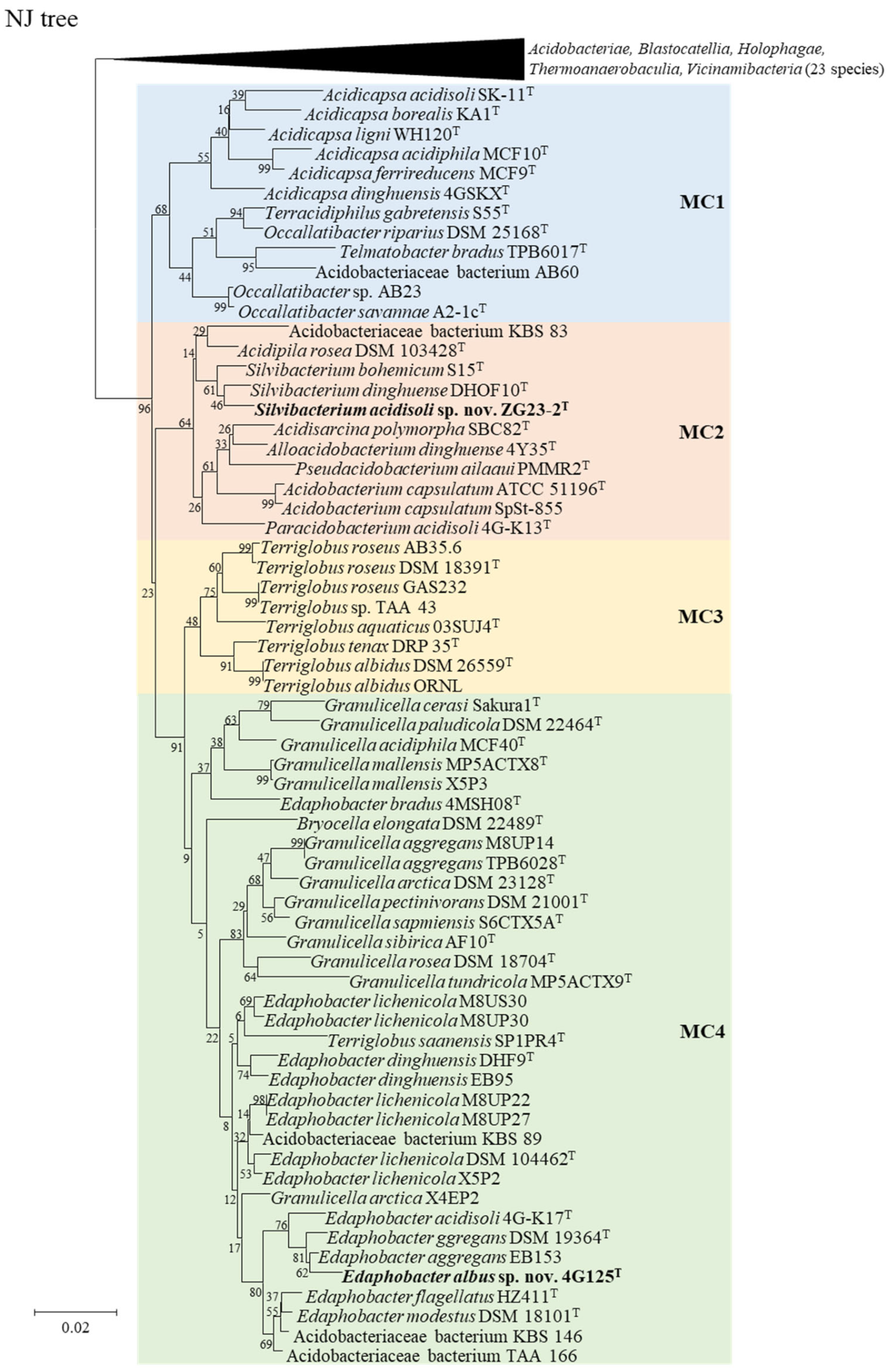
Figure 1. A neighbor-joining phylogenetic tree based on the 16S rRNA gene reproduced from the 88 strains of the phylum Acidobacteriota.
Taxonomy 05 00040 g001
Figure 2. Phylogenomic trees of 67 strains of the phylum Acidobacteriota based on 92 concatenated nucleotide sequences using UBCGs (a) and amino acid sequences inferred from 389 conserved proteins using the PhyloPhlAn pipeline (b). a, gene support indices (GSIs) are given at branching points. b, bootstrap values expressed as percentages of 1000 replications are given at branching points.
Taxonomy 05 00040 g002aTaxonomy 05 00040 g002b
Figure 3. ANI and AAI from pairwise genome comparisons. The heat map shows ANI and AAI values between genomes, along with the Phylophlan tree cladogram to show relationships. Boxed regions indicate inferred genus clusters based on AAI comparisons, as well as monophyly in the genome-based phylogeny. Tree scale: 0.1. Upper triangle is AAI and lower triangle is ANI.
Taxonomy 05 00040 g003
Figure 4. The heat map shows POCP and 16S rRNA gene sequence identity values between strains along with the Phylophlan tree cladogram to show relationships. Boxed regions indicate inferred genus clusters based on AAI comparisons, as well as monophyly in the genome-based phylogeny. Tree scale: 0.1. Upper triangle is POCP and lower triangle is 16S rRNA gene sequence identity.
Taxonomy 05 00040 g004
Figure 5. Comparison between the original and the proposed taxonomy system for the family of Acidobacteriaceae based on ANI and AAI values. Blue and red filled circles indicate intra-genus and inter-genus, respectively.
Taxonomy 05 00040 g005
Table 1. Genomic features of the strains included in this study.
StrainGenome AccessionGenome Size (Mb)Scaffold countN50 Scaffold (bp)(G+C)%Gene Number
Acidicapsa dinghuensis 4GSKXTJAGSYH0000000006.1717816,15656.475125
Acidicapsaligni WH120TJAGSYG0000000005.6817961,24955.414697
Acidicapsa acidisoli SK-11TJAGSYI0000000007.11181,906,82757.135905
Terracidiphilus gabretensis S55TGCA_001449115.15.3517729,62257.304426
Occallatibacter riparius DSM 25168TCP0933136.7916,794,54760.235637
Acidobacteriaceae bacterium AB60GCA_008682415.16.6533466,25861.865574
Occallatibacter sp. AB23GCA_003131205.16.2818795,66259.105356
Acidobacteriaceae bacterium KBS 83GCA_000381585.16.2521826,80159.145419
Acidipilarosea DSM 103428TGCA_004339725.14.21151,147,13758.813513
Silvibacterium bohemicum S15TGCA_001006305.16.4625883,33758.225345
Acidipila dinghuensis DHOF10TGCA_004123295.15.1291,191,39660.503998
Silvibacterium acidisolisp. nov. ZG23-2TCP0853235.3615,359,90058.404344
Acidisarcina polymorpha SBC82TGCA_003330725.17.6057,112,01156.876702
Pseudacidobacterium dinghuensesp. nov. 4Y35TGCA_014274465.16.2716,271,11455.795475
Acidobacterium ailaaui PMMR2TGCA_000688455.13.6913,686,52356.493163
Acidobacterium capsulatum ATCC 51196TGCA_000022565.14.1314,127,35660.503365
Acidobacterium capsulatum SpSt-855GCA_011333575.13.168166,93760.992719
Paracidobacterium acidisoligen. nov., sp. nov. 4G-Kl3TGCA_003428625.25.0016846,09060.464022
Terriglobus roseus AB35.6GCA_900105625.14.8124,742,75759.894003
Terriglobus roseus DSM 18391TGCA_000265425.15.2315,227,85860.284347
Terriglobus roseus GAS232GCA_900102185.14.8514,852,06656.454097
Terriglobus sp. TAA 43GCA_000800015.14.9573,477,25756.694213
Terriglobus aquaticus 03SUJ4TJAGSYB0000000004.1624,126,41662.713452
Terriglobus saanensis SP1PR4TGCA_000179915.25.1015,095,22657.344267
Terriglobus tenax DRP35TJAGSYA0000000004.3643,021,85259.763697
Teriglobus albidus DSM26559TJAGTAS0000000006.1058220,28457.564963
Terriglobus albidus ORNLGCA_008000815.16.4116,405,58257.905217
Granulicella cerasi SakuralTJAGSYD0000000004.087978,91660.823418
Granulicella paludicola DSM22464TJAGSYC0000000004.9626630,16258.994031
Granulicella mallensis MP5ACTX8TGCA_000178955.26.2416,237,57757.914899
Granulicella mallensis X5P3GCA_014203225.16.4752253,60257.805186
Bryocella elongata DSM 22489TGCA_900108185.15.6716661,24762.004535
Granulicella aggregans M8UP14GCA_014203275.17.3083844,69058.596188
Granulicella aggregans TPB6028TJAGSYE0000000005.74141,416,59059.834653
Granulicella pectinivorans DSM 21001TGCA_900114625.15.2834,439,41361.284282
Grandicella sibirica AF10TGCA_004115155.16.14161,421,51359.825140
Grandicella rosea DSM 18704TGCA_900188085.15.2924586,56662.914296
Granulicella arctica DSM 23128TJAGTUT0000000005.8594,736,69257.575138
Granulicella arctica X4EP2GCA_013410065.14.3132,159,44758.263645
Granulicella tundricola MP5ACTX9TGCA_000178975.25.5064,309,15359.984715
Acidobacteriaceae bacterium KBS 89GCA_000381605.16.0113943,63157.555055
Edaphobacter lichenicola DSM 104462TCP0736965.6615,662,23956.484891
Edaphobacter lichenicola X5P2GCA_014201335.16.1823507,84756.415415
Edaphobacter lichenicola M8UP30GCA_013410115.15.2942,888,93257.564466
Edaphobacter lichenicola M8UP22GCA_013410875.15.0752,909,13357.734322
Edaphobacter lichenicola M8UP27GCA_014201315.14.88151,190,61358.404146
Edaphobacter lichenicola M8US30GCA_014201375.14.8315711,31158.184171
Edaphobacter dinghuensisDHF9TJAGSYJ0000000004.54111,438,06356.933732
Edaphobacter dinghuensis EB95GCA_003633965.14.4623,541,52955.563854
Edaphobacter acidisoli 4G-K17TJAGSYK0000000004.2091,158,79958.443493
Edaphobacter aggregans DSM 19364TGCA_000745965.18.1834576,81158.707425
Edaphobacter aggregans EB153GCA_003945235.16.1516,152,25657.255109
Edaphobacter bradus 4MSH08TJAGSYF0000000004.75131,458,50859.444076
Edaphobacter albussp.nov.4G125TGCA_014274685.14.2514,254,61455.033605
Edaphobacter flagellatus HZ411TCP0736974.4614,445,62757.413727
Edaphobacter modestus DSM 18101TGCA_004217555.17.4586,121,18057.106642
Acidobacteriaceae bacterium KBS 146GCA_000688615.15.0024,996,38456.744161
Acidobacteriaceae bacterium TAA 166GCA_000421065.16.1434,712,21058.835269
Candidatus Koribacter versatilis Ellin345GCA_000014005.15.6515,650,36858.384995
Candidatus Solibacter usitatus Ellin6076GCA_000014905.19.9719,965,64061.908193
Bryobacter aggregatus MPL3TGCA_000702445.15.7544,257,80957.955060
Chloracidobacterium thermophilum BTGCA_000226295.13.7022,683,36261.343046
Pyrinomonas methylaliphatogenes K22TGCA_000820845.23.7916443,59859.373165
Luteitalea pratensis DSM 100886TGCA_001618865.17.4817,480,31467.226260
Holophagafoetida DSM 6591TGCA_000242615.34.1333,443,19262.903586
Geothrix fermentans DSM 14018TGCA_000428885.13.2936164,72268.852903
Thermoanaerobaculum aquaticum MP-01TGCA_000687145.12.6668115,89263.012324
Genomes sequenced in this study are marked in bold.
Table 2. Morphological, physiological and growth properties of Acidobacteriaceae type strains. Strains: 1, Acidicapsa ferrireducens MCF9T [58]; 2, Acidicapsa acidiphila MCF10T [58]; 3, Acidicapsa borealis KA1T [59]; 4, Acidicapsa ligni WH120T [59]; 5, Acidicapsa acidisoli SK-11T [60]; 6, Acidicapsa dinghuensis 4GSKXT [61]; 7, Telmatobacter bradus TPB6017T [18]; 8, Occallatibacter riparius 277T [25]; 9, Occallatibacter savannae A2-1cT [25]; 10, Terracidiphilus gabretensis S55T [24]; 11, Acidobacteriaceae bacterium KBS 83 [62]; 12, Acidisarcina polymorpha SBC82T [21]; 13, Acidipila rosea AP8T [16]; 14, Pseudoacidobacterium ailaaui PMMR2T [63]; 15, Alloacidobacterium dinghuense 4Y35T [64]; 16, Paraacidobacterium acidisoli 4G-K13T [64]; 17, Acidobacterium capsulatum ATCC 51196T [4,18]; 18, Silvibacterium acidisoli sp. nov. ZG23-2T; 19, Silvibacterium bohemicum S15T [23]; 20, Silvibacterium dinghuense DHOF10T [65]; 21, Terriglobus albidus Ac_26_B10T [66]; 22, Terriglobus tenax DRP 35T [67]; 23, Terriglobus saanensis SP1PR4T [68]; 24, Terriglobus aquaticus 03SUJ4T [69]; 25, Terriglobus roseus DSM 18391T [62]; 26, Terriglobus sp. TAA 43 [62]; 27, Bryocella elongata DSM 22489T [70]; 28, Granulicella paludicola OB1010T [71]; 29, Granulicella acidiphila MCF40T [58]; 30, Granulicella mallensis MP5ACTX8T [72]; 31, Granulicella cerasi Sakura1T [73]; 32, Granulicella pectinivorans DSM 21001T [71]; 33, Granulicella rosea DSM 18704T [71]; 34, Granulicella aggregans TPB6028T [71]; 35, Granulicella arctica MP5ACTX2T [72]; 36, Granulicella sibirica AF10T [74]; 37, Granulicella sapmiensis S6CTX5AT [72]; 38, Granulicella tundricola MP5ACTX9T [72]; 39, Edaphobacter acidisoli 4G-K17T [75]; 40, Edaphobacter modestus Jbg-1T [17]; 41, Edaphobacter aggregans DSM 19364T [17]; 42, Edaphobacter flagellatus HZ411T [76]; 43, Edaphobacter albus sp. nov. 4G125T; 44, Edaphobacter bradus 4MSH08T [76]; 45, Edaphobacter dinghuensis DHF9T [77]; 46, Edaphobacter lichenicola SBC68T [78]; 47, Acidobacteriaceae bacterium KBS 89 [62]; 48, Acidobacteriaceae bacterium TAA 166 [62]. All the data, except those for strains 18 and 43, were obtained from the original publication.
StrainColorMotilityRelation to OxygenCapsuleTemperature
Range
pH RangeNaCl (w/v,%)
1Pink+AerobicND10–323.2–5.2>300 mM
2White+AerobicND10–322.3–5.1>200 mM
3PinkAerobic+10–332.5–7.30–2.0
4WhiteAerobic+10–333.5–6.40–2.0
5WhiteAerobic+10–354.0–5.50–0.4
6CreamAerobic12–374.0–6.50–1.0
7Opaque+Facultatively
anaerobic
4–353.0–6.00–0.06
8White+Aerobic+11–403.5–8.50–1.0
9Pink+Aerobic+11–403.5–6.50–0.5
10White+Aerobic+12–303.0–6.00–0.5
11WhiteMicroaerophilic+12–374.5–6.0ND
12WhiteFacultatively
anaerobic
5–364.0–7.70–1.5
13PinkAerobic+22–373.0–6.00–1.0
14CreamFacultatively
anaerobic
+15–554.5–7.00–1.0
15WhiteAerobic12–373.5–6.50–2.0
16Pale yellowAerobic12–373.5–6.50–1.0
17Orange+Aerobic+25–373.0–6.00–3.5
18TransparentAerobic+12–333.5–6.00–2.5
19White+Aerobic20–303.0–6.00–0.1
20WhiteAerobic+10–373.5–8.00–1.0
21WhiteAerobic10–423.9–9.8ND
22CreamAerobicND15–453.5–7.00–1.0
23CreamAerobic4–304.5–7.5ND
24PinkAerobicND15–306.0–7.00–1.0
25PinkFacultatively
anaerobic
+12–235.0–7.0ND
26WhiteFacultatively
anaerobic
+12–235.0–6.5ND
27PinkAerobic+6–323.2–6.60–3.0
28RedAerobic2–333.0–7.50–3.5
29WhiteAerobicND10–322.3–5.3>300 mM
30WhiteAerobic4–283.5–6.50–1.5
31PinkAerobic10–304.5–8.50–1.0
32RedAerobic2–333.0–7.50–3.5
33RedAerobic2–333.0–7.50–3.5
34PinkAerobic2–333.0–7.50–3.5
35PinkAerobic4–283.5–6.50–1.0
36Pale–pinkAerobic20–254.5–5.00–1.5
37WhiteAerobic4–263.5–7.00–1.0
38PinkAerobic4–283.5–6.50–1.0
39CreamAerobic10–423.0–7.00–2.5
40Cream+Aerobic15–304.5–7.0ND
41CreamAerobic15–374.0–7.0ND
42Faint yellow+Aerobic10–373.5–6.50–2.5
43White+Aerobic+12–374.0–7.00–3.0
44Faint yellowAerobic10–373.0–6.50–3.0
45CreamAerobic10–333.5–5.50–2.0
46PinkAerobic7–373.4–7.00–1.0
47WhiteFacultatively
anaerobic
+12–23NDND
48WhiteFacultatively
anaerobic
+12–235.0–7.0ND
+, Positive; −, Negative; ND, Not Determined.
Table 3. Fatty acid profiles of Acidobacteriaceae species. Strains: 1, Acidicapsa ferrireducens MCF9T [58]; 2, Acidicapsa acidiphila MCF10T [58]; 3, Acidicapsa borealis KA1T [59]; 4, Acidicapsa ligni WH120T [59]; 5, Acidicapsa acidisoli SK-11T [60]; 6, Acidicapsa dinghuensis 4GSKXT [61]; 7, Telmatobacter bradus TPB6017T [18]; 8, Occallatibacter riparius 277T [25]; 9, Occallatibacter savannae A2-1cT [25]; 10, Terracidiphilus gabretensis S55T; 11, Acidisarcina polymorpha SBC82T [21]; 12, Acidipila rosea AP8T [16]; 13, Pseudacidobacterium ailaaui PMMR2T; 14, Alloacidobacterium dinghuense 4Y35T; 15, Paracidobacterium acidisoli 4G-K13T; 16, Acidobacterium capsulatum ATCC 51196T; 17, Silvibacterium acidisoli sp. nov. ZG23-2T; 18, Silvibacterium bohemicum S15T; 19, Silvibacterium dinghuense DHOF10T [65]; 20, Terriglobus albidus Ac_26_B10T; 21, Terriglobus tenax DRP 35T; 22, Terriglobus aquaticus 03SUJ4T [69]; 23, Terriglobus roseus DSM 18391T [62]; 24, Terriglobus saanensis SP1PR4T [68]; 25, Terriglobus sp. TAA 43 [62]; 26, Bryocella elongata DSM 22489T; 27, Granulicella paludicola OB1010T; 28, Granulicella acidiphila MCF40T [58]; 29, Granulicella mallensis MP5ACTX8T; 30, Granulicella cerasi Sakura1T; 31, Granulicella pectinivorans DSM 21001T [71]; 32, Granulicella rosea DSM 18704T; 33, Granulicella aggregans TPB6028T; 34, Granulicella arctica MP5ACTX2T; 35, Granulicella tundricola MP5ACTX9T [72]; 36, Granulicella sibirica AF10T [74]; 37, Granulicella sapmiensis S6CTX5AT [72]; 38, Edaphobacter acidisoli 4G-K17T [75]; 39, Edaphobacter modestus Jbg-1T [17]; 40, Edaphobacter aggregans DSM 19364T [79]; 41, Edaphobacter flagellatus HZ411T [76]; 42, Edaphobacter albus sp. nov. 4G125T; 43, Edaphobacter bradus 4MSH08T [76]; 44, Edaphobacter dinghuensis DHF9T [77]; 45, Edaphobacter lichenicola SBC68T; 46, Acidobacteriaceae bacterium KBS 89 [62]; 47, Acidobacteriaceae bacterium TAA 166 [62]. Strains of 8, 10, 13–15, 17, 18, 20, 21, 26, 27, 29, 30, 32–34, 42 and 45 were detected in this study. Others were obtained from the original papers.
Analyses MethodStrainC14:0C15:0C16:0C17:0C18:0iso-C13:0iso-C15:0C14:0 anteisoC14:1ω5ciso-C15:0 3-OHC16:1ω7cC17:1ω8cC18:1ω9ciso-C17:0C17:0 cycloiso-C17:1ω7c13,16-dimethyl-octacosanedioic acidSummed Features:1Summed Features:8Summed Features:9
MIDI10.30.10.80.80.2 69.3 0.1 2.90.50.15.9 17.2 0.7
MIDI2 0.5 80.7 0.70.9 1.9 13.0 1.8
GC-MS3 0.9 13 45.9 3.4 21.921.9
GC-MS40.4 1.4 0.7 40.7 3.9 1332.8
MIDI5 2.90.50.5 55.4 2.2B 0.516.7 0.6 17.7
MIDI6 7.3 48.8 2.1B 5.28.1 3.514.7
MIDI70.1 1.50.20.5 69.8 0.4A 5.4 14.8C 1.9
MIDI8 0.5 61.9 0.4B 5.3 29
MIDI9 0.6 54.8 0.9B 9.5 30.7
MIDI10 3.94 0.76 45.78 4.0B 12.17 22.37
GC-MS110.4 8.1 0.9 29.4 20.4 1.5 30.7
GC-MS120.5–1.6 8.7–13.31.5–2.11.3–1.9 49.9–53.1 25.3–26.5 1.5–2.4
MIDI130.8 2.50.13.8 60.61.10.20.14.2B0.10.411.4 1 8.8
MIDI141.14 6.490.971.430.0952.32.410.15 10.47B1.775.394.74 3.12 5.77
MIDI150.4 32.693.8 56.61 0.08 6.24B3.13.256.09 1.47 8.08
MIDI161.36 4.210.862.050.1250.91 0.18 5.09B 16.251.760.1 0.57 1.59
MIDI171.37 5.861.694 37.66 0.37 15.23B3.198.541.64 1.742.963.51
MIDI180.96 5.280.21.99 54.56 0.340.0713.31B0.927.284.30.99 1.5 4.58
MIDI191.7 8.62.73.7 48.6 11.5B 17
MIDI200.6 3.520.070.6 60.191.880.320.1423.6B0.08 0.76 0.721.641.59
MIDI210.94 6.10.080.720.0553.340.730.450.0732.79B0.03 0.65 0.421.240.65
GC-MS222.10.48.40.50.49.839.9 28.4 1.7
MIDI236.8 9.1 2.844.5 2.7 22.5B 0.8
MIDI243.4 7.77 43.88 27.08B
MIDI254.250.749.14 0.68 36.33 0.67 30.24A 0.79
MIDI260.96 5.02 0.970.2747.58 0.46 30.15B 1.47 0.710.881.83
MIDI271.8 13.20.90.513.143.31.10.30.118.3B0.10.11.9 0.10.20.4
MIDI280.6 3.2 68.5 21B 0.8 3.7 0.9
MIDI291.39 11.150.110.4710.6445.26 0.370.1422.54B 1.49 0.170.760.67
MIDI304.45 6.29 0.226.8349.24 2.45 24.6B 0.10.5 0.36 0.29
MIDI312.50.460.50.30.452.8 27.7B 1.5
MIDI324.04 14.630.660.850.0843.76 0.89 25.64B0.151.431.31 0.13 0.5
MIDI331.07 7.440.31.160.8462.28 0.250.1518.81B0.08 3.160.02 0.340.831.04
MIDI341.09 5.990.50.84 31.03 1.136.9535.63B0.220.190.87 0.180.92
GC-MS353 6.6 0.60.446.4 35 3.3
GC-MS361.9 7.50.20.7 18.7 27 1.6 38.9
GC-MS371.6 4.6 0.251.5 35.3 1.8
MIDI382.3 17.3 6.344.9 20.2B 1.8
MIDI392.25 11.290.530.49 38.93 0.83 35.831.67 3.05 1.84C
MIDI401.98 7.130.10.49 44.09 1.520.2534.30.16 2 0.370.12.11
MIDI412 9.62.2 39.8 38.1
MIDI420.99 8.950.671.52 40.444.7 0.2129.37B0.180.392.18 2.17
MIDI431.9 81.2 46.1 29.5
MIDI441.6 10.22.24.3 36.7 31.6B
MIDI451.58 13.580.170.72.9452.05 0.5 20.56B 0.242.54 0.70.380.76
MIDI461.16 7.33 0.754.1247.09 1 26.18A 1.25C
MIDI473.68 8.71 0.59 37.06 0.57 26.99A
Major components (≥5.0%) are highlighted in bold. The summed feature 3 was determined as C16:1 ω7c and/or C16:1 ω6c in this study by the MIDI method but as C16:1 ω7c and/or iso-C15:0 2-OH in several papers [17,18,62]. GC-MS data for this summed feature indicated that in species of the phylum Acidobacteria subdivision 1, C16:1 ω7c was the dominant fatty acid and that C16:1 ω6c and iso-C15:0 2-OH did not occur [68,79]. Both were defined to C16:1 ω7c for comparison. #, summed features represent groups of two or more fatty acids that could not be separated by gas chromatography using the MIDI system. Summed feature 1: iso-C15:1 and/or C13:0 3-OH; summed feature 8: C18:1 ω7c/C18:1 ω6c; summed feature 9: iso-C17:1 ω9c/C16:0 10-methyl. A, determined as summed feature 3 (C16:1 ω7c and/or iso-C15:0 2-OH) by the MIDI method; B, determined as summed feature 3 (C16:1 ω7c and/or C16:1 ω6c) by the MIDI method; C, determined as iso-C17:1 ω9c by the MIDI method.
Disclaimer/Publisher’s Note: The statements, opinions and data contained in all publications are solely those of the individual author(s) and contributor(s) and not of MDPI and/or the editor(s). MDPI and/or the editor(s) disclaim responsibility for any injury to people or property resulting from any ideas, methods, instructions or products referred to in the content.