Article

Scale Sensilla in the Snakes of the Genus Natrix, and in the Old and New World Natricids

by
Alessandro Paterna
OPHIS Museo Paleontologico e Centro Erpetologico, 64100 Teramo, Italy
Taxonomy 2025, 5(3), 34; https://doi.org/10.3390/taxonomy5030034
Submission received: 18 April 2025 / Revised: 13 June 2025 / Accepted: 23 June 2025 / Published: 25 June 2025

Abstract

Among European colubroids, scale sensilla—mechanoreceptors present in the head integument—are more expressed in natricids. The presence of protruded sensilla, observable with the naked eye, is found in the cephalic shields of all species belonging to the genus Natrix. The identification of these sense organs in this genus determines its correlation in aquatic and semi-aquatic species, in which these traits are more developed and recognizable than in terrestrial species. As hypothesized for elapoids, this differentiation could be due to the fact that in natricids, like sea snakes, these can perform a hydrodynamic function in addition to the mechanosensory one. In support of this thesis, within the genus Natrix, the most aquatic species of the five, Natrix tessellata, features the most expressed sensilla. This specificity represents a further analogy in the evolutionary convergences involving the cephalic region that this species shares with marine elapids. Still in the genus Natrix, a second trait involving the shields has been identified, expressing itself in the opposite condition to the protruded sensilla, occurring as a pitting arranged mainly within the shields along the upper portion of the labial arches. In vivo examinations and microscopy were performed on different species of the Natrix genus, and comparative analyses have been carried out on other natricid taxa from the New and Old World, where the presence of protruded scale sensilla has been found in several species.

Graphical Abstract

1. Introduction

The presence and, in some cases, the function of skin sense organs in the integument of reptiles belonging to the Lepidosauria group have been widely documented and described to date [1,2,3,4]. In the scales of some species, small mechanoreceptors are present, manifesting themselves as small integumental protrusions, known as scale sensilla. In snakes, these tactile sensory structures may be referred to as head pitting, touch corpuscles, tactile sense organs, skin sense organs, touch papillae, sensory pustules, and tubercles [1,5,6]. These structures consist of a cluster of innervated central cells forming a dermal papilla evaginating from the dermis to the epidermis, resulting in a reduction in the last layer of corneous beta proteins and the α-keratin layer [7,8]. These scale organs, present in the head of snakes, seem to have a role in several biological and ecological aspects of these animals. Their mechanosensory function seems to be linked to exploration and predation, as in the sensilla found in dorsal scales [9,10], and those present in the chin region seem to be further involved in courtship [6,9]. The number and distribution of sensilla within the cephalic shields show intraspecific variations, which do not appear to be linked to sexual dimorphism [10]. Modern studies focused on these organs are mostly oriented towards elapoids, in particular marine elapids, where these traits differ from the ones present in terrestrial species [7,11,12]. In the literature, references to these sensory structures in Old World species are scarce. In this study, through observations in the field and in controlled environments, and the examination of photographic material produced by citizen science available on digital platforms, the presence of protruded scale sensilla is discussed mainly in the context of European aquatic and semi-aquatic species—the natricids of the genus Natrix—while taking into consideration snakes of the same family occurring in other regions of the world.

2. Materials and Methods

The current analysis on the genus Natrix was carried out both through field studies, through the in vivo observation of wild individuals, and through the re-examination of photographic material previously acquired in various European locations. A further inspection for the presence of protruded scale sensilla in a part of the natricids belonging to the genera Natrix, Nerodia and Thamnophis, was carried out through the in vivo examination of individuals kept in captivity in private collections. Photographic comparisons with all the species of colubroids from the New and Old World discussed in this work were possible by consulting the material uploaded to the digital platform iNaturalist (Table 1). On this platform, reports of natricids from the New and Old World of the genera Afronatrix, Amphiesma, Aspidura, Atretium, Clonophis, Hebius, Helophis, Herpetoreas, Hydraethiops, Limnophis, Liodytes, Lycognathophis, Natriciteres, Natrix, Nerodia, Opisthotropis, Pseudagkistrodon, Regina, Rhabdophis, Sahyadriophis, Storeria, Thamnophis, Trachischium, Trimerodytes, Tropidoclonion, Virginia and Xenochrophis amount to almost 540,000 at the time of writing this paper. The largest number of materials come from the American continent, with over 249,000 reports for the genus Thamnophis, which represents over 46% of the total. This number is followed by Nerodia (over 134,000) and Storeria (over 71,000), and then moving on to Europe with the genus Natrix (over 47,000). Reports concerning the genera Thamnophis, Nerodia, Storeria, Virginia and Natrix alone represent 96% of all observations of individuals belonging to the Natricidae family. There are diametrically opposed cases where for some taxa (from Africa and Asia), there are reports corresponding to few units (under a dozen). The examination of the photographic material was managed by making an initial selection that discarded the reports attaching photos acquired from a significant distance, preferring those in which the head and cephalic shields of the subjects were clearly visible. From this initial screening, only a small part of the photographic material proved to be functional to the research, further discarding the out-of-focus photos and those in which the environmental parameters did not allow a correct visualization of the cephalic shields and the features present. This equates to just under 400 images, excluding redundant records in species with the greatest amounts of photographic material and sightings, and in reports that include multiple photographs of a single subject. However, in rare cases, some users uploaded close-up portraits of the individuals found, which were very useful for the investigation of the protruded sensilla. Preserved adult and subadult specimens of the species N. natrix (2), N. helvetica (4) and N. tessellata (2) used for microscopy were donated to OPHIS by private individuals, together with locally collected deceased specimen utilized for the current and past studies during the years 2022–2024. Further in vivo examinations were carried out in a controlled environment on N. helvetica specimens bred and reproduced at OPHIS, and on adult specimens and hatchlings of N. tessellata and N. helvetica recovered and subsequently released together with the Carabinieri Forestali command of the province of Teramo. Microphotographs of the scale sensilla were taken using an Optika SZX-121 stereomicroscope together with an Optika C-H4K camera at the Faculty of Veterinary Medicine of the University of Teramo.

3. Results

Within the genus Natrix, protruding scale sensilla are observable with the naked eye, and are expressed in different frequency and distribution values among the various specimens of the different species. In the five species ascribed to the genus, this type of sensilla can be found in all the dorsal, lateral and ventral cephalic shields. At a first visual examination, Natrix tessellata is the species that features the most distinguishable and most protruded sensilla (Figure 1A). Like this one, Natrix maura can also present a significant quantity of protruded sensilla within all the shields (Figure 1B). In many individuals belonging to the species Natrix helvetica and Natrix natrix, the protruded sensilla appears to be less evident than in the previous two, while Natrix astreptophora seems to be the species in which these external traits are less expressed or more rarely observable (Figure 1C,D). Scale sensilla are also present in the juveniles of these species, but these are difficult to distinguish with the naked eye. Through the use of photography, their identification is possible and facilitated, also by orienting the light in a certain way, or by using photographic filters. These traits, however, as in adults, seem to express themselves in a more defined manner only in some individuals.
Also, following a naked-eye inspection of live specimens, a second type of structure, diffuse within the shields and easily distinguishable, was identified in all five species. This trait expresses in a way opposite to the protruded sensilla, appearing as circumscribed pits that notch the surface of the cephalic shield. These are co-occurring with the protruded sensilla in the shields arranged along the upper labial arch: supralabial, nasal, loreal and preocular shields, mainly around the lower perimeter of the eye to the nostril. In the species N. natrix, N. helvetica and N. astreptophora, pits appear to occur in higher numbers (Figure 1C,D), with the larger ones located in the preocular shields (Figure 1B,D).
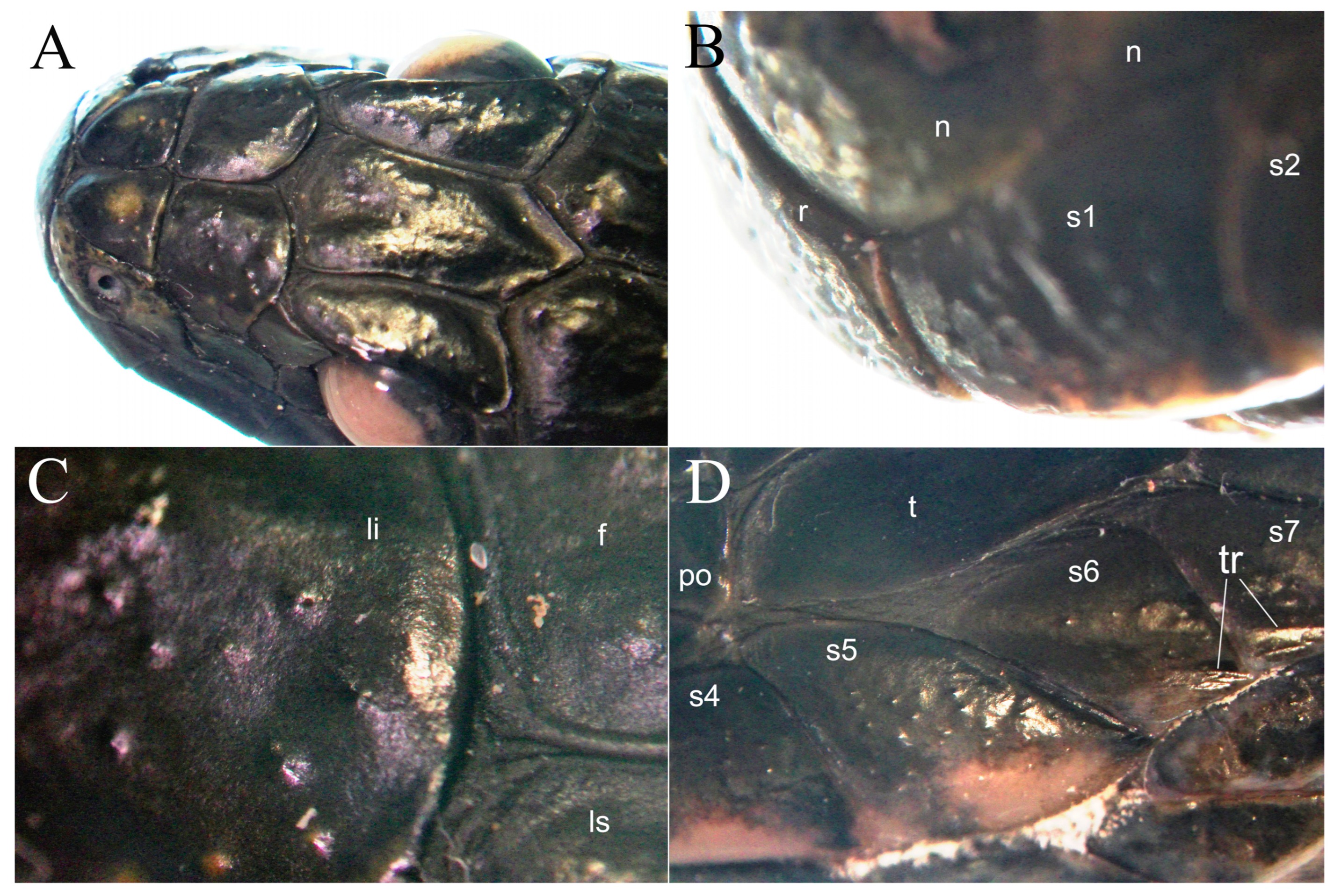
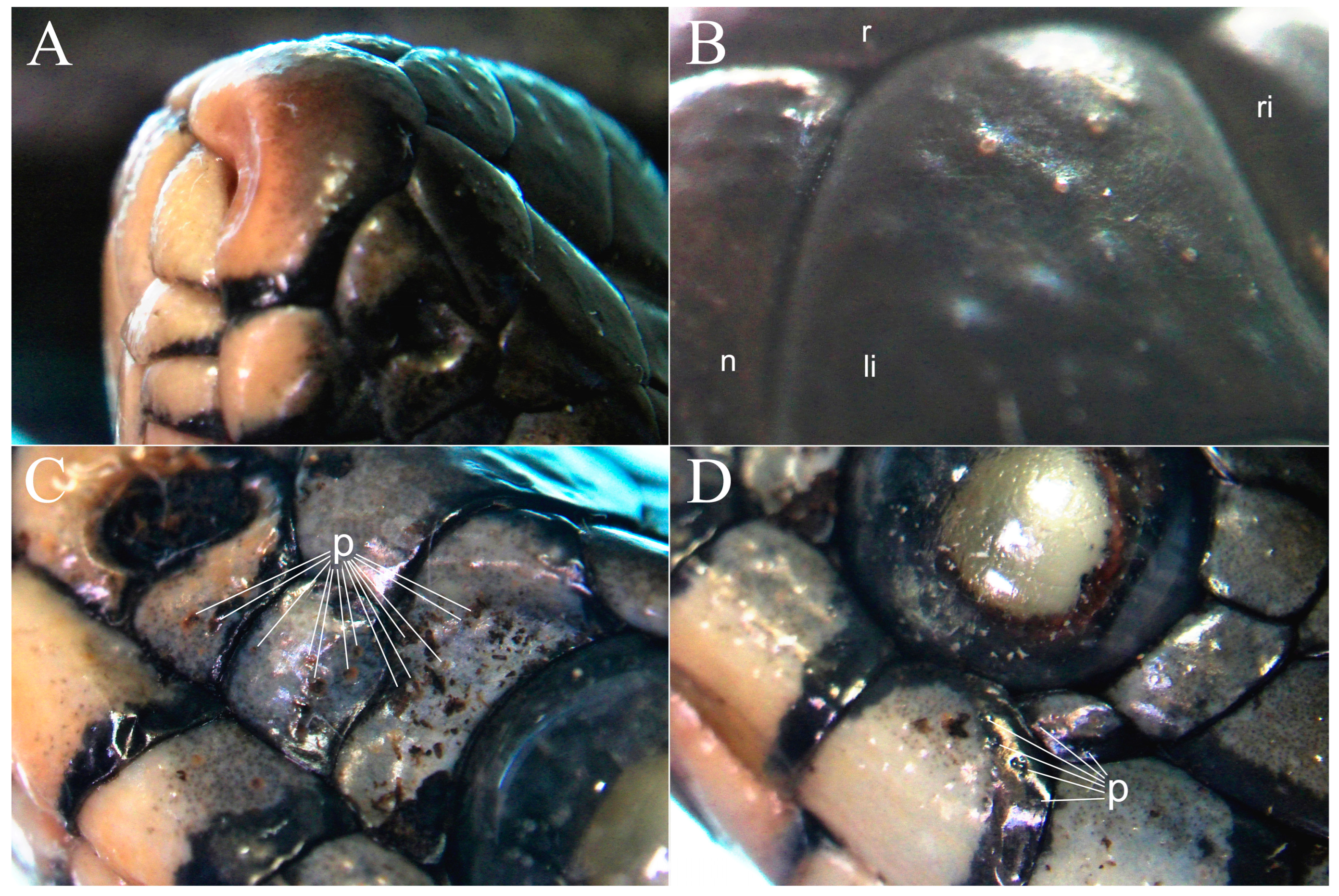
The use of microscopy or macrophotography makes the three-dimensionality of the scale sensilla more tangible and comprehensible. In N. tessellata, these are distributed within all the cephalic shields (Figure 2A), with a major density on the rostrum, specifically the rostral, anterior supralabial and nasal shields, conferring a rough surface to the rostrum (Figure 2B). The most posteromedial shields, i.e., frontal and parietal, are the ones which present the lesser number of sensilla. In this species, the more defined sensilla, present in the dorsal cephalic shields, appear as domes that may present a small pit or lateral concavity (Figure 2C). Other protrusions may be present in the most posterior shields. In the penultimate supralabial, where the silhouette of the ventral margin curves sharply dorsally, transverse ridges may occur close to the posterior ventrolateral margin (Figure 2D). Even in the following shield, the last and most posterior supralabial, such ridges may be present close to the longitudinal keel that runs along its ventral margin. Other transverse ridges, but more similar to the carinae of the dorsal scales, may be present in the center of the posterior temporal shields. In-depth examinations reveal a significant diffusion of small sensilla in the rostrum also in N. natrix, and N. helvetica (Figure 3A), similarly to what was observed in N. tessellata. The high concentration of sensilla in the shields covering the rostrum of these snakes confirms what has been described by other authors in other ophidians [2,13,14]. Always dorsally, but more posteriorly, the sensilla become sparser in the internasal and prefrontal shields, but still appear well defined, where the apex of the dome of the sensillum can be highlighted by a lighter coloration (Figure 3B). In the N. helvetica, N. natrix and N. astreptophora group, the most easily identifiable sensilla occur in the large supralabial shields that distinguish these species, and in the infralabial shields appearing as orange or darker freckles. Always laterally, in the preocular and loreal shields, defined pits are present, often characterized by an orange or dark coloration (Figure 3C). Always laterally, but more posteriorly, the pitting can appear more diffuse, particularly from the supralabials in contact with the eye to the posterior supralabials. The pitting in these shields may co-occur with the protruded sensilla, and contrasts with the more defined sensilla present in the adjacent and more dorsal, postocula, and temporal shields (Figure 3D). Both sensilla and pits are also easily distinguishable in the shed of these species, which aids in their identification.

4. Discussion

4.1. Protruted Scale Sensilla in European Natricids

In European colubroids, the most distinct and naked-eye-recognizable protruded sensilla are present in natricids. Within the genus Natrix, the most aquatic taxon differs from the others by presenting a more evident expression of these traits. The fact that the sensilla are more externally developed in Natrix tessellata, the most aquatic of the taxa, confirms what was hypothesized by Crowe-Riddell et al. [7,11], according to which, similarly to marine elapoids, they may perform a hydrodynamic–mechanosensory function in this genus. According to parallel studies, protruded scale sensilla or dome-shaped sensilla seem to be more developed in aquatic and semi-aquatic snakes, where these probably evolved independently several times [11,15], and in the literature, in addition to elapids, descriptions of similar sense organs can be found in other families/subfamilies of aquatic snakes, such as Acrochordidae [16,17] and Dipsadinae/Dipsadidae [13]. In support of this hypothesis, among European colubroids, these structures are more widespread and more developed in aquatic and semi-aquatic snakes, such as the examined species of the genus Natrix, and less marked in the sympatric terrestrial species. This is also supported by phylogenetic analyses where within the genus Natrix, the most basal taxon, N. maura, and the more derived N. natrix, N. helvetica and N. astreptophora show less protruded sensilla than N. tessellata [18,19,20], suggesting that the development of these traits is mainly linked to the ecology of a given taxon, where intraspecific variations are nevertheless present within the five species ascribed to this genus, likely related to both individual and population expression within certain ecological–environmental contexts. A similar correlation is also found outside Lepidosauria, such as in crocodiles (both modern and extinct taxa), where similar “dome pressure receptor” structures, linked to the presence of foramina in the cranial bones, are present exclusively in aquatic or semi-aquatic species (as in the extant ones) [21,22,23,24,25]. Among terrestrial European colubroids, however, under certain conditions these can still be observed with the naked eye, as in the case of Hierophis viridiflavus, where scale sensilla are particularly recognizable in the anterior dorsal cephalic shields of hatchlings (internasal, prefrontal and parietal), and in the other species with the support of macrophotography. Like other traits present in the cephalic shields of colubroids, the presence of defined protruded sensilla, as well as the number or arrangement of these within certain shields, does not represent a constant within the species analyzed. This peculiarity has been observed and described in other studies, where the diversity of these values is hypothesized to be the consequence of differences in foraging and/or reproductive strategies between species and sexes [8].
A second trait identified in the cephalic shields is the pitting. This is easily observed in N. helvetica, where it is distributed in the lateral shields along a transverse axis at the base of the eye (nasal, loreal, preocular and postocular shields, and in the dorsal portion of the anterior supralabials) (Figure 4A–D). The pits appear asymmetrically and in different numbers among the various specimens examined. As for the sensilla, this work aims to provide only a first anatomical description following the identification of these traits involving the cephalic shields of this group of reptiles, in the direction of in-depth analyses that can determine their origin and if they present a different function from the sensilla.

4.2. Preliminary Comparative Examination in Extra-European Natricids

A similar scenario to the European one occurs among the large natricids of the New World, where protruded sensilla can be easily observed in the aquatic species of the genus Nerodia, such as Nerodia sipedon, Nerodia erythrogaster and Nerodia taxispilota, and are less evident in the genus Thamnophis, which instead present fewer aquatic habits. N. taxispilota features a further interesting condition. In some specimens, on the posterior edges of certain cephalic shields (supralabial, postocular and temporal), sensilla may be present in association with circular chromatic spots similar to those present in the dorsal scales, the apical pits (which in this species are more marked than in the others of the same genus) (Figure 5A). Always in the New World, scale sensilla are easily observable in the cephalic shields of other natricids such as Liodytes and Regina, but not in the smaller taxa, such as Clonophis, Virginia, Tropidoclonion and Storeria, where Noble [9] describes their presence under the epidermis in the scales in correspondence with the cloaca, with differences between the sexes. This observation seems to be in line with what was supported by the same author, who suggested that the hypertrophy of tactile organs in American natricine snakes is correlated with the important role of tactile sense during courtship in the genus. The fact that the presence of scale sensilla has not been found in the last taxon mentioned does not necessarily mean that such snakes are devoid of them. The investigation carried out on the extra-European species is purely preliminary, since it is almost exclusively an examination of photographic material generated by citizen science. Especially in small-sized species, as well as in the rest of the natricids and colubroids, in-depth inspections in vivo or of specimens belonging to museum collections are necessary to establish as correctly as possible the diffusion and morphology of these mechanoreceptors. Cephalic scale sensilla are also present in the Asian natricids of the genera Amphiesma, Atretium, Hebius (Figure 5B), Herpetoreas, Opisthotropis, Pseudagkistrodon, Rhabdophis (Figure 5C), Sahyadriophis, Trimerodytes and Xenochrophis (Table 1). In the species Hebius pryeri, Herpetoreas platyceps, Rhabdophis helleri and Rhabdophis himalayanus in addition to the presence of protruded sensilla, the presence of pitting is also observable. On the African continent, protruded scale sensilla are present in the natricids Afronatrix (Figure 5D), Hydraethiops, Lycognathophis and Natriciteres (Table 1).
It is interesting that the European and American taxa that present more evident sensilla are all aquatic, followed by semi-aquatic genera. This homology is consistent with the evolutionary path of this group of natricids, originating from a common ancestor from which, about 26 million years ago, the species that led to the current group of the genus Natrix (in Europe, North Africa and Central Asia) and the clade of natricids of the New World (where most of the representatives are aquatic and semi-aquatic) derived [26]. Natricids from sub-Saharan Africa also present aquatic and semi-aquatic habits. Among these, scale sensilla have been identified in all genera except Helophis, a gap most likely linked to the poor availability of photographic material regarding the latter genus. Although the Asian clades include several terrestrial and burrowing snakes, the most protruded sensilla are still observed in semi-aquatic taxa, and taxa that live in humid environments, where some genera present very evident sensilla, such as Pseudagkistrodon and Rhabdophis, where the impressions of these structures are also easily visible in the exuviae.

4.3. Protruded Scale Sensilla in Non-Aquatic Snakes

Pronounced scale sensilla are also present in another group of African colubroids, the egg-eating snakes of the genus Dasypeltis. Having studied the species D. medici in the past [27], I had noticed the presence of defined and numerous sensilla in all the cephalic shields of this snake. Following this observation, through in-depth investigations, I was able to verify the presence of similar sensilla in the cephalic shields of all the species included in the genus (D. atra, D. confusa, D. fasciata, D. gansi, D. inornata, D. medici, D. sahelensis and D. scabra). The presence of these structures in this taxon had already been described by Gans [5] under the name of “head pitting”, where he found two different typologies in this genus: the first characterized by the presence of sensilla arranged along the margins of the cephalic shields (the frontal shield in the text), and the second one characterized by the presence of sensilla along the whole shield. Gans reports, “these dots are often raised above the scale surface giving it a pimpled appearance”. An evaluation of the species phylogenetically related to this latter taxon confirmed the presence of these structures in the catsnake complex, where these may be less pronounced and not as explicit as in Dasypeltis. In this latter clade, such traits are observable in Toxicodryas blandingii, Boiga dendrophila, Telescopus semiannulatus and Telescopus fallax. The presence of very marked sensilla in Dasypeltis, and in other catsnakes, certainly does not represent the only exception to the trend analyzed in this study. This is because, very likely, these mechanoreceptors have different roles based on biological, ecological and environmental factors, where even the differences in the types of sensilla, in their density and position, seem to have different functions within the same organism [7,9,14]. A possible preliminary hypothesis could attribute the selection or development of these traits to the different nature or habits of a given taxon (e.g., crepuscular, nocturnal, arboreal, fossorial) for which the presence of defined sensilla could always be linked to a mechanosensory function, but which plays different roles in contexts other than aquatic and semi-aquatic ones. In confirmation of this, small sensory structures (papillae) have also been described in the rostra of Typhlopids [28], blind snakes, small and purely fossorial ophidians.
By expanding the research on these sensory organs in other clades, future investigations will surely confirm the presence of these traits in many other species in which they have so far gone unnoticed or have not been taken into account, leading to a major understanding of the role that these structures may have in the biology and ecology of the different species.

Funding

This research received no external funding.

Data Availability Statement

Preserved specimens used in this study were deposited at OPHIS Museo Paleontologico e Centro Erpetologico.

Acknowledgments

I thank Josef Beck, Luca Palazzese and the Veterinary Medicine Faculty of Teramo for their support in this investigation. I thank the iNaturalist users Manuel Raab, Thibaud Fournier, Nacho Perez, Tony Colbert, Kris Yan, Kim Hyun-Tae and Alexandre Douglas Rebelo for their pictures of wild specimens used in this study. I thank the Associate Editor and the anonymous reviewers for the improvements made to the manuscript following their considerations and comments.

Conflicts of Interest

The author declares no conflicts of interest.

References

  1. Jackson, M.K. Histology and distribution of cutaneous touch corpuscles in some Leptotyphlopid and colubrid snakes (Reptilia, Serpentes). J. Herpetol. 1977, 11, 7–15. [Google Scholar] [CrossRef]
  2. Jackson, M.K.; Sharawy, M. Scanning electron microscopy and distribution of specialized mechanoreceptors in the Texas rat snake, Elaphe obsoleta lindheimeri (Baird and Girard). J. Morphol. 1980, 163, 59–67. [Google Scholar] [CrossRef]
  3. Ananjeva, N.B.; Dilmuchamedov, M.E.; Matveyeva, T.N. The skin sense organs of some iguanian lizards. J. Herpetol. 1991, 25, 186–199. [Google Scholar] [CrossRef]
  4. Bucklitsch, Y.; Böhme, W.; Koch, A. Scanning electron micros copy (SEM) of monitor Lizards’ scale ultrastructure: Systematic implications. Biawak 2012, 6, 57–60. [Google Scholar]
  5. Gans, C. A Taxonomic Revision of the African Snake Genus Dasypeltis (Reptilia: Serpentes); Carnegie Museum: Tervuren, Belgium; Pittsburg, PA, USA, 1959. [Google Scholar]
  6. Keathley, V.L. Tactile Discrimination in Three Species of Garter Snake (Thamnophis); University of Texas at Arlington: Arlington, TX, USA, 2004. [Google Scholar]
  7. Crowe-Riddell, J.M.; Williams, R.; Chapuis, L.; Sanders, K.L. Ultrastructural evidence of a mechanosensory function of scale organs (sensilla) in sea snakes (Hydrophiinae). Roy. Soc. Open Sci. 2019, 6, 182022. [Google Scholar] [CrossRef] [PubMed]
  8. García-Cobos, D.; Gómez-Sánchez, D.A.; Crowe-Riddell, J.M.; Sanders, K.L.; Molina, J. Ecological and sexual roles of scale mechanoreceptors in two species of Neotropical freshwater snake (Dipsadinae: Helicops). Biol. J. Linn. Soc. 2021, 134, 958–974. [Google Scholar] [CrossRef]
  9. Noble, G.K. The sense organs involved in the courtship of Storeria, Thamnophis and other snakes. Bull. Am. Mus. Nat. Hist. 1937, 73, 673–725. [Google Scholar]
  10. Velasquez-Cañon, V.; Bravo-Vega, C.; Galeano, S.P.; Molina, J.; Salazar-Guzman, A.M.; García-Cobos, D. The mechanosensory world in aquatic snakes: Corporal scale sensilla in three species of Neotropical freshwater dipsadine. Front. Amphib. Reptile Sci. 2024, 2, 1412004. [Google Scholar] [CrossRef]
  11. Crowe-Riddell, J.M.; Snelling, E.P.; Watson, A.P.; Suh, A.K.; Partridge, J.C.; Sanders, K.L. The evolution of scale sensilla in the transition from land to sea in elapid snakes. Open Biol. 2016, 6, 160054. [Google Scholar] [CrossRef]
  12. Lillywhite, H.B.; Menon, G.K. Structure and function of skin in the pelagic sea snake, Hydrophis platurus. J. Morphol. 2019, 280, 544–554. [Google Scholar] [CrossRef]
  13. García-Cobos, D.; Vásquez-Restrepo, J.D. Notes on the distribution, morphological variation, and mechanoreceptors of Tretanorhinus nigroluteus (Serpentes: Dipsadidae) in Colombia. Biota Colomb. 2022, 23, e943. [Google Scholar] [CrossRef]
  14. Wagner, A.; Johnson, C.; Hoon Ha, M.; Sanders, K.L.; Collin, S.P.; Crowe-Riddell, J.M. Morphology and distribution of a new scale mechanoreceptor type in olive-headed sea snakes (Hydrophis major). BioRxviv 2025. preprint. [Google Scholar] [CrossRef]
  15. Udyawer, V.; Barnes, P.; Bonnet, X.; Brischoux, F.; Crowe-Riddell, J.M.; D’Anastasi, B.; Fry, B.G.; Gillett, A.; Goiran, C.; Guinea, M.L.; et al. Future Directions in the Research and Management of Marine Snakes. Front. Mar. Sci. 2018, 5, 399. [Google Scholar] [CrossRef]
  16. Schmidt, W. Studien am Integument der Reptilien. VII. Über die Haut der Acrochordinen. Zool. Jahrb. Allg. Zool. 1918, 40, 155–202. [Google Scholar]
  17. Povel, D.; Van der Kooij, J. Scale sensillae of the file snake (Serpentes: Acrochordidae) and some other aquatic and burrowing snakes. Neth. J. Zool. 1997, 47, 443–456. [Google Scholar] [CrossRef]
  18. Guicking, D.; Lawson, R.; Joger, U.; Wink, M. Evolution and phylogeny of the genus Natrix (Serpentes: Colubridae). Biol. J. Linn. Soc. 2006, 87, 127–143. [Google Scholar] [CrossRef]
  19. Kindler, C.; de Pous, P.; Carranza, S.; Beddek, M.; Geniez, P.; Fritz, U. Phylogeography of the Ibero-Maghrebian red-eyed grass snake (Natrix astreptophora). Org. Divers. Evol. 2018, 18, 143–150. [Google Scholar] [CrossRef]
  20. Figueroa, A.; McKelvy, A.D.; Grismer, L.L.; Bell, C.D.; Lailvaux, S.P. A species-level phylogeny of extant snakes with description of a new colubrid subfamily and genus. PLoS ONE 2016, 11, e0161070. [Google Scholar] [CrossRef]
  21. von Düring, M. The Ultrastructure of Lamellated Mechanoreceptors in the Skin of Reptiles. Z. Anat. Entwicklungs. 1973, 143, 81–94. [Google Scholar] [CrossRef]
  22. von Düring, M.; Miller, M. Sensory nerve endings of the skin and deeper structures. In Biology of the Reptilia; Gans, C., Ed.; Academic Press: New York, NY, USA, 1979; pp. 407–441. [Google Scholar]
  23. Jackson, M.K.; Butler, D.G.; Youson, J.H. Morphology and ultrastructure of possible integumentary sense organs in the estuarine crocodile (Crocodylus porosus). J. Morphol. 1996, 229, 315–324. [Google Scholar] [CrossRef]
  24. Soares, D. An ancient sensory organ in crocodilians. Nature 2002, 417, 241. [Google Scholar] [CrossRef] [PubMed]
  25. Leitch, D.B.; Catania, K.C. Structure, innervation and response properties of integumentary sensory organs in crocodilians. The J. Exp. Biol. 2012, 215, 4217–4230. [Google Scholar] [CrossRef] [PubMed]
  26. Deepak, V.; Cooper, N.; Poyarkov, N.A.; Kraus, F.; Burin, G.; Das, A.; Narayanan, S.; Streicher, J.W.; Smith, S.J.; Gower, D.J. Multilocus phylogeny, natural history traits and classification of natricine snakes (Serpentes: Natricinae). Zool. J. Linn. Soc.-Lond. 2021, 195, 279–298. [Google Scholar] [CrossRef]
  27. Paterna, A. Reproduction cycle of the Eastern egg-eater snake Dasypeltis medici medici (Bianconi, 1859) in captivity. Russ. J. Herpetol. 2017, 24, 228–234. [Google Scholar] [CrossRef]
  28. Young, B.A.; Wallach, V. Description of a papillate tactile organ in the Typhlopidae. S. Afr. J. Zool. 1998, 33, 249–253. [Google Scholar] [CrossRef]
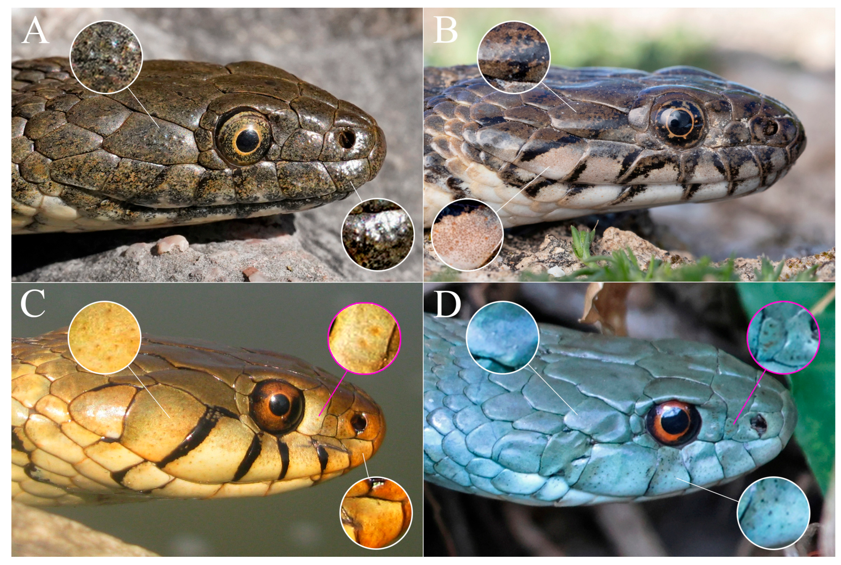
Figure 1. Portraits of in vivo European natricids in lateral view showing scale sensilla within the cephalic shields. (A) Natrix tessellata from Arnoldstein, Austria (photo by Manuel Raab); (B) Natrix maura from Lansargues, France (photo by Thibaud Fournier); (C) Natrix helvetica from Abruzzo, Italy (photo by Alessandro Paterna); (D) Natrix astreptophora from Astrurias, Spain (photo by Nacho Perez). White crops correspond to a zoom on the sensilla; magenta crops correspond to a zoom on the pitting.
Taxonomy 05 00034 g001
Figure 2. Microscopy of the cephalic shields of a Natrix tessellata preserved adult specimen: (A) close up of the dorsal shields, showing scale sensilla; (B) close up of the rostrum. From left to right, rostral scale, nasals and anterior supralabials; (C) detail of the scale sensilla within the left prefrontal shield; (D) detail of the scale sensilla within posterior supralabial and infralabial shields. Abbreviations: f, frontal; n, nasal; li, left internasal; ls, left supraocular; po, postocular; r, rostral; s1, first supralabial; s2, second supralabial; s4, fourth supralabial; s5, fifth supralabial, s6, sixth supralabial, s7, seventh supralabial; t, temporal; tr, transversal ridges.
Taxonomy 05 00034 g002
Figure 3. Microscopy of the cephalic shields of a Natrix natrix preserved adult specimen: (A) close up of the rostrum, showing small sensilla on the rostral, infralabial, internasal and prefrontal shields; (B) detail of the sensilla within the left internasal shield. Microscopy of the cephalic shields of a Natrix helvetica preserved adult specimen: (C) close up of the pitting in the nasal, loreal, preocular and supralabial shields; (D) close up of sensilla and pitting within supralabial, and postocular shields. Abbreviations: li, left internasal; n, nasal; r, rostral; ri, right internasal; p, pitting.
Taxonomy 05 00034 g003
Figure 4. Scale sensilla and pitting in the cephalic shields of Natrix helvetica: (A) portrait of an adult female from the OPHIS collection; (B) position and density of the scale sensilla (magenta) and pits (cyan); (C) detail of the rostral sensilla on the external surface of the shed of N. helvetica; (D) detail of scale sensilla and pits on the interior surface of the shed of N. helvetica. Abbreviations: l, loreal; r, rostral; s1, first supralabial; s3, third supralabial; s4, fourth supralabial; s5, fifth supralabial; so, subocular; p, pits; po, postocular; s, sensilla.
Taxonomy 05 00034 g004
Figure 5. Portraits of in vivo Old and New World natricids in lateral view, showing different patterns and types of sensilla in the cephalic shields. (A) Nerodia taxispilota from Dade County, Florida (photo by Tony Colbert); (B) Hebius craspedogaster from Ningde, China (photo by Kris Yan); (C) Rhabdophis tigrinus from Gangwon, South Korea (photo by Kim Hyun-Tae); (D) Afronatrix anoscopus from Silver Hills Concession, Liberia (photo by Alexandre Douglas Rebelo). White crops correspond to a zoom on the sensilla.
Taxonomy 05 00034 g005
Table 1. This table shows a preliminary study on the protruded sensilla among natricids. Since most of the latter have been exclusively evaluated through photographic examination (“photogr. exam.” column), where the presence of protruded sensilla has not been observed in a given genus, it does not necessarily mean that such traits may be categorically absent. Within the “protruded sensilla” column, the values reported correspond to the following: 1, slight: the protruded sensilla can be observed after careful examination, in particular with the use of macrophotography (e.g., Figure 5B); 2, distinguishable: the protruded sensilla are observable with the naked eye in live specimens and easily recognizable during photographic material examination (e.g., Figure 1B–D and Figure 5D); 3, evident: the protruded sensilla are more expressed, easily identifiable with the naked eye in live specimens and in photographic material, even when the subjects are portrayed from a distance. (e.g., Figure 1A and Figure 5C). * The genus Natrix has been assigned to Europe as the species belonging to it are mostly distributed in this continent, despite N. maura and N. tessellata also occurring in Africa and Asia, respectively. • Indicates which taxa have been investigated.
ContinentGenusProt. SensillaIn Vivo Exam.Photogr. Exam.
AsiaAmphiesma1
AsiaAspidura
AsiaAtretium1
AsiaHebius1, 2
AsiaHerpetoreas1
AsiaOpisthotropis1, 2
AsiaPseudagkistrodon2, 3
AsiaRhabdophis2, 3
AsiaSahyadriophis2
AsiaTrachischium
AsiaTrimerodytes1, 2
AsiaXenochrophis1
AfricaAfronatrix1, 2
AfricaHelophis
AfricaHydraethiops1
AfricaLimnophis1
AfricaLycognathophis1
AfricaNatriciteres1, 2
Europe *Natrix2, 3
AmericaClonophis
AmericaLiodytes1
AmericaNerodia2, 3
AmericaRegina1
AmericaStoreria
AmericaThamnophis1, 2
AmericaTropidoclonion
AmericaVirginia
Disclaimer/Publisher’s Note: The statements, opinions and data contained in all publications are solely those of the individual author(s) and contributor(s) and not of MDPI and/or the editor(s). MDPI and/or the editor(s) disclaim responsibility for any injury to people or property resulting from any ideas, methods, instructions or products referred to in the content.