Article

Disentangling the Taxonomic History of the Widespread and Overlooked Centric Diatom Stephanodiscus makarovae and Its Transfer to Cyclostephanos

1
Department of Botany, Institute of Biological Sciences, University of Rostock, Wismarsche Str. 44/45, 18051 Rostock, Germany
2
Laboratoire de Paleoecologie Aquatique (LPA), Departement de Geographie, Universite Laval, Quebec, QC G1V 0A6, Canada
3
Medical Biology and Electron Microscopy Centre, Rostock University Medical Center, Strempelstraße 14, 18057 Rostock, Germany
4
Institute of Biological Sciences, Applied Ecology and Phycology, University of Rostock, Albert-Einstein-Str. 3, 18059 Rostock, Germany
*
Authors to whom correspondence should be addressed.
Taxonomy 2021, 1(4), 425-437; https://doi.org/10.3390/taxonomy1040030
Submission received: 23 November 2021 / Revised: 2 December 2021 / Accepted: 3 December 2021 / Published: 13 December 2021

Abstract

:
Stephanodiscus makarovae, a taxon originally described from Russia, is morphologically similar to several other taxa within Cyclostephanos, namely C. invisitatus, C. delicatus and C. tholiformis. However, it has not yet been transferred into Cyclostephanos, perhaps due to the difficulty in identifying it, as its original description is available only in the Russian language. To investigate its morphology, a detailed morphological comparison of S. makarovae and C. invisitatus was done from 286 SEM micrographs of 12 monoclonal strains. We performed a three-gene phylogenetic analysis with strains from eight additional taxa to independently confirm the position of S. makarovae. The morphology of S. makarovae shows key features of the genus Cyclostephanos and this attribution is supported by the phylogeny. Here we propose the transfer of the taxon S. makarovae to Cyclostephanos, considering the morphological and molecular data. According to both the molecular and morphological data, C. delicatus has a unique position within the genus; S. makarovae and C. invisitatus are morphologically very similar but genetically distinct. Furthermore, based upon the results, it was possible to reassign the authority of the transfer of S. delicatus into Cyclostephanos.

1. Introduction

Scientific and methodical advancements in diatom taxonomy have given rise to conflicting species and generic concepts, resulting in frequent transfers. This of course also applies to centric diatoms. These difficulties are especially clear when examining the taxonomic relationships within the many genera erected in the mid-19th century, many of which have since been revised, forgotten or resurrected. In this paper, we will examine the links between four taxa derived from or still in the genus Stephanodiscus Ehrenberg 1845: Stephanodiscus makarovae Genkal 1978, Cyclostephanos invisitatus (M.H.Hohn and Hellermann) E.C.Theriot, Stoermer and Håkasson 1988, C. tholiformis Stoermer, Håkansson and Theriot 1988 and C. delicatus (Genkal) S.J.Casper and W.Scheffler 1990. All these taxa are small and morphologically quite similar.
Ehrenberg first published the genus Stephanodiscus in 1845 [1]. The type species, named by Boyer 1927 [2], is Stephanodiscus niagarae Ehrenberg. Round 1982 the authors of [3] invalidly erected the genus Cyclostephanos mainly due to the alveolar structures in some species, but this lacked a valid type reference, which was subsequently established as C. novae-zeelandiae (Cleve) in 1987 [4]. Theriot et al. 1987 [5] then highlighted the significance of the position and form of the external opening of the rimoportula to distinguish between both genera, which is inconspicuous and below the spines in Cyclostephanos and spine-like in Stephanodiscus. This was also the basis for their subsequent transfer of S. invisitatus to the genus Cyclostephanos. Shortly after in 1990, both Casper and Scheffler [6] and Håkansson and Kling [7] published, in close succession, revisions of S. delicatus to C. delicatus. Håkansson and Kling also amended the description of C. tholiformis to separate it from C. delicatus. Dreßler and Hübener 2006 [8] later attributed the transfer of C. delicatus to Casper and Scheffler and also assumed that C. tholiformis is conspecific to C. delicatus.
S. makarovae is morphologically quite similar to the aforementioned taxa. It has, however, not been transferred to the genus Cyclostephanos and has had little to no recognition in taxonomic publications outside of Russia, possibly due to the fact that the original publication [9] was written in Russian. Based on the results of this study, we will also clarify the priority for the transfer of C. delicatus to the genus Cyclostephanos. Furthermore, we will use integrative morphological and molecular data from new isolates from Canada and Europe to transfer the taxon currently known as S. makarovae to the genus Cyclostephanos.

2. Materials and Methods

2.1. Sampling, Cultivation and Electron Microscopy

Table 1 lists the strains used and their relevant metadata. Water samples of 0.5–1 L were taken from each waterbody near the surface. Single cells were isolated using an inverse microscope–micromanipulator–micropipette system and clonal cultures from these cells cultivated for at least two weeks in 0.2 µm filtered mineral water enriched with 4 mL × L−1 f/2 medium (Guillard and Ryther 1962) and 60 mg × L−1 metasilicate (Na2SiO3 × 9 H2O). Liquid cultures were maintained in 50–150 mL Erlenmeyer flasks at 14–18 °C in a light/dark photocycle of 14.5:9.5 h and moderate shaking. Finally, subsamples of each clonal culture were used for light microscopy (LM) and scanning electron microscopy (SEM) analyses. The rest of each culture was concentrated by centrifugation and the resulting pellets were resuspended with dH2O in 1.5 mL tubes and stored at −20 °C for DNA analysis.
For LM and SEM, ~5 mL of each culture were oxidized with 35% H2O2 for four to six weeks and finally the suspensions were washed by centrifugation four times with distilled water. These cleaned cell suspensions were pipetted onto coverslips and glued on aluminum stubs for SEM analysis. Prepared stubs were coated with ~25 nm Au and viewed under a ZEISS Merlin VP compact SEM.
In valve view the following parameters were recorded: undulation of the valve, maximum number of areolae per fascicle at the valve-mantle junction, total diameter, number of striae, number of marginal fultoportulae, number and position of central fultoportulae and number of rimoportulae. The striae density is given as the number of striae in 10 µm circumference [10]. Internal views additionally revealed the orientation of the labium, the number of accompanying cowlings [8,11] of the marginal and central fultoportulae (satellite pores) and the structure of the cribra.

2.2. DNA Extraction, Purification, Amplification

Genomic DNA of frozen cultures was extracted using the salt-extraction technique modified after Aljanabi and Martinez [12]. For DNA-amplification the following primer combinations were used: T16N and T24U [13] for D2 and D3 regions of the large rDNA subunit (LSU), Wawrik_for and Wawrik_rev [14] for partial rbcL, and CoxF and CoxR [15] as well as CO1_for and CO1_rev [16] for partial cox1. The respective PCR programs for the primers were implemented as described in their references.
PCR products were visualized in 1.5% agarose gel and relevant bands were cut out. Gel extraction and purification of PCR products were conducted by applying an innuPREP Gel Extraction kit (Analytik Jena, Jena, Germany). Final products were sequenced using Sanger sequencing with PCR primers as sequence primers. Sequences were edited and aligned with the software BioEdit v7.2.5 [17].
The molecular phylogeny was calculated using the software Geneious 8.1.9 (Biomatters Ltd., v. 8.1.9, Auckland, New Zealand) with the add-ons for RAxML (ML) and MrBayes (BI), respectively. Both ML and BI analyses used the GTR + G + I model with four rate categories, with the –f a option with 10,000 bootstrap replicates to calculate branch support for the best-scoring tree in ML. All analyses were conducted under random seed 12354. The following settings were used for BI: runs with four incrementally heated Metropolis-coupled Monte-Carlo Markov Chains with five million generations, burn-in 1,250,000 generations, with a subsampling frequency of 1000; heated chains = 4, temp. = 0.2; random seed = 10,464. The effective sample size (ESS) value was > 200 and the trace plot indicated convergence. Introns were ignored for the phylogenetic analyses, since they contributed to artificially high differences in the resulting trees. Final tree files were edited in Powerpoint (Microsoft Office, Standard 2013, Redmond, WA, USA). The outgroups were Lindavia sp. and Pantocsekiella ocellata.

3. Results

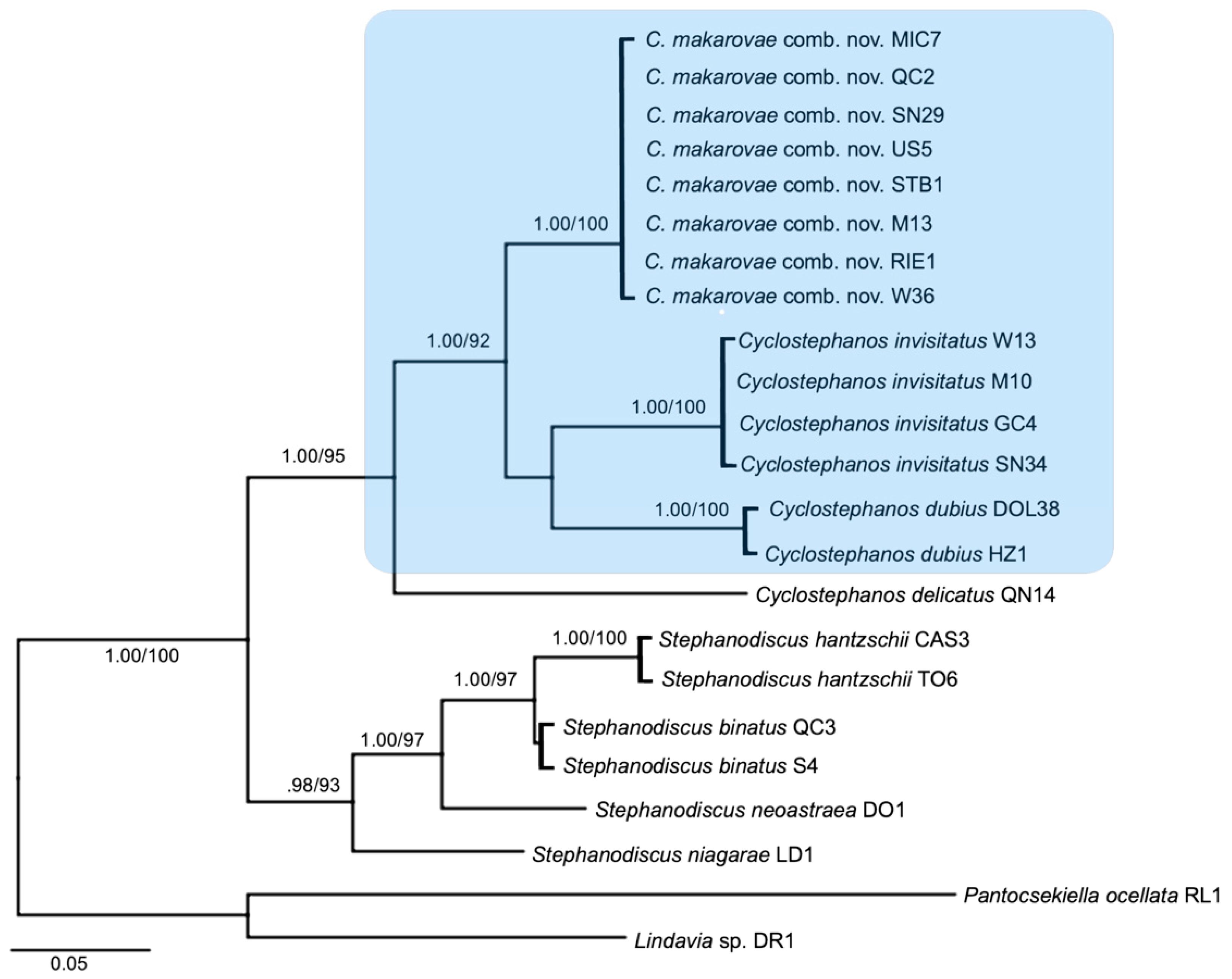
3.1. Molecular Phylogeny
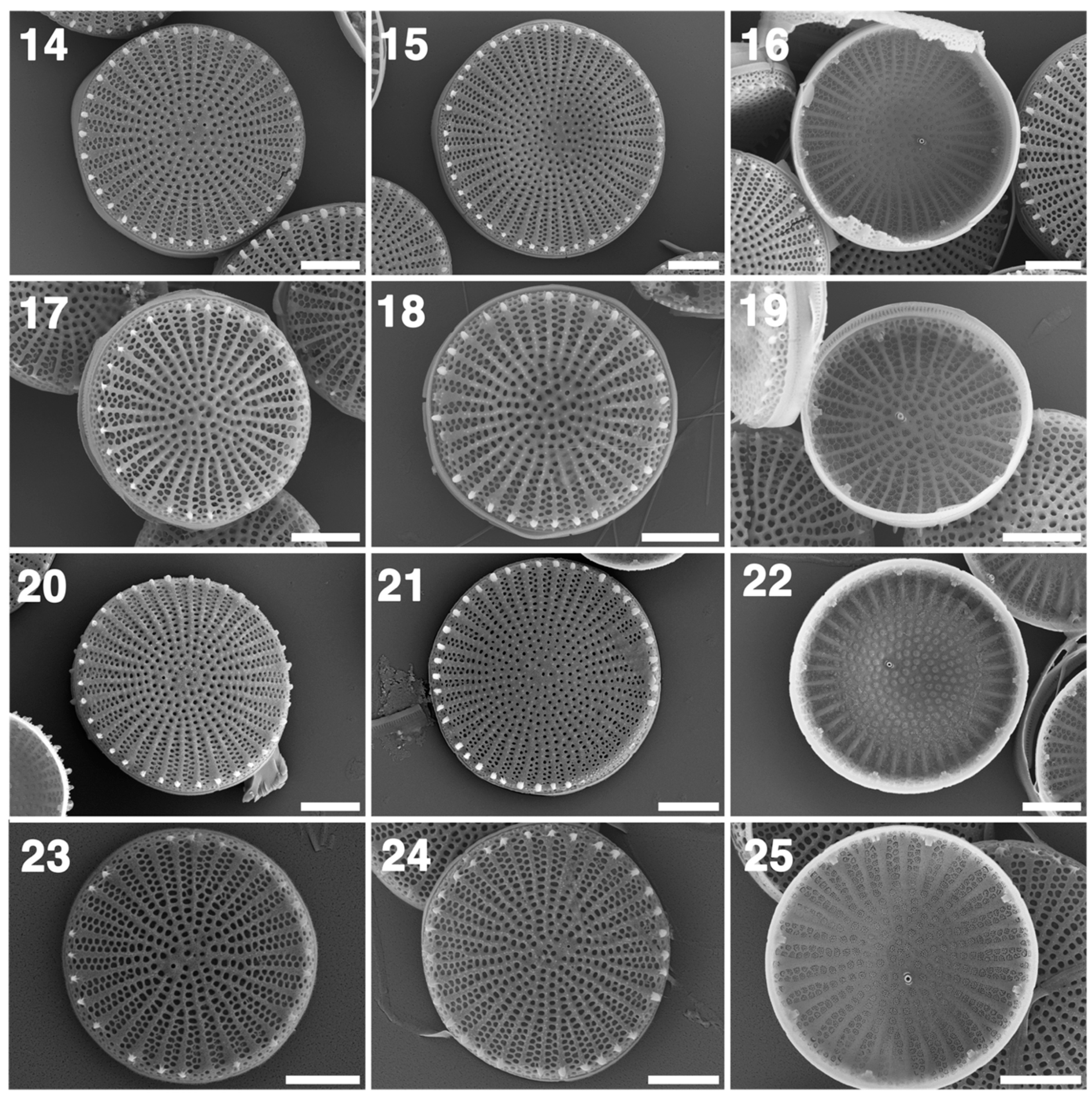
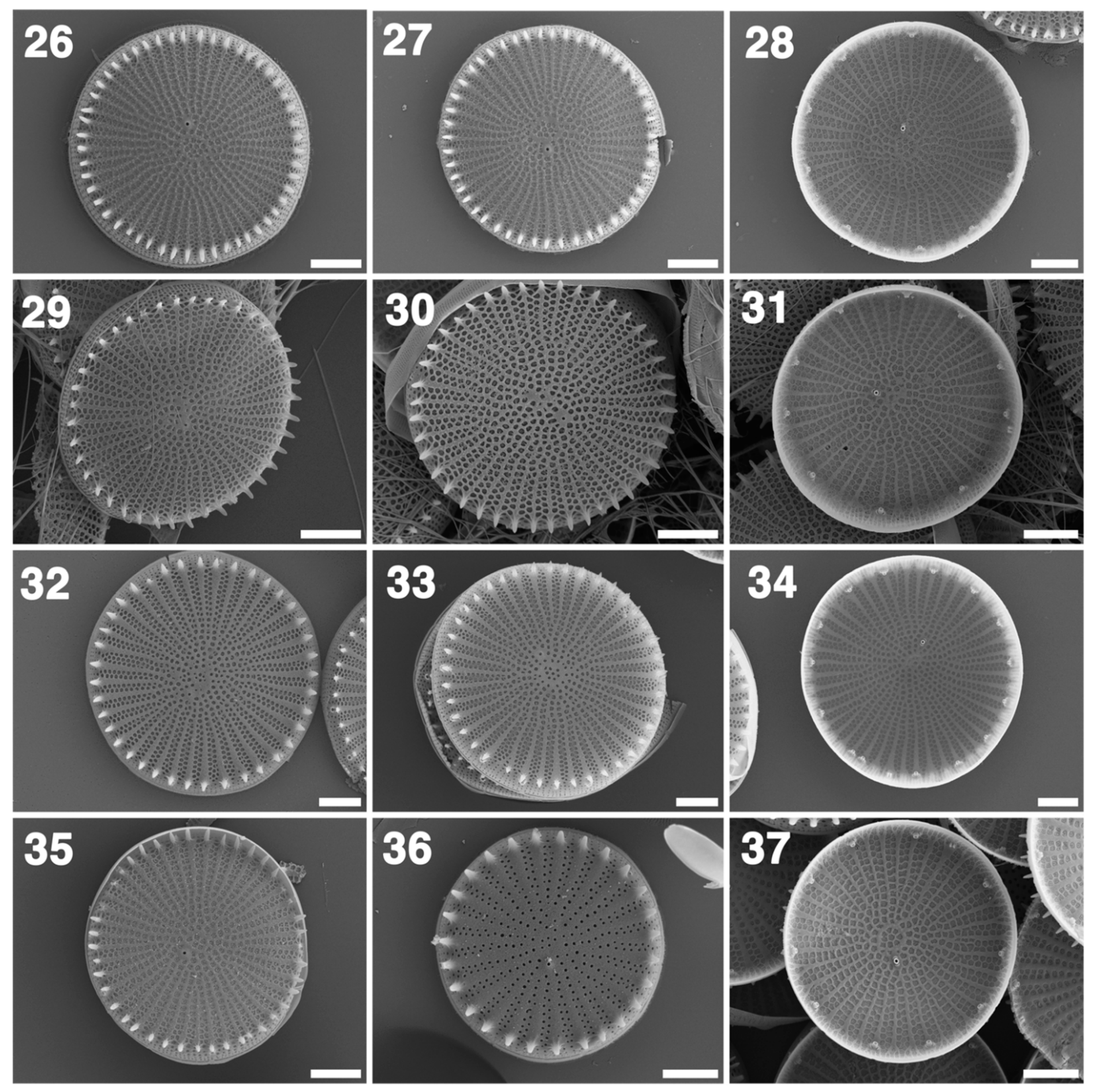
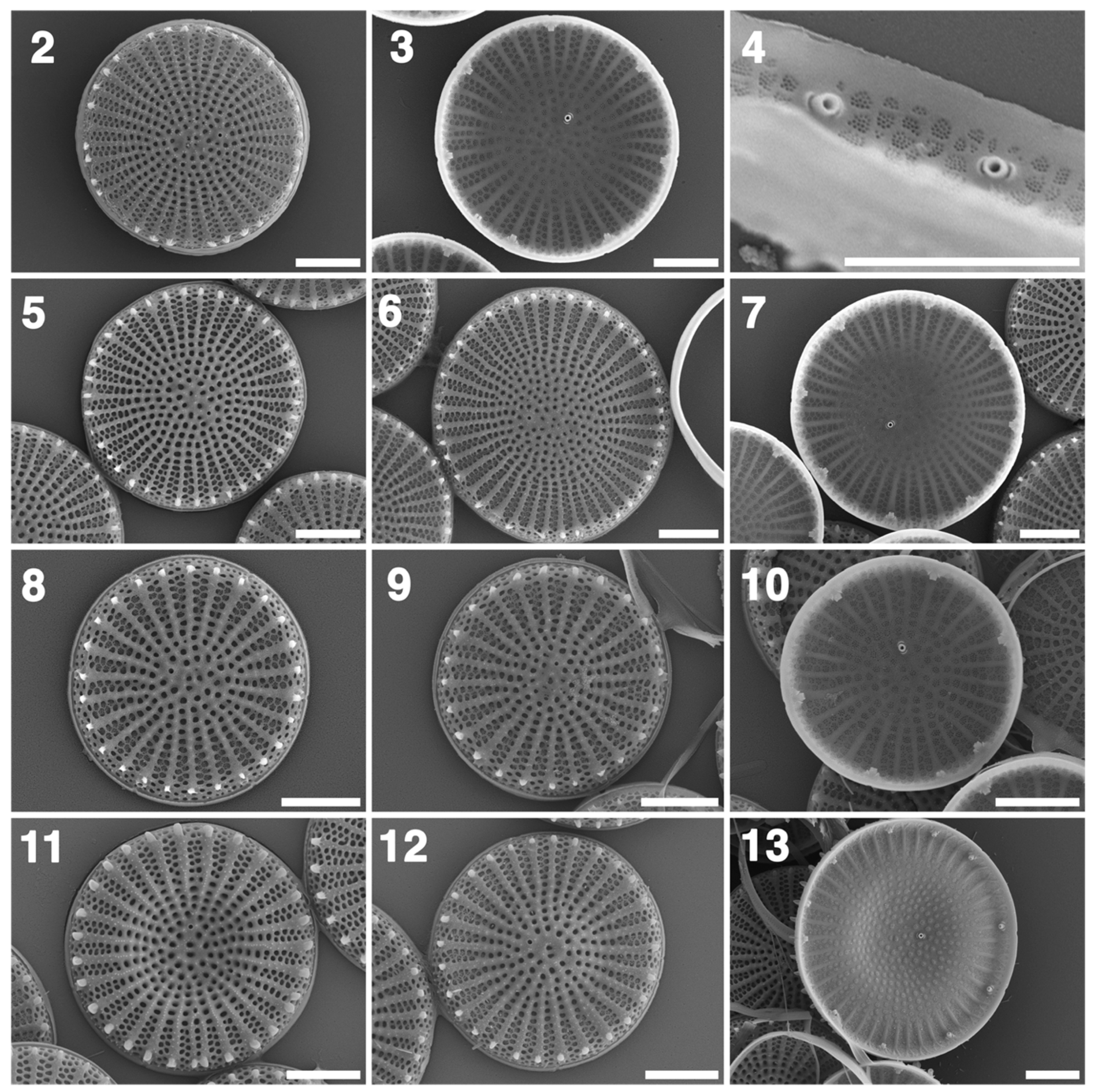
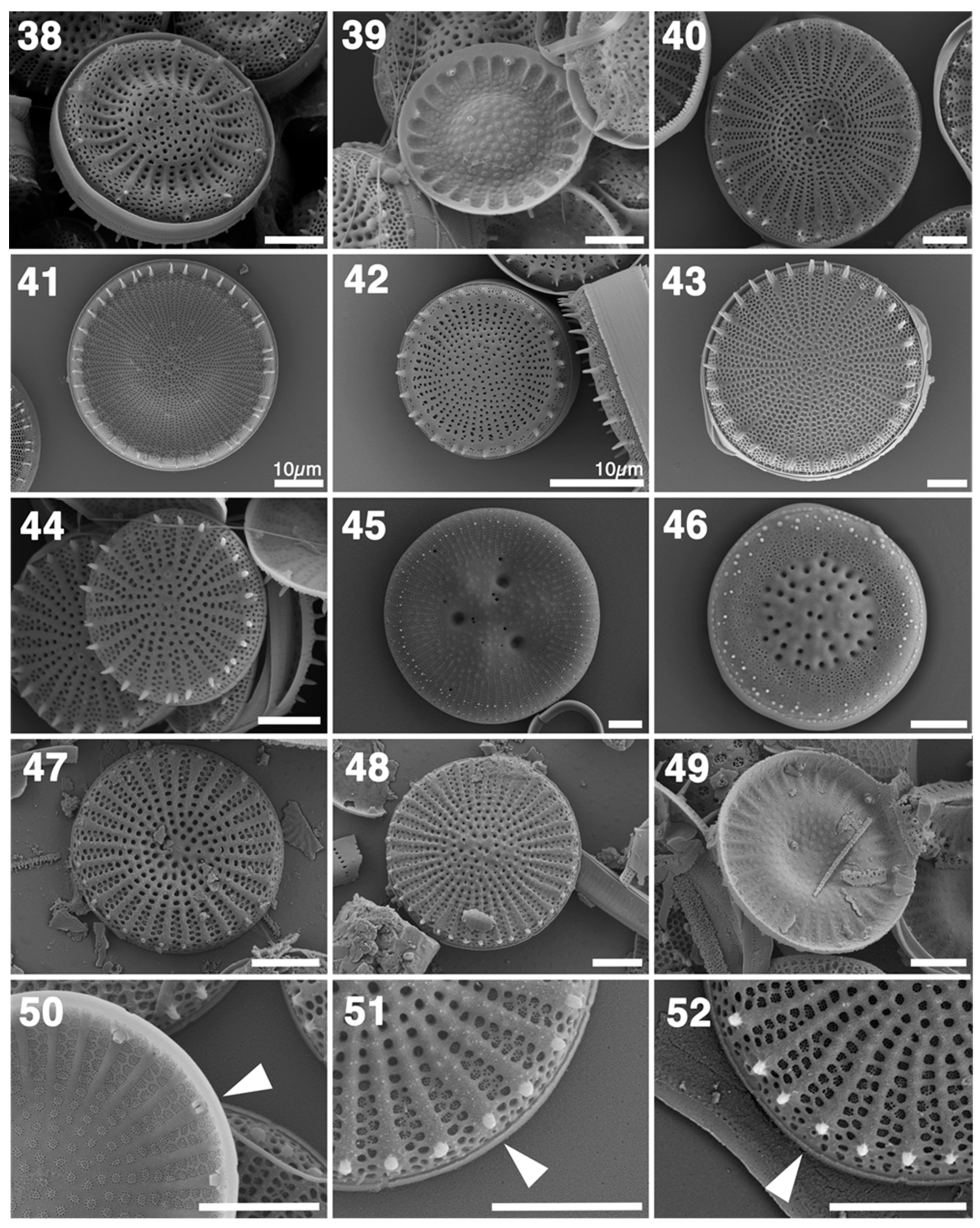
Figure 1 shows the phylogeny generated from the concatenated dataset (D2D3, rbcL and cox1). All branches had exceptionally high support, above 0.9 BI and 90 ML. Two genera, Cyclostephanos and Stephanodiscus, were recovered from the phylogeny with high support (1.00/100), with Stephanodiscus clearly separate from Cyclostephanos. S. niagarae, the type species for Stephanodiscus, fell within the Stephanodiscus clade. Cyclostephanos consisted of four clades (1.00/95)—representing C. delicatus, S. makarovae, C. invisitatus and C. dubius, with high support for all the nodes.

3.2. LM and SEM

Figure 2 and Figure 3 (225) detail the morphological structures of C. makarovae and Figure 4 (2637) that of C. invisitatus; Figure 5 (38–46) is an overview of other relevant centrics. Table 2 shows a summary of the measured morphometrical parameters of all the studied monoclonal cultures as well as references from past morphological studies on C. makarovae and C. invisitatus. The results of this study are in good agreement with the literature references (Table 2). However, looking at most parameters it becomes clear that both taxa share many morphological traits. The number of central fultoportulae, the number of areolae per fascicle at the valve-mantle junction and the density of marginal fultoportulae are basically identical between both taxa. The diameter of C. invisitatus is on average higher as well as the number of marginal fultoportulae, which is correlated to the diameter. However, the ranges of both parameters widely overlap in both taxa, leaving only the undulation and descriptive features such as the areolation pattern as distinguishing traits.
The number of satellite pores accompanying the marginal fultoportulae was always two for all strains in the Cyclostephanos clade, except for one C. delicatus strain (QN14), which always had three satellite pores (N = 15).

4. Discussion

4.1. The Transfer of S. makarovae into Cyclostephanos

Based on the results of this study we find that S. makarovae should be transferred to Cyclostephanos, as has been done with the similar taxa S. invisitatus and S. delicatus in the past. The issue first came to light when eight strains from eight distinct water bodies first identified in the light microscope as C. invisitatus were molecularly distinct from other C. invisitatus strains in culture as well as from all other sequenced Cyclostephanos strains (Figure 1). These strains had morphological characters of the genus Cyclostephanos but were yet unidentified. Upon combing the literature for a diagnosis for similar small-valved Cyclostephanos, a description of the taxon S. makarovae was discovered and fitted the strains in question well. The diagnosis was also confirmed by Genkal, who originally described the taxon (personal communication).
The concatenated three-gene phylogeny also indicates that isolates identified as S. makarovae are distinct from Stephanodiscus, including S. niagarae, the generitype of the genus (1.00/100), as well as other strains within Cyclostephanos. Furthermore, the gene locus sequences for all eight newly identified strains are identical, which strongly suggests that they belong to one taxon.
Delineating S. makarovae from Stephanodiscus requires a detailed look at their morphological features and the subsequent comparison to Cyclostephanos. S. makarovae has multiple morphological features in common with Cyclostephanos that are distinct from Stephanodiscus:
  • Two satellite pores accompanying the marginal fultoportulae (Figure 2 (2)).
  • External openings of the rimoportula are inconspicuous, not spine-like (Figure 5 (5152)).
  • Slight alveolar structures can be present (Figure 2 (13)).
This morphological separation is echoed by the phylogeny (Figure 1). We hence make the following taxonomic and nomenclatural transfer:
Cyclostephanos makarovae (Genkal) Schultz comb. nov.
Basionym:Stephanodiscus makarovae Genkal 1978: Novyi vid iz roda Stephanodiscus Ehr. (Bacillariophyta) [New species from the genus Stephanodiscus Ehr. (Bacillariophyta)]. Novosti Sistematiki Nizshykh Rasteniy 15: 11–14, 2 pls. pl. I [1]: figs. 3–6; pl. II [2]: figs. 1–6. The Latin diagnosis of S.makarovae is on pages 13–14).
Type locality: Russian Federation, Tver region, Ivankovo Reservoir on Volga River below Tver town (old name–Kalinino).
(Genkal 1978: 11–14); Type: S.I. Genkel; ix. 1972; Inst. Biol. Aquarum Internarum Acad. Sc. URSS, Borok district, Yaroslavl Oblast (Index Nominum Algarum).

4.2. Reprioritizing the Name of C. delicatus

Because S. makarovae has been largely ignored outside of Russia, it can be assumed that it has been mistaken for similar taxa in the past. The example of the transfer of S. delicatus into Cyclostephanos illustrates this.
Within a week of each other in November 1990, both Casper and Scheffler and Håkansson and Kling transferred Stephanodiscus delicatus into the genus Cyclostephanos. In 2006, Dreßler and Hübener assigned priority to the transfer made by Casper and Scheffler. However, based on examination of the valve morphology of the specimen material from Casper and Sheffler, it appears to belong to C. makarovae, not C. delicatus as the depicted valves are mostly biseriate and the marginal fultoportulae have two satellite pores (also see 4.3 Comparative morphology). The authors themselves pointed these differences out but attributed it to intraspecific variation. Genkal, in 2007, had already come to the same conclusion that their material shows C. Makarovae [18].
This resulted in a valid nomenclatural transfer but invalid in terms of the true identity of the specimen. Genkal [20] stated that S. delicatus was indeed similar to S. makarovae and both taxa have probably been confused on numerous occasions.
The feature that most clearly distinguishes the two taxa is the number of satellite pores associated with the marginal fultoportulae. C. delicatus has three while S. makarovae has two. However, C. delicatus has a confusing history concerning the number of satellite pores: The original description [21] does not mention this feature. When C. tholiformis was first described by Stoermer et al. 1987 12/3/2021 8:56:00 AM [22], three satellite pores accompanying the marginal fultoportulae of this taxon were mentioned. In 1990 Håkansson and Kling amended the species description of C. tholiformis and pointed out that in the SEM material at least two Cyclostephanos species could be found: C. delicatus with three and C. tholiformis with two satellite pores.
In 2006 Dreßler and Hübener compared environmental samples containing C. delicatus (Lake Vielbecker See, Germany) and the type of material of C. tholiformis (Lazy Lagoon, USA). In both materials they found Cyclostephanos valves with two or three satellite pores and concluded that both taxa can have two or three. However, in the case of the C. tholiformis material, this had already been called into question by Håkansson and Kling 1990 as mentioned before.
In this study we re-examined the material from Lake Vielbecker a(s used by Dreßler and Hübener in 2006). Amongst C. delicatus, C. makarovae was abundant in the samples (Figure 5 (4749)), providing a likely explanation as to why marginal fultoportulae with three, as well as two, satellite pores can be found in the material.
Our study confirms Håkansson and Kling 1990 in that all investigated valves of the C. delicatus strain (QN14) have three satellite pores associated with the marginal fultoportulae while all valves of all other Cyclostephanos taxa present in this study always had two. This is also reflected in the basal position and long branch length of C. delicatus in the phylogeny (Figure 1). In addition, we know of no other verified example of intraspecific variation of this feature within the Stephanodiscaceae. This example also highlights the difficulties working solely with environmental material, where single cells may be isolated and decontextualized from its true taxonomic placement.
Complicating things further, the specimen chosen by Håkansson and Kling appears to be the correct specimen matching the diagnosis of C. delicatus, with but not given priority in terms of publication time. To avoid further confusion, we propose to prioritize the specimen and nomenclatural transfer made by Håkansson and Kling and to designate the specimen from Casper and Scheffler to be of C. makarovae. Based on the reprioritization of the Håkansson and Kling 1990 specimen, the taxon C. delicatus will reprise its former epithet:
Cyclostephanos delicatus (S.I. Genkal) H. Kling and H. Håkansson in H. Håkansson and H. Kling 1990

4.3. Comparative Morphology

In SEM the distinction between C. makarovae and C. delicatus is relatively simple. The marginal fultoportulae of C. delicatus are accompanied by three satellite pores, while C. makarovae and C. invisitatus, like most other Cyclostephanos species, have two satellite pores (Figure 2 (4)). Moreover, C. delicatus has broader striae with 3–4 areolae per fascicle (e.g., Figure 5 (40)) whereas C. makarovae and C. invisitatus have mostly biseriate (more rarely triseriate) fascicles (Figure 2 and Figure 3 (225)). The concentric undulation is usually even more distinct in C. delicatus.
In contrast, C. invisitatus and C. makarovae are differentiated mainly by habitus. C. makarovae is mostly somewhat undulated but can also be flat (e.g., Figure 2 (2)) or distinctly undulated (e.g., Figure 2 (11)). The valve face of C. invisitatus is always flat (Figure 4 (2637)). In most cases the central area of valve faces of C. makarovae is more strongly silicified with the areolae becoming increasingly round and small towards the center, giving C. makarovae a coarser appearance in SEM images. Due to this feature, a ring-like structure, sometimes referred to as annulus, in the center of the valve may infrequently be seen in C. makarovae, but is common in C. invisitatus (e.g., Figure 4 (33)). The fascicles in both C. makarovae and C. invisitatus are typically biseriate at the valve–mantle junction. While in C. makarovae triseriate fascicles occur relatively rarely, they seem to be more frequent in C. invisitatus (Figure 4 (33)) and may even be the dominating type in some valves. Furthermore, the spines of C. makarovae are often less developed and shorter than those of C. invisitatus (e.g., Figure 3 (23)). A direct comparison of the strains in our study to the type of material is unfortunately not possible, because there is no type material suitable for SEM (Genkal, personal communication).
Another taxon that is potentially similar to these two is C. tholiformis. According to the amended description of Håkansson and Kling 1990, this taxon has, in contrast to C. delicatus, two satellite pores associated with the marginal fultoportulae. The taxon has often been confused with C. delicatus and there is generally not much data available. It could be conspecific with C. makarovae or C. invisitatus and in both cases the latter taxa would have priority. However, only judging by the micrographs available in Håkansson and Kling 1990, it could well be a distinct taxon. In contrast to C. makarovae and C. invisitatus, the striae seem to be triseriate and the interstriae are distinctly raised. More morphological and molecular data for this taxon is needed. In future investigations, morphological diagnoses should always be complemented by molecular gene sequence data for small Stephanodiscaceae.

4.4. Environmental Distribution

C. makarovae was described by Genkal 2007 [18] to be widespread throughout Russia in meso-eutrophic water bodies of 10–16 °C, with pH of 6–8.8, although valves also have been found in brackish waters of the Northern Caspian Sea. In this study the sampling sites where C. makarovae are found include Germany, Sweden, and Canada; it is thus possible that the taxon occurs throughout North America. It is especially common in the nutrient-rich water bodies of the German lowlands. Even considering that the ecological data on this taxon is still sparse, these conditions are so common in the northern hemisphere, that it is likely that C. makarovae is quite common.
C. makarovae is also found in the same water bodies as C. invisitatus (e.g., River Warnow, Lake Schweriner See, Lake Mälaren) hinting at similar ecological preferences and further complicating species delimitation.

Author Contributions

Conceptualization, K.S.; methodology, K.S., T.H., M.D., M.F., A.S. and O.J.; software, K.S. and A.T.V.; validation, K.S.; formal analysis, K.S.; investigation, K.S.; resources, T.H., M.F. and A.S.; data curation, K.S., T.H., M.D. and O.J.; writing—original draft preparation, K.S. and A.T.V.; writing—review and editing, K.S. and A.T.V.; visualization, A.T.V. All authors have read and agreed to the published version of the manuscript.

Funding

This research was funded by the State Postgraduate Scholarship Program of the State of Mecklenburg-Vorpommern. We acknowledge financial support by Deutsche Forschungsgemeinschaft and Universität Rostock within the funding programme Open Access Publishing (project number 325496636.

Institutional Review Board Statement

Not applicable.

Informed Consent Statement

Not applicable.

Data Availability Statement

Not applicable.

Acknowledgments

We would like to thank Karin Fink, Steffi Gottschalk, Sarah Haupt, Matthias Knie, Udo Nitschke, Susanne Schultz for their sampling efforts. We also thank Malin Alf for her dedicated work on the genus Cyclostephanos as part of her B.Sc. thesis. Furthermore, we thank Sergey Genkal for his correspondence on the matter.

Conflicts of Interest

The authors declare no conflict of interest.

References

  1. Ehrenberg, C.G. Neue Untersuchungen Uber Das Kleinste Leben Als Geologisches Moment. Mit Kurzer Charakteristik von 10 Neuen Genera Und 66 Neuen Arten. Ber. Uber Die Zur Bekanntm. Geeigneten Verh. Der K.-Preuss. Akad. Der Wiss. Zu Berl. 1846, 1845, 53–88. [Google Scholar]
  2. Boyer, C.S. Supplement: Synopsis of North American Diatomaceae. Part II. Naviculatae, Surirellatae. Proc. Acad. Nat. Sci. Phila. 1927, 79, 228–583. [Google Scholar]
  3. Round, F.E. Cyclostephanos—A New Genus within the Sceletonemaceae. Arch. Für Protistenkd. 1982, 125, 323–329. [Google Scholar] [CrossRef]
  4. Theriot, E.; Håkansson, H.; Kociolek, J.P.; Round, F.E.; Stoermer, E.F. Validation of the Centric Diatom Genus Name Cyclostephanos. Br. Phycol. J. 1987, 22, 345–347. [Google Scholar] [CrossRef] [Green Version]
  5. Theriot, E.; Stoermer, E.; Håkansson, H. Taxonomic interpretation of the rimoportula of freshwater genera in the centric diatom family Thalassiosiraceae. Diatom Res. 1987, 2, 251–265. [Google Scholar] [CrossRef]
  6. Casper, S.J.; Scheffler, W. Cyclostephanos delicatus (Genkal) Casper et Scheffler Comb. Nov. from Waters in the Northern Part of Germany. Arch. Für Protistenkd. 1990, 138, 304–312. [Google Scholar] [CrossRef]
  7. Håkansson, H.; Kling, H. The current status of some very small freshwater diatoms of the genera Stephanodiscus and Cyclostephanos. Diatom Res. 1990, 5, 273–287. [Google Scholar] [CrossRef]
  8. Dreßler, M.H.; Hübener, T. Morphology and Ecology of Cyclostephanos delicatus (Genkal) Casper Scheffler (Bacillariophyceae) in Comparison with C. tholiformis Stoermer, Hakansson Theriot. Nova Hedwig. 2006, 82, 409–434. [Google Scholar] [CrossRef]
  9. Genkal, S.I. New Taxa of the Genus Stephanodiscus Ehr. (Bacillariophyta). Nov. Sist. Nizshykh Rasteniy 1978, 15, 11–14. [Google Scholar]
  10. Genkal, S.I. Method of Calculation of Some Taxonomically Important Structural Elements of the Diatom Valve from Family Thalassiosiraceae Lebour Emend Hasle (Bacillariophyta). Bot. Zhurnal 1977, 62, 848–851. [Google Scholar]
  11. Theriot, E.; Serieyssol, K. Phylogenetic Systematics as a Guide to Understanding Features and Potential Morphological Characters of the Centric Diatom Family Thalassiosiraceae. Diatom Res. 1994, 9, 429–450. [Google Scholar] [CrossRef]
  12. Aljanabi, S.M.; Martinez, I. Universal and Rapid Salt-Extraction of High Quality Genomic DNA for PCR-Based Techniques. Nucleic Acids Res. 1997, 25, 4692–4693. [Google Scholar] [CrossRef] [PubMed]
  13. Hamsher, S.E.; Evans, K.M.; Mann, D.G.; Poulíčková, A.; Saunders, G.W. Barcoding Diatoms: Exploring Alternatives to COI-5P. Protist 2011, 162, 405–422. [Google Scholar] [CrossRef] [PubMed]
  14. Wawrik, B.; Paul, J.H.; Tabita, F.R. Real-Time PCR Quantification of RbcL (Ribulose-1, 5-Bisphosphate Carboxylase/Oxygenase) MRNA in Diatoms and Pelagophytes. Appl. Environ. Microbiol. 2002, 68, 3771–3779. [Google Scholar] [CrossRef] [Green Version]
  15. Iwatani, N.; Murakami, S.; Suzuki, Y. A Sequencing Protocol of Some DNA Regions in Nuclear, Chloroplastic and Mitochondrial Genomes with an Individual Colony of Thalassiosira nordenskioeldii Cleve (Bacillariophyceae). Polar Biosci. 2005, 18, 35–45. [Google Scholar]
  16. Kistenich, S.; Dreßler, M.; Zimmermann, J.; Hübener, T.; Bastrop, R.; Jahn, R. An Investigation into the Morphology and Genetics of Cyclotella comensis and Closely Related Taxa. Diatom Res. 2014, 29, 423–440. [Google Scholar] [CrossRef]
  17. Hall, T.A. BioEdit: A User-Friendly Biological Sequence Alignment Editor and Analysis Program for Windows 95/98/NT. Nucleic Acids Symp. Ser. 1999, 41, 95–98. [Google Scholar]
  18. Genkal, S.I. Morphology, Taxonomy, Ecology and Distribution of Small-Sized Stephanodiscus Species (Bacillariophyta). 2. Stephanodiscus Makarovae. Bot. Zhurnal 2007, 92, 241–248. [Google Scholar]
  19. Houk, V.; Klee, R.; Tanaka, H. Atlas of Freshwater Centric Diatoms with a Brief Key and Descriptions: Stephanodiscus, Cyclostephanos, Pliocaenicus, Hemistephanos, Stephanocostis, Mesodictyon & Spicaticribra. Stephanodiscaceae B; Czech Phycological Society: Prague, Czech Republic, 2014. [Google Scholar]
  20. Genkal, S. Morphological Variability and Taxonomy of Diatoma Tenue Ag. (Bacillariophyta). Int. J. Algae 2004, 6, 319–330. [Google Scholar] [CrossRef]
  21. Genkal, S.; Makarova, V. Diatomvye Vodorosli, Novye Dlya Planktona Kapiiiskogo i Azoskogo Morei (Bacillariophyta Planctonica Nova e Maribus Caspio et Maeotico). Nov. Sist. Nizhi. Rastenii 1985, 22, 35–37. [Google Scholar]
  22. Stoermer, E.F.; Håkansson, H.; Theriot, E.C. Cyclostephanos Species Newly Reported from North America: C. tholiformis Sp. Nov. and C. Costatilimbus Comb. Nov. Br. Phycol. J. 1987, 22, 349–358. [Google Scholar] [CrossRef] [Green Version]
Figure 1. Concatenated gene tree of LSU D2/D3, rbcL and cox1 sequences of the dataset strains. Strains within the blue box possess 2 satellite pores marginal to the fultoportulae. Left branch support values = BI, right branch support values = ML; BI/ML branch support values over 90/50 are displayed.
Taxonomy 01 00030 g001
Figure 2. (213) SEM photographs of the internal and external ultrastructure of Cyclostephanos makarovae comb. nov. (24) strain MIC7; (58) strain QC3; (810) strain SN29; (1113) strain US5. Scale bars = 2 µm.
Taxonomy 01 00030 g002
Figure 3. (1425) SEM photographs of the internal and external ultrastructure of Cyclostephanos makarovae comb. nov. (1416) strain STB1; (1719) strain M13; (2022) strain RIE1; (2325) strain W36. Scale bars = 2 µm.
Taxonomy 01 00030 g003
Figure 4. (2637) SEM photographs of the internal and external ultrastructure of Cyclostephanos invisitatus. (2628) strain W13; (2931) strain M10; (3234) strain GC4; (3537) strain SN34. Scale bars = 2 µm.
Taxonomy 01 00030 g004
Figure 5. (3852) SEM photographs of the internal and external ultrastructure of various strains. (3839) strain HZ1 Cyclostephanos dubius; (40) strain QN14 C. delicatus; (41) strain LD1 Stepha-nodiscus niagarae; (42) strain DOL10 S. neoastraea; (43) strain TO6 S. hantzschii; (44) strain QC3 S. binatus; (45) strain RL1 Pantocsekiella ocellata; (46) strain DR1 Lindavia sp.; (4749) C. makarovae from Lake Vielbecker See, Germany; (5052) arrowheads mark the position of the internal and external openings of the rimoportula in C. makarovae comb. nov., (5051) strain US5, (52) strain RIE1. Scale bars = 2 µm unless otherwise marked.
Taxonomy 01 00030 g005
Table 1. The strains used in this study and their strain ID, localities, coordinates, collector and isolator information, and GenBank accession numbers for the respective gene loci.
TaxonStrainLocalityCoordinatesCollectorIsolationD2D3 LSUrbcLcox1
Cyclostephanos makarovaeMIC7Lake Mickowsee, Germany53.70459°, 11.62498°K. SchultzK. SchultzOL436661OL493032OL628836
QC2Lake Lac Saint-Augustin, Canada46.75047°–71.39313°O. JacquesK. SchultzOL436662OL493031OL628834
SN29Lake Schweriner See, Germany53.63405°, 11.46698°S. SchultzK. SchultzOL436665OL493033OL628837
US5Lake Untersee, Germany52.94595°, 12.44498°M. DreßlerK. SchultzOL436664OL493034OL628835
STB1Lake Sternberger See, Germany53.71738°, 11.84062°K. SchultzK. SchultzOL436668OL493037OL628832
M13Lake Mälaren, Sweden59.4325°, 17.70446°S. GottschalkM. DreßlerOL436666OL493036OL628833
RIE1Lake Scharmützelsee, Germany52.21957°, 14.02552°M. KnieT. HübenerOL436663OL493038OL628831
W36River Warnow, Germany54.04522°, 12.16332°T. HübenerT. Hübener OL436667OL493035 OL628838
Cyclostephanos invisitatusW13River Warnow, Germany54.04522°, 12.16332°T. HübenerT. HübenerOL436670OL493042OL628841
M10Lake Mälaren, Sweden59.4325°, 17.70446°S. GottschalkM. DreßlerOL436671OL493041OL628840
GC4River Guadalquivir, Spain37.86848°, 4.78506°S. HauptK. SchultzOL436672OL493040OL628842
SN34Lake Schweriner See, Germany53.63405°, 11.46698°S. SchultzK. Schultz OL436669OL493039OL628839
Cyclostephanos dubiusHZ1Lake Herzsee, Austria47.24854°, 11.45539°E. RottM. DreßlerOL436674OL493045OL628843
DOL38Lake Dolgener See, Germany53.95012°, 12.24597°T. HübenerT. HübenerOL436673OL493044OL628844
Cyclostephanos delicatusQN14St. Lawrence River, Canada46.75062°–71.26741°O. JacquesK. Schultz OL436675OL493043OL628845
Stephanodiscus niagaraeLD1Lake Lac Daviault, Canada52.81044°–67.07239°O. JacquesK. Schultz OL436676OL493051OL628851
Stephanodiscus neoastraeaDOL10Lake Dolgener See, Germany53.95012°, 12.24597°T. HübenerT. Hübener OL436679OL493046OL628848
Stephanodiscus hantzschiiCAS3Lake Cambser See, Germany53.68989°, 11.53306°M. DreßlerM. DreßlerOL436677OL493049OL628846
TO6Lake Lago di Toblino, Italy46.05274°, 10.96444°K. FinkM. DreßlerOL436678OL493050OL628847
Stephanodiscus binatusQC3Lake Lac Saint-Augustin, Canada46.75047°–71.39313°O. JacquesK. Schultz OL436681OL493048OL628849
S4Lake Stechlinsee, Germany53.15284°, 13.02772°L. KrienitzT. Hübener OL436680OL493047OL628850
Pantocsekiella ocellataRL1Ross Lake, Ireland53.37257°–9.21186°U. NitzschkeT. Hübener OL436682OL493052OL628852
Lindavia sp.DR1Lago di Landro, Italy46.63116°, 12.23037°K. FinkK. Schultz OL436683OL493053OL628853
Table 2. Morphometric measurements of the strains studied and summaries, compared with literature references by Genkal 2007 for C. makarovae, Houk et al. 2014 for C. invisitatus and C. delicatus and Håkansson and Kling 1990 for C. delicatus. Und = undulation (1 = flat, 2 = somewhat undulated, 3 = distinctly undulated); Arae/Fas = maximum number of rows of areolae per fascicle at the valve-mantle-junction; D = diameter in µm; S/D = striae in 10 µm circumference [10]; N MFP = number of marginal fultoportulae; N CFP = number central fultoportulae; MFP/D = marginal fultoportulae in 10 µm circumference.
TaxonNStrainUnd.Arae/FasDS/DN MFPN CFPMFP/D
C. makarovae22MIC71–226.7–8.314.1–18.07–101 (–2)3.1–4.3
21QC21–32 (–3)6.0–8.614.3–18.05–812.5–3.8
25SN291–32 (–3)4.9–7.012.9–16.65–712.7–3.7
21US51–32 (–3)6.1–9.312.7–17.36–1012.6–3.7
22STB11–327.8–10.413.5–16.27–912.5–3.3
26M131–32 (–3)5.8–7.612.4–16.75–812.5–4.0
21RIE11–32 (–3)6.4–9.212.2–16.26–111 (–2)2.8–4.8
24W361–22 (–3)5.8–7.413.0–17.16–913.0–4.4
182all1–32 (–3)4.9–10.412.2–18.05–111 (–2)2.5–4.8
C. makarovae [18]447 2–33–1014–25
C. invisitatus28W1312 (–3)9.5–10.014.3–16.98–1112.9–3.7
36M1012–38.9–9.712.5–16.18–111 (–2)2.6–3.6
20GC412–311.3–12.210.6–15.012–161 (–2)3.2–4.4
20SN3412 (–3)8.4–9.810.2–15.57–1112.7–3.8
104all12–38.4–12.210.2–16.97–161 (–2)2.6–4.4
C. invisitatus [19] 126–189–19 1
C. delicatus [7] 2–32–46–148–20 1
C. delicatus [19]2–3(2–) 3–45–151 (–2)
Publisher’s Note: MDPI stays neutral with regard to jurisdictional claims in published maps and institutional affiliations.

