Review

Osteoporosis: Focus on Bone Remodeling and Disease Types

1
UOSD Reumatologia, Ospedale San Camillo De Lellis, 02100 Rieti, Italy
2
Molecular Medicine Department, Sapienza University of Rome, 00185 Roma, Italy
*
Author to whom correspondence should be addressed.
BioChem 2025, 5(3), 31; https://doi.org/10.3390/biochem5030031
Submission received: 12 May 2025 / Revised: 20 August 2025 / Accepted: 28 August 2025 / Published: 11 September 2025
(This article belongs to the Special Issue Feature Papers in BioChem, 2nd Edition)

Abstract

Osteoporosis is a common skeletal disease that leads to increased bone fragility, associated with increased risk of fracture and consequent significant morbidity and mortality, and is a global public health problem. It results from a chronic imbalance in bone remodeling, where bone resorption by osteoclasts exceeds bone formation by osteoblasts. Aging, hormonal changes, comorbidities, and drugs influence the process that leads to osteoporosis. In this review, we delve into the pathogenesis of primary and secondary osteoporosis after a summary of the normal physiology of bone homeostasis. Primary osteoporosis includes postmenopausal osteoporosis, driven largely by estrogen deficiency, and age-related (senile) osteoporosis, associated with reduced bone formation. An insight into male osteoporosis and osteoporosis treatment is also provided. Secondary osteoporosis can derive from underlying conditions, such as endocrine disorders, chronic inflammatory and genetic diseases, or prolonged use of glucocorticoids. Clinically, osteoporosis is often unacknowledged, underlining the importance of early risk assessment and diagnosis. A thorough understanding of the disease, its subtypes, and its underlying pathogenetic mechanisms is essential for early diagnosis and individualized treatment, all targeted to effective fracture prevention.

1. Introduction

Osteoporosis (OP) is a systemic skeletal disorder characterized by reduced bone mass and deterioration of bone microarchitecture, resulting in increased fragility and fracture risk [1,2]. Fragility fractures, especially in the proximal femur, vertebrae, and wrist, are major contributors to disability and loss of independence in older adults [2]. The global burden of OP is significant: in 2019, there were approximately 178 million new fracture cases worldwide (95% UI 162–196), a 33.4% increase since 1990 [3]. As the population ages, the incidence of OP is expected to rise further, with substantial health and economic impacts [3].
OP can have various causes: it is classified as primary (post-menopausal and senile) or secondary. Secondary OP results from underlying conditions or medications, including endocrine, genetic, and gastrointestinal diseases [1,2,3]. This review summarizes the main pathological mechanisms behind this increasingly common disease with diverse causes, after examining bone homeostasis physiology. Therapeutic strategies will also be discussed. Greater understanding of OP can improve treatment options and prevention, emphasizing early diagnosis and personalized care to address this growing health issue.

2. Bone Physiology

Key regulators of calcium-phosphate homeostasis include parathyroid hormone (PTH), calcitonin, fibroblast growth factor 23 (FGF-23), and active vitamin D (calcitriol or 1,25-dihydroxyvitamin D–1,25 (OH)2D) [4]. These hormones control calcium and phosphate metabolism by acting on the kidneys, intestine, and bones [4].
PTH, produced by the parathyroid glands, responds to hypocalcemia [5]. When serum calcium drops, the parathyroid glands secrete more PTH, which is triggered by calcium-sensing receptors (CaSR) detecting reduced serum ionized calcium [4]. PTH elevates serum calcium and phosphate levels by increasing calcium reabsorption from bones and reducing phosphate reabsorption in kidneys, while also stimulating renal synthesis of 1,25(OH)2D [6]. Elevated calcium levels inhibit PTH release through a negative-feedback loop [5]. PTH also boosts 1,25(OH)2D levels by stimulating CYP27B1 (1α-hydroxylase) and downregulating CYP24A1 (24-hydroxylase) [4].
Calcitonin, produced by thyroid parafollicular cells, responds to increased serum calcium by inhibiting osteoclast activity—reducing bone resorption—by promoting the internalization of hydrolyses within cells. In the kidneys, calcitonin decreases calcium reabsorption [7].
1,25(OH)2D promotes calcium and phosphate absorption from the intestine [4]. It is synthesized in the liver from vitamin D3 (produced from 7-dehydrocholesterol via sunlight exposure or obtained through diet and supplements) and then converted to calcitriol in the kidneys. This active form binds to vitamin D receptors in organs such as the parathyroid glands, intestines, kidneys, and bones, increasing serum calcium by absorption and release from bones. PTH and 1,25(OH)2D stimulate bone resorption by activating osteoclasts and promoting RANKL production by osteoblasts [8].
FGF23, secreted by osteocytes and stimulated by PTH, regulates phosphate elimination and influences renal synthesis of 1,25(OH)2D, forming part of the feedback loops that integrate calcium homeostasis [4,9,10,11].
The primary cells involved in bone health are osteoblasts, osteoclasts, and osteocytes. Osteoblasts originate from bone mesenchymal stem cells, deposit on bone surfaces, and produce collagen to form hydroxyapatite, leading to bone formation. They can become osteocytes by embedding into the matrix or remain as lining cells [12]. Osteocytes, residing in lacunae, regulate mineral metabolism and bone mass; their death causes skeletal fragility. Conditions such as immobilization and glucocorticoid therapy can induce osteocyte necrosis or apoptosis [13]. Osteoclasts resorb bone by secreting acids and proteases, forming trenches on trabeculae where osteoblasts subsequently synthesize new matrix. Excessive osteoclast activity can lead to bone erosion and loss, especially through the RANL/RANKL pathway [14]. Moreover, the mevalonate pathway influences bone physiology by generating intermediates like FPP and GGPP, which activate GTPases Ras and Rho, essential for osteoclast function, and produce isoprenoids that regulate osteoblast differentiation and mineralization [15,16,17].
Osteocytes secrete RANKL, which binds to its specific receptor (RANK) on osteoclasts, prompting their differentiation and activation [18]. After RANKL and RANK bind, NF-kB is activated through tumor necrosis factor receptor-related factor-6 (TRAF-6), regulating osteoclast maturation and differentiation [19]. Osteoprotegerin (OPG), a decoy receptor produced by osteoblasts, competes with RANKL to bind RANK, thus diminishing osteoclast differentiation [20] (Figure 1A).
IL-1 can induce TNFα to stimulate osteoblasts to produce GM-CSF and IL-6 and to induce osteoclast precursor differentiation [21,22], enhancing the role of inflammation in bone disruption. TNFα can also bind to TNFR-1 expressed by osteoclast precursors, activate NF-κB, JNK, p38, or ERK, and promote the differentiation of osteoclast precursors into osteoclasts [23].
Wnt protein in osteoblasts binds to LRP5/6 and Fz (frizzled) receptors, located on the osteoblast membrane. The binding stabilizes intracellular β-catenin by inhibiting the β-catenin degradation complex. Then, β-catenin can translocate into the nucleus, interact with a group of genes (TCF/LEF) encoding for transcription factors, and regulate the expression of osterix and Runx2, transcription factors for osteogenesis. When Wnt does not bind to LRP5/6 and Fz, β-catenin is phosphorylated and stays bound to its degradation complex, leading to disruption in the proteasome, thus not prompting gene transcription [24,25] (Figure 1B).
Another important pathway is that of Hedgehog (Hh), which binds to receptors on MSC membranes and activates intracellular signaling molecules, which are translocated into the nucleus to upregulate the expression of Runx2 [26].
Figure 1A,B summarize the main pathways involved in osteoclasts and osteoblasts differentiation.

3. Osteoporosis Definition and Epidemiology

Bone density values are expressed relative to a reference population in standard deviation (SD) units, with the standard being a young, healthy population. The use of SD in relation to this group is called the T-score [2]. Based on these criteria, the WHO established diagnostic thresholds: normal (T-score ≥ 1 SD), low bone mass or osteopenia (T-score between −1.0 and −2.5 SD), and osteoporosis (T-score ≤ −2.5 SD) [2]. A lower T-score indicates a higher fracture risk. WHO defines osteoporosis as affecting roughly 6.3% of men and 21.2% of women over 50 worldwide, equating to about 500 million affected individuals [27]. It can cause fractures, including vertebral and non-vertebral, like hip fractures [3]. Hip fractures carry a mortality rate of 20–24% in the first year post-fracture, with increased mortality risk persisting over five years [28]. They also lead to loss of function and independence [28]. Nearly one-third of vertebral fractures go unnoticed, with up to 29% unrecognized in Europe [28].

4. Primary Osteoporosis

Primary OP is further divided into two groups: type I, which corresponds to post-menopausal OP (50–70 years of age), and type II, age-related OP, often occurring after 70 years of age [29].
Estrogens’ actions are mediated by their receptors, ERα and Erβ, also expressed by osteocytes, osteoblasts, BMSCs, and osteoclasts. Estrogens inhibit RANKL and prompt the secretion of OPG and growth hormone; this results in inhibited osteoclast activity. Also, estrogens stimulate an osteogenic differentiation of BMSCs and regulate the number of osteoblasts. For all these actions mediated by estrogens, menopause leads to bone loss and eventually OP [30,31,32,33].
While the decline in estrogen levels initiates bone loss in postmenopausal women, deterioration of skeletal integrity continues also after the moment of menopause, thus suggesting the involvement of additional mechanisms, such as senescent osteoblasts, increased oxidative stress, and alterations in the bone remodeling milieu associated with aging. Many mechanisms have been suggested for the pathogenesis of age-related OP, almost all related to loss of cellular function or decrease in number, and finally, to cell senescence.
Cultured human bone MSCs (mesenchymal stem cells) have shown increased expression of markers related to senescence, such as β-galactosidase, suggesting that MSCs could have an impaired proliferation capacity with aging [34,35]. Zhou et al. demonstrated an increase in the number of apoptotic cells in the same MSC cultures, suggesting that increased apoptosis, and thus an age-related decline in the lifespan of cellular precursors, could be implicated in the aging of bone [35]. Contrasting data on the adipocyte differentiation are available, with some authors suggesting that there could be a preferential polarization of MSC in the sense of adipocyte, instead of osteoblast, differentiation [34].
Reactive Oxygen Species (ROS) have also been implicated in the pathogenesis of age-related OP, since ROS damage is a well-known component of tissue degeneration in the whole body [36]. In mice, oxidative stress decreases osteoblast and osteocyte lifespan [37], and ROS are also able to inhibit the Wnt/β-catenin pathway, fundamental for osteoblastogenesis, by interacting with Forkhead box transcription factors (FoxO) transcription factors. ROS promotes FoxO-mediated transcription factors, which can sequester β-catenin. This causes a diminished proliferation and differentiation of osteoblasts and their progenitors, resulting in decreased bone formation in aging [38].
Other data show an age-dependent decrease in the production of collagen in osteoblast cultures, suggesting that these cells could experience a loss of function and insufficient turnover in the aging bone [34]. Also, an insufficient response to growth factors has been demonstrated in an age-related manner, suggesting that aging osteoblastic cells from cultures are less prone to respond to anabolic pathways in the senescent bone [39]. Not only do they respond less, but they also secrete less growth factors and show an enhanced secretion of inflammatory cytokines and matrix-degrading proteases that alter the bone microenvironment [40].
Genetic senescence mechanisms have also been implicated in the pathogenesis of age-related OP. Recent evidence shows the role of telomere length in the pathogenesis of OP and its link to environmental and lifestyle elements: indeed, a network between genetic predisposition, early smoke exposure, and telomere shortening has been shown to constitute a risk for OP onset [41].
Sirtuins, a family of 7 genes (from SIRT1 to 7), are involved in a plethora of biological activities, such as mitochondrial function, inflammation, antioxidative stress, cellular senescence, and OP [42]. As for bone biology, in murine models, sirtuins regulate bone MSCs, osteoblast function, and osteogenic differentiation by interacting with β-catenin and FoxO, decreasing ROS production, and promoting the transcriptional activity of anabolic genes [42].
Data on the quantity and role of bone MSCs remain inconsistent. Autophagy is crucial for stem cell self-renewal and differentiation, but its specific effects on bone MSCs are not entirely clear. Some studies indicate that autophagy declines during osteogenic differentiation, and proper autophagy activation is necessary to facilitate this process. Regarding aging, findings on bone MSCs’ number and function are conflicting: some reports suggest an age-related decrease, while others find no change [43,44].
Growing evidence concerning the role of microRNA is available, showing a potential role of the small, single-stranded RNA in the pathogenesis of OP, with up- or downregulated microRNAs that seem to target various molecules implicated in bone physiology [45,46,47,48].

5. Male Osteoporosis

OP is often overlooked and undertreated in men because it is more common in women. However, it also occurs frequently in men, with a global prevalence of about 12%, reaching up to 20% in some regions [49,50,51]. Men experience higher morbidity and mortality from fractures, likely because these happen 5 to 10 years later than in women, often alongside older age and more comorbidities [52,53]. The lower prevalence of male OP can be explained by mechanical and hormonal factors: men generally have higher peak bone mineral density (BMD) and larger bones, due to androgen effects on periosteal bone growth. This creates a more even distribution of mechanical forces across a wider area, providing mechanical benefits. Androgens stimulate periosteal bone apposition, potentially compensating for endocortical thinning and increased cortical porosity, a key difference between aging men and women [54,55]. The structure of trabecular bone also influences male osteoporosis: men tend to have more trabeculae, and aging causes thinning rather than loss of these structures. Conversely, women experience loss of connectivity and perforation in trabeculae with age [54,55]. Androgen levels decline more gradually in men and usually at older ages; however, illness or medications inducing hypogonadism can accelerate this decline, leading to increased bone resorption, rapid bone loss, and higher fracture risk, like estrogen drops during women’s menopause [56]. Estrogen also plays a role in reaching peak bone mass, and deficiency is linked to increased bone remodeling and loss in men. Lower levels of dehydroepiandrosterone (DHEA) are associated with reduced BMD, muscle weakness, falls, and fractures in older men [57,58,59]. Other risk factors include excessive alcohol intake, chronic glucocorticoid use, vitamin D deficiency (due to diet, less sun exposure, and lower skin conversion capacity), declining muscle function causing more falls, and increased osteoclast activity coupled with inhibited osteoblasts driven by inflammatory cytokines in the elderly [60,61,62,63].

6. Secondary Osteoporosis

When OP is caused or exacerbated by other disorders or drug exposure, it is referred to as secondary OP. Identifying secondary causes of osteoporosis is critical, as the treatment depends on the underlying condition. A paragraph is also dedicated to the male’s OP, which slightly differs from the women’s OP. Table 1 presents the main causes of secondary OP.

6.1. Glucocorticoids

Glucocorticoid (GC)-induced OP is the most common secondary cause of osteoporosis; bone loss and risk of fracture increase in a dose-dependent manner within the first three to six months of assumption [64]. GC prolonged use is associated with an overall higher risk of vertebral (more frequent) and non-vertebral fractures [65]. Direct effects of GC on bone formation are mediated through upregulation of peroxisome proliferator-activated receptor gamma receptor 2 (PPARγ2) [66] and effects on the Wnt/β- catenin signaling pathway [67,68], resulting in the differentiation of precursor cells to adipocytes with respect to osteoblasts, thus leading to a decrease in bone formation. GCs also have direct effects on bone resorption: they increase the production of macrophage colony-stimulating factor (M-CSF) and RANKL and decrease the production of OPG in osteoblasts and osteocytes, increasing the number and activity of osteoclasts [69,70].
Other important factors to consider regarding GC-induced OP are an increased inflammatory state, associated with GC therapy administration, which contributes to bone loss through increased pro-resorptive cytokines [71] and reduced physical activity, increased renal and intestinal losses of calcium, and reduced production of growth hormone, insulin-like growth factor 1 (IGF1) and IGF1 binding protein (IGF-BP) as well as myopathy, with loss of muscle mass and consequent increased risk of fall [72].

6.2. Endocrine Disorders

6.2.1. Diabetes Mellitus

Diabetes mellitus (DM) can impact not only the eyes, kidneys, and heart, but also bone density and bone turnover, causing OP, known as diabetic bone disease [73]. In a cohort of patients with Type 2 DM, 35% reported bone loss, of which approximately 20% could satisfy the criteria for OP diagnosis [74].
Available data reports reduced bone density in children with Type 1 DM [75]. In adult cohorts, a decrease in BMD was predominantly observed in the femur, with a marginal diminution in vertebral BMD [76]. The hyperglycemic environment causes reduced bone turnover due to a slowed bone matrix maturation and mineralization rate [77,78,79].
Patients with either Type 1 or 2 DM have impaired bone microarchitecture, in consideration of a decrease in the number of trabeculae and morphological disorders. Trabecular thickness of the femoral head is significantly lower in patients with Type 2 DM than in non-diabetic patients [80,81,82,83].
The pathogenesis of diabetic bone disease involves three main mechanisms: first, insulin receptors on the surface of osteoblasts trigger intracellular signaling pathways that stimulate cell proliferation and the production of osteocalcin and collagen, both of which are crucial for bone tissue development [84]. Insulin enhances the expression of RUNX2, which influences bone metabolism, boosting osteoblast differentiation and maturation of the bone matrix [85]. Furthermore, it promotes IGF-1, known for its pro-anabolic impacts on osteoblasts, which speeds up collagen production and mineralization of the bone matrix [86]. These events are notably diminished in diabetic individuals. The second mechanism involves the build-up of advanced glycation end products (AGEs), which form cross-links with collagen in the bone matrix, resulting in decreased collagen elasticity and heightened bone fragility [87]. Elevated levels of extracellular free sugars adversely affect the activity of both osteoblasts and osteocytes, leading to osteoblast apoptosis [88] and stimulating osteoclast activation [89]. Lastly, the third mechanism is linked to the chronic inflammatory state associated with Type 2 DM, which promotes the lipogenic differentiation of bone marrow MSC and fat accumulation in the marrow, coupled with the release of free fatty acids and a significant number of inflammatory cytokines [90].

6.2.2. Thyroid and Parathyroid

Among endocrine disorders leading to reduced bone formation, thyroid and parathyroid dysfunction play an important role. Both hyperthyroidism and hyperparathyroidism increase bone turnover [91,92]. Primary hyperparathyroidism generally affects individuals aged more than 50 years and has a prevalence of 233 per 100,000 in women and 85 per 100,000 in men in US cohorts [93]. It is characterized by persistent hypercalcemia with an elevated or inappropriately normal PTH serum level, and it is often associated with a single gland adenoma [94] or with syndromes including multiple endocrine neoplasia (MEN) type 1, MEN type 4, MEN2A [95]. Current guidelines recommend surgery in asymptomatic patients who satisfy the following criteria: age less than 50 years, serum calcium level of >1 mg/dL or >0.25 mmol/L above the upper limit of the reference interval for total calcium (>0.12 mmol/L for ionized calcium), BMD T-score ≤−2.5 at any site, fragility fracture, glomerular filtration rate less than 60 mL/min, nephrocalcinosis, renal calculi, or high stone risk [96].

6.2.3. Hypogonadism

Several conditions associated with sexual hormone alterations are directly related to secondary OP. Estrogen deficiency is the most common, as in primary amenorrhea, premature ovarian insufficiency, or Turner syndrome: the latter is due to total or partial X chromosome monosomy that leads to gonadal dysgenesis. To note, testosterone alterations lead to OP, as in Klinefelter syndrome or androgen insensitivity syndrome, the latter caused by a lack of response to androgens due to mutations of their receptors [97].

6.2.4. Obesity

Although the association of increased fracture risk with low body mass index (BMI) is well recognized, a relationship between fracture risk and increased BMI can be identified. Many factors contribute to an increased fracture risk, such as the coexistence of comorbidities such as DM, vitamin D deficiency, inflammation and oxidative stress associated with adipose tissue, increased fall risk, altered gut microbiome, sarcopenic obesity, reduced physical activity, and obesity-induced hypogonadism in men. On the other hand, protective factors include the effect of adipose tissue–derived estrogens, which may contribute to reduced hip fracture risk [98].
Given the growth of bariatric surgery, malabsorption derived from it is becoming another common cause of secondary OP. Gastroesophageal reflux and changes in gut microbiome are other consequences of bariatric surgery that result in negative skeletal effects [99].
Bariatric surgery includes Roux-en-Y gastric bypass (RYGB) (bypassing duodenum and proximal jejunum), vertical sleeve gastrectomy, adjustable gastric band, and biliopancreatic diversion with duodenal switch (that combines both gastric reduction and extreme intestinal bypass) [100]. Longitudinal studies demonstrate declining BMD, altered cortical and trabecular microarchitecture, reduced bone strength, and increased levels of bone turnover markers in the first 6 months after surgery, that continue for more than 5 years despite stabilization of body weight [101,102,103].

6.2.5. Miscellaneous

Among other endocrine disorders associated with secondary OP, hemochromatosis, a genetic disease in which there is excessive intestinal iron absorption with subsequent intracellular iron accumulation, that leads to multiple organ dysfunction (hypogonadism, diabetes, anterior pituitary dysfunction) and consequential bone loss, can be mentioned [104]. In acromegaly, an excessive production of Growth Hormone (GH), OP is described, and it is due to the hypogonadism often associated with this disorder [105].

6.3. GI Disease

Conditions that interfere with normal intestinal absorption of calcium, phosphorus, and vitamin D can cause secondary hyperparathyroidism, stimulating osteoclastic bone resorption and resulting in bone loss. Moreover, some gastrointestinal (GI) diseases are characterized by inflammatory states in which inflammatory cytokines contribute to bone microarchitectural deterioration and are treated with GCs, which contribute to bone loss.
Celiac disease is an important cause of secondary OP: gluten ingestion and related proteins cause inflammatory injury to the mucosa of the small intestine, thus resulting in malabsorption. The most common features of celiac disease are diarrhea, steatorrhea in severe cases, and iron deficiency [97].
Age at diagnosis and time when gluten-free diet is initiated are crucial determinants of bone mass in patients with celiac disease: at diagnosis, about 80% of adults have reduced BMD, and 60% of children have growth retardation. After the initiation of a gluten-free diet, BMD often improves, although some patients have persistent osteopenia despite treatment [106]. Peripheral fractures are more frequent, especially radius fractures. Some studies also indicate a greater fracture prevalence in men and individuals with GI symptoms compared with patients with extra-intestinal or subclinical manifestations [107].
26% to 34% of patients with celiac disease reportedly have lumbar spine OP; femoral OP is less common [108,109]. Higher risk of fracture is reported in patients with celiac disease who are not following a gluten-free diet [110].
The presence of inflammatory bowel disease (IBD), i.e., Crohn’s disease (CD) or ulcerative colitis (CU), is another crucial cause of secondary OP due to malabsorption and eventual ileal resection in CD. Moreover, GC treatment is often prolonged in IBD patients [97]. The negative impact of IBD on bone health is multifactorial, resulting in lower peak bone mass and increased bone loss in adults [111]. In addition, falls risk may be increased; low muscle mass and sarcopenia were observed in 21% and 12%, respectively, among 137 IBD patients [112].
Pancreatic insufficiency associated with cholestasis and alcohol abuse should be considered in patients with bone loss [106]. Haaber et al. reported decreased bone mass in 62% of patients with chronic pancreatitis. However, among the patients included in their studies, in 79% pancreatic disease was associated with alcohol abuse; to distinguish between the effects of alcohol and pancreatic insufficiency with OP was thus not possible [113]. Children and adults with cystic fibrosis (CF) can develop exocrine pancreatic insufficiency. Notably, decreased BMD and extremely high fracture rates are also frequent complications of CF. However, patients with CF have many additional risk factors for OP, including calcium and vitamin D malabsorption, delayed puberty, reduced sex steroid production, and increased serum concentrations of cytokines secondary to chronic pulmonary infections [114,115].
Hepatic function is also crucial in bone normal structure, as the liver converts vitamin D to 25-OHD, synthesizes vitamin D transport proteins, and promotes intestinal vitamin D and calcium absorption by bile. Primary biliary cirrhosis (PBC) is associated with OP due to malabsorption of calcium, phosphorus, and vitamin D and increased urinary losses of vitamin D conjugates. Moreover, PBC is predominantly in middle-aged women, and its skeletal effects may be added to losses related to postmenopausal estrogen deficiency [106].

6.4. Transplantation

OP is one of the most common long-term complications after organ transplantation. Medications such as loop diuretics or heparin, end-stage renal or hepatic disease, and especially immunosuppressive therapy with GCs, cyclosporine A, or tacrolimus all impact bone health [116]. Since bone loss occurs immediately after transplantation, preventive therapy against OP is recommended for all patients during the early post-transplant period, regardless of BMD. A meta-analysis found that bisphosphonate treatment during the first year after solid organ transplantation reduces fractures by 47% and vertebral fractures by 76% [117].

6.5. Cancer

Osteolytic lesions, osteoporosis (OP), and osteopenia occur in about 80% of people with Multiple Myeloma (MM) [118]. Serious MM complications include pathological fractures leading to hypercalcemia, spinal cord compression, and pain, all increasing morbidity and mortality [119]. Fractures at diagnosis or later are linked to a higher risk of death [120]. Myeloma cells release cytokines and osteoclast-activating factors that promote bone loss [121]. Bisphosphonates effectively prevent lytic lesions, vertebral fractures, and bone pain [119]. Concerns have recently been raised about adverse bone effects from hormonal deprivation therapy used in prostate and breast cancer treatment. Androgen-deprivation therapy (ADT) for prostate cancer causes systemic bone loss, microarchitecture deterioration, and increased fracture risk [122]. The osteoporosis risk varies with the type, duration, and dose of ADT. Bilateral orchidectomy causes more bone loss than pharmacological ADT, which in turn causes less than GnRH ADT [123,124].
Breast cancer can induce bone loss directly through tumor effects or systemically, and via estrogen deprivation from treatments like chemotherapy, ovarian ablation, or endocrine therapy, plus natural menopause [125]. Tamoxifen, an estrogen receptor antagonist, has partial agonist effects on bone, whereas aromatase inhibitors improve disease-free survival but are associated with significant bone loss and higher fracture risk [126]. Aromatase inhibitors block androgen conversion to estrogens, resulting in serum estradiol levels much lower than in healthy postmenopausal women, leading to faster and more severe bone loss compared to natural menopause [127]. All women with breast cancer should follow OP screening and prevention guidelines similar to the general population.

6.6. Other Drugs

The link between OP, fractures, and medications is debated. Data mainly concern: Depot medroxyprogesterone acetate (DMPA), a progestogen-only contraceptive given as an intramuscular injection every 3 months, which can cause hypoestrogenism and glucocorticoid effects [128]; anti-epileptic drugs, which interfere with calcium and vitamin D metabolism, cause OP through hyponatremia, and increase fall risk [129]; selective serotonin reuptake inhibitors (SSRIs), which have opposing effects of serotonin on bone [130]; proton pump inhibitors, which reduce gastric acidity and thus calcium absorption [131]; and heparin, which stimulates bone resorption and suppresses osteoblast function [132].

6.7. Skeletal Development Disorders and Genetic Causes

Among other causes of secondary osteoporosis, we also highlight rare conditions that should be excluded when diagnosing atypical osteoporosis, especially in young patients. These include inherited skeletal development disorders such as osteogenesis imperfecta, Homocystinuria, Marfan syndrome, Ehlers-Danlos syndrome, rheumatic diseases, neurological conditions like Cerebral Palsy, spina bifida, Duchenne dystrophy, multiple sclerosis, Parkinson’s disease, and systemic mastocytosis [97,133]. Genetic factors play a significant role in osteoporosis, with many related genes identified through genome-wide association studies (GWAS) [134,135]. The latest classification of genetic skeletal disorders lists 55 conditions associated with skeletal fragility [136]. Osteogenesis imperfecta (OI), a monogenic disorder caused by mutations in extracellular matrix proteins—mainly type I collagen [137,138]—results in bone weakness due to impaired osteoblast and osteoclast activity, defective matrix mineralization, and disrupted calcium and phosphate balance. Several critical genes for early-onset osteoporosis include LRP5, where biallelic mutations cause childhood osteoporosis and blindness; heterozygous loss-of-function variants lead to milder forms [139,140]. Biallelic WNT1 mutations cause severe skeletal fragility, similar to OI type III, while heterozygous variants produce milder, later-onset cases [141,142]. PLS3, on the X chromosome, encodes Plastin3, an actin-binding protein involved in cytoskeleton remodeling, and its mutations can cause severe male osteoporosis due to its role in bone mineralization [143,144]. Recently, other monogenic osteoporosis forms have been identified, such as SGMS2, which encodes sphingomyelin synthase 2 (SMS2). Heterozygous mutations in this gene are linked to childhood fractures, hyperostotic lesions, osteoporosis, and neurological symptoms like transient facial nerve palsy [145,146]. Among genetic syndromes, Marfan syndrome is strongly associated with severe osteoporosis resulting from connective tissue alterations caused by autosomal dominant mutations in the fibrillin-1 (FBN1) gene on chromosome 15q21.1 [147]. Three new FBN1 mutations and ten FBN3 single-nucleotide polymorphisms (SNPs) have been identified [148,149]. The FBN1 gene encodes fibrillin, a glycoprotein that forms microfibrils, essential components of the suspensory ligament of the lens, and a substrate for elastin in the aorta and other tissues. Abnormal fibrillin can impair bone mineralization and alter the distribution of mechanical strain [150]. Bone mineral density (BMD) studies in children with Marfan syndrome report reductions at the femoral neck, and in women, BMD is decreased in the lumbar spine and total hip [151]. Similar BMD decreases are found in men and women at the hip and radius, demonstrating how abnormal fibrillin impacts both bone mineralization and the distribution of mechanical strain [151].

7. Treatment

The treatment of OP must primarily consider non-pharmacological strategies, whose aim is to remove all the modifiable risk factors that increase the risk of OP. These are: alcohol, smoking, low dietary calcium intake and vitamin D deficiency, low body mass index, frequent falls, and insufficient exercise [152]. Secondary OP causes must be investigated, and their treatment is part of the therapeutic strategy of this type of OP [153].
Pharmacological therapies for OP are divided into two main groups: anti-resorptive and anabolic. The first category is composed of bisphosphonates, denosumab and selective estrogen receptor modulators. Teriparatide, abaloparatide and romosozumab are anabolic drugs. To summarize, anti-resorptive preserve already existing bone, while anabolic drugs aim to restore lost bone [154].
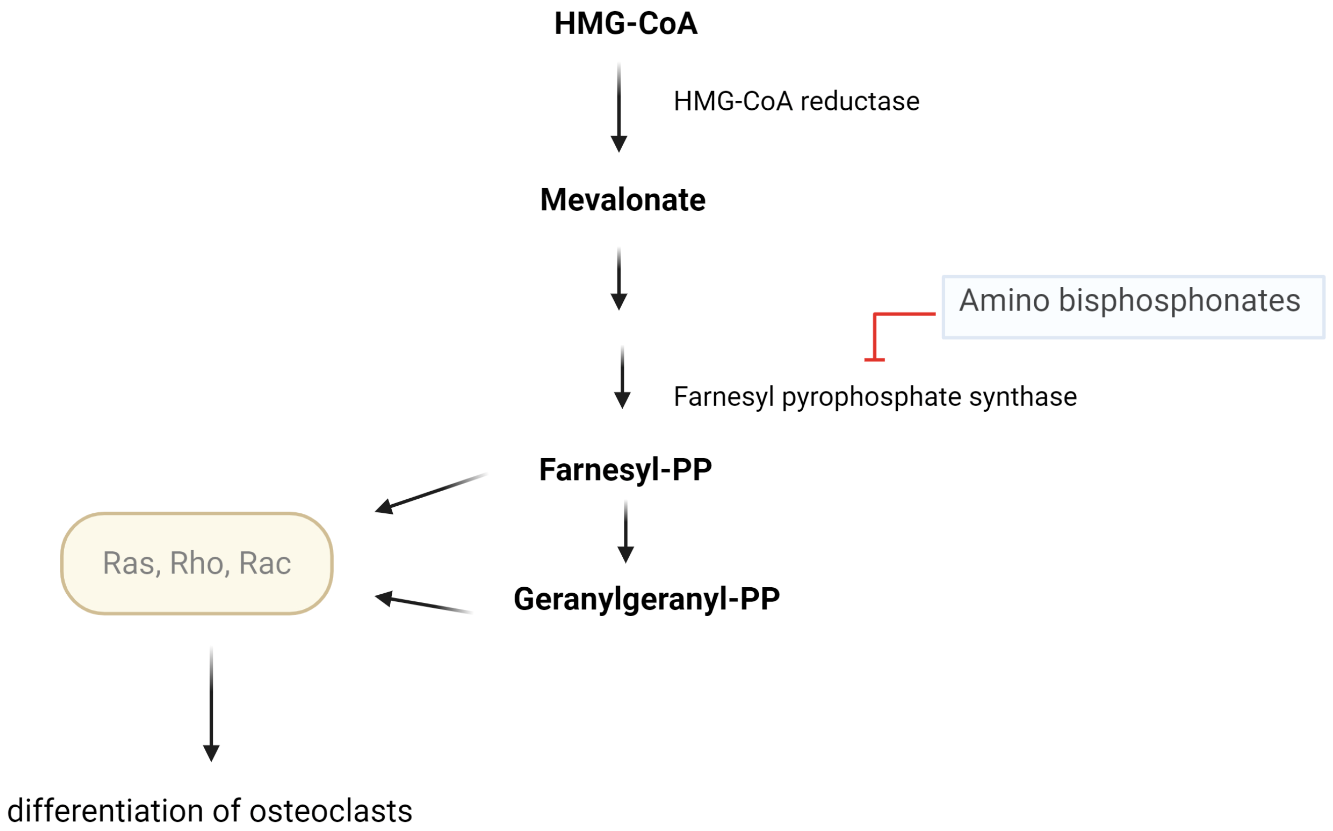
During bone resorption, bisphosphonates are taken up by osteoclasts, where they block the mevalonate pathway. The amino bisphosphonates, such as alendronate, risedronate, and zoledronate, inhibit farnesyl pyrophosphate synthase, thus impairing the resorptive activity of osteoclasts and enhancing apoptosis [154] (Figure 2).
The mevalonate pathway is responsible for the formation of prenyl pyrophosphates (farnesyl and geranylgeranyl pyrophosphates), implicated in the activation of small guanosine triphosphate (GTP)-binding proteins (GTPases—Ras, Rho, Rac, Rab). These GTPases are involved in the differentiation of the osteoclasts and hinder the activation of osteoblasts [154].
Instead, denosumab binds RANKL and prevents it from activating RANK. This mechanism impedes the stimulation of RANK by RANKL, which would prompt the differentiation of osteoclast precursor cells into mature osteoclasts. [154]. As stated in previous sections of this review, osteoclasts’ reabsorbing activity is increased by the fall in estrogen concentration after the menopause. This offers the rationale for the use of selective estrogen receptor modulators (raloxifene). [154].
Anabolic drugs can be divided into agonists of parathyroid hormone type 1 receptors (PTH1R) (teriparatide and abaloparatide) and anti-sclerostin antibodies (romosozumab). When the activation of the PTH receptor is continuous, as in primary hyperparathyroidism, when there is an excess of PTH, bone loss is predominant. When PTH receptor activators are administered in pulses, such as teriparatide and abaloparatide, bone gain is achieved [154]. Indeed, when PTH1R is transiently stimulated by PTH or PTHrP (parathyroid hormone-related protein), cAMP-dependent protein kinase A (cAMP/PKA) and phospholipase C-protein kinase C (PKC) pathways are activated. These lead to the transcription of proteins implicated in bone formation, such as OPG [155], the decoy receptor for RANKL [156]. PTHR1 activates Ras and MAPK through the PKC pathway and stimulates bone proliferation in marrow mesenchymal progenitor cells [154]. The PKC pathway is also involved in the estrogen inhibition of PTH-stimulated osteoclasts [138]. As for sclerostin, it binds to the Wnt LRP 5/6 coreceptors, thus inhibiting the canonical Wnt-signaling pathway, which normally prompts differentiation of mesenchymal stem cells to osteoblasts [155]. After the inactivation of the canonical Wnt-signaling, OPG production and bone formation are reduced [156]. Being the decoy receptor of RANKL, in normal conditions, OPG reduces the activity of RANK-RANKL, thus reducing osteoclast differentiation and bone resorption [156]. Romosozumab is an anti-sclerostin antibody: by inhibiting sclerostin, the canonical Wnt-signaling pathway can work by favoring bone formation [156]. Wnt stimulation of the LRP 5/6 and frizzled membrane co-receptor complex allows the migration of β-catenin from the cytoplasm into the nucleus, permitting protein transcription and bone anabolic effects. The same pathways prompt OPG upregulation and are inhibited by sclerostin, which, in turn, is inhibited by PTH [155]. In the FRAME trial of romosozumab, 7180 women were enrolled and randomly assigned to receive either a monthly subcutaneous dose of 210 mg of romosozumab for one year or a placebo. This was followed by denosumab administered every six months for 36 months. The study found a 73% reduction in the risk of new vertebral fractures and a 25% decrease in non-vertebral fractures in the romosozumab group. At 12 months, severe cardiovascular events occurred in 1.1% of patients in the placebo group and 1.2% in the romosozumab group, with these figures rising to 2.2% and 2.3%, respectively, at 24 months. Mortality was 0.5% in the romosozumab group compared to 0.4% in the placebo group [157]. Recent data also indicate that romosozumab induces early increases in bone formation and decreases in bone resorption, as shown by histomorphometric analysis of bone biopsies from the FRAME trial [158]. In Eriksen et al.’s study, higher osteoid and mineralized wall thickness were observed early in the romosozumab group compared to placebo (p = 0.005 and p = 0.004, respectively), demonstrating the positive effect of the anti-sclerostin antibody on bone mass and fracture risk reduction [158].

8. Conclusions

OP is a complex, multifactorial, often underdiagnosed condition that is a leading cause of morbidity and disability among aging populations, with significant socioeconomic impacts worldwide. Differentiating between primary and secondary OP is not just academically important but essential for accurate diagnosis and effective, targeted treatment. Failing to distinguish these can result in therapeutic errors, underestimating risks, and less effective interventions. Although advancements have been made in understanding the molecular mechanisms—such as specific pathways, senescence roles, and new pharmacological options like anti-sclerostin monoclonal antibodies and anabolic drugs—much remains to be performed. Emphasis is needed on prevention, early diagnosis, personalized therapy, and management of modifiable risk factors. It is crucial to detect OP early, ideally prevent its development, and address risk factors. As shown in this review, its multidisciplinary nature intersects many fields of medicine, making it a common challenge across specialties. Ultimately, managing OP requires a comprehensive, multidisciplinary approach that considers hormonal, genetic, and environmental interactions. Only through personalized, evidence-based strategies can we improve clinical outcomes and lessen the global and individual burdens of this silent, insidious skeletal disease.

Author Contributions

Conceptualization, C.C. and E.D.M.; methodology, E.D.M.; software, C.C.; validation, C.C., P.S. and E.D.M.; formal analysis, C.C., P.S. and E.D.M.; investigation, C.C., P.S. and E.D.M.; resources, C.C. and E.D.M.; data curation, C.C., P.S. and E.D.M.; writing—original draft preparation, C.C. and E.D.M.; writing—review and editing, C.C., P.S. and E.D.M.; visualization, C.C. and E.D.M.; supervision, P.S. All authors have read and agreed to the published version of the manuscript.

Funding

This research received no external funding.

Conflicts of Interest

The authors declare no conflicts of interest.

References

  1. Consensus Development Conference. Diagnosis, prophylaxis, and treatment of osteoporosis. Am. J. Med. 1993, 94, 646–650. [Google Scholar] [CrossRef] [PubMed]
  2. World Health Organization. Guidelines for Preclinical Evaluation and Clinical Trials in Osteoporosis; World Health Organization: Geneva, Switzerland, 1998; Available online: https://iris.who.int/handle/10665/42088 (accessed on 20 April 2025).
  3. Wu, A.M.; Bisignano, C.; James, S.L.; Abady, G.G.; Abedi, A.; Abu-Gharbieh, E.; Alhassan, R.K.; Alipour, V.; Alipour, J.; Asaad, M.; et al. Global, regional, and national burden of bone fractures in 204 countries and territories, 1990–2019: A systematic analysis from the global burden of disease study 2019. Lancet Healthy Longev. 2021, 2, e580–e592. [Google Scholar] [CrossRef]
  4. Murray, S.L.; Wolf, M. Calcium and Phosphate Disorders: Core Curriculum 2024. Am. J. Kidney Dis. 2024, 83, 241–256. [Google Scholar] [CrossRef]
  5. Khan, M.; Jose, A.; Sharma, S. Physiology, Parathyroid Hormone. In StatPearls; StatPearls Publishing: Treasure Island, FL, USA, 2025. [Google Scholar]
  6. Leung, E.K.Y. Parathyroid hormone. Adv. Clin. Chem. 2021, 101, 41–93. [Google Scholar]
  7. Srinivasan, A.; Wong, F.K.; Karponis, D. Calcitonin: A useful old friend. J. Musculoskelet. Neuronal Interact. 2020, 20, 600–609. [Google Scholar] [PubMed]
  8. Lung, B.E.; Komatsu, D.E.E. Calcitriol. In StatPearls; StatPearls Publishing: Treasure Island, FL, USA, 2025. [Google Scholar]
  9. Columbu, C.; Rendina, D.; Gennari, L.; Pugliese, F.; Carnevale, V.; Salcuni, A.S.; Chiodini, I.; Battista, C.; Tabacco, P.; Guarnieri, V.; et al. Phosphate metabolism in primary hyperparathyroidism: A real-life long-term study. Endocrine 2025, 88, 571–580. [Google Scholar] [CrossRef]
  10. Ho, B.B.; Bergwitz, C. FGF23 signalling and physiology. J. Mol. Endocrinol. 2021, 66, R23–R32. [Google Scholar] [CrossRef]
  11. Edmonston, D.; Grabner, A.; Wolf, M. FGF23 and klotho at the intersection of kidney and cardiovascular disease. Nat. Rev. Cardiol. 2024, 21, 11–24. [Google Scholar] [CrossRef]
  12. Blair, H.C.; Larrouture, Q.C.; Li, Y.; Lin, H.; Beer-Stoltz, D.; Liu, L.; Tuan, R.S.; Robinson, L.J.; Schlesinger, P.H.; Nelson, D.J. Osteoblast differentiation and bone matrix formation in vivo and in vitro. Tissue Eng. Part B Rev. 2017, 23, 268–280. [Google Scholar] [CrossRef]
  13. Nahian, A.; AlEssa, A.M. Histology, Osteocytes. In StatPearls; StatPearls Publishing: Treasure Island, FL, USA, 2025. [Google Scholar]
  14. Kitazawa, S.; Haraguchi, R.; Kitazawa, R. Roles of osteoclasts in pathological conditions. Pathol. Int. 2025, 75, 55–68. [Google Scholar] [CrossRef] [PubMed]
  15. Hasan, W.N.W.; Chin, K.Y.; Jolly, J.J.; Ghafar, N.A.; Soelaiman, I.N. Identifying potential therapeutics for osteoporosis by exploiting the relationship between the mevalonate pathway and bone metabolism. Endocr. Metab. Immune Disord. Drug Targets 2018, 18, 450–457. [Google Scholar] [CrossRef]
  16. Jung, J.; Park, J.S.; Chun, J.; Al-Nawas, B.; Ziebart, T.; Kwon, Y.D. Geranylgeraniol application in human osteoblasts and osteoclasts for reversal of the effect of bisphosphonates. Life 2023, 13, 1353. [Google Scholar] [CrossRef]
  17. Xu, W.; Gong, L.; Tang, W.; Lu, G. Nitrogen-containing bisphosphonate induces enhancement of OPG expression and inhibition of RANKL expression via inhibition of farnesyl pyrophosphate synthase to inhibit the osteogenic differentiation and calcification in vascular smooth muscle cells. BMC Cardiovasc. Disord. 2024, 24, 494. [Google Scholar] [CrossRef]
  18. McDonald, M.M.; Khoo, W.H.; Ng, P.Y.; Xiao, Y.; Zamerli, J.; Thatcher, P.; Kyaw, W.; Pathmanandavel, K.; Grootveld, A.K.; Moran, I.; et al. Osteoclasts recycle via osteomorphs during RANKL-stimulated bone resorption. Cell 2021, 184, 1330–1347.e13. [Google Scholar] [CrossRef]
  19. Tobeiha, M.; Moghadasian, M.H.; Amin, N.; Jafarnejad, S. RANKL/RANK/OPG pathway: A mechanism involved in exercise-induced bone remodeling. Biomed. Res. Int. 2020, 2020, 6910312. [Google Scholar] [CrossRef] [PubMed]
  20. Qin, S.; Zhang, Q.; Zhang, L. Effect of OPG gene mutation on protein expression and biological activity in osteoporosis. Exp. Ther. Med. 2017, 14, 1475–1480. [Google Scholar] [CrossRef]
  21. Wang, T.; He, C. TNF-alpha and IL-6: The link between immune and bone system. Curr. Drug Targets 2020, 21, 213–227. [Google Scholar]
  22. Amarasekara, D.S.; Yun, H.; Kim, S.; Lee, N.; Kim, H.; Rho, J. Regulation of osteoclast differentiation by cytokine networks. Immune Netw. 2018, 18, e8. [Google Scholar] [CrossRef]
  23. Marahleh, A.; Kitaura, H.; Ohori, F.; Kishikawa, A.; Ogawa, S.; Shen, W.R.; Qi, J.; Noguchi, T.; Nara, Y.; Mizoguchi, I. TNF-alpha directly enhances osteocyte RANKL expression and promotes osteoclast formation. Front. Immunol. 2019, 10, 2925. [Google Scholar] [CrossRef] [PubMed]
  24. Agostino, M.; Pohl, S. The structural biology of canonical Wnt signalling. Biochem. Soc. Trans. 2020, 48, 1765–1780. [Google Scholar] [CrossRef] [PubMed]
  25. Sawakami, K.; Robling, A.G.; Ai, M.; Pitner, N.D.; Liu, D.; Warden, S.J.; Li, J.; Maye, P.; Rowe, D.W.; Duncan, R.L.; et al. The Wnt co-receptor LRP5 is essential for skeletal mechanotransduction but not for the anabolic bone response to parathyroid hormone treatment. J. Biol. Chem. 2006, 281, 23698–23711. [Google Scholar] [CrossRef]
  26. Komori, T. Molecular mechanism of Runx2-dependent bone development. Mol. Cells 2020, 43, 168–175. [Google Scholar]
  27. Kanis, J.A.; McCloskey, E.V.; Johansson, H.; Oden, A.; Melton, L.J., 3rd; Khaltaev, N. A reference standard for the description of osteoporosis. Bone 2008, 42, 467–475. [Google Scholar] [CrossRef]
  28. Osteoporosis Foundation. Epidemiology of Osteoporosis and Fragility Fractures. Available online: https://www.osteoporosis.foundation/facts-statistics/epidemiology-of-osteoporosis-and-fragility-fractures (accessed on 20 April 2025).
  29. Patel, D.; Saxena, B. Decoding osteoporosis: Understanding the disease, exploring current and new therapies, and emerging targets. J. Orthop. Rep. 2025, 4, 100472. [Google Scholar] [CrossRef]
  30. Li, L.; Wang, Z. Ovarian aging and osteoporosis. Adv. Exp. Med. Biol. 2018, 1086, 199–215. [Google Scholar]
  31. Khosla, S.; Oursler, M.J.; Monroe, D.G. Estrogen and the skeleton. Trends Endocrinol. Metab. 2012, 23, 576–581. [Google Scholar] [CrossRef]
  32. Cauley, J.A. Estrogen and bone health in men and women. Steroids 2015, 99, 11–15. [Google Scholar] [CrossRef] [PubMed]
  33. Vilaca, T.; Eastell, R.; Schini, M. Osteoporosis in men. Lancet Diabetes Endocrinol. 2022, 10, 273–283. [Google Scholar] [CrossRef]
  34. Kassem, M.; Marie, P.J. Senescence-associated intrinsic mechanisms of osteoblast dysfunctions. Aging Cell 2011, 10, 191–197. [Google Scholar] [CrossRef]
  35. Zhou, S.; Greenberger, J.S.; Epperly, M.W.; Goff, J.P.; Adler, C.; Leboff, M.S.; Glowacki, J. Age-related intrinsic changes in human bone-marrow-derived mesenchymal stem cells and their differentiation to osteoblasts. Aging Cell 2008, 7, 335–343. [Google Scholar] [CrossRef] [PubMed]
  36. Almeida, M. Aging mechanisms in bone. Bonekey Rep. 2012, 1, 102. [Google Scholar] [CrossRef] [PubMed]
  37. Jilka, R.L.; Almeida, M.; Ambrogini, E.; Han, L.; Roberson, P.K.; Weinstein, R.S.; Manolagas, S.C. Decreased oxidative stress and greater bone anabolism in the aged, as compared to the young, murine skeleton by parathyroid hormone. Aging Cell 2010, 9, 851–867. [Google Scholar] [CrossRef] [PubMed]
  38. Manolagas, S.C.; Almeida, M. Gone with the Wnts: Beta-catenin, T-cell factor, forkhead box O, and oxidative stress in age-dependent diseases of bone, lipid, and glucose metabolism. Mol. Endocrinol. 2007, 21, 2605–2614. [Google Scholar] [CrossRef]
  39. Pfeilschifter, J.; Diel, I.; Pilz, U.; Brunotte, K.; Naumann, A.; Ziegler, R. Mitogenic responsiveness of human bone cells in vitro to hormones and growth factors decreases with age. J. Bone Miner. Res. 1993, 8, 707–717. [Google Scholar] [CrossRef]
  40. Campisi, J. Senescent cells, tumor suppression, and organismal aging: Good citizens, bad neighbors. Cell 2005, 120, 513–520. [Google Scholar] [CrossRef]
  41. Han, M.H.; Kwon, H.S.; Hwang, M.; Park, H.H.; Jeong, J.H.; Park, K.W.; Kim, E.J.; Yoon, S.J.; Yoon, B.; Jang, J.W.; et al. Association between osteoporosis and the rate of telomere shortening. Aging 2024, 16, 11151–11161. [Google Scholar] [CrossRef]
  42. Li, Q.; Cheng, J.C.; Jiang, Q.; Lee, W.Y. Role of sirtuins in bone biology: Potential implications for novel therapeutic strategies for osteoporosis. Aging Cell 2021, 20, e13301. [Google Scholar] [CrossRef]
  43. Stolzing, A.; Jones, E.; McGonagle, D.; Scutt, A. Age-related changes in human bone marrow-derived mesenchymal stem cells: Consequences for cell therapies. Mech. Ageing Dev. 2008, 129, 163–173. [Google Scholar] [CrossRef]
  44. Xing, Y.; Liu, C.; Zhou, L.; Li, Y.; Wu, D. Osteogenic effects of rapamycin on bone marrow mesenchymal stem cells via inducing autophagy. J. Orthop. Surg. Res. 2023, 18, 129. [Google Scholar] [CrossRef] [PubMed]
  45. Suarjana, I.N.; Isbagio, H.; Soewondo, P.; Rachman, I.A.; Sadikin, M.; Prihartono, J.; Malik, S.G.; Soeroso, J. The role of serum expression levels of microRNA-21 on bone mineral density in hypostrogenic postmenopausal women with osteoporosis: Study on level of RANKL, OPG, TGFβ-1, sclerostin, RANKL/OPG ratio, and physical activity. Acta Med. Indones 2019, 51, 245–252. [Google Scholar]
  46. Zuo, B.; Zhu, J.F.; Li, J.; Wang, C.D.; Zhao, X.Y.; Cai, G.Q.; Li, Z.; Peng, J.; Wang, P.; Shen, C.; et al. MicroRNA-103a functions as a mechanosensitive microRNA to inhibit bone formation through targeting Runx2. J. Bone Miner. Res. 2015, 30, 330–345. [Google Scholar] [CrossRef]
  47. Liu, H.; Yue, X.; Zhang, G. Downregulation of miR-146a inhibits osteoporosis in the jaws of ovariectomized rats by regulating the Wnt/β-catenin signaling pathway. Int. J. Mol. Med. 2021, 47, 6. [Google Scholar] [CrossRef]
  48. Trojniak, J.; Sendera, A.; Banaś-Ząbczyk, A.; Kopańska, M. The microRNAs in the pathophysiology of osteoporosis. Int. J. Mol. Sci. 2024, 25, 6240. [Google Scholar] [CrossRef]
  49. Zamani, A.; Zamani, V.; Heidari, B.; Parsian, H.; Esmaeilnejad-Ganji, S.M. Prevalence of osteoporosis with the World Health Organization diagnostic criteria in the Eastern Mediterranean Region: A systematic review and meta-analysis. Arch. Osteoporos. 2018, 13, 129. [Google Scholar] [CrossRef]
  50. Salari, N.; Ghasemi, H.; Mohammadi, L.; Behzadi, M.H.; Rabieenia, E.; Shohaimi, S.; Mohammadi, M. The global prevalence of osteoporosis in the world: A comprehensive systematic review and meta-analysis. J. Orthop. Surg. Res. 2021, 16, 609. [Google Scholar] [CrossRef]
  51. Chen, P.; Li, Z.; Hu, Y. Prevalence of osteoporosis in China: A meta-analysis and systematic review. BMC Public Health 2016, 16, 1039. [Google Scholar] [CrossRef]
  52. Bliuc, D.; Nguyen, N.D.; Milch, V.E.; Nguyen, T.V.; Eisman, J.A.; Center, J.R. Mortality risk associated with low-trauma osteoporotic fracture and subsequent fracture in men and women. J. Am. Med. Assoc. 2009, 301, 513–521. [Google Scholar] [CrossRef] [PubMed]
  53. Bandeira, L.; Bilezikian, J.P. Novel Therapies for Postmenopausal Osteoporosis. Endocrinol. Metab. Clin. N. Am. 2017, 46, 207–219. [Google Scholar] [CrossRef] [PubMed]
  54. Khosla, S.; Riggs, B.L.; Atkinson, E.J.; Oberg, A.L.; McDaniel, L.J.; Holets, M.; Peterson, J.M.; Melton, L.J., 3rd. Effects of sex and age on bone microstructure at the ultradistal radius: A population-based noninvasive in vivo assessment. J. Bone Miner. Res. 2006, 21, 124–131. [Google Scholar] [CrossRef] [PubMed]
  55. Khosla, S.; Monroe, D.G. Regulation of Bone Metabolism by Sex Steroids. Cold Spring Harb. Perspect. Med. 2018, 8, a031211. [Google Scholar] [CrossRef]
  56. Szulc, P.; Claustrat, B.; Marchand, F.; Delmas, P.D. Increased risk of falls and increased bone resorption in elderly men with partial androgen deficiency: The MINOS study. J. Clin. Endocrinol. Metab. 2003, 88, 5240–5247. [Google Scholar] [CrossRef] [PubMed]
  57. Cauley, J.A.; Ewing, S.K.; Taylor, B.C.; Fink, H.A.; Ensrud, K.E.; Bauer, D.C.; Barrett-Connor, E.; Marshall, L.; Orwoll, E.S.; Osteoporotic Fractures in Men Study (MrOS) Research Group. Sex steroid hormones in older men: Longitudinal associations with 4.5-year change in hip bone mineral density—The osteoporotic fractures in men study. J. Clin. Endocrinol. Metab. 2010, 95, 4314–4323. [Google Scholar] [CrossRef]
  58. LeBlanc, E.S.; Nielson, C.M.; Marshall, L.M.; Lapidus, J.A.; Barrett-Connor, E.; Ensrud, K.E.; Hoffman, A.R.; Laughlin, G.; Ohlsson, C.; Orwoll, E.S.; et al. The effects of serum testosterone, estradiol, and sex hormone binding globulin levels on fracture risk in older men. J. Clin. Endocrinol. Metab. 2009, 94, 3337–3346. [Google Scholar] [CrossRef] [PubMed]
  59. Kirby, D.J.; Buchalter, D.B.; Anil, U.; Leucht, P. DHEA in bone: The role in osteoporosis and fracture healing. Arch. Osteoporos. 2020, 15, 84. [Google Scholar] [CrossRef] [PubMed]
  60. D’Amelio, P.; Roato, I.; D’Amico, L.; Veneziano, L.; Suman, E.; Sassi, F.; Bisignano, G.; Ferracini, R.; Gargiulo, G.; Castoldi, F.; et al. Bone and bone marrow pro-osteoclastogenic cytokines are up-regulated in osteoporosis fragility fractures. Osteoporos. Int. 2011, 22, 2869–2877. [Google Scholar] [CrossRef]
  61. Lips, P. Vitamin D deficiency and secondary hyperparathyroidism in the elderly: Consequences for bone loss and fractures and therapeutic implications. Endocr. Rev. 2001, 22, 477–501. [Google Scholar] [CrossRef]
  62. Metter, E.J.; Conwit, R.; Tobin, J.; Fozard, J.L. Age-associated loss of power and strength in the upper extremities in women and men. J. Gerontol. Ser. A Biol. Sci. Med. Sci. 1997, 52, B267–B276. [Google Scholar] [CrossRef]
  63. Izquierdo, M.; Ibañez, J.; Gorostiaga, E.; Garrues, M.; Zúniga, A.; Antón, A.; Larriòn, J.L.; Hakkinen, K. Maximal strength and power characteristics in isometric and dynamic actions of the upper and lower extremities in middle-aged and older men. Acta Physiol. Scand. 1999, 167, 57–68. [Google Scholar] [CrossRef]
  64. Compston, J. Glucocorticoid-induced osteoporosis: An update. Endocrine 2018, 61, 7–16. [Google Scholar] [CrossRef]
  65. van Staa, T.P.; Leufkens, H.G.M.; Abenhaim, L.; Zhang, B.; Cooper, C. Oral corticosteroids and fracture risk: Relationship to daily and cumulative doses. Rheumatology 2000, 39, 1383–1389. [Google Scholar] [CrossRef]
  66. Wu, Z.; Bucher, N.L.R.; Farmer, S.R. Induction of peroxisome proliferator-activated receptor γ during the conversion of 3T3 fibroblasts into adipocytes is mediated by C/EBPβ, C/EBPγ, and glucocorticoids. Mol. Cell Biol. 1996, 16, 4128–4136. [Google Scholar] [CrossRef]
  67. Weinstein, R.S.; Jilka, R.L.; Parfitt, A.M.; Manolagas, S.C. Inhibition of osteoblastogenesis and promotion of apoptosis of osteoblasts and osteocytes by glucocorticoids—Potential mechanisms of their deleterious effects on bone. J. Clin. Investig. 1998, 102, 274–282. [Google Scholar] [CrossRef]
  68. Ohnaka, K.; Tanabe, M.; Kawate, H.; Nawata, H.; Takayanagi, R. Glucocorticoid suppresses the canonical Wnt signal in cultured human osteoblasts. Biochem. Biophys. Res. Commun. 2005, 329, 177–181. [Google Scholar] [CrossRef]
  69. Swanson, C.; Lorentzon, M.; Conaway, H.H.; Lerner, U.H. Glucocorticoid regulation of osteoclast differentiation and expression of receptor activator of nuclear factor-kappaB (NF-kappaB) ligand, osteoprotegerin, and receptor activator of NF-kappaB in mouse calvarial bones. Endocrinology 2006, 147, 3613–3622. [Google Scholar] [CrossRef]
  70. Hofbauer, L.C.; Gori, F.; Riggs, B.L.; Lacey, D.L.; Dunstan, C.R.; Spelsberg, T.C.; Khosla, S. Stimulation of osteoprotegerin ligand and inhibition of osteoprotegerin production by glucocorticoids in human osteoblasts: Potential paracrine mechanisms of glucocorticoid-induced osteoporosis. Endocrinology 1999, 140, 4382–4389. [Google Scholar] [CrossRef]
  71. Mazziotti, G.; Formenti, A.M.; Adler, R.A.; Bilezikian, J.P.; Grossman, A.; Sbardella, E.; Minisola, S.; Giustina, A. Glucocorticoid-induced osteoporosis: Pathophysiological role of GH/IGF-I and PTH/vitamin D axes, treatment options and guidelines. Endocrine 2016, 54, 603–611. [Google Scholar] [CrossRef]
  72. Sato, A.Y.; Richardson, D.; Cregor, M.; Davis, H.M.; Au, E.D.; McAndrews, K.; Zimmers, T.A.; Organ, J.M.; Peacock, M.; Plotkin, L.I.; et al. Glucocorticoids induce bone and muscle atrophy by tissue-specific mechanisms upstream of E3 ubiquitin ligases. Endocrinology 2017, 158, 664–677. [Google Scholar] [CrossRef] [PubMed]
  73. Skowrońska-Jóźwiak, E.; Lewandowski, K. Editorial: Osteoporosis secondary to endocrine disorders. Front. Endocrinol. 2023, 14, 1194241. [Google Scholar] [CrossRef] [PubMed]
  74. Xu, Y.; Wu, Q. Trends in osteoporosis and mean bone density among type 2 diabetes patients in the US from 2005 to 2014. Sci. Rep. 2021, 11, 3693. [Google Scholar] [CrossRef] [PubMed]
  75. Chen, S.C.; Shepherd, S.; McMillan, M.; McNeilly, J.; Foster, J.; Wong, S.C.; Robertson, K.J.; Ahmed, S.F. Skeletal fragility and its clinical determinants in children with type 1 diabetes. J. Clin. Endocrinol. Metab. 2019, 104, 3585–3594. [Google Scholar] [CrossRef]
  76. Shah, V.N.; Harrall, K.K.; Shah, C.S.; Gallo, T.L.; Joshee, P.; Snell-Bergeon, J.K.; Kohrt, W.M. Bone mineral density at femoral neck and lumbar spine in adults with type 1 diabetes: A meta-analysis and review of the literature. Osteoporos. Int. 2017, 28, 2601–2610. [Google Scholar] [CrossRef] [PubMed]
  77. Hofbauer, L.C.; Busse, B.; Eastell, R.; Ferrari, S.; Frost, M.; Müller, R.; Burden, A.M.; Rivadeneira, F.; Napoli, N.; Rauner, M. Bone fragility in diabetes: Novel concepts and clinical implications. Lancet Diabetes Endocrinol. 2022, 10, 207–220. [Google Scholar] [CrossRef] [PubMed]
  78. Holloway-Kew, K.L.; De Abreu, L.L.F.; Kotowicz, M.A.; Sajjad, M.A.; Pasco, J.A. Bone turnover markers in men and women with impaired fasting glucose and diabetes. Calcif. Tissue Int. 2019, 104, 599–604. [Google Scholar] [CrossRef]
  79. Kitaura, H.; Ogawa, S.; Ohori, F.; Noguchi, T.; Marahleh, A.; Nara, Y.; Pramusita, A.; Kinjo, R.; Ma, J.; Kanou, K.; et al. Effects of incretin-related diabetes drugs on bone formation and bone resorption. Int. J. Mol. Sci. 2021, 22, 6578. [Google Scholar] [CrossRef]
  80. Schacter, G.I.; Leslie, W.D. Diabetes and osteoporosis: Part I, epidemiology and pathophysiology. Endocrinol. Metab. Clin. N. Am. 2021, 50, 275–285. [Google Scholar] [CrossRef]
  81. Sheu, A.; Greenfield, J.R.; White, C.P.; Center, J.R. Assessment and treatment of osteoporosis and fractures in type 2 diabetes. Trends Endocrinol. Metab. 2022, 33, 333–344. [Google Scholar] [CrossRef]
  82. Martínez-Montoro, J.I.; García-Fontana, B.; García-Fontana, C.; Muñoz-Torres, M. Evaluation of quality and bone micro-structure alterations in patients with type 2 diabetes: A narrative review. J. Clin. Med. 2022, 11, 2206. [Google Scholar] [CrossRef] [PubMed]
  83. Sihota, P.; Yadav, R.N.; Dhaliwal, R.; Bose, J.C.; Dhiman, V.; Neradi, D.; Karn, S.; Sharma, S.; Aggarwal, S.; Goni, V.G.; et al. Investigation of mechanical, material, and compositional determinants of human trabecular bone quality in type 2 diabetes. J. Clin. Endocrinol. Metab. 2021, 106, e2271–e2289. [Google Scholar] [CrossRef]
  84. Yuan, S.; Wan, Z.H.; Cheng, S.L.; Michaëlsson, K.; Larsson, S.C. Insulin-like growth factor-1, bone mineral density, and fracture: A Mendelian randomization study. J. Clin. Endocrinol. Metab. 2021, 106, e1552–e1558. [Google Scholar] [CrossRef]
  85. Ouquerke, A.; Blulel, J. Osteoblasts and insulin: An overview. J. Biol. Regul. Homeost. Agents 2021, 35, 27–35. [Google Scholar]
  86. Govoni, K.E. Insulin-like growth factor-I molecular pathways in osteoblasts: Potential targets for pharmacological manipulation. Curr. Mol. Pharmacol. 2012, 5, 143–152. [Google Scholar] [CrossRef]
  87. Karim, L.; Bouxsein, M.L. Effect of type 2 diabetes-related non-enzymatic glycation on bone. Bone 2016, 82, 21–27. [Google Scholar] [CrossRef]
  88. Gortázar, A.R.; Ardura, J.A. Osteocytes and diabetes: Altered function of diabetic osteocytes. Curr. Osteoporos. Rep. 2020, 18, 796–802. [Google Scholar] [CrossRef]
  89. Kalaitzoglou, E.; Popescu, I.; Bunn, R.C.; Fowlkes, J.L.; Thrailkill, K.M. Effects of type 1 diabetes on osteoblasts, osteocytes, and osteoclasts. Curr. Osteoporos. Rep. 2016, 14, 310–319. [Google Scholar] [CrossRef] [PubMed]
  90. Palermo, A.; D’Onofrio, L.; Buzzetti, R.; Manfrini, S.; Napoli, N. Pathophysiology of bone fragility in patients with diabetes. Calcif. Tissue Int. 2017, 100, 122–132. [Google Scholar] [CrossRef] [PubMed]
  91. Skowrońska-Jóźwiak, E.; Krawczyk-Rusiecka, K.; Lewandowski, K.C.; Adamczewski, Z.; Lewiński, A. Successful treatment of thyrotoxicosis is accompanied by a decrease in serum sclerostin levels. Thyroid Res. 2012, 5, 14. [Google Scholar] [CrossRef] [PubMed]
  92. Skowrońska-Jóźwiak, E.; Lewandowski, K.C.; Adamczewski, Z.; Krawczyk-Rusiecka, K.; Lewiński, A. Mechanisms of normalisation of bone metabolism during recovery from hyperthyroidism: Potential role for sclerostin and parathyroid hormone. Int. J. Endocrinol. 2015, 2015, 948384. [Google Scholar] [CrossRef]
  93. Yeh, M.W.; Ituarte, P.H.; Zhou, H.C.; Nishimoto, S.; Liu, I.L.A.; Harari, A.; Haigh, P.I.; Adams, A.L. Incidence and prevalence of primary hyperparathyroidism in a racially mixed population. J. Clin. Endocrinol. Metab. 2013, 98, 1122–1129. [Google Scholar] [CrossRef]
  94. Pallan, S.; Rahman, M.O.; Khan, A.A. Diagnosis and management of primary hyperparathyroidism. BMJ 2012, 344, e1013. [Google Scholar] [CrossRef]
  95. Thakker, R.V. Genetics of parathyroid tumours. J. Intern. Med. 2016, 280, 574–583. [Google Scholar] [CrossRef]
  96. Bilezikian, J.P.; Brandi, M.L.; Eastell, R.; Silverberg, S.J.; Udelsman, R.; Marcocci, C.; Potts, J.T., Jr. Guidelines for the management of asymptomatic primary hyperparathyroidism: Summary statement from the Fourth International Workshop. J. Clin. Endocrinol. Metab. 2014, 99, 3561–3569. [Google Scholar] [CrossRef]
  97. Stein, E.; Shane, E. Secondary osteoporosis. Endocrinol. Metab. Clin. N. Am. 2003, 32, 115–134. [Google Scholar] [CrossRef] [PubMed]
  98. Lespessailles, E.; Paccou, J.; Javier, R.M.; Thomas, T.; Cortet, B.; GRIO Scientific Committee. Obesity, bariatric surgery, and fractures. J. Clin. Endocrinol. Metab. 2019, 104, 4756–4768. [Google Scholar] [CrossRef] [PubMed]
  99. Gagnon, C.; Schafer, A.L. Bone health after bariatric surgery. J. Bone Miner. Res. Plus 2018, 2, 121–133. [Google Scholar] [CrossRef] [PubMed]
  100. Mechanick, J.I.; Apovian, C.; Brethauer, S.; Garvey, W.T.; Joffe, A.M.; Kim, J.; Kushner, R.F.; Lindquist, R.; Pessah-Pollack, R.; Seger, J.; et al. Clinical practice guidelines for the perioperative nutrition, metabolic, and nonsurgical support of patients undergoing bariatric procedures—2019 update: Cosponsored by American Association of Clinical Endocrinologists/American College of Endocrinology, The Obesity Society, American Society for Metabolic & Bariatric Surgery, Obesity Medicine Association, and American Society of Anesthesiologists. Surg. Obes. Relat. Dis. 2020, 16, 175–247. [Google Scholar]
  101. Schafer, A.L.; Kazakia, G.J.; Vittinghoff, E.; Stewart, L.; Rogers, S.J.; Kim, T.Y.; Carter, J.T.; Posselt, A.M.; Pasco, C.; Shoback, D.M.; et al. Effects of gastric bypass surgery on bone mass and microarchitecture occur early and particularly impact postmenopausal women. J. Bone Miner. Res. 2018, 33, 975–986. [Google Scholar] [CrossRef]
  102. Lindeman, K.G.; Greenblatt, L.B.; Rourke, C.; Bouxsein, M.L.; Finkelstein, J.S.; Yu, E.W. Longitudinal 5-year evaluation of bone density and microarchitecture after Roux-en-Y gastric bypass surgery. J. Clin. Endocrinol. Metab. 2018, 103, 4104–4112. [Google Scholar] [CrossRef]
  103. Hansen, S.; Jørgensen, N.R.; Hermann, A.P.; Støving, R.K. Continuous decline in bone mineral density and deterioration of bone microarchitecture 7 years after Roux-en-Y gastric bypass surgery. Eur. J. Endocrinol. 2020, 182, 303–311. [Google Scholar] [CrossRef]
  104. Diamond, T.; Stiel, D.; Posen, S. Osteoporosis in hemochromatosis: Iron excess, gonadal deficiency, or other factors? Ann. Intern. Med. 1989, 110, 430–436. [Google Scholar] [CrossRef]
  105. Schneider, A.; Shane, E. Osteoporosis secondary to illnesses and medications. In Osteoporosis; Marcus, R., Feldman, D., Kelsey, J., Eds.; Academic Press: San Diego, CA, USA, 2001; pp. 303–327. [Google Scholar]
  106. Bikle, D. Osteoporosis in gastrointestinal, pancreatic, and hepatic diseases. In Osteoporosis; Marcus, R., Feldman, D., Kelsey, J., Eds.; Academic Press: San Diego, CA, USA, 2001; pp. 237–258. [Google Scholar]
  107. Sánchez, M.I.; Mohaidle, A.; Baistrocchi, A.; Matoso, D.; Vazquez, H.; Gonzalez, A.; Mazure, R.; Maffei, E.; Ferrari, G.; Smecuol, E.; et al. Risk of fracture in celiac disease: Gender, dietary compliance, or both? World J. Gastroenterol. 2011, 17, 3035–3042. [Google Scholar] [CrossRef]
  108. Pritchard, L.; Wilson, S.; Griffin, J.; Pearce, G.; Murray, I.A.; Lewis, S. Prevalence of reduced bone mineral density in adults with coeliac disease—Are we missing opportunities for detection in patients below 50 years of age? Scand. J. Gastroenterol. 2018, 53, 1433–1436. [Google Scholar] [CrossRef] [PubMed]
  109. Meyer, D.; Stavropolous, S.; Diamond, B.; Shane, E.; Green, P.H. Osteoporosis in a North American adult population with celiac disease. Am. J. Gastroenterol. 2001, 96, 112–119. [Google Scholar] [CrossRef]
  110. Vazquez, H.; Mazure, R.; Gonzalez, D.; Flores, D.; Pedreira, S.; Niveloni, S.; Smecuol, E.; Maurino, E.; Bai, J.C. Risk of fractures in celiac disease patients: A cross-sectional, case-control study. Am. J. Gastroenterol. 2000, 95, 183–189. [Google Scholar] [CrossRef]
  111. Hernandez, C.J.; Guss, J.D.; Luna, M.; Goldring, S.R. Links between the microbiome and bone. J. Bone Miner. Res. 2016, 31, 1638–1646. [Google Scholar] [CrossRef]
  112. Bryant, R.V.; Ooi, S.; Schultz, C.G.; Goess, C.; Grafton, R.; Hughes, J.; Lim, A.; Bartholomeusz, F.D.; Andrews, J.M. Low muscle mass and sarcopenia: Common and predictive of osteopenia in inflammatory bowel disease. Aliment. Pharmacol. Ther. 2015, 41, 895–906. [Google Scholar] [CrossRef] [PubMed]
  113. Haaber, A.B.; Rosenfalck, A.M.; Hansen, B.; Hilsted, J.; Larsen, S. Bone mineral metabolism, bone mineral density, and body composition in patients with chronic pancreatitis and pancreatic exocrine insufficiency. Int. J. Pancreatol. 2000, 27, 21–27. [Google Scholar] [CrossRef]
  114. Stoltz, D.A.; Meyerholz, D.K.; Welsh, M.J. Origins of cystic fibrosis lung disease. N. Engl. J. Med. 2015, 372, 351–362. [Google Scholar] [CrossRef]
  115. Paccou, J.; Zeboulon, N.; Combescure, C.; Gossec, L.; Cortet, B. The prevalence of osteoporosis, osteopenia, and fractures among adults with cystic fibrosis: A systematic literature review with meta-analysis. Calcif. Tissue Int. 2010, 86, 1–7. [Google Scholar] [CrossRef]
  116. Ebeling, P.R. Transplantation osteoporosis. In Primer on the Metabolic Bone Diseases and Disorders of Mineral Metabolism, 9th ed.; John, P., Bilezikian, J.W., Eds.; Wiley & Sons, Inc.: Hoboken, NJ, USA, 2019. [Google Scholar]
  117. Stein, E.M.; Ortiz, D.; Jin, Z.; McMahon, D.J.; Shane, E. Prevention of fractures after solid organ transplantation: A meta-analysis. J. Clin. Endocrinol. Metab. 2011, 96, 3457–3465. [Google Scholar] [CrossRef]
  118. Rajkumar, S.V.; Dimopoulos, M.A.; Palumbo, A.; Blade, J.; Merlini, G.; Mateos, M.V.; Kumar, S.; Hillengass, J.; Kastritis, E.; Richardson, P.; et al. International Myeloma Working Group updated criteria for the diagnosis of multiple myeloma. Lancet Oncol. 2014, 15, e538–e548. [Google Scholar] [CrossRef] [PubMed]
  119. Thorsteinsdottir, S.; Gislason, G.; Aspelund, T.; Sverrisdottir, I.; Landgren, O.; Turesson, I.; Bjorkholm, M.; Kristinsson, S.Y. Fractures and survival in multiple myeloma: Results from a population-based study. Haematologica 2020, 105, 1067–1073. [Google Scholar] [CrossRef]
  120. Nador, G.; Ramasamy, K.; Panitsas, F.; Pratt, G.; Sadler, R.; Javaid, M.K. Testing and management for monoclonal gammopathy of uncertain significance and myeloma patients presenting with osteoporosis and fragility fractures. Rheumatology 2019, 58, 1142–1153. [Google Scholar] [CrossRef] [PubMed]
  121. Kumar, S.K.; Rajkumar, V.; Kyle, R.A.; van Duin, M.; Sonneveld, P.; Mateos, M.V.; Gay, F.; Anderson, K.C. Multiple myeloma. Nat. Rev. Dis. Primers 2017, 3, 17046. [Google Scholar] [CrossRef]
  122. Hamilton, E.J.; Ghasem-Zadeh, A.; Gianatti, E.; Lim-Joon, D.; Bolton, D.; Zebaze, R.; Seeman, E.; Zajac, J.D.; Grossmann, M. Structural decay of bone microarchitecture in men with prostate cancer treated with androgen deprivation therapy. J. Clin. Endocrinol. Metab. 2010, 95, E456–E463. [Google Scholar] [CrossRef]
  123. Ng, H.S.; Koczwara, B.; Roder, D.; Vitry, A. Development of comorbidities in men with prostate cancer treated with androgen deprivation therapy: An Australian population-based cohort study. Prostate Cancer Prostatic Dis. 2018, 21, 403–410. [Google Scholar] [CrossRef]
  124. Lassemillante, A.C.; Doi, S.A.; Hooper, J.D.; Prins, J.B.; Wright, O.R. Prevalence of osteoporosis in prostate cancer survivors: A meta-analysis. Endocrine 2014, 45, 370–381. [Google Scholar] [CrossRef] [PubMed]
  125. Milat, F.; Vincent, A.J. Management of bone disease in women after breast cancer. Climacteric 2015, 18, 47–55. [Google Scholar] [CrossRef]
  126. Kim, D.; Oh, J.; Lee, H.S.; Jeon, S.; Park, W.C.; Yoon, C.I. Association between tamoxifen and incidence of osteoporosis in Korean patients with ductal carcinoma in situ. Front. Oncol. 2024, 13, 1236188. [Google Scholar] [CrossRef]
  127. Coates, A.S.; Keshaviah, A.; Thürlimann, B.; Mouridsen, H.; Mauriac, L.; Forbes, J.F.; Paridaens, R.; Castiglione-Gertsch, M.; Gelber, R.D.; Colleoni, M.; et al. Five years of letrozole compared with tamoxifen as initial adjuvant therapy for postmenopausal women with endocrine-responsive early breast cancer: Update of study BIG 1-98. J. Clin. Oncol. 2007, 25, 486–492. [Google Scholar] [CrossRef]
  128. Hadji, P.; Colli, E.; Regidor, P.A. Bone health in estrogen-free contraception. Osteoporos. Int. 2019, 30, 2391–2400. [Google Scholar] [CrossRef] [PubMed]
  129. Diemar, S.S.; Sejling, A.S.; Eiken, P.; Andersen, N.B.; Jørgensen, N.R. An explorative literature review of the multifactorial causes of osteoporosis in epilepsy. Epilepsy Behav. 2019, 100, 106511. [Google Scholar] [CrossRef]
  130. Ducy, P.; Karsenty, G. The two faces of serotonin in bone biology. J. Cell Biol. 2010, 191, 7–13. [Google Scholar] [CrossRef]
  131. Poly, T.N.; Islam, M.M.; Yang, H.C.; Wu, C.C.; Li, Y.J. Proton pump inhibitors and risk of hip fracture: A meta-analysis of observational studies. Osteoporos. Int. 2019, 30, 103–114. [Google Scholar] [CrossRef]
  132. Hosein-Woodley, R.; Hirani, R.; Issani, A.; Hussaini, A.S.; Stala, O.; Smiley, A.; Etienne, M.; Tiwari, R.K. Beyond the surface: Uncovering secondary causes of osteoporosis for optimal management. Biomedicines 2024, 12, 2558. [Google Scholar] [CrossRef] [PubMed]
  133. Ebeling, P.R.; Nguyen, H.H.; Aleksova, J.; Vincent, A.J.; Wong, P.; Milat, F. Secondary osteoporosis. Endocr. Rev. 2022, 43, 240–313. [Google Scholar] [CrossRef]
  134. Trajanoska, K.; Morris, J.A.; Oei, L.; Zheng, H.F.; Evans, D.M.; Kiel, D.P.; Ohlsson, C.; Richards, J.B.; Rivadeneira, F. Assessment of the genetic and clinical determinants of fracture risk: Genome-wide association and mendelian randomisation study. BMJ 2018, 362, k3225. [Google Scholar] [CrossRef]
  135. Formosa, M.M.; Bergen, D.J.M.; Gregson, C.L.; Maurizi, A.; Kämpe, A.; Garcia Giralt, N.; Zhou, W.; Grinberg, D.; Ovejero Crespo, D.; Zillikens, M.C.; et al. A roadmap to gene discoveries and novel therapies in monogenic low and high bone mass disorders. Front. Endocrinol. 2021, 12, 709711. [Google Scholar] [CrossRef]
  136. Unger, S.; Ferreira, C.R.; Mortier, G.R.; Ali, H.; Bertola, D.R.; Calder, A.; Cohn, D.H.; Cormier Daire, V.; Girisha, K.M.; Hall, C.; et al. Nosology of genetic skeletal disorders: 2023 revision. Am. J. Med. Genet. A 2023, 191, 1164–1209. [Google Scholar] [CrossRef]
  137. Gistelinck, C.; Weis, M.; Rai, J.; Schwarze, U.; Niyazov, D.; Song, K.M.; Byers, P.H.; Eyre, D.R. Abnormal bone collagen cross-linking in osteogenesis imperfecta/Bruck syndrome caused by compound heterozygous PLOD2 mutations. J. Bone Miner. Res. Plus 2021, 5, e10454. [Google Scholar] [CrossRef] [PubMed]
  138. Jovanovic, M.; Guterman Ram, G.; Marini, J.C. Osteogenesis Imperfecta: Mechanisms and signaling pathways connecting classical and rare OI types. Endocr. Rev. 2022, 43, 61–90. [Google Scholar] [CrossRef] [PubMed]
  139. Baron, R.; Kneissel, M. WNT signaling in bone homeostasis and disease: From human mutations to treatments. Nat. Med. 2013, 19, 179–192. [Google Scholar] [CrossRef]
  140. Gong, Y.; Slee, R.B.; Fukai, N.; Rawadi, G.; Roman Roman, S.; Reginato, A.M.; Wang, H.; Cundy, T.; Glorieux, F.H.; Lev, D.; et al. LDL receptor related protein 5 (LRP5) affects bone accrual and eye development. Cell 2001, 10, 513–523. [Google Scholar] [CrossRef]
  141. Hu, J.; Lin, X.; Gao, P.; Zhang, Q.; Zhou, B.; Wang, O.; Jiang, Y.; Xia, W.; Xing, X.; Li, M. Genotypic and phenotypic spectrum and pathogenesis of WNT1 variants in a large cohort of patients with OI/osteoporosis. J. Clin. Endocrinol. Metab. 2023, 108, 1776–1786. [Google Scholar] [CrossRef]
  142. Peris, P.; Monegal, A.; Mäkitie, R.E.; Guañabens, N.; González Roca, E. Osteoporosis related to WNT1 variants: A not infrequent cause of osteoporosis. Osteoporos. Int. 2023, 34, 405–411. [Google Scholar] [CrossRef] [PubMed]
  143. van Dijk, F.S.; Zillikens, M.C.; Micha, D.; Riessland, M.; Marcelis, C.L.; de Die Smulders, C.E.; Milbradt, J.; Franken, A.A.; Harsevoort, A.J.; Lichtenbelt, K.D.; et al. PLS3 mutations in X-linked osteoporosis with fractures. N. Engl. J. Med. 2013, 369, 1529–1536. [Google Scholar] [CrossRef]
  144. Kämpe, A.J.; Costantini, A.; Levy Shraga, Y.; Zeitlin, L.; Roschger, P.; Taylan, F.; Lindstrand, A.; Paschalis, E.P.; Gamsjaeger, S.; Raas Rothschild, A.; et al. PLS3 deletions lead to severe spinal osteoporosis and disturbed bone matrix mineralization. J. Bone Miner. Res. 2017, 32, 2394–2404. [Google Scholar] [CrossRef] [PubMed]
  145. Pekkinen, M.; Terhal, P.A.; Botto, L.D.; Henning, P.; Mäkitie, R.E.; Roschger, P.; Jain, A.; Kol, M.; Kjellberg, M.A.; Paschalis, E.P.; et al. Osteoporosis and skeletal dysplasia caused by pathogenic variants in SGMS2. JCI Insight 2019, 4, e126180. [Google Scholar] [CrossRef] [PubMed]
  146. Formosa, M.M.; Christou, M.A.; Mäkitie, O. Bone fragility and osteoporosis in children and young adults. J. Endocrinol. Investig. 2024, 47, 285–298. [Google Scholar] [CrossRef]
  147. Gray, J.R.; Bridges, A.B.; Mole, P.A.; Pringle, T.; Boxer, M.; Paterson, C.R. Osteoporosis and the Marfan syndrome. Postgrad. Med. J. 1993, 69, 373–375. [Google Scholar] [CrossRef]
  148. Liu, R.; Weng, G.; Zheng, F.; Chen, J.; Wang, K.; Han, J.; Huang, J.; Yan, L.; Jin, J. Generation of an integration-free induced pluripotent stem cell line, FJMAi001-A, from a Marfan syndrome patient with a heterozygous mutation c.2777G > A (p.Cys926Tyr) in FBN1. Stem Cell Res. 2024, 81, 103591. [Google Scholar] [CrossRef]
  149. Racine, C.; Callier, P.; Touraine, R.; Vitobello, A.; Hanna, N.; Arnaud, P.; Jondeau, G.; Boileau, C.; Thauvin-Robinet, C.; Creveaux, I.; et al. De novo balanced translocations disrupting the FBN1 gene diagnosed by genome sequencing: An uncommon cause of Marfan syndrome modifying genetic counseling. Am. J. Med. Genet. Part A 2025, 197, e63923. [Google Scholar] [CrossRef]
  150. Charoenngam, N.; Rittiphairoj, T.; Ponvilawan, B.; Jaroenlapnopparat, A.; Waitayangkoon, P.; Suppakitjanusant, P.; Prasitsumrit, V.; Pongchaiyakul, C.; Holick, M.F. Bone fragility in hereditary connective tissue disorders: A systematic review and meta-analysis. Endocr. Pract. 2023, 29, 589–600. [Google Scholar] [CrossRef]
  151. Le Parc, J.M.; Plantin, P.; Jondeau, G.; Goldschild, M.; Albert, M.; Boileau, C. Bone mineral density in sixty adult patients with Marfan syndrome. Osteoporos. Int. 1999, 10, 475–479. [Google Scholar] [CrossRef] [PubMed]
  152. International Osteoporosis Foundation. Modifiable Risk Factors. Available online: https://www.osteoporosis.foundation/health-professionals/about-osteoporosis/risk-factors/modifiable-risks (accessed on 21 April 2025).
  153. Reid, I.R.; Billington, E.O. Drug therapy for osteoporosis in older adults. Lancet 2022, 399, 1080–1092. [Google Scholar] [CrossRef]
  154. Tong, G.; Meng, Y.; Hao, S.; Hu, S.; He, Y.; Yan, W.; Yang, D. Parathyroid hormone activates phospholipase C (PLC)-independent protein kinase C signalling pathway via protein kinase A (PKA)-dependent mechanism: A new defined signalling route would induce alternative consideration to previous conceptions. Med. Sci. Monit. 2017, 23, 1896–1906. [Google Scholar] [CrossRef] [PubMed]
  155. Vasiliadis, E.S.; Evangelopoulos, D.-S.; Kaspiris, A.; Benetos, I.S.; Vlachos, C.; Pneumaticos, S.G. The role of sclerostin in bone diseases. J. Clin. Med. 2022, 11, 806. [Google Scholar] [CrossRef] [PubMed]
  156. Liu, B.Y.; Wu, P.W.; Bringhurst, F.R.; Wang, J.T. Estrogen inhibition of PTH-stimulated osteoclast formation and attachment in vitro: Involvement of both PKA and PKC. Endocrinology 2002, 143, 627–635. [Google Scholar] [CrossRef]
  157. Cosman, F.; Crittenden, D.B.; Adachi, J.D.; Binkley, N.; Czerwinski, E.; Ferrari, S.; Hofbauer, L.C.; Lau, E.; Lewiecki, E.M.; Miyauchi, A.; et al. Romosozumab treatment in postmenopausal women with osteoporosis. N. Engl. J. Med. 2016, 375, 1532–1543. [Google Scholar] [CrossRef]
  158. Eriksen, E.F.; Boyce, R.W.; Shi, Y.; Brown, J.P.; Betah, D.; Libanati, C.; Oates, M.; Chapurlat, R.; Chavassieux, P. Reconstruction of remodeling units reveals positive effects after 2 and 12 months of romosozumab treatment. J. Bone Miner. Res. 2024, 39, 729–736. [Google Scholar] [CrossRef]
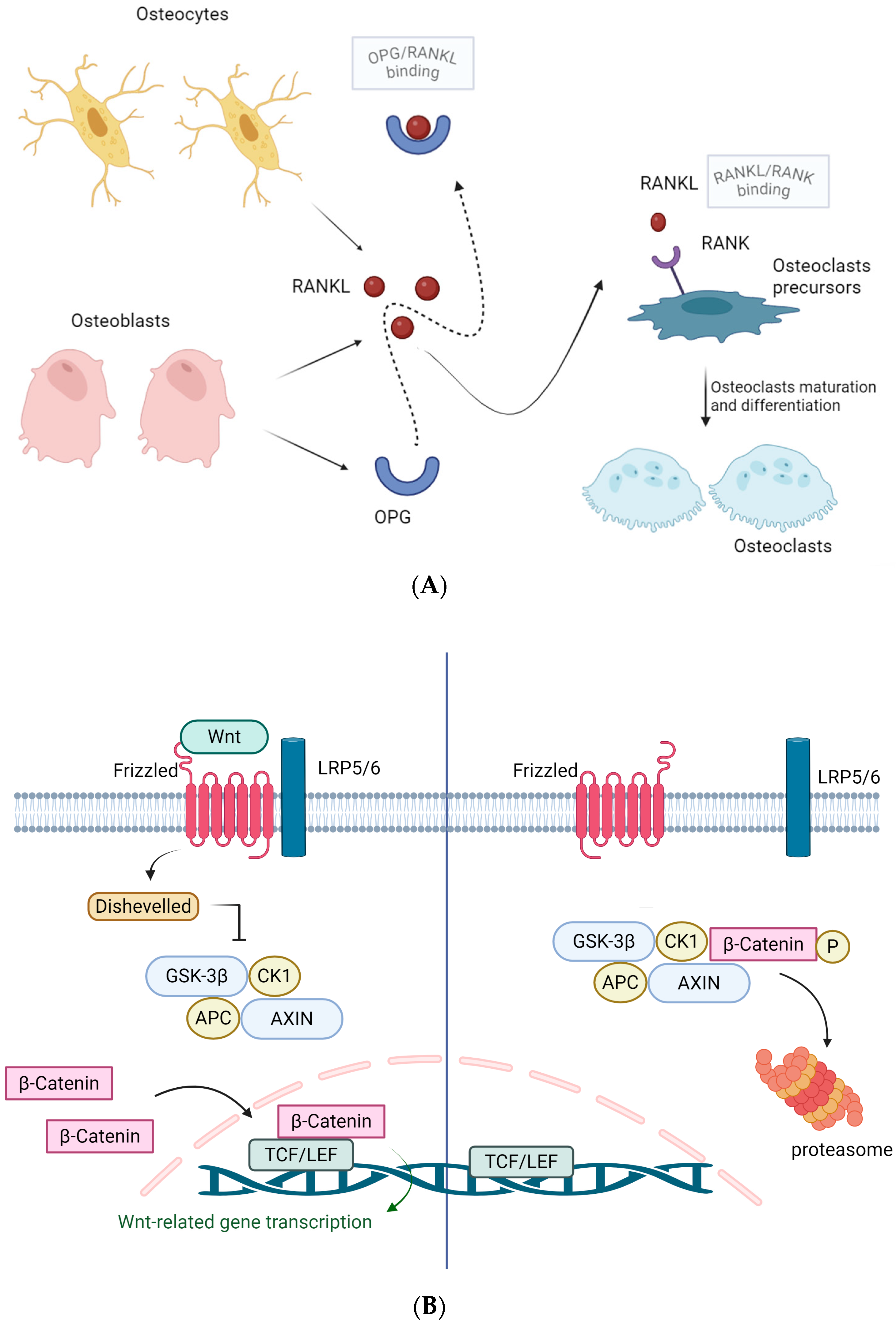
Figure 1. Overview of the main pathways involved in osteoclasts differentiation and bone resorption, and osteoblasts differentiation and proliferation. (A) RANKL/RANK/OPG (RANK/L, receptor activator of nuclear factor kb/ligand; OPG, osteoprotegerin), Created in BioRender, https://BioRender.com/ef7fdte (Accessed on 5 September 2025). (B) Wnt/βcatenin pathway. Created in BioRender, https://BioRender.com/9vt6eud BioRender.com (Accessed on 5 September 2025). Abbreviations: APC, Adenomatous Polyposis Coli; CK1, casein kinase-1; GSK-3β, Glycogen synthase kinase-3 beta; LRP5/6, lipoprotein receptor-related proteins 5/6; OPG, osteoprotegerin; RANK/L, receptor activator of nuclear factor kb/ligand; TCF/LEF, T cell factor/lymphoid enhancer factor family.
Biochem 05 00031 g001
Figure 2. Mevalonate pathway and the action of bisphosphonates. Abbreviations: HMG-CoA, 3-hydroxy-3-methyl-glutaryl-coenzyme A; PP, pyrophosphate. Created in BioRender, https://BioRender.com/n12j0m9 (Accessed on 5 September 2025).
Biochem 05 00031 g002
Table 1. Main causes of secondary OP.
Causes of Secondary Osteoporosis
Hormonal deficiency (Androgen deficiency -> hypogonadism, diseases, medications; estrogen and DHEA deficiency)
Chronic alcohol use
Medications (antiepileptics, SSRIs, PPIs, heparin, glucocorticoids)
Vitamin D deficiency
Sarcopenia and inflammation
Diabetes mellitus (type 1 and type 2)
Hyperthyroidism and hyperparathyroidism
Hypogonadism (including Turner and Klinefelter syndromes)
Obesity and related factors
Gastrointestinal disorders causing malabsorption (e.g., celiac disease, inflammatory bowel disease, bariatric surgery)
Organ transplants and immunosuppressive therapy
Multiple myeloma and other cancers
Rare genetic diseases (osteogenesis imperfecta, Marfan syndrome, Ehlers-Danlos syndrome, homocystinuria)
Rheumatic and neurological diseases (e.g., Parkinson’s disease, multiple sclerosis)
Systemic mastocytosis
DHEA, dehydroepiandrosterone; SSRIs, selective serotonin reuptake inhibitors; PPIs, proton pump inhibitors.
Disclaimer/Publisher’s Note: The statements, opinions and data contained in all publications are solely those of the individual author(s) and contributor(s) and not of MDPI and/or the editor(s). MDPI and/or the editor(s) disclaim responsibility for any injury to people or property resulting from any ideas, methods, instructions or products referred to in the content.