Case Report

Odontogenic Keratocyst in an Edentulous Patient: Report of an Unusual Case

by
Alexandre Perez
1,*,
Valentina Calcoen
1 and
Tommaso Lombardi
2
1
Unit of Oral Surgery and Implantology, Division of Oral and Maxillofacial Surgery, Department of Surgery, Geneva University Hospitals, Faculty of Medicine, University of Geneva, 1205 Geneva, Switzerland
2
Unit of Oral Medicine and Oral Maxillofacial Pathology, Division of Oral and Maxillofacial Surgery, Department of Surgery, Geneva University Hospitals, Faculty of Medicine, University of Geneva, 1205 Geneva, Switzerland
*
Author to whom correspondence should be addressed.
Oral 2023, 3(3), 307-315; https://doi.org/10.3390/oral3030025
Submission received: 31 May 2023 / Revised: 20 June 2023 / Accepted: 11 July 2023 / Published: 13 July 2023

Abstract

:
The purpose of this study was to report a rare case of an odontogenic keratocyst occurring in the edentulous jaw area. A 64-year-old man presented with a painful swelling of the right posterior mandibular vestibule that prevented him from wearing a complete lower denture. Upon intraoral clinical examination, the patient was totally edentulous and had two removable complete dentures. He had a fistula in the vestibular mucosa of edentulous site 48 that was painful upon palpation. Radiological examination revealed an unilocular radiolucent lesion with a continuous peripheral sclerotic border, centered on both the mandibular angle and right branch. Differential diagnosis mainly included a residual cyst and an odontogenic cystic tumor. The biopsy and the excisional material allowed a diagnosis of an odontogenic keratocyst to be made, the cyst being lined by a uniform parakeratinized squamous epithelium, corrugated in places, showing intercellular edema in places, with a well differentiated basal cell layer ranging from cuboidal to columnar in shape, a relatively thin, inflammation-free fibrous wall, and a cyst lumen that contained varying amounts of desquamated keratin. In this case, the surgical risk was represented by paresthesia of both the inferior alveolar and the lingual nerves. The lesion was enucleated without any complications, and the follow-up after 1 year did not reveal any nerve functional damage. Our case underlines the importance for the clinicians to consider a keratocyst in the differential diagnosis of cyst-like lesions presenting in an edentulous area.

1. Introduction

Odontogenic keratocysts (OKCs), developmental cysts that account for up to 11.7% of cysts of the jaws, are benign intraosseous lesions that primarily develop from the rests of the dental lamina [1]. From 2005 to 2017, an OKC was classified by the WHO (World Health Organization) as a “keratocystic odontogenic tumor” due to its association with a mutation of the tumor suppressor gene (PTCH1) located on chromosome 9, the presence of satellite cysts, its aggressive character, and its high risk of recurrence [2]. However, OKCs have now been reclassified as cysts, as there is insufficient evidence to consider this pathology a neoplastic lesion [3]. About 5% of OKCs are associated with nevoid basal cell carcinoma syndrome (NBCCS) [3,4,5]. The prevalence of OKCs is higher in men than in women, in individuals aged between 20 and 40 years, and in the Caucasian population of Northern Europe [6]. In 65% to 80% of cases it is located in the region of the mandibular angle [7], and in 33% of the cases it presents in the maxilla [8]. Clinically, OKCs are usually asymptomatic, and their discovery is often fortuitous, mainly during radiological investigations. In the event of superinfection, or when a cyst becomes of large size, some signs and/or symptoms may appear, such as pain, swelling, or even nerve compression [6]. Radiologically, it presents as a unilocular or multilocular radiolucent lesion, with a well defined sclerotic border that may have a scalloped appearance. The lesion is usually visible as a single cyst, whereas in patients with NBCS multiple OKCs are found [7]. Most of the OKC lesions reported in the literature were diagnosed in dentate patients. We report here a rare case of an OKC centered on the mandibular right-angle region in a fully edentulous patient who sought (emergency) dental advice because of pain that prevented him from wearing his dentures. Only a very few cases have been described where teeth were missing [8,9]. It is therefore important for practitioners to also include OKCs in the differential diagnosis of cystic lesions in edentulous patients.

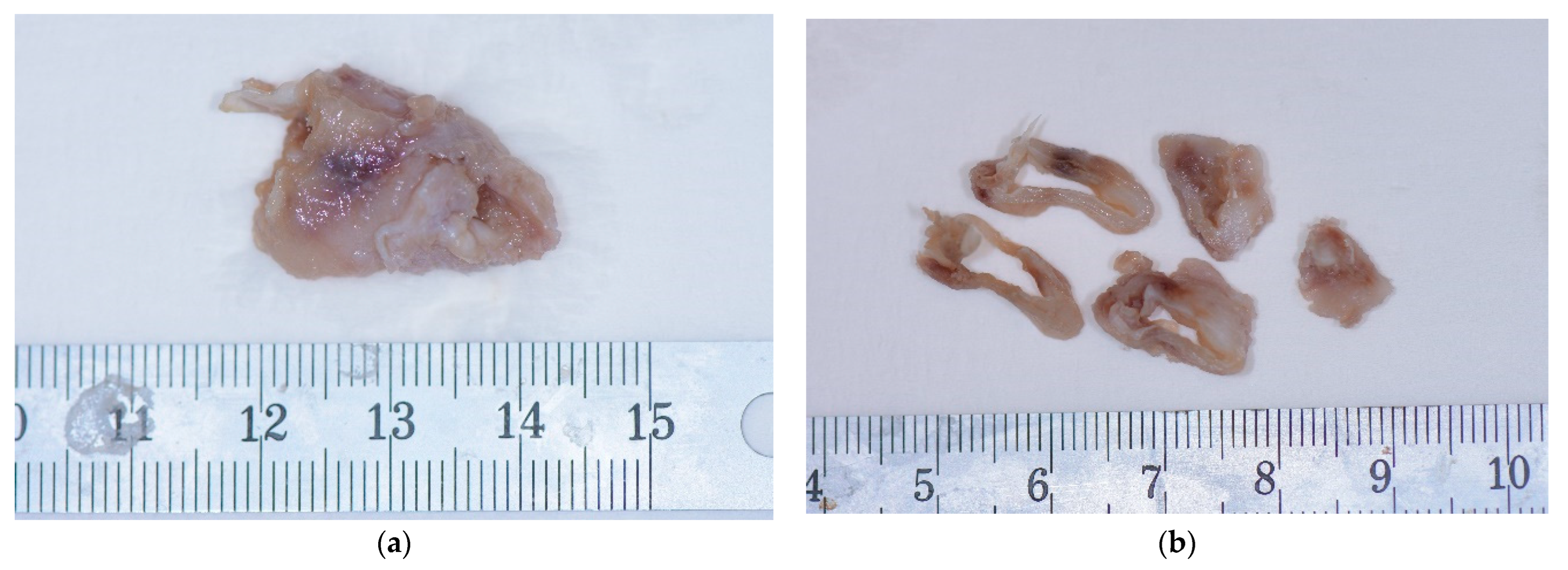
2. Case Presentation

A 64-year-old man consulted the Oral Surgery and Implantology Unit of the Geneva University Hospital (HUG) with a painful swelling of the right posterior mandibular vestibule. The pain, preventing him from wearing a complete lower denture, had been present for about two weeks. He was in fairly good general health, with no known allergies, and he was an active smoker, with 80 packs per year. The extraoral examination did not show any anomalies. Upon an intraoral examination, the patient was totally edentulous and had two complete dentures. A fistula was present in the vestibular mucosa of edentulous site 48 that was painful upon palpation. On the orthopantomogram (OPG) taken as part of a routine investigation, a unilocular radiolucency with a continuous peripheral sclerotic border and a length of approximately 20 mm × 30 mm was visible, centered in the right angle of the mandible (Figure 1).
The lesion displaced the inferior alveolar nerve canal posteriorly and was responsible for the thinning of the vestibular cortex. A computed tomography scan (CT) examination showed an expansive, unilocular osteolytic lesion in the right mandibular angle, measuring 28 mm × 17 mm × 10 mm, in contact with the superior margin of the inferior alveolar nerve canal, which was dehiscent (Figure 2).
The central part of the lesion had a low density and contained some air bubbles. There was also cortical thinning and multiple breaches of the lingual cortex, the largest being present in the posteroinferior and anterosuperior sides. There was no soft tissue involvement. The differential diagnosis included residual cysts, aneurysmal cysts, cystic ameloblatoma, OKCs, and malignant tumor lesions, such as osteosarcoma or metastases from primary tumors in other sites, albeit rare. The patient gave consent to perform both the surgery for histological evaluation and the surgery for total excision. A biopsy was performed under local anesthesia (3M Ubistesin forte 1/100.000) (Figure 3).
A crestal incision was made at the level of edentulous space # 47, extending to the horizontal branch on the external oblique line, followed by a vestibular mucoperiosteal flap for visualization of the lesion that had blown the cortical bone away. A fragment of the overlying bone was removed together with a sample of the lesion for histopathological examination. The histological aspect of the sample revealed the presence of an OKC. In fact, the biopsied specimen exhibited a cyst lined by a rather uniform, parakeratinized stratified squamous epithelium, wavy in places, made up of eight to ten cells, showing intercellular edema in some places, and a well defined palisaded basal cell layer comprised of cuboidal to columnar cells. The fibrous wall was relatively thin and almost devoid of inflammation. The cyst cavity contained varying amounts of desquamated keratin. The enucleation of the lesion concomitant to cryotherapy was then planned. The enucleation was performed 2 weeks later under local anesthesia using 3M Ubistesin forte 1/100.000) (Figure 4).
A crestal incision at the level of the edentulous space of tooth # 47, going up to the horizontal branch along the external oblique line, was carried out, followed by a double vestibular and lingual mucoperiosteal flap. A lingual retractor was inserted, and the lesion was enucleated according to the cleavage planes, paying particular attention to not impinging on both the inferior alveolar and the lingual nerves because of the absence of cranial cortical bone and because of the dehiscence of the lingual cortical bone. The lesion was enucleated in toto and sent for histopathological analysis (Figure 5).
Revision of the bone cavity with a ball burr and a thick curette was performed while protecting the inferior alveolar nerve and the lingual nerve with a retractor. Liquid nitrogen in spray form was applied to the internal cavity walls, followed by sodium chloride rinsing, wound closure with 4-0 Supramid, hemostasis, and routine post-op instructions. The histopathological results confirmed the diagnosis of an OKC (Figure 6).
The histopathological analysis of the excised specimen showed the same histological features found on the biopsy as described above. The patient was regularly followed up, and at the 1-year follow-up appointment there was no recurrence or other abnormalities, such as loss of sensitivity (Figure 7).

3. Discussion

The large range of potential differential diagnoses makes the diagnosis of an OKC difficult, even though it has its own radiological features. Radiologically, it presents as an unilocular or multilocular radiolucent lesion, though usually unilocular, with a well defined sclerotic border that may have a scalloped appearance. Upon radiological examination, the location of an OKC in the mandible accounts for up to 80% of cases, [6,10] most often in the posterior half, very often extending into the ascending ramus [6,7,10,11,12]. When an OKC is located in the mandible, its center is always above the alveolar canal (NAC) [7,11]. An OKC frequently surrounds the crown of an impacted tooth and is difficult to distinguish radiologically from a dentigerous cyst [6,7,8,11]. It should be noted that more than 50% of OKCs surround the crown of an impacted tooth, in most cases a mandibular third molar [6,8]. OKCs appear as a unilocular or occasionally as a multilocular radioluceny, with well defined borders that indicate an osseous reaction [6,8]. As to the consequences on adjacent structures, a mandibular OKC tends to grow in the space that offers the least resistance, i.e., in an anteroposterior axis. In the maxilla, growth is more likely to be centrifugal due to the presence of the sinuses, nasal cavities, and relatively thin bone cortices. As a result, an OKC is usually detected earlier in the maxilla than in the mandible [10]. An OKC more often results in lysis rather than blowing of the cortices [6,7,8,11], except in the upper part of the ramus or in the coronoid process, where significant blow-outs are often observed [7,11]. An OKC can lead to dental displacement and, more rarely, to rhizalysis [7,10,11]. It may displace the alveolar canal downwards, usually without associated neurological signs, or develop in the maxillary sinus and nasal cavity [12]. On an OPG X-ray, a differential diagnosis may primarily suggest a dentigerous cyst, especially when it surrounds the crown of an impacted tooth, without being able to rule out an OKC, an ameloblastoma, or other, even more rare entities [7].
In the present case, we discussed the possibility of a residual cyst, an aneurysmal cyst, a cystic ameloblatoma, an OKC, and a malignant tumor lesion, such as osteosarcoma or metastases from primary tumors in other sites, albeit rare because the patient was edentulous. A CT scan further widened the array of the differential diagnoses, going up to an aneurysmal cyst. The CT scan made cystic ameloblastoma less likely to be included in the differential diagnosis, considering the density values of the tissue of the lesion [12]. A biopsy was then harvested, and it allowed a final diagnosis of an OKC to be made. The OKC histological examination is commonly characterized by the following characteristics: a thin, uniform lining of stratified squamous parakeratinized epithelium, corrugated in places; a spinous cell layer of eight cells or less in thickness, sometimes showing intercellular edema; a well differentiated basal cell layer, ranging from cuboidal to columnar in shape; a relatively thin, inflammation-free fibrous wall; and a lumen that contains varying amounts of desquamated keratin [4]. This cyst may exhibit either parakeratosis, orthokeratosis, or a combination of the two [7].
The management of OKCs still remains a controversial issue in oral and maxillofacial surgery. Different treatments are proposed for OKCs: curettage, marsupialization, enucleation, and resection [9]. Theoretically, the ideal treatment for OKCs should be the most conservative option with the lowest morbidity and risk of recurrence. Many studies showed the lowest recurrence rates with aggressive resection, but at the cost of high morbidity, increased risk of local complications, such as pathologic bone fractures, and reduced quality of life [4,13]. Therefore, some authors criticize this approach and point out that epithelial residues or microcysts may persist at soft tissue extensions [14]. Enucleation is the treatment of choice because of its lower recurrence rate and conservative technique [12]. However, the additional use of adjuvants has been suggested, such as Carnoy’s solution, as has the use of liquid nitrogen during cryotherapy, especially when treating cysts that difficult to remove due to their localization or multilocular cysts. Carnoy’s solution or cryotherapy is applied to reduce potential cyst recurrence. Carnoy’s solution has a recurrence rate of 14.5%, whilst cryotherapy has one of 11.5% [15]. Carnoy’s solution induces superficial tissue necrosis and helps eliminate tumor remnants. The rationale for using Carnoy’s solution is similar to that of cryosurgery. The goal of both techniques is to remove epithelial rests of the dental lamina in the bone margin. One of the advantages of cryotherapy using liquid nitrogen is that it maintains the bone architecture and facilitates bone formation [16,17]. It takes 2 to 3 weeks for the cellular elements to repopulate the bone [16]. The use of liquid nitrogen during cryotherapy allows the reaching of temperatures below −20 °C in order to obtain cellular necrosis [17]. This necrosis is due to the formation of ice crystals, which will disrupt the osmotic pressure and induce the denaturation of proteins and vascular stasis [17]. Cryotherapy also has the advantage of promoting good healing and reducing the amount of bleeding. The main disadvantage of liquid nitrogen is the difficulty for the practitioner to precisely control the amount of product sprayed into the cavity, so care must be taken to protect the adjacent soft tissue [18]. Post-operatively, cryotherapy tends to decrease the mechanical strength of the bone, making it vulnerable to potential fractures. It is therefore recommended that patients adopt a soft diet for the first 2 months following surgery [19]. Additionally, patients must be warned about the possibility of an alteration in sensitivity if the operation was performed near a nerve bundle, although this sensitivity should be recovered in the following months [17]. When using such procedures, the patient must be followed up with regular clinical and radiological control visits, especially during the first 5 years, when the risk of recurrence is high [6]. There are multiple mechanisms of recurrence [6,20,21]. Some are related to the surgical technique: incomplete enucleation, due to its thin, friable membrane, may leave small pieces of the lesion or satellite microcysts in place. Attempting to preserve teeth adjacent to the cyst or curettage of a lesion with scalloped margins are difficult maneuvers that may result in the persistence of lesional tissue with a high intrinsic growth potential, facilitating the development of recurrence [6,20,21]. Other mechanisms are related to lesion characteristics, such as location. OKCs located in the posterior mandibular region are prone to recurrence because of the difficult access to remove all of the cystic wall fragments hidden in the bone. Aggressive OKCs perforate the cortical bone and also involve the adjacent mucosa [6,20,21]. Finally, large lesions and multilocular lesions also have an increased risk of recurrence [22]. In our presented case, the chosen treatment was enucleation followed by cryotherapy. The biopsy showed a thick, firm cystic membrane that easily detached from the bone cavity, as opposed to the particularly thin, friable membrane of an OKC [23]. The thick, firm membrane also facilitated enucleation, with macroscopically healthy bone margins. This finding was similar to that described by some authors [24]. Although the lesion was located posteriorly, access was easy because the patient was completely edentulous. In addition, the lesion had perforated the lingual cortex, and the opposite mucosa was removed/reflected while protecting the lingual nerve. Another peculiarity was the contact with the inferior alveolar nerve due to the resorption of its cranial cortex and the contact of the lesion with the lingula of the mandible (spine of Spix). The patient was informed of the risk of neurologic complications associated with enucleation and cryotherapy. The lesion was gently separated from the nerve followed by cryotherapy, and no neurological or other complications occurred. In cases of edentulism, complete removable dentures are often used for rehabilitation [25]. These prostheses, especially in the mandible, can sometimes become unstable due to anatomical reasons, such as mouth floor enlargement and destabilizing movements of the muscles of the tongue [26]. To overcome these problems, the complete hybrid prostheses stabilized by two inter-foraminal implants are considered the gold standard in such cases [26]. Our patient had complete removable dentures for several years, and he was not keen to opt for a lower implant-supported complete overdenture. The limitation of this case report is represented by the short follow-up (one year).

4. Conclusions

This OKC case is quite unusual since it occurred in a fully edentulous patient complaining of not being able to wear his prosthesis anymore. The lesion was initially found on an OPG, and then further investigated through other radiological exams. Routine radiographs should be taken in edentulous patients in order to rule out the presence of bony pathologies which may grow over time. Histopathologic examination allowed a definitive diagnosis. Cryotherapy was used as a complementary treatment after the conservative excision of the lesion. Follow-up was uneventful after one year without functional neurological damages.

Author Contributions

Draft of the manuscript, preparation of images, patient treatment, A.P.; draft of the manuscript and radiological interpretation preparation of images, V.C.; conception, drafting, and critical review of the manuscript, T.L. All authors have read and agreed to the published version of the manuscript.

Funding

This research received no external funding.

Institutional Review Board Statement

The presented study adhered to the Helsinki Declaration of ethical principles by the World Medical Association. The study did not require approval of the Ethics Commission on Human Research of Geneva (CCER-Geneva) according to the Federal Human Research Act (Art.3al.a), since the study involved less than five patients.

Informed Consent Statement

Written informed consent was obtained from the patient to publish this paper.

Data Availability Statement

Not applicable.

Conflicts of Interest

The authors declare no conflict of interest.

References

  1. Madras, J.; Lapointe, H. Keratocystic odontogenic tumour: Reclassification of the odontogenic keratocyst from cyst to tumour. J. Can. Dent. Assoc. 2008, 74, 165. [Google Scholar] [PubMed]
  2. Avril, L.; Lombardi, T.; Ailianou, A.; Burkhardt, K.; Varoquaux, A.; Scolozzi, P.; Becker, M. Radiolucent lesions of the mandible: A pattern-based approach to diagnosis. Insights Imaging 2014, 5, 85–101. [Google Scholar] [CrossRef] [Green Version]
  3. Manfredi, M.; Vescovi, P.; Bonanini, M.; Porter, S. Nevoid basal cell carcinoma syndrome: A review of the literature. Int. J. Oral Maxillofac. Surg. 2004, 33, 117–124. [Google Scholar] [CrossRef]
  4. Gianfranco, F.; Spirito, F.; Lo Muzio, E.; Capodiferro, S.; Tempesta, A.; Limongelli, L.; Lo Muzio, L.; Maiorano, E. Histopathological Comparative Analysis between Syndromic and Non-Syndromic Odontogenic Keratocysts: A Retrospective Study. Oral 2022, 3, 198–204. [Google Scholar]
  5. Passi, D.; Singhal, D.; Singh, M.; Mishra, V.; Panwar, Y.; Sahni, A. Odontogenic keratocyst (OKC) or keratocystic odontogenic tumor (KCOT)—Journey of OKC from cyst to tumor to cyst again: Comprehensive review with recent updates on WHO classification. Int. J. Curr. Res. 2017, 9, 54080–54086. [Google Scholar]
  6. Myoung, H.; Hong, S.P.; Hong, S.D.; Lee, J.l.; Lim, C.Y.; Choung, P.H.; Lee, J.H.; Choi, J.Y.; Seo, B.M.; Kim, M.J. Odontogenic keratocyst: Review of 256 cases for recurrence and clinicopathologic parameters. Oral Surg. Oral Med. Oral Pathol. Oral Radiol. Endod. 2001, 91, 328–333. [Google Scholar] [CrossRef] [PubMed]
  7. White, S.C.; Pharoah, M.J. Oral Radiology: Principles and Interpretation; Mosby Elsevier: St. Louis, MO, USA, 2009. [Google Scholar]
  8. Thamizhchelvan, H.; Malathi, N.; Radhika, T.; Padmanabhan, V.; Nandakumar, N.; Santhosh Kumar, K. Incidental discovery of odontogenic keratocyst in an edentulous patient: Importance of routine pre-prosthetic radiographic evaluation. J. Indian Prosthodont. Soc. 2011, 3, 199–201. [Google Scholar] [CrossRef]
  9. Nurhan, G.; Sençift, K.; Demirkol, O. Conservative management of keratocystic odontogenic tumors of jaws. Sci. World J. 2012, 2012, 680397. [Google Scholar] [CrossRef] [Green Version]
  10. Mohammad, A.; Baughman, R.A. Maxillary odontogenic keratocyst: A common and serious clinical misdiagnosis. J. Am. Dent. Assoc. 2003, 134, 877–883. [Google Scholar]
  11. Chow, H.T. Odontogenic keratocyst: A clinical experience in Singapore. Oral Surg. Oral Med. Oral Pathol. Oral Radiol. Endod. 1998, 86, 573–577. [Google Scholar]
  12. Zachariades, N.; Papanicolaou, S.; Triantafyllou, D. Odontogenic keratocysts: Review of the literature and report of sixteen cases. J. Oral Maxillofac. Surg. 1985, 43, 177–182. [Google Scholar] [CrossRef]
  13. Zhao, Y.; Li, Z.; Sun, Y.; Liu, B.; JiaJia, J. Pathological fractures of the mandible: A report of 27 cases. Clin. Surg. 2017, 2, 1839. [Google Scholar]
  14. Fidele, N.B.; Bing, L.; Sun, Y.; Wu, T.; Zheng, Y.; Zhao, Y. Management of mandibular odontogenic keratocyst through radical resection: Report of 35 cases. Oncol. Lett. 2019, 18, 733–741. [Google Scholar] [CrossRef] [PubMed] [Green Version]
  15. Stoelinga, P.J. The treatment of odontogenic keratocysts by excision of the overlying, attached mucosa, enucleation, and treatment of the bony defect with Carnoy solution. J. Oral Maxillofac. Surg. 2005, 63, 1662–1666. [Google Scholar] [CrossRef] [PubMed]
  16. Sharif, F.N.; Oliver, R.; Sweet, C.; Sharif, M.O. Interventions for the treatment of keratocystic odontogenic tumours. Cochrane Database Syst. Rev. 2015, 2016, CD008464. [Google Scholar] [CrossRef] [PubMed]
  17. Whittaker, D.K. Mechanisms of tissue destruction following cryosurgery. Ann. R. Coll. Surg. Engl. 1984, 66, 313–318. [Google Scholar]
  18. Pogrel, M.A. The use of liquid nitrogen cryotherapy in the management of locally aggressive bone lesions. J. Oral Maxillofac. Surg. 1993, 51, 269–273. [Google Scholar] [CrossRef]
  19. Janas-Naze, A.; Zhang, W.; Szuta, M. Modified Carnoy’s Versus Carnoy’s Solution in the Management of Odontogenic Keratocysts—A Single Center Experience. J. Clin. Med. 2023, 3, 1133. [Google Scholar] [CrossRef]
  20. Kinard, B.E.; Chuang, S.K.; August, M.; Dodson, T.B. How well do we manage the odontogenic keratocyst? J. Oral Maxillofac. Surg. 2013, 71, 1353–1358. [Google Scholar] [CrossRef]
  21. Tarakji, B.; Baroudi, K.; Hanouneh, S.; Azzeghaiby, S.N.; Nassani, M.Z. Possible recurrence of keratocyst in nevoid basal cell carcinoma syndrome: A review of literature. Eur. J. Dent. 2013, 7, S126–S134. [Google Scholar] [CrossRef] [Green Version]
  22. Jung, H.D.; Lim, J.H.; Kim, H.J.; Nam, W.; Cha, I.H. Appropriate follow-up period for odontogenic keratocyst: A retrospective study. Maxillofac. Plast. Reconstr. Surg. 2021, 43, 16. [Google Scholar] [CrossRef] [PubMed]
  23. Lima, J.L.; Dias-Ribeiro, E.; Honfi, E.S.; De Araujo, T.N.; Honorio de Goes, K.K.; Do Socorro Aragao, M. Odontogenic keratocyst of mandible. Indian J. Otolaryngol. Head. Neck Surg. 2006, 58, 373–376. [Google Scholar] [CrossRef] [PubMed] [Green Version]
  24. Vallejo-Rosero, K.A.; Camolesi, G.V.; de Sá, P.L.D.; Bernaola-Paredes, W.E. Conservative management of odontogenic keratocyst with long-term 5-year follow-up: Case report and literature review. Int. J. Surg. Case Rep. 2020, 66, 8–15. [Google Scholar] [CrossRef] [PubMed]
  25. Lee, D.J.; Saponaro, P.C. Management of Edentulous Patients. Dent. Clin. N. Am. 2019, 63, 249–261. [Google Scholar] [CrossRef]
  26. Feine, J.S.; Carlsson, G.E.; Awad, M.A. The McGill consensus statement on overdentures. Mandibular two-implant overdentures as first choice standard of care for edentulous patients. Gerodontology 2002, 19, 3–4. [Google Scholar] [CrossRef]
Figure 1. Clinical (a) view and panoramic X-ray (b) at initial examination.
Oral 03 00025 g001
Figure 2. Computed tomography scan (CT): frontal (a) and axial views (b). Orange triangle: Spina mandibulae.
Oral 03 00025 g002
Figure 3. Intraoperative biopsy view: (a) vestibular flap, (b) detachment and dissection of the medial portion of the lesion, (c) cavity after biopsy, and (d) biopsied specimen.
Oral 03 00025 g003
Figure 4. Intraoperative view: (a) dissection of the lesion at the lingual part, (b) enucleation in toto, and (c) cryotherapy.
Oral 03 00025 g004
Figure 5. Removed tissue intact (a) and sectioned (b).
Oral 03 00025 g005
Figure 6. Histopathological aspects of the excised specimen. Red arrow: parakeratinized layer; blue arrow: flat interface; and yellow arrow: cuboidal and columnar palisading basal cells (H&E, ×20).
Oral 03 00025 g006
Figure 7. Panoramic X-ray at 1-year follow-up recall appointment.
Oral 03 00025 g007
Disclaimer/Publisher’s Note: The statements, opinions and data contained in all publications are solely those of the individual author(s) and contributor(s) and not of MDPI and/or the editor(s). MDPI and/or the editor(s) disclaim responsibility for any injury to people or property resulting from any ideas, methods, instructions or products referred to in the content.

