Case Report

Introduction to Surgical Navigation in Oral Surgery: A Case-Series

by
Giorgio Novelli
1,2,*,
Mattia Moretti
1,3,
Maria Costanza Meazzini
1,
Cristina Maria Angela Cassé
2,
Fabio Mazzoleni
1 and
Davide Sozzi
1,2
1
Operative Unit of Maxillo-Facial Surgery, Fondazione IRCCS San Gerardo dei Tintori, 20900 Monza, Italy
2
Department of Medicine and Surgery, School of Medicine and Surgery, University of Milano Bicocca, 20900 Monza, Italy
3
Postgraduate School of Maxillofacial Surgery, University of Milan, 20122 Milan, Italy
*
Author to whom correspondence should be addressed.
Oral 2023, 3(2), 146-154; https://doi.org/10.3390/oral3020013
Submission received: 9 January 2023 / Revised: 14 March 2023 / Accepted: 17 March 2023 / Published: 28 March 2023

Abstract

:
The application of surgical navigation in oral and maxillo-facial surgery has been increasing over time. In fact, computer-assisted surgery provides real-time, precise, and accurate position and guidance during surgery. The purpose of our work is to introduce the evolution of surgical navigation in recent decades, describe some technical aspects of this technology, explore new possibilities of application of surgical navigation in oral surgery, and validate the accuracy of computer-assisted surgery. We included four patients in our sample who underwent virtual planning on the cone beam CT data set and surgical navigation using non-invasive fiducial markers. The first patient presented a dislocated orthodontic arch in the soft tissues of the cheek, while the other patients presented supernumerary and impacted dental elements. Among them, two patients were affected by craniofacial synostosis. We evaluated the accuracy of computer-assisted surgery, calculating the discrepancy between the real and virtual target. In all cases, the target registration error was less than or equal to 1 mm. We can affirm that surgical navigation is a valid tool to enhance oral surgery, guaranteeing an undoubted advantage in terms of the reliability and predictability of the results, especially in complex cases.

1. Introduction

The surgical navigation can be considered the evolution of stereotaxic surgery. The principles of stereotaxic navigation were developed more than 100 years ago by Horsley and Clarke [1], who first described an instrument able to locate intracranial structures. In 1947, Spingel et al. applied this technique in humans to orient and guide the instruments during surgery [2]. The set-up included the pre-operative positioning of a helmet made up from a metal frame fixed to the patient’s head. The helmet had the purpose of maintaining a constant position during the acquisition of the images and during the surgical phase, limiting the surgical access even when working in small or deeply located brain areas. The direction and depth of the surgical instruments, fixed on the helmet, were originally determined by mathematical calculations based on stereotaxic anatomy atlases, in which each internal structure was correlated with external points defined by a spatial reference system.
The main limits of this procedure were that the individual variability of each patient and the presence of pathological tissues were not considered [3].
Only with the development of CT and MRI since 1980 has it been possible to provide more accurate and specific data for each patient [4].
However, the restricted access to the operating field due to the presence of the helmet and the related artifacts on CT scan remained strong deterrents to clinical application. In 1987, Watanabe et al. introduced, for the first time, frameless stereotaxic surgery [5]. This technique was able to locate anatomical structures intra-operatively using data acquired from pre-operative CT and MRI and allowing real-time information on the position of surgical instruments without the presence of the bulky helmet.
In the first surgical navigation systems, the instruments were fixed on a mechanical arm connected to a computer, thus reducing the range of motion and their field of application. The development of ultrasonic and electromagnetic navigation systems made it possible to eliminate the mechanical arm thanks to the principle of satellite tracking [3].
The improvement and the evolution of this technology led to its application in different medical fields, such as oncology, reconstructive surgery, and traumatology. In particular, in maxillo-facial surgery navigation was used for the first time in 1994 for the removal of a tumor of the skull base and then has become, over the last 20 years, an aid to surgeons in complex cases of traumatic injuries as well as oncological demolition and reconstruction. Actually, articles on the use of surgical navigation for the management of complex fractures of the facial skeleton are increasingly widespread in scientific literature, with particular attention paid to the orbitozygomatic-maxillary complex [6,7,8], as well as for reconstruction following oncological surgery, both for the maxilla and the mandible [9,10]. The use of this technology, in fact, makes it possible to considerably increase the accuracy of the interventions, thus improving the surgical outcomes, especially if associated with new techniques, such as piezo-electric surgery [11].
The enthusiasm for surgical navigation has also spread to the dental world, which is already prone to the use of new devices and technologies [12,13]. However, the most described application in oral surgery is computer-assisted surgery for dental implant placement [14,15,16]. In fact, only a few articles describe other uses of navigation systems; some case reports analyze the results of computer-assisted surgery in the removal of impacted teeth [14,17] and foreign bodies [18,19].
The purpose of this work is to lay the foundations for a preliminary assessment of the different indications in oral surgery for the use of computer-assisted surgery.

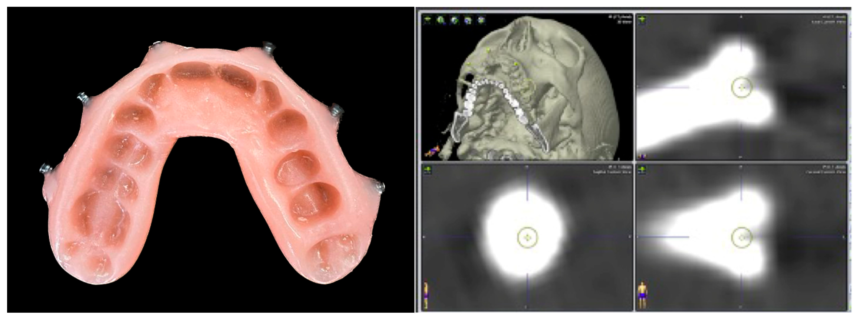
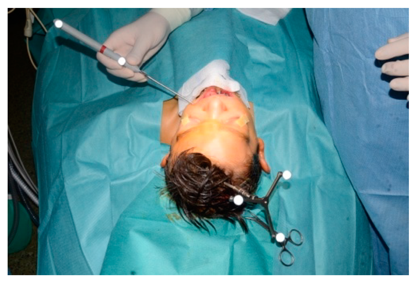
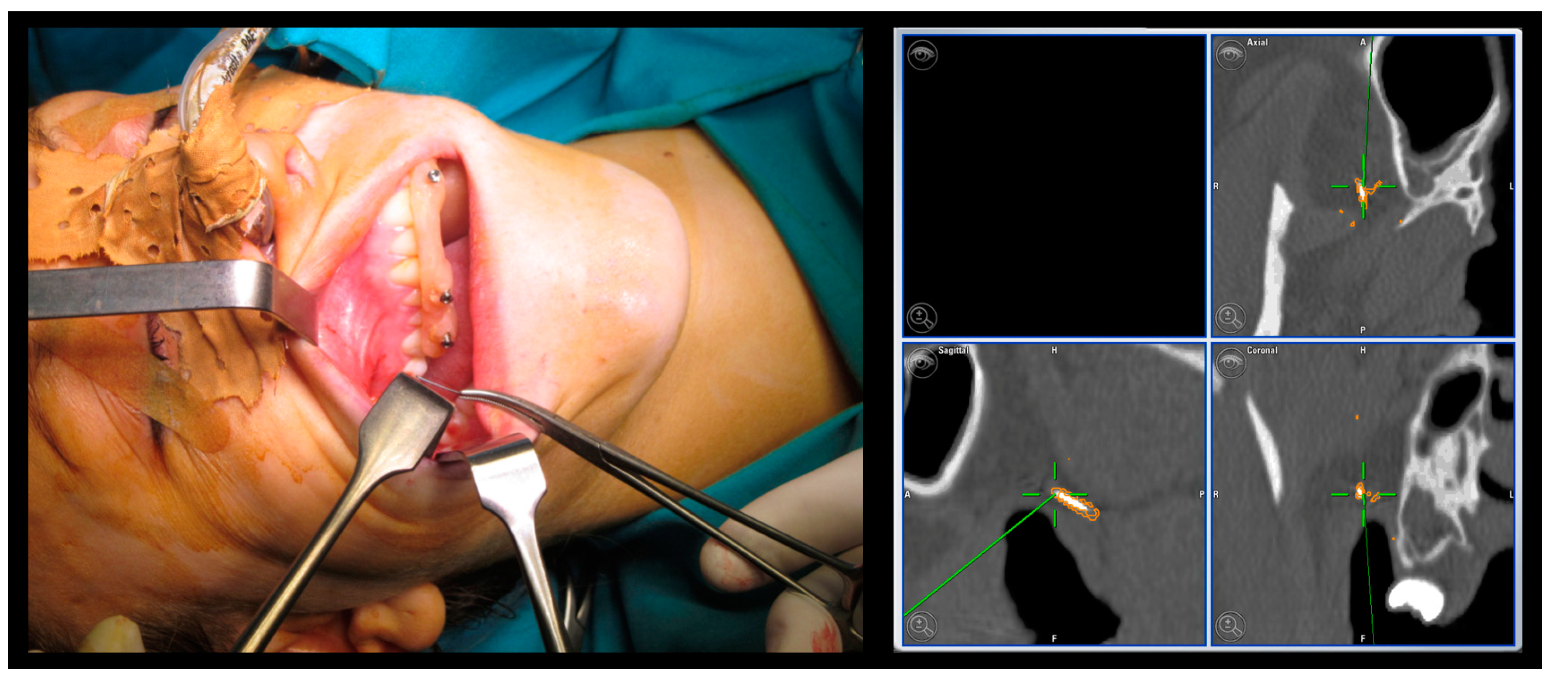
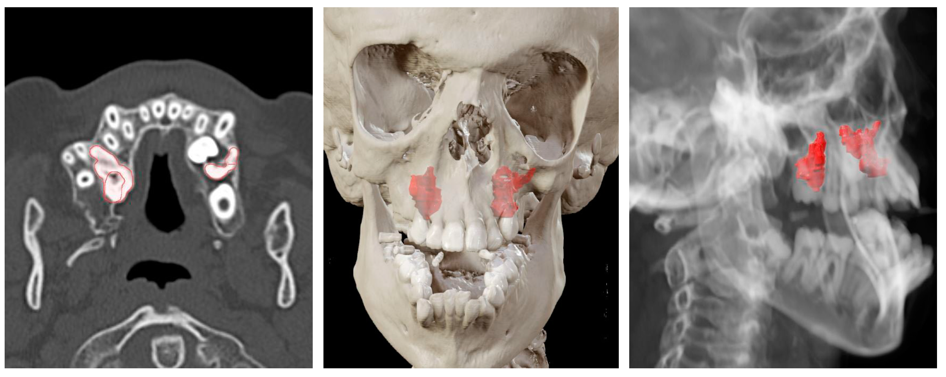
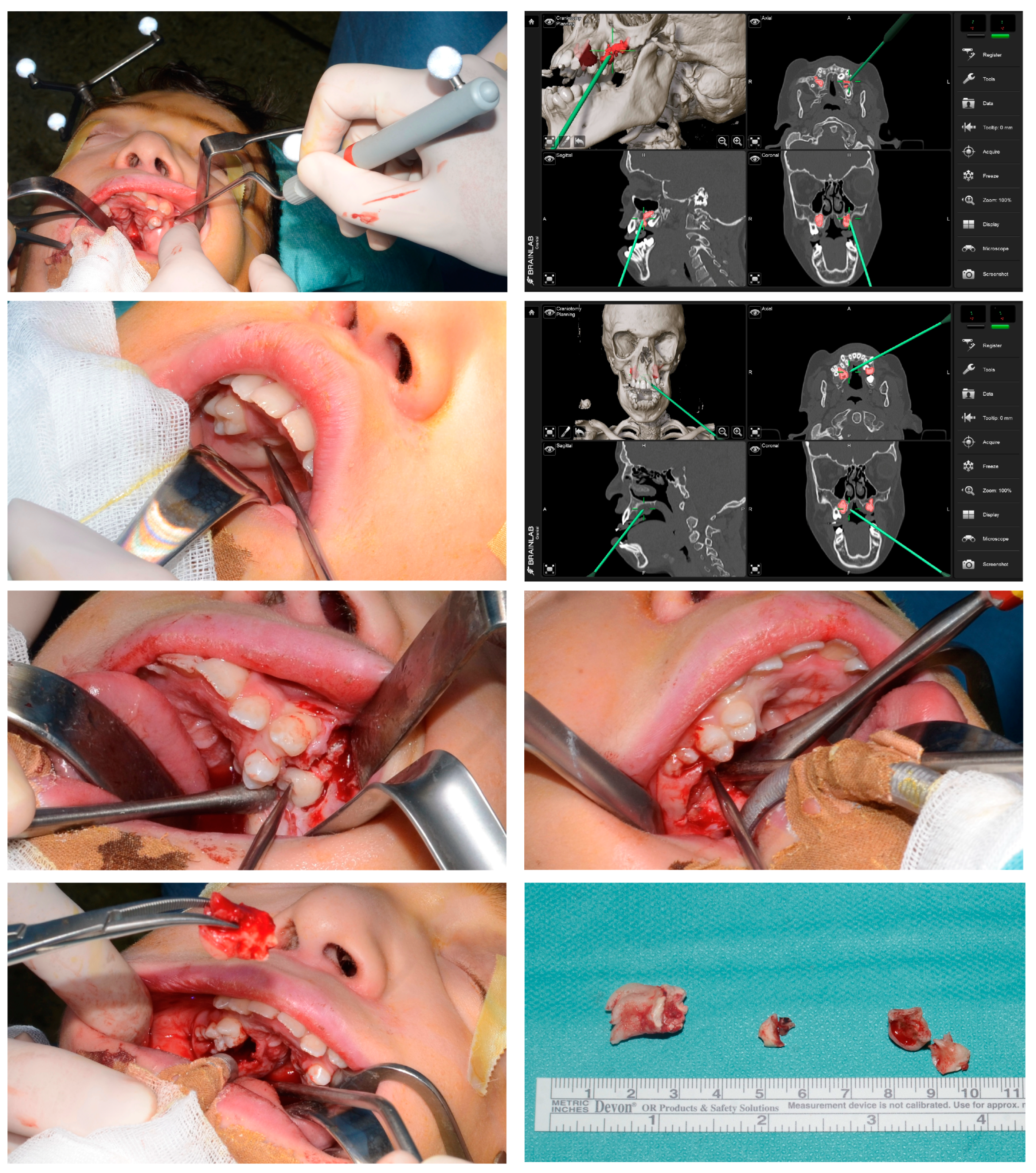
2. Materials and Methods
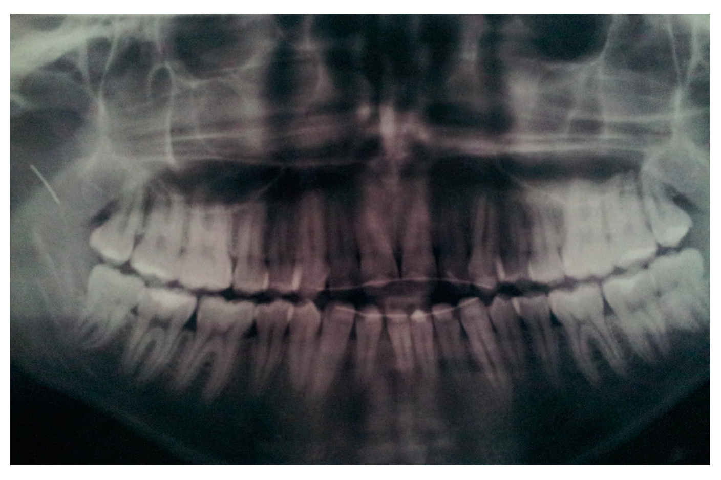
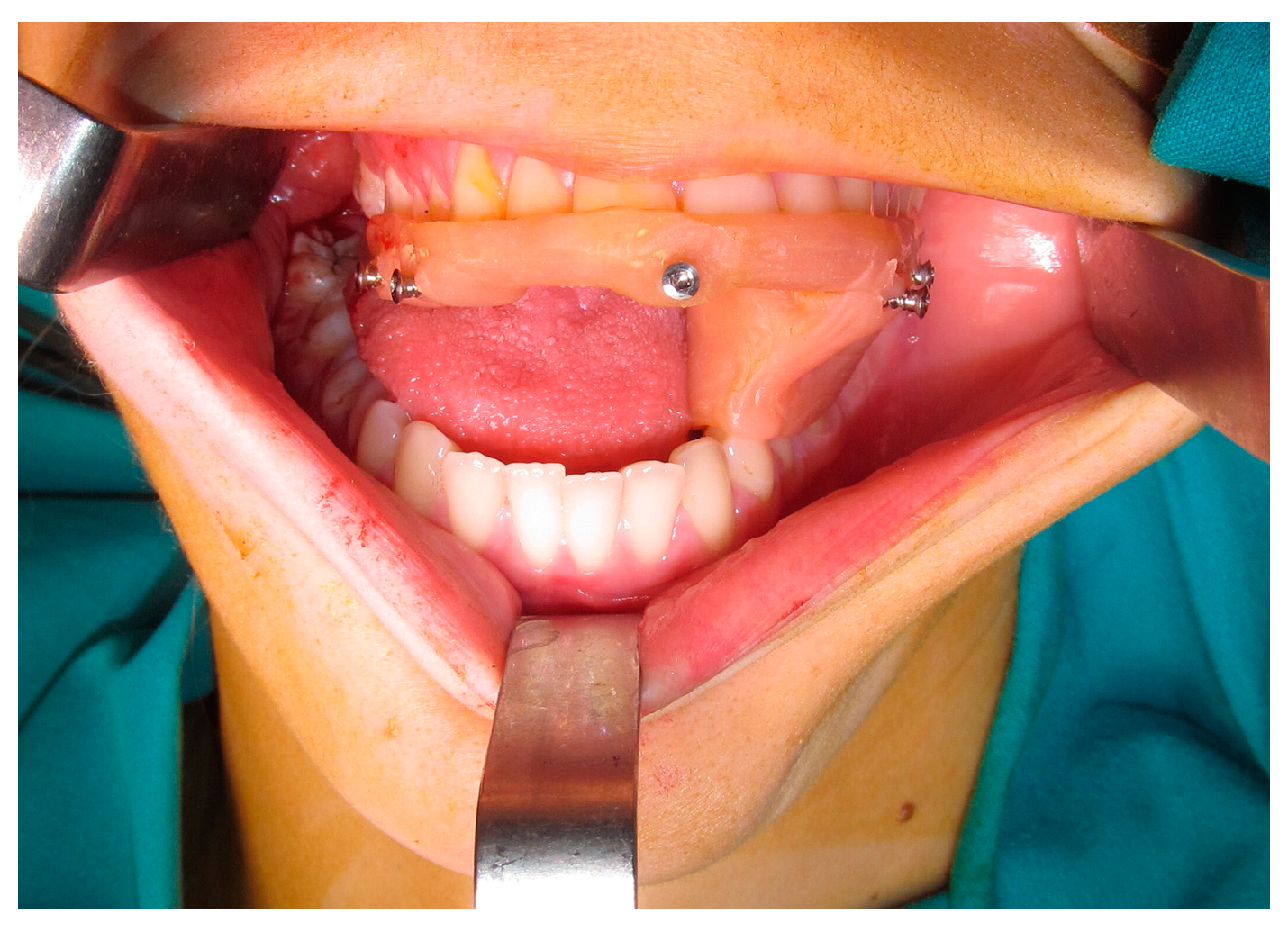
In our preliminary study, we enrolled four patients treated in our center. The first patient presented a dislocated orthodontic arch in the soft tissues of the cheek. In other three cases, patients presented supernumerary and included dental elements. Among them, two patients were affected by craniofacial synostosis (Pfeiffer Syndrome and Apert Syndrome).
The process of computer-assisted surgery began with the fabrication of a personalized maxillary occlusal splint, in which five titanium hexagonal-headed screws were inserted in a non-coplanar position, as fiducial markers for patient’s orientation during surgery (Figure 1). Then, a high resolution cone beam CT (0.8–1 mm slices) was performed, after positioning the splint on the maxilla.
Surgical planning and navigation were performed according to the protocol previously described by Novelli et al. [6,7]. Pre-surgical planning on DICOM data was performed with iPlan 3.0 CMF software (BrainLab, Feldkirchen, Germany). After orienting the data set on the axial, sagittal, and coronal planes, the fiducial markers and the surgical targets were identified. These landmarks assume a specific spatial position in the three-dimensional reconstruction of CT data (Figure 1).
Vector Vision II (BrainLab, Feldkirchen, Germany) was used for recording and for surgical navigation. It is an optical navigation system that involves the use of a reflective system fixed to the patient’s skull, called the dynamic reference frame, identifiable by an infrared video camera (Figure 2).
The software, using the dynamic reference frame and the CT data set, is able to determine the real position on the patient of the fiducial markers. The accuracy of the recording was assessed during surgery, by checking target structures in vivo with CT images.

3. Results

We described two cases of our sample in order to explain the procedure proposed: the first patient treated is a twenty-three year-old woman, who came to the attention of our department due to the evidence on orthopantomography of a foreign body in the right infra-temporalis region, as visible in Figure 3.
Since it was difficult to clinically investigate the anatomical region, a cone beam CT scan was required in order to localize the foreign body and plan the surgical navigation. In this case, we fabricated a splint with an extension in order to keep the mouth open during surgery, as visible in Figure 4. In fact, mouth movements should be avoided to permit the correct triangulation of the navigation system.
The surgery was performed under general anesthesia with an intra-oral incision in the right upper vestibular fornix to reach the infra-temporalis fossa. The foreign body was identified and removed after soft-tissue dissection with the aid of surgical navigation (Figure 5). It was a fragment of a dislocated orthodontic arch.
The second patient treated is a twelve-year-old boy affected by Pfeiffer Syndrome with dental crowding and impaired eruption of dental elements 1.6, 6.5, and 2.6. The patient was referred to our center in order to proceed with the extractions under general anesthesia.
Before surgery, we identified, on the cone beam CT, the dental elements to be removed with iPlan 3.0 CMF software (BrainLab, Feldkirchen, Germany), as visible in Figure 6.
The surgery was performed under general anesthesia with the aid of the surgical navigation system, as previously described. An intra-sulcular palatal incision was made, extending from dental element 1.3 to the right tuber maxillae. We performed an ostectomy to identify the dental element 1.6 in palatal position by using the navigation system pointer. Then, we safely proceeded with the avulsion of the dental element 1.6. On the left side, in order to remove the dental elements 6.5 and 2.6, we performed a vestibular incision extending from dental element 2.3 to the left tuber maxillae and a similar access ostectomy to identify the correct teeth before the avulsion. The intra-operative images are visible in Figure 7.
The data obtained from the analysis of accuracy in the four patients are reported in Table 1.

4. Discussion

Surgical navigation instruments give the surgeons the opportunity to be guided through the anatomical structures, verifying, pre-, intra-, and post-operatively, the adequacy of their actions and results.
The purpose of pre-operative surgical planning is to identify, in the virtual model, the target of surgery in order to increase the predictability, reproducibility, and safety of the surgical act [20,21]. In order to obtain these goals, several factors influencing the accuracy of surgical navigation should be analyzed:
  • Geometric accuracy is an index of image recording precision and indicates the technical quality of the image. Marmulla et al. [22] and Eggers et al. [23] analyzed the geometric accuracy of cone beam CT compared to Spiral CT. These studies showed that the geometric accuracy of cone beam CT was lower than Spiral CT, in the range of fractions of millimeters; moreover, it was unequally distributed over the region of interest. However, cone beam CT appeared to be adequate for the purpose of oral surgery.
  • The type of navigation systems influences the accuracy of computer-assisted surgery:
    Electromagnetic systems superimpose a magnetic field over the surgical site. The position of a tracking probe is determined by analyzing the effect of its ferromagnetic parts on the magnetic field. They have a fast intra-operative registration, but there are interferences with metal instruments [24].
    Optical systems measure the time that it takes a transducer-emitted tone to reach the microphone-bearing frame. They have a higher technical precision without interference with the metal instruments commonly present in the operating room. However, they must be positioned in a linear way with the patient in order to interact with the surgical field [25,26].
  • Target registration error is an index of accuracy of the navigation system. It is defined as the discrepancy, in millimeters, between the real and virtual anatomical points and the fiducial markers [27,28]. The factors that can most influence the target registration error are the type and position of fiducial markers [29]:
    Invasive fiducial markers: they are normally made up of self-tapping titanium screws inserted into bone through a cutaneous or mucosal access. They are extremely precise, small in size, and do not undergo modifications from the image acquisition phase to the operative time. Due to their characteristics, they can be applied in large numbers. The disadvantages are linked to the invasiveness of their positioning.
    Non-invasive fiducial markers: specific occlusal dental splints in which radiopaque landmarks are placed in such a way that they can be worn during the CT scan acquisition and used as non-invasive fiducial markers. It is an easy technique, but it requires the additional time to fabricate the splint. It cannot be used in edentulous patients, and its accuracy tends to be reduced above the plane passing through the orbital floor. The splint can be safely removed after registration and re-used during surgery and if the recording has to be repeated to verify the post-operative accuracy.
    Dental landmarks: they can be used as an alternative to the occlusal splint. This method requires the presence of at least 4 to 5 non-periodontopathic dental elements in the upper dental arch on which dental brackets could be placed.
    Surface scanner: it allows one to scan the surface of soft tissues, recording an average of 100–200 surface points. The advantage of this method is to allow a rapid acquisition of a large number of reference points in order to obtain an accurate superimposition with the virtual images, without the need of a CT scan with landmarks.
According to Metzger et al. [29], the most accurate registration method is based on invasive landmarks. Collyer [3] and Novelli et al. [6,7] validated the use of occlusal splints for orbital surgery. We can state that in oral surgery the use of occlusal splints, dental markers, or bone anchoring screws can be considered valid and reliable due to the degree of precision required and to the location of the surgical field.
The extraction of impacted teeth is a commonly performed procedure associated with several potentially significant complications. Many of these complications arise as a result of iatrogenic damage due to the operator’s inability to directly visualize anatomical structures, such as inferior alveolar nerve, maxillary sinus, nasal cavity, and adjacent teeth [30].
Although an easy but necessary learning curve for the surgical planning and registration process is needed, thus leading to an initial lengthening of the preoperative times, computer-assisted surgery offers an undoubted advantage in terms of the reliability and predictability of the results, optimizing operating times and reducing post-operative complications [9].
In our sample, the target registration error was always less than a 1 mm, in the case of impacted teeth. It reached 1 mm when the target was in the soft tissues and subjected to variations of its position in relation to the jaws.
Our results show that the correspondence between the intra-operative target position and pre-operative target planning has an average margin of error of less than 1 mm. We can affirm that the target position can be faithfully predicted by the object planning.

5. Conclusions

Surgical navigation is also taking on an ever-increasing role in oral surgery. In fact, the possibility of accurately identifying both impacted dental elements and foreign bodies makes it possible to reduce the surgical invasiveness of the accesses and the risk of error or injuries to anatomical structures adjacent to the surgical target. Our study is a theoretical introduction to anyone who wants to approach surgical navigation, providing ideas for its application in a clinical context and demonstrating its accuracy even in settings different from those in which it was conceived. Obviously, other studies with a higher number of cases are needed to validate its use in oral surgery and in order to exploit its potential and justify the costs.
Until then, according to our experience in the field of computer-assisted surgery and to these preliminary published results, it can be stated that surgical navigation is a valid tool to enhance oral surgery.

Author Contributions

Conceptualization, G.N. and M.M.; methodology, G.N. and F.M.; validation, G.N. and M.C.M.; formal analysis, D.S.; investigation, G.N., M.M. and F.M.; resources, D.S. and C.M.A.C.; data curation, G.N., M.M. and F.M.; writing—original draft preparation, G.N. and M.M.; writing—review and editing, G.N. and M.M.; visualization, G.N. and M.C.M.; supervision, D.S.; and project administration, G.N. and D.S. All authors have read and agreed to the published version of the manuscript.

Funding

This research received no external funding.

Institutional Review Board Statement

This research study was conducted retrospectively from data obtained for clinical purposes. We consulted extensively with our Institutional Review Board, who determined that our study did not need ethical approval. The study was conducted in accordance with the Declaration of Helsinki.

Informed Consent Statement

Informed consent was obtained from all subjects involved in the study.

Data Availability Statement

The data presented in this study are available on request from the corresponding author.

Conflicts of Interest

The authors declare no conflict of interest.

References

  1. Horsley, V.; Clarke, R.H. The structure and function of the cerebellum esamine by a new method. Brain 1908, 31, 45–124. [Google Scholar] [CrossRef]
  2. Spiegel, E.A.; Wycis, H.T.; Marks, M.; Lee, A.J. Stereotaxic Apparatus for Operations on the Human Brain. Science 1947, 106, 349–350. [Google Scholar] [CrossRef] [PubMed]
  3. Collyer, J. Stereotactic navigation in oral and maxillofacial surgery. Br. J. Oral Maxillofac. Surg. 2010, 48, 79–83. [Google Scholar] [CrossRef] [PubMed]
  4. Leksell, L.; Jernberg, B. Stereotaxis and tomography a technical note. Acta Neurochir. 1980, 52, 1–7. [Google Scholar] [CrossRef] [PubMed]
  5. Watanabe, E.; Watanabe, T.; Manaka, S.; Mayanagi, Y.; Takakura, K. Three-dimensional digitizer (neuronavigator): New equipment for computed tomography-guided stereotaxic surgery. Surg. Neurol. 1987, 27, 543–547. [Google Scholar] [CrossRef]
  6. No, G.; Tonellini, G.; Mazzoleni, F.; Sozzi, D.; Bozzetti, A. Surgical Navigation Recording Systems in Orbitozygomatic Traumatology. J. Craniofac. Surg. 2012, 23, 890–892. [Google Scholar] [CrossRef] [Green Version]
  7. Novelli, G.; Tonellini, G.; Mazzoleni, F.; Bozzetti, A.; Sozzi, D. Virtual surgery simulation in orbital wall reconstruction: Integration of surgical navigation and stereolithographic models. J. Cranio-Maxillofacial Surg. 2014, 42, 2025–2034. [Google Scholar] [CrossRef]
  8. Dubron, K.; Van Camp, P.; Jacobs, R.; Politis, C.; Shaheen, E. Accuracy of virtual planning and intraoperative navigation in zygomaticomaxillary complex fractures: A systematic review. J. Stomatol. Oral Maxillofac. Surg. 2022, 123, e841–e848. [Google Scholar] [CrossRef]
  9. Azarmehr, I.; Stokbro, K.; Bell, R.B.; Thygesen, T. Surgical Navigation: A Systematic Review of Indications, Treatments, and Outcomes in Oral and Maxillofacial Surgery. J. Oral Maxillofac. Surg. 2017, 75, 1987–2005. [Google Scholar] [CrossRef] [Green Version]
  10. Sozzi, D.; Filippi, A.; Canzi, G.; De Ponti, E.; Bozzetti, A.; Novelli, G. Surgical Navigation in Mandibular Reconstruction: Accuracy Evaluation of an Innovative Protocol. J. Clin. Med. 2022, 11, 2060. [Google Scholar] [CrossRef]
  11. Dean, A.; Heredero-Jung, S.; Solivera, J.; Sanjuan, A.; Alamillos-Granados, F.J. Computer-assisted and navigated piezoelectric surgery: A new technology to improve precision and surgical safety in craniomaxillofacial surgery. Laryngoscope Investig. Otolaryngol. 2022, 7, 684–691. [Google Scholar] [CrossRef]
  12. Minervini, G.; Russo, D.; Herford, A.S.; Gorassini, F.; Meto, A.; D’Amico, C.; Cervino, G.; Cicciù, M.; Fiorillo, L. Teledentistry in the Management of Patients with Dental and Temporomandibular Disorders. BioMed Res. Int. 2022, 2022, 7091153. [Google Scholar] [CrossRef] [PubMed]
  13. Reddy, L.K.V.; Madithati, P.; Narapureddy, B.R.; Ravula, S.R.; Vaddamanu, S.K.; Alhamoudi, F.H.; Minervini, G.; Chaturvedi, S. Perception about Health Applications (Apps) in Smartphones towards Telemedicine during COVID-19: A Cross-Sectional Study. J. Pers. Med. 2022, 12, 1920. [Google Scholar] [CrossRef]
  14. Landaeta-Quinones, C.G.; Hernandez, N.; Zarroug, N.K. Computer-Assisted Surgery: Applications in Dentistry and Oral and Maxillofacial Surgery. Dent. Clin. N. Am. 2018, 62, 403–420. [Google Scholar] [CrossRef]
  15. Pellegrino, G.; Ferri, A.; Del Fabbro, M.; Prati, C.; Gandolfi, M.; Marchetti, C. Dynamic Navigation in Implant Dentistry: A Systematic Review and Meta-analysis. Int. J. Oral Maxillofac. Implant. 2021, 36, e121–e140. [Google Scholar] [CrossRef] [PubMed]
  16. Gargallo-Albiol, J.; Barootchi, S.; Salomó-Coll, O.; Wang, H.-L. Advantages and disadvantages of implant navigation surgery. A systematic review. Ann. Anat. 2019, 225, 1–10. [Google Scholar] [CrossRef]
  17. Retana, A.; Emery, R.W.; Keir, V. Removal of Impacted Supernumerary Teeth Using a Dynamic Surgical Navigation System: A Case Report. J. Oral Maxillofac. Surg. 2019, 77, 1130–1134. [Google Scholar] [CrossRef] [PubMed]
  18. Cheng, Y.; Li, Q.B.; Li, Z.; Cheng, G. Needle Removal in the Deep Maxillofacial Region Assisted by Computerized Navigation Technique and Digital Guiding Plate. J. Craniofac. Surg. 2023; Epub ahead of print. [Google Scholar] [CrossRef]
  19. Lan, L.; He, Y.; An, J.; Zhang, Y. Application of Computer-Aided Navigation Technology in the Extraction of Foreign Body from the Face. J. Craniofac. Surg. 2020, 31, e166–e169. [Google Scholar] [CrossRef] [PubMed]
  20. Strong, E.B.; Rafii, A.; Holhweg-Majert, B.; Fuller, S.C.; Metzger, M.C. Comparison of 3 Optical Navigation Systems for Computer-Aided Maxillofacial Surgery. Arch. Otolaryngol. Neck Surg. 2008, 134, 1080–1084. [Google Scholar] [CrossRef] [PubMed] [Green Version]
  21. Schramm, A.; Suarez-Cunqueiro, M.M.; Rücker, M.; Kokemueller, H.; Bormann, K.-H.; Metzger, M.C.; Gellrich, N.-C. Computer-assisted therapy in orbital and mid-facial reconstructions. Int. J. Med Robot. Comput. Assist. Surg. 2009, 5, 111–124. [Google Scholar] [CrossRef]
  22. Marmulla, R.; Wörtche, R.; Mühling, J.; Hassfeld, S. Geometric accuracy of the NewTom 9000 Cone Beam CT. Dentomaxillofac. Radiol. 2005, 34, 28–31. [Google Scholar] [CrossRef] [PubMed]
  23. Eggers, G.; Klein, J.; Welzel, T.; Mühling, J. Geometric accuracy of digital volume tomography and conventional computed tomography. Br. J. Oral Maxillofac. Surg. 2008, 46, 639–644. [Google Scholar] [CrossRef]
  24. Fried, M.P.; Kleefield, J.; Gopal, H.; Reardon, E.; Ho, B.T.; Kuhn, F.A. Image-Guided Endoscopic Surgery: Results of Accuracy and Performance in a Multicenter Clinical Study Using an Electromagnetic Tracking System. Laryngoscope 1997, 107, 594–601. [Google Scholar] [CrossRef] [PubMed]
  25. Heilbrun, M.P.; McDonald, P.; Wiker, C.; Koehler, S.; Peters, W. Stereotactic Localization and Guidance Using a Machine Vision Technique. Ster. Funct. Neurosurg. 1992, 58, 94–98. [Google Scholar] [CrossRef]
  26. Schramm, A.; Gellrich, N.C.; Schmelzeisen, R. Navigational Surgery of the Facial Skeleton; Springer: Berlin/Heidelberg, Germany, 2007. [Google Scholar]
  27. Widmann, G.; Stoffner, R.; Bale, R. Errors and error management in image-guided craniomaxillofacial surgery. Oral Surg Oral Med. Oral Pathol. Oral Radiol. Endod. 2009, 107, 701–715. [Google Scholar] [CrossRef] [PubMed]
  28. Fitzpatrick, J.; West, J. The distribution of target registration error in rigid-body point-based registration. IEEE Trans. Med Imaging 2001, 20, 917–927. [Google Scholar] [CrossRef]
  29. Metzger, M.C.; Rafii, A.; Holhweg-Majert, B.; Pham, A.M.; Strong, B. Comparison of 4 Registration Strategies for Computer-Aided Maxillofacial Surgery. Otolaryngol. Neck Surg. 2007, 137, 93–99. [Google Scholar] [CrossRef]
  30. Shah, A.; Gill, D.S.; Tredwin, C.; Naini, F.B. Diagnosis and Management of Supernumerary Teeth. Dent. Updat. 2008, 35, 510–520. [Google Scholar] [CrossRef] [Green Version]
Figure 1. A personalized maxillary occlusal splint and the identification of hexagonal-headed screws during pre-surgical planning.
Oral 03 00013 g001
Figure 2. A dynamic reference frame fixed to a skull patients.
Oral 03 00013 g002
Figure 3. Patient’s orthopantomography.
Oral 03 00013 g003
Figure 4. A modified maxillary occlusal splint with a mandibular extension.
Oral 03 00013 g004
Figure 5. Intra-operative images.
Oral 03 00013 g005
Figure 6. Virtual planning of the case.
Oral 03 00013 g006
Figure 7. Intra-operative images.
Oral 03 00013 g007
Table 1. Accuracy—target registration error.
CaseTarget 1Target 2Target 3Target 4
Case 1
Foreign body
1.00 mm
Case 2
Impacted teeth in Pfeiffer Syndrome
0.5 mm0.3 mm0.6 mm
Case 3
Impacted teeth in Apert Syndrome
0.3 mm0.5 mm0.4 mm0.5 mm
Case 4
Impacted teeth
0.3 mm0.2 mm
Disclaimer/Publisher’s Note: The statements, opinions and data contained in all publications are solely those of the individual author(s) and contributor(s) and not of MDPI and/or the editor(s). MDPI and/or the editor(s) disclaim responsibility for any injury to people or property resulting from any ideas, methods, instructions or products referred to in the content.

