A Case of a Giant Sublingual Epidermoid Cyst Removed by Content Reducing Surgery
Abstract
1. Introduction
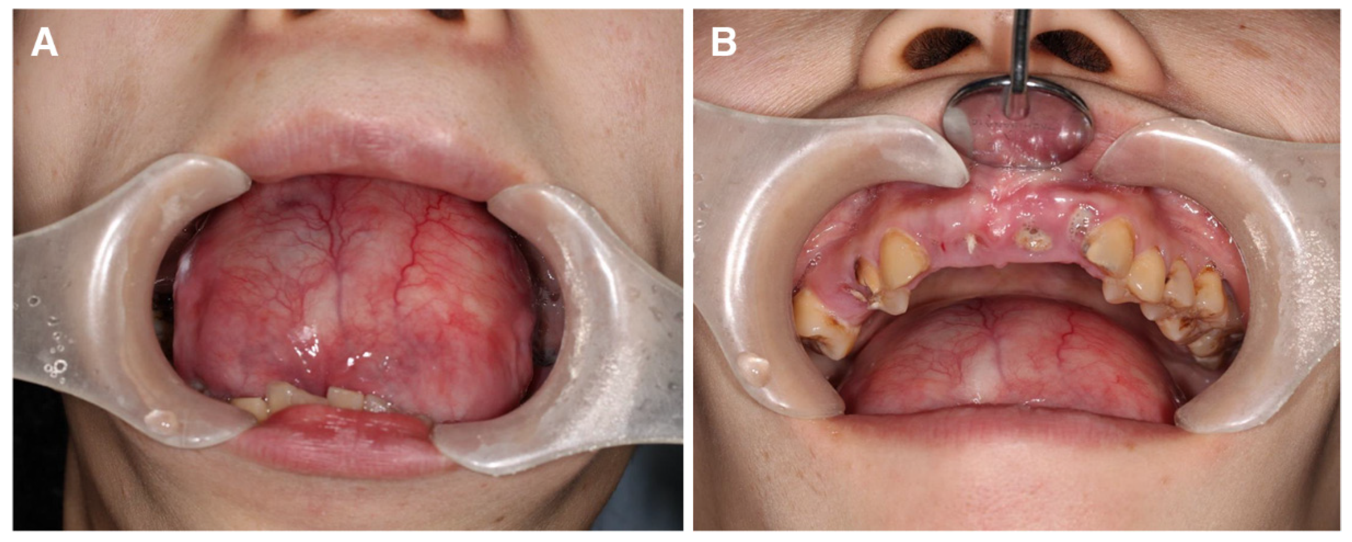
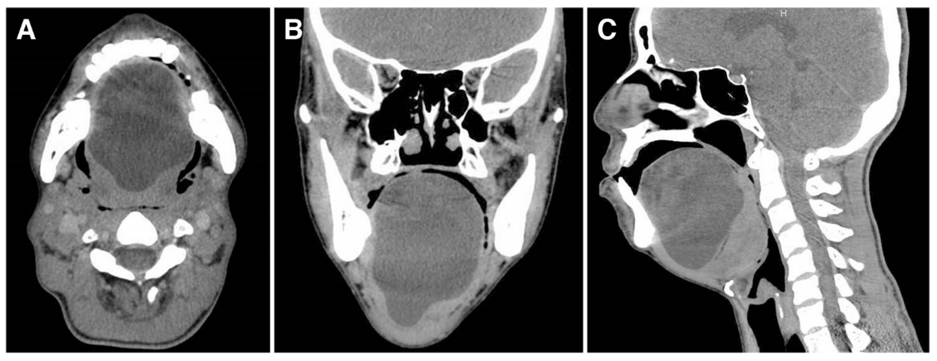
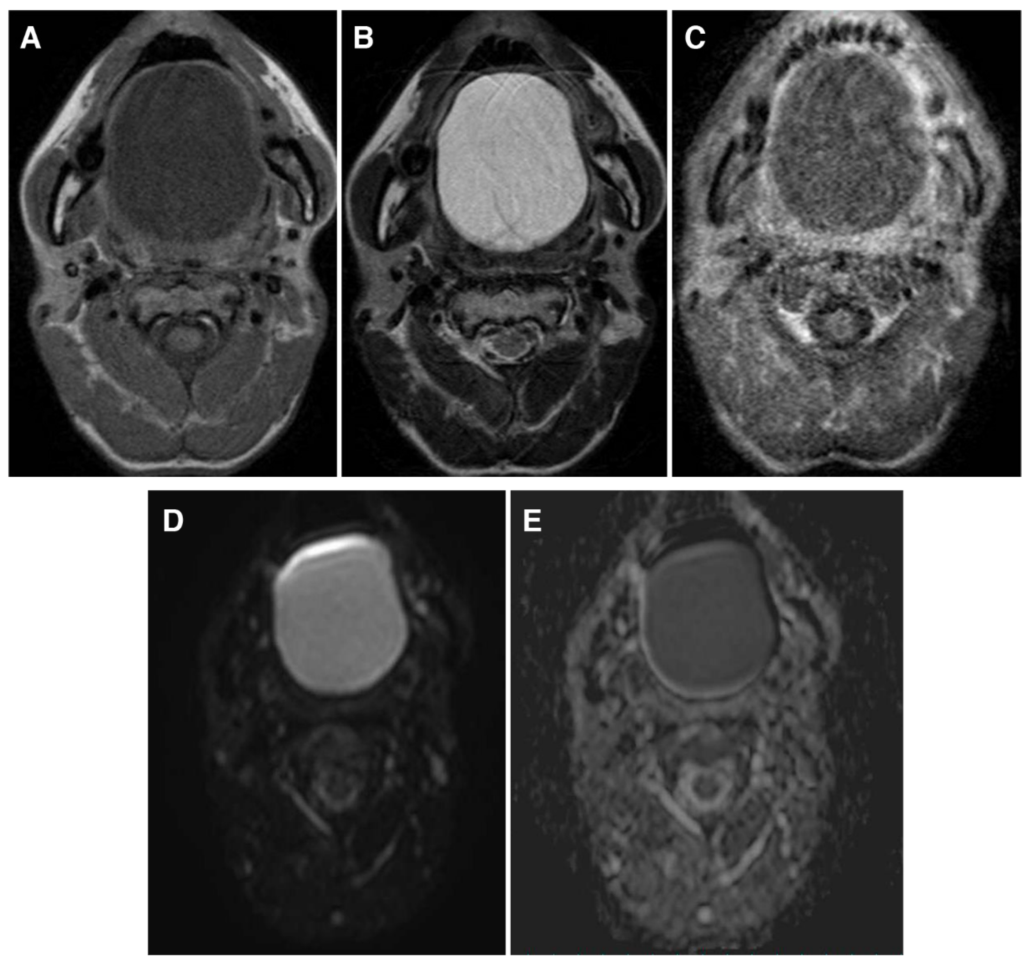
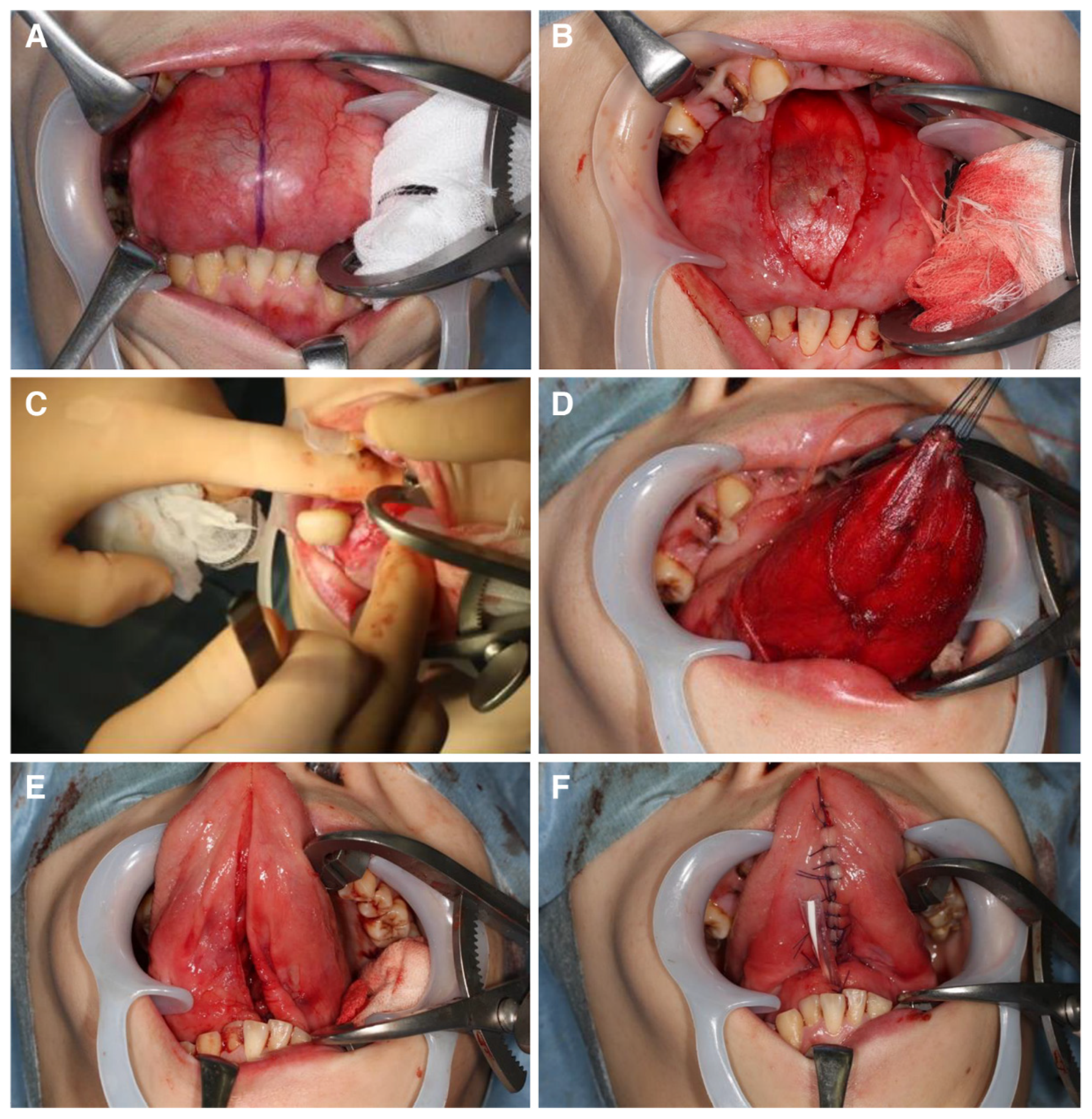
2. Case Presentation
3. Histological Diagnosis
4. Discussion
5. Conclusions
Author Contributions
Funding
Informed Consent Statement
Data Availability Statement
Acknowledgments
Conflicts of Interest
References
- New, G.B.; Erich, J.B. Dermoid cysts of the head and neck. Surg. Gynecol Obs. 1937, 65, 48–55. [Google Scholar]
- Kobayashi, Y.; Kino, K.; Manita, H.; Satou, R.; Kikuchi, K.; Yoshimasu, H.; Amagasa, T. Clinical Observation of Dermoid Cyst and Epidermoid Cyst in the Oral and Maxillofacial Region. J. Jpn. Stomatol. Soc. 1998, 47, 101–107. [Google Scholar]
- Ege, G.; Akman, H.; Senvar, A.; Kuzucu, K. Case report: Sublingual dermoid cyst. Tani. Gir. Radyol. 2003, 9, 57–59. [Google Scholar]
- De Ponte, F.S.; Brunelli, A.; Marchetti, E.; Bottini, D. Sublingual epidermoid cyst. J. Craniofac. Surg. 2002, 13, 308–310. [Google Scholar] [CrossRef] [PubMed]
- Kandogan, T.; Koc, M.; Vardar, E.; Selek, E.; Sezgin, O. Sublingual epidermoid cyst: A case report. J. Med. Case Rep. 2007, 1, 87. [Google Scholar] [CrossRef]
- Meyer, I. Dermoid cysts (dermoids) of the floor of the mouth. Oral Surg. Oral Med. Oral Pathol. 1955, 8, 1149–1164. [Google Scholar] [CrossRef]
- Kyriakidou, E.; Howe, T.; Veale, B.; Atkins, S. Sublingual dermoid cysts: Case report and review of the literature. J. Laryngol. Otol. 2015, 129, 1036–1039. [Google Scholar] [CrossRef]
- Berbel, P.; Ostrosky, A.; Tosti, F. Large Sublingual Dermoid Cyst: A Case of Mandibular Prognathism. Craniomaxillofac Trauma Reconstr. 2016, 9, 345–348. [Google Scholar] [CrossRef]
- Ueda, M.; Dehari, H.; Nakamori, K.; Shiratori, K.; Suzuki, R.; Hiratsuka, H. A case of a large epidermoid cyst in the floor of the mouth associated with dyspnea. Jpn. J. Oral Maxillofac. Surg. 2010, 56, 476–479. [Google Scholar] [CrossRef][Green Version]
- Longo, F.; Maremonti, P.; Mangone, G.M.; De Maria, G.; Califano, L. Midline (dermoid) cysts of the floor of the mouth: Report of 16 cases and review of surgical techniques. Plast. Reconstr. Surg. 2003, 112, 1560–1565. [Google Scholar] [CrossRef]
- Ho, M.W.; Crean, S.J. Simultaneous occurrence of sublingual dermoid cyst and oral alimentary tract cyst in an infant: A case report and review of the literature. Int. J. Paediatr. Dent. 2003, 13, 441–446. [Google Scholar] [CrossRef] [PubMed]
- Di Francesco, A.; Chiapasco, M.; Biglioli, F.; Ancona, D. Intraoral approach to large dermoid cysts of the floor of the mouth: A technical note. Int. J. Oral Maxillofac. Surg. 1995, 24, 233–235. [Google Scholar] [CrossRef]
- Obiechina, A.E.; Arotiba, J.T.; Ogunbiyi, J.O. Coexisting congenital sublingual dermoid and bronchogenic cyst. Br. J. Oral Maxillofac. Surg. 1999, 37, 58–60. [Google Scholar] [CrossRef] [PubMed]
- Seah, T.E.; Sufyan, W.; Singh, B. Case report of a dermoid cyst at the floor of the mouth. Ann. Acad. Med. Singap. 2004, 33 (Suppl. 4), 77–79. [Google Scholar] [PubMed]
- Jham, B.C.; Duraes, G.V.; Jham, A.C.; Santos, C.R. Epidermoid cyst of the floor of the mouth: A case report. J. Can. Dent. Assoc. 2007, 73, 525–528. [Google Scholar] [PubMed]
- El-Hakim, I.E.; Alyamani, A. Alternative surgical approaches for excision of dermoid cyst of the floor of mouth. Int. J. Oral Maxillofac. Surg. 2008, 37, 497–499. [Google Scholar] [CrossRef]
- Papadogeorgakis, N.; Kalfarentzos, E.F.; Vourlakou, C.; Alexandridis, C. Surgical management of a large median dermoid cyst of the neck causing airway obstruction. Case Rep. Oral Maxillofac. Surg. 2009, 13, 181–184. [Google Scholar] [CrossRef]
- Tsirevelou, P.; Papamanthos, M.; Chlopsidis, P.; Zourou, I.; Skoulakis, C. Epidermoid cyst of the floor of the mouth: Two case reports. Cases J. 2009, 2, 9360. [Google Scholar] [CrossRef]
- Patil, K.; Mahima, V.G.; Malleshi, S.N. Sublingual epidermoid cyst: A case report. Cases J. 2009, 2, 8848. [Google Scholar] [CrossRef]
- Jadwani, S.; Misra, B.; Kallianpur, S.; Bansod, S. Dermoid cyst of the floor of the mouth with abundant hair: A case report. J. Maxillofac. Oral Surg. 2009, 8, 388–389. [Google Scholar] [CrossRef][Green Version]
- Anantanarayanan, P.; Manikandhan, R.; Bhargava, D.; Sivapathasundara, B. Sub-lingual epidermoid cyst. Head Neck Pathol. 2010, 4, 136–138. [Google Scholar] [CrossRef] [PubMed][Green Version]
- Pan, M.; Nakamura, Y.C.; Clark, M.; Eisig, S. Intraoral dermoid cyst in an infant: A case report. J. Oral Maxillofac. Surg. 2011, 69, 1398–1402. [Google Scholar] [CrossRef] [PubMed]
- Jain, H.; Singh, S.; Singh, A. Giant sublingual dermoid cyst in floor of the mouth. J. Maxillofac. Oral Surg. 2012, 11, 235–237. [Google Scholar] [CrossRef] [PubMed]
- Ohta, N.; Watanabe, T.; Ito, T.; Kubota, T.; Suzuki, Y.; Ishida, A.; Kakehata, S.; Aoyagi, M. A case of sublingual dermoid cyst: Extending the limits of the oral approach. Case Rep. Otolaryngol. 2012, 2012, 634949. [Google Scholar] [CrossRef] [PubMed]
- Assaf, A.T.; Heiland, M.; Blessmann, M.; Friedrich, R.E.; Zustin, J.; Al-Dam, A. Extensive sublingual epidermoid cyst--diagnosis by immunohistological analysis and proof by podoplanin. In Vivo 2012, 26, 323–326. [Google Scholar] [PubMed]
- Verma, S.; Kushwaha, J.K.; Sonkar, A.A.; Kumar, R.; Gupta, R. Giant sublingual epidermoid cyst resembling plunging ranula. Natl. J. Maxillofac. Surg. 2012, 3, 211–213. [Google Scholar] [CrossRef] [PubMed]
- Dutta, M.; Saha, J.; Biswas, G.; Chattopadhyay, S.; Sen, I.; Sinha, R. Epidermoid cysts in head and neck: Our experiences, with review of literature. Indian J. Otolaryngol. Head Neck Surg. 2013, 65 (Suppl. 1), 14–21. [Google Scholar] [CrossRef]
- Aydın, S.; Demir, M.G.; Demir, N.; Şahin, S.; Kayıpmaz, S.S. A Giant Plunging Sublingual Dermoid Cyst Excised by Intraoral Approach. J. Maxillofac. Oral Surg. 2016, 15, 277–280. [Google Scholar] [CrossRef]
- Oginni, F.O.; Oladejo, T.; Braimah, R.O.; Adenekan, A.T. Sublingual epidermoid cyst in a neonate. Ann. Maxillofac. Surg. 2014, 4, 96–98. [Google Scholar] [CrossRef]
- Vieira, E.M.M.; Borges, A.H.; Volpato, L.E.R.; Porto, A.N.; Carvalhosa, A.A.; Botelho, G.A.; Bandeca, M.C. Unusual dermoid cyst in oral cavity. Case Rep. Pathol. 2014, 2014, 389752. [Google Scholar] [CrossRef]
- Yoshida, N.; Kodama, K.; Iino, Y. Sublingual epidermoid cyst presenting with distinctive magnetic resonance imaging findings. Clin. Pract. 2014, 4, 664. [Google Scholar] [CrossRef] [PubMed]
- Gordon, P.E.; Faquin, W.C.; Lahey, E.; Kaban, L.B. Floor-of-mouth dermoid cysts: Report of 3 variants and a suggested change in terminology. J. Oral Maxillofac. Surg. 2013, 71, 1034–1041. [Google Scholar] [CrossRef] [PubMed]
- Gulati, U.; Mohanty, S.; Augustine, J.; Gupta, S.R. Potentially fatal supramylohyoid sublingual epidermoid cyst. J. Maxillofac. Oral Surg. 2015, 2015 (Suppl. 1), 355–359. [Google Scholar] [CrossRef] [PubMed]
- Dabán, R.P.; Díez, E.G.; Navarro, B.G.; López-López, J. Epidermoid cyst in the floor of the mouth of a 3-year-old. Case Rep. Dent. 2015, 2015, 172457. [Google Scholar] [CrossRef][Green Version]
- Nishar, C.C.; Ambulgekar, V.K.; Gujrathi, A.B.; Chavan, P.T. Unusually Giant Sublingual Epidermoid Cyst: A Case Report. Iran J. Otorhinolaryngol. 2016, 28, 291–296. [Google Scholar]
- Basterzi, Y.; Sari, A.; Ayhan, S. Giant Epidermoid Cyst on the Forefoot. Dermatol. Surg. 2002, 28, 639–640. [Google Scholar]
- Brunet-Garcia, A.; Lucena-Rivero, E.; Brunet-Garcia, L.; Faubel-Serra, M. Cystic mass of the floor of the mouth. J. Clin. Exp. Dent. 2018, 10, e287–e290. [Google Scholar] [CrossRef]
- Silveira, H.A.; Almeida, L.Y.; Dominguete, M.H.L.; Graciano, K.P.P.; Bufalino, A.; León, J.E. Intraoral epidermoid cyst with extensive elastofibromatous changes: An unusual finding. Oral Maxillofac. Surg. 2019, 23, 493–497. [Google Scholar] [CrossRef]
- Baliga, M.; Shenoy, N.; Poojary, D.; Mohan, R.; Naik, R. Epidermoid cyst of the floor of the mouth. Natl. J. Maxillofac. Surg. 2014, 5, 79–83. [Google Scholar] [CrossRef]
- Oluleke, O.O.; Akau, K.S.; Godwin, A.I.; Kene, A.I.; Sunday, A.O. Sublingual Dermoid Cyst: Review of 14 Cases. Ann. Maxillofac. Surg. 2020, 10, 279–283. [Google Scholar] [CrossRef]
- Misch, E.; Kashiwazaki, R.; Lovell, M.A.; Herrmann, B.W. Pediatric sublingual dermoid and epidermoid cysts: A 20-year institutional review. Int. J. Pediatr. Otorhinolaryngol. 2020, 138, 110265. [Google Scholar] [CrossRef] [PubMed]
- Klibngern, H.; Pornchaisakuldee, C. A large sublingual epidermoid cyst with parapharyngeal space extension: A case report. Int. J. Surg. Case Rep. 2020, 72, 233–236. [Google Scholar] [CrossRef] [PubMed]
- Vélez-Cruz, M.E.; Gómez-Clavel, J.F.; Licéaga-Escalera, C.J.; Pérez, L.A.M.; Fandiño, J.J.T.; Iriarte, C.J.T.; Ramírez-Cano, M.F.; García-Muñoz, A. Sublingual dermoid cyst in an infant: A case report and review of the literature. Clin. Case Rep. 2020, 8, 1403–1408. [Google Scholar] [CrossRef] [PubMed]
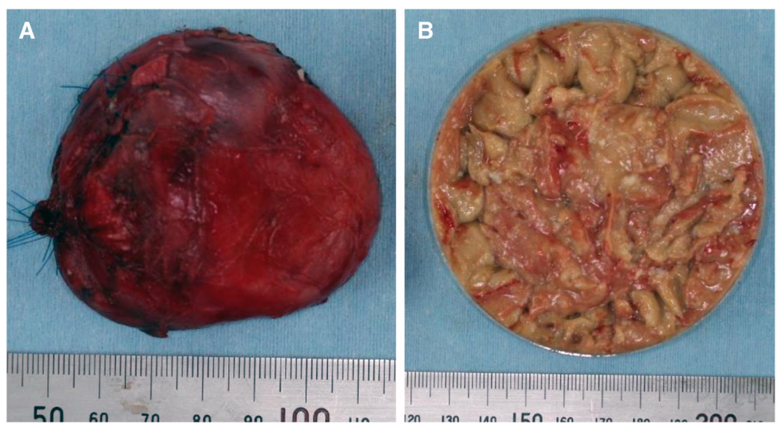
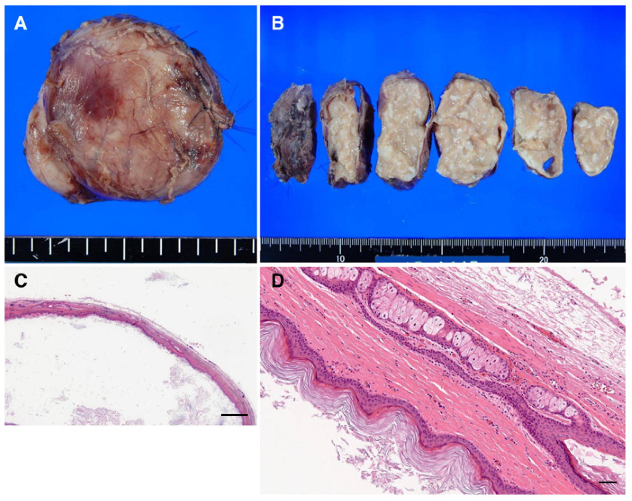
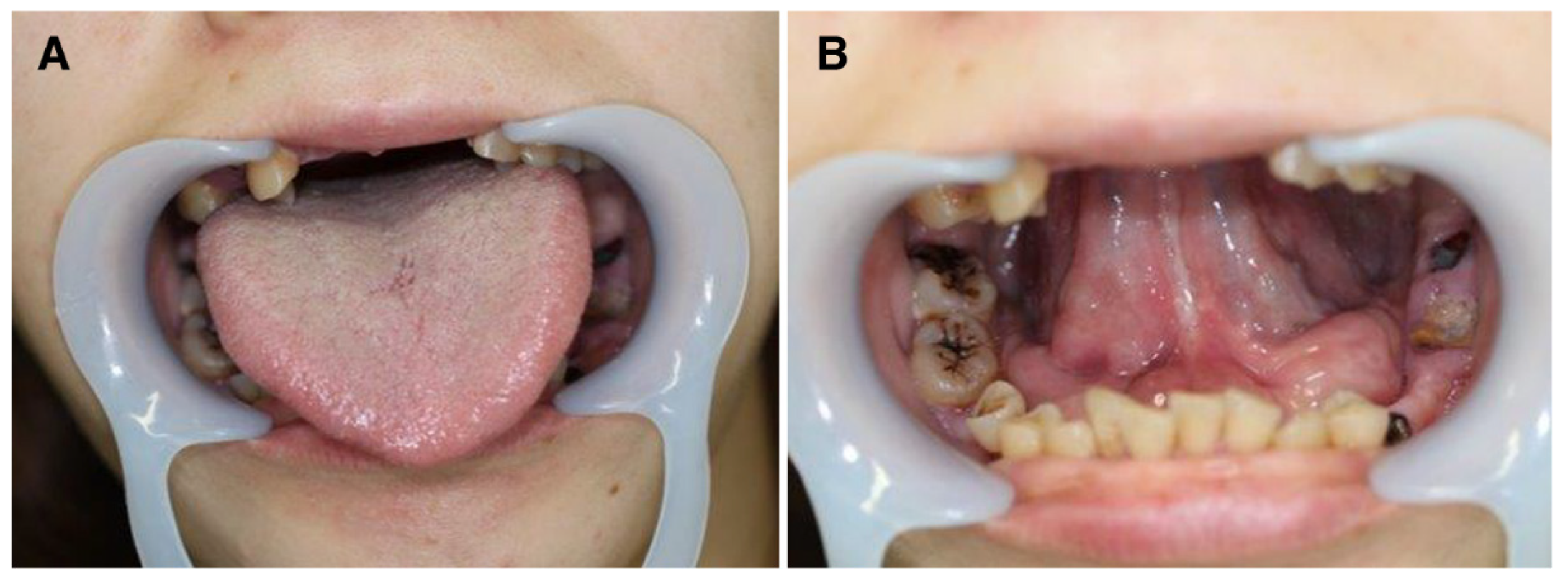
| Year | Author | Age (Years) | Sex | Disease Duration (Months) | Tracheostomy | Category 1 | Size (mm) | Surgical Method | Reduction Performed | Dysarthria | Dysphagia | Breathing Disorder |
|---|---|---|---|---|---|---|---|---|---|---|---|---|
| 1999 | Obiechina [13] | 5 | M | 60 | (-) | D | 45 × 50 × 35 | intraoral | (-) | (+) | (+) | (-) |
| 2003 | Ho [11] | 0.125 (1.5 months) | M | 0.07 | (-) | D | 11 × 10 × 7 | intraoral | (-) | (-) | (+) | (-) |
| 2004 | Seah [14] | 19 | M | 0.1 | (-) | D | 35 × 26 | intraoral | (-) | (-) | (-) | (-) |
| 2007 | Jham [15] | 25 | M | 2 | (-) | E | 50 × 50 | intraoral | (-) | (+) | (+) | (-) |
| 2008 | El-Hakim [16] | 22 | M | 120 | (-) | D | 120 × 120 | extraoral | (-) | (+) | (-) | (-) |
| 2009 | Papadogeorgakis [17] | 21 | F | 120 | (+) | D | 75 × 55 × 35 | intraoral/extraoral | (-) | (+) | (+) | (+) |
| 2009 | Tsirevelou [18] | 14 | F | 120 | (-) | E | 14 × 12 | intraoral | (-) | (-) | (-) | (-) |
| 2009 | Patil [19] | 28 | M | 5 | (-) | E | 30 × 20 × 20 | intraoral | (-) | (-) | (-) | (-) |
| 2009 | Jadwani [20] | 22 | M | 12 | (-) | D | 40 × 20 | intraoral | (-) | (-) | (-) | (-) |
| 2010 | Anantanarayanan [21] | 12 | F | 12 | (-) | E | 40 | intraoral | (-) | (+) | (-) | (-) |
| 2011 | Pan [22] | 0.4 (5 months) | M | 0 | (-) | D | 30 × 20 × 10 | intraoral | (-) | (+) | (-) | (+) |
| 2012 | Jain [23] | 17 | F | 48 | (-) | D | 90 × 90 | intraoral | (-) | (-) | (+) | (+) |
| 2012 | Ohta [24] | 21 | F | 24 | (+) | D | 55 × 56 × 45 | intraoral | (-) | (-) | (+) | (-) |
| 2012 | Assaf [25] | 39 | M | no data | (-) | E | no data | intraoral | (-) | (+) | (+) | (+) |
| 2012 | Verma [26] | 16 | F | 5 | (-) | E | 70 × 50 × 45 | intraoral | (-) | (-) | (+) | (-) |
| 2013 | Dutta [27] | 23 | F | 360 | (+) | E | 75 × 60 × 45 | extraoral | (-) | (+) | (+) | (-) |
| 2013 | Dutta [27] | 36 | M | 60 | (-) | E | 60 × 40 × 30 | intraoral | (-) | (-) | (-) | (-) |
| 2014 | Aydin [28] | 30 | M | 36 | (-) | D | 36 × 39 | intraoral | (-) | (-) | (-) | (-) |
| 2014 | Oginni [29] | 0.67 (8 months) | no data | 0.9 | (-) | E | 40 × 30 | intraoral | (-) | (-) | (-) | (-) |
| 2014 | Viera [30] | 29 | M | no data | (-) | D | 45 × 55 | intraoral | (-) | no data | no data | no data |
| 2014 | Yoshida [31] | 39 | F | 48 | (-) | E | 95 × 80 × 40 | intraoral | (-) | (+) | (-) | (+) |
| 2015 | Gordon [32] | 79 | F | 300 | (-) | D | 53 × 29 × 27 | intraoral | (-) | (-) | (-) | (-) |
| 2015 | Kyriakidou [7] | 17 | F | 3 | (-) | D | 60 × 50 × 35 | intraoral | (-) | (+) | (-) | (-) |
| 2015 | Gulati [33] | 16 | M | 3 | (-) | E | 62 × 60 × 57 | intraoral/extraoral | (-) | (-) | (+) | (-) |
| 2015 | Dabán [34] | 3 | F | 2 | (-) | E | 20 × 15 | intraoral | (-) | (-) | (-) | (-) |
| 2016 | Nishar [35] | 60 | M | 360 | (+) | E | 100 × 80 | extraoral | (-) | (-) | (-) | (-) |
| 2016 | Berbel [8] | 8 | F | 36 | (-) | D | 60 × 50 | intraoral | (-) | (+) | (-) | (-) |
| 2017 | Basterzi [36] | 10 | M | no data | (-) | E | 30 × 40 × 40 | extraoral | (-) | (-) | (+) | (+) |
| 2018 | Brunet-Garcia [37] | 43 | M | 24 | (-) | D | 40 × 32 × 34 | intraoral | (-) | (-) | (-) | (-) |
| 2019 | Silveira [38] | 26 | M | 24 | (-) | E | 70 × 70 | intraoral | (-) | (+) | (+) | (-) |
| 2020 | Baliga [39] | 26 | F | 36 | (-) | E | 30 | intraoral | (-) | (+) | (+) | (-) |
| 2020 | Oluleke [40] | 0.002 (1 day) | M | Birth | (-) | D | no data | intraoral | (-) | (-) | (+) | (+) |
| 2020 | Oluleke [40] | 0.002 (1 day) | F | Birth | (-) | D | no data | intraoral | (-) | (-) | (+) | (+) |
| 2020 | Oluleke [40] | 0.002 (1 day) | M | Birth | (-) | D | no data | intraoral | (-) | (-) | (+) | (+) |
| 2020 | Oluleke [40] | 0.002 (1 day) | M | Birth | (-) | D | no data | intraoral | (-) | (-) | (+) | (+) |
| 2020 | Oluleke [40] | 0.002 (1 day) | F | Birth | (-) | D | no data | intraoral | (-) | (-) | (+) | (+) |
| 2020 | Oluleke [40] | 0.16 | M | 2 | (-) | D | no data | intraoral | (-) | (-) | (+) | (+) |
| 2020 | Oluleke [40] | 1 | F | 12 | (-) | D | no data | intraoral | (-) | (-) | (+) | (+) |
| 2020 | Oluleke [40] | 7 | M | 84 | (-) | E | no data | intraoral | (-) | (-) | (-) | (+) |
| 2020 | Oluleke [40] | 9 | M | 108 | (-) | E | no data | intraoral | (-) | (-) | (-) | (-) |
| 2020 | Oluleke [40] | 10 | F | 120 | (-) | E | no data | intraoral | (-) | (-) | (-) | (-) |
| 2020 | Oluleke [40] | 13 | M | 156 | (-) | E | no data | intraoral | (-) | (-) | (-) | (-) |
| 2020 | Misch [41] | 12 | F | no data | (-) | D | 54 × 38 × 41 | intraoral | no data | no data | no data | no data |
| 2020 | Misch [41] | 8 | F | no data | (-) | D | 14 × 10 × 15 | intraoral | no data | no data | no data | no data |
| 2020 | Misch [41] | 1 | F | no data | (-) | E | 4 × 4×2 | intraoral | no data | no data | no data | no data |
| 2020 | Misch [41] | 16 | F | no data | (-) | D | 44 × 21 × 26 | intraoral | no data | no data | no data | no data |
| 2020 | Misch [41] | 11 | M | no data | (-) | D | 43 × 20 × 28 | intraoral | no data | no data | no data | no data |
| 2020 | Misch [41] | 0.17 | M | no data | (-) | E | 22 × 16 × 20 | intraoral | no data | no data | no data | no data |
| 2020 | Misch [41] | 14 | F | no data | (-) | D | 37 × 38 × 31 | intraoral | no data | no data | no data | no data |
| 2020 | Misch [41] | 8 | F | no data | (-) | D | 19 × 12 × 24 | intraoral | no data | no data | no data | no data |
| 2020 | Misch [41] | 0.5 | M | no data | (-) | E | 6 × 5×5 | intraoral | no data | no data | no data | no data |
| 2020 | Misch [41] | 2 | F | no data | (-) | D | 10 × 10 × 10, 12 × 10 × 12 | intraoral/extraoral | no data | no data | no data | no data |
| 2020 | Misch [41] | 6 | F | no data | (-) | E | 12 × 11 × 5 | intraoral | no data | no data | no data | no data |
| 2020 | Misch [41] | 3 | M | no data | (-) | E | 6 × 5×3 | intraoral | no data | no data | no data | no data |
| 2020 | Klibngern [42] | 26 | F | no data | (-) | E | 65 × 32 × 25 | intraoral | (-) | (+) | (+) | (+) |
| 2020 | Vélez-Cruz [43] | 0.58 | F | 8 | (-) | D | 25 × 20 × 10 | intraoral | (-) | (-) | (+) | (-) |
| Present Case: Hikasa | 38 | F | 6 | (-) | E | 65 × 76 × 54 | intraoral | (-) | (+) | (+) | (+) | |
Publisher’s Note: MDPI stays neutral with regard to jurisdictional claims in published maps and institutional affiliations. |
© 2022 by the authors. Licensee MDPI, Basel, Switzerland. This article is an open access article distributed under the terms and conditions of the Creative Commons Attribution (CC BY) license (https://creativecommons.org/licenses/by/4.0/).
Share and Cite
Hikasa, H.; Sakata, K.-i.; Mizuno, T.; Kato, T.; Kitagawa, Y.; Sakakibara, N. A Case of a Giant Sublingual Epidermoid Cyst Removed by Content Reducing Surgery. Oral 2022, 2, 126-136. https://doi.org/10.3390/oral2010013
Hikasa H, Sakata K-i, Mizuno T, Kato T, Kitagawa Y, Sakakibara N. A Case of a Giant Sublingual Epidermoid Cyst Removed by Content Reducing Surgery. Oral. 2022; 2(1):126-136. https://doi.org/10.3390/oral2010013
Chicago/Turabian StyleHikasa, Hiroshi, Ken-ichiro Sakata, Takayuki Mizuno, Takumi Kato, Yoshimasa Kitagawa, and Noriyuki Sakakibara. 2022. "A Case of a Giant Sublingual Epidermoid Cyst Removed by Content Reducing Surgery" Oral 2, no. 1: 126-136. https://doi.org/10.3390/oral2010013
APA StyleHikasa, H., Sakata, K.-i., Mizuno, T., Kato, T., Kitagawa, Y., & Sakakibara, N. (2022). A Case of a Giant Sublingual Epidermoid Cyst Removed by Content Reducing Surgery. Oral, 2(1), 126-136. https://doi.org/10.3390/oral2010013


