Review

Primary Cutaneous B-Cell Lymphoma: An Update on Pathologic and Molecular Features

1
Anatomic Pathology Unit, Department of Molecular Medicine, University of Pavia, Fondazione IRCCS Policlinico San Matteo, Via Forlanini 14, 27100 Pavia, Italy
2
Anatomic Pathology Unit, Department of Molecular Medicine, University of Pavia, 27100 Pavia, Italy
*
Author to whom correspondence should be addressed.
Hemato 2022, 3(2), 318-340; https://doi.org/10.3390/hemato3020023
Submission received: 23 March 2022 / Revised: 26 May 2022 / Accepted: 27 May 2022 / Published: 29 May 2022

Abstract

:
Primary cutaneous B-cell lymphomas (PCBCLs) account for 25% of all primary cutaneous lymphomas. Three major types are currently recognized by the WHO classification: primary cutaneous marginal zone B-cell lymphoma (PCMZL), primary cutaneous follicle centre lymphoma (PCFCL) (both considered indolent lymphomas) and primary cutaneous diffuse large B-cell lymphoma, leg-type (PCDLBCL-LT), which is, instead, a very aggressive disease. Nowadays, the PCBCL’s category also includes some rare entities such as intravascular B-cell lymphoma (IVBL) and the EBV+ mucocutaneous ulcer (EBVMCU). Furthermore, controversies still exist concerning the category of primary cutaneous diffuse large B-cell lymphoma (PCDLBCL), because some cases may present with clinical and histological features between PCFCL and PCDLBCL-LT. Therefore, some authors proposed introducing another category called PCDLBCL, not otherwise specified (NOS). Regardless, PCBCLs exhibit distinct features and differ in prognosis and treatment from their nodal/systemic counterparts. Therefore, clinicopathologic analysis is a key diagnostic element in the work-up of these lymphomas.

1. Introduction

Primary cutaneous B-cell lymphomas (PCBCLs) are a clinically and pathologically heterogeneous group of extranodal non-Hodgkin’s lymphomas (NHLs) that primarily involve the skin, do not have evidence of extracutaneous disease at diagnosis and do not exhibit extracutaneous spread for a long time (or for the entire course of the disease) [1].
PCBCLs make up about 25% of all primary cutaneous lymphomas, but less than 1% of all NHLs [2,3]. PCBCLs exhibit distinct clinical, histological, immunophenotypic and genetic features and differ in prognosis and treatment from their nodal/systemic counterparts [4,5].
PCBCL diagnosis and management is a multidisciplinary task, involving dermatologists, pathologists, haemato-oncologists and radiation oncologists [6]. The diagnosis of PCBCL needs careful histomorphological and immunophenotypical analyses, corroborated by clinical data and, when necessary, by molecular and cytogenetic investigations. The pre-analytical phase is crucial, and adequate (in size and quality) lesional samples should be obtained. Whenever possible, excisional biopsies should be recommended [2].
Careful clinical examination and staging at presentation are mandatory to exclude secondary cutaneous localization of systemic B-cell lymphomas—these can be histologically indistinguishable from their primary cutaneous counterparts [7].
Current WHO (2017) classification [8] recognizes three types of PCBCLs as being most frequent: (1) primary cutaneous marginal zone B-cell lymphoma (PCMZL); (2) primary cutaneous follicle centre lymphoma (PCFCL); and (3) primary cutaneous diffuse large B-cell lymphoma, leg-type (PCDLBCL-LT). PCMZL and PCFCL are considered to be indolent lymphomas with a good prognosis and a five-year disease-specific survival rate of >95%. In contrast, PCDLBCL-LT is an aggressive lymphoma, with a five-year survival rate between 40 and 60%. PCBCL also includes some rare entities such as intravascular B-cell lymphoma (IVBL) and the EBV+ mucocutaneous ulcer (EBVMCU), a provisional entity [3].
In spite of these classificational advances, the diagnosis of PCBCL can be challenging and some issues are still a matter of debate. In some cases, a clear-cut distinction between PCMZL and/or PCFCL and reactive cutaneous lymphoid infiltrates (so-called “pseudolymphoma”) may be difficult through histology alone [9].
Furthermore, controversies still exist concerning the category of primary cutaneous diffuse large B-cell lymphoma (PCDLBCL) [10]. The border between centroblast-rich PCFCL and PCDLBCL is not clear; similarly, the term “PCDLBCL-LT” sounds inadequate for describing the whole spectrum of PCDLBCLs. Cases of PCDLBCL that are mostly composed of centroblast-like cells may also arise in sites other than the legs and pursue a less aggressive clinical course. Based on such findings, some authors proposed the definition of PCDLBCL, not otherwise specified (NOS) for these cases [11].

2. Primary Cutaneous Marginal Zone B-Cell Lymphoma

PCMZL is a very indolent type of extranodal marginal zone lymphoma (MZL), originating from the skin associated lymphoid tissue (SALT) that presents in the skin with some distinctive features [3,12]. It accounts for about 20–40% of all PCBCLs in western countries (0.4 per 1,000,000 per year in the U.S.A.) [8]. It affects mainly adults and the elderly (median age at diagnosis >50 years with a male predominance) [13] and, exceptionally, children and adolescents. However, the occurrence of primary cutaneous MZL in the paediatric age range is a matter of debate [14,15].
As in other extranodal MZL, a link with chronic antigenic stimulation has also been suggested for PCMZL, including bacterial and viral agents, tattoo pigments, vaccines and iatrogenic agents (fluoxetine). Borrelia burgdorferi infection has been associated with PCBCL, including the PCMZL type, according to studies from some European countries (Scotland and Austria); however, other studies, mostly from Asia and the U.S.A., did not confirm such an association, suggesting geographic variability [16,17]. Other infections possibly associated with the development of PCMZL are herpes simplex virus type 1 and hepatitis virus [18]. Notably, hepatitis C virus (HCV) infections have been found in association with up to 43% of PCMZL, according to one Italian study, and rare cases responding to antiviral therapy have been reported [19,20,21,22]. PCMZL may also arise in an autoimmune disease setting, such as Sjögren syndrome or Hashimoto’s thyroiditis [23,24,25].

2.1. Clinical Features

PCMZL usually presents on the arms and trunk, but other sites, including the head and neck, may also be involved. Lesions are often multifocal, in contrast to PCFCL, and consist of red to purple papules, nodules and/or plaques. Ulceration is exceptional [26,27]. In some cases, mostly in the context of autoimmune disorders, the lesion may spontaneously regress, leaving a localized area of flaccid skin (so-called anetoderma) due to loss of dermal elastic tissue [28,29]. One peculiar subset of PCMZL, which is associated with HCV infection and has a female predilection, presents with confined subcutaneous nodular lesions, clinically mimicking lipoma [22].

2.2. Pathology

PCMZL infiltrates initially involve the reticular dermis and may subsequently extend throughout the whole dermis and hypodermis—the epidermis is spared. Periadnexal infiltration around eccrine glands and hair follicles is frequently seen, but lymphoepithelial lesions are uncommon and not critical for diagnosis [24].
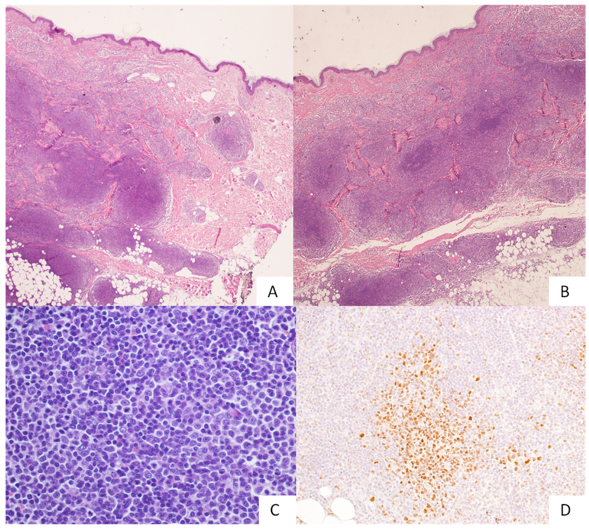
The overall architecture is usually nodular, but it may also be diffuse (Figure 1). Lymphoid follicles with reactive germinal centres and preserved mantle zones are frequently seen. In the initial lesion, the neoplastic marginal zone (MZ) circumscribes the reactive follicles. At disease progression, the follicles can be colonized by MZ B cells with partial and/or diffuse effacement of dendritic meshwork [12]. The lymphoma population includes small- to medium-sized centrocyte-like MZ B cells, monocytoid B cells (Figure 1), lymphoplasmacytic cells, scattered centroblasts and/or immunoblast-like cells. A variable degree of plasmacytic differentiation may occur (Figure 1), the plasma cells usually being located at the periphery of the infiltrate. Amyloid deposition can be found in cases with prominent plasmacytic differentiation. Some morphological variants of PCMZL have been described, including small-cell lymphocytic variants, monocytoid variants and variants with diffuse plasmacytic differentiation [30]. A variable amount of inflammatory cells may be admixed with the neoplastic population, including T-cell lymphocytes, histiocytes, mast cells and eosinophils. The reactive inflammatory population may be prominent, obscuring the lymphoma cells [31].
No specific immunophenotype profile exists for MZLs in general, including cutaneous ones. Lymphoma cells express B-cell antigens (CD19, CD20, CD22 and CD79a), and bcl-2 being negative for CD5, CD10, bcl-6 and Cyclin D1. CD23 expression is variable. Plasma cell components express CD38, CD138 and CD79a, but not CD20. Light chain clonal restrictions can be demonstrated with immunohistochemistry, as well as by in situ hybridization. Follicular markers (CD10 and bcl-6), coupled with Ki-67, are helpful in highlighting residual reactive germinal centres, in which CD21 and CD23 immunostainings may evidentiate the disruption of the dendritic meshwork [32].
Increased numbers of plasmacytoid dendritic cells (CD123+) have been reported in PCMZL when compared with very few or absent plasmacytoid dendritic cells in PCFCL and PCLBCL, respectively [33].
Recently, PCMZL has been subdivided into two subtypes, one carrying class-switched immunoglobulins, the other carrying non-class-switched ones [34]. The class-switched form of the disease (90% of cases) contains monotypic plasma cells with expressions of IgG and, to a lesser extent, IgA or IgE, which are mainly localized at the periphery of the infiltrate. It lacks CXCR3, shows a predominant type 2 helper T cell environment and lacks reactive germinal centre colonization [35]. In contrast, the non-class-switched PCMZL (10% of cases) shows CXCR3 and IgM expression and more frequently reveals a diffuse proliferation of large nodules of neoplastic B cells. The class-switched subtype seems to pursue an indolent course, with an extremely low risk of progression to large B-cell lymphoma and/or of extracutaneous spread [36].
On such a basis, it seems reasonable to retain the class-switched subtype as a clonal chronic lymphoproliferative disorder, rather than as a lymphoma. Up to 40% of IgG-positive PCMZL cases express IgG4, but they are unrelated to systemic IgG4-related diseases [37].

2.3. Molecular and Cytogenetic Features

Clonal IGH or IGK gene rearrangements may be detected in up to 80–92% of PCMZLs [24,38]. Cytogenetic abnormalities usually associated with MALT lymphoma have been variably reported in PCMZL: the t(14;18)(q32;q21) (IGH-MALT1) is the most frequent one (in up to 25% of PCMZL), whereas t(11;18)(q21;q21) (BIRC3-MALT1) and t(3;14)(p14.1;q32) (IGH-FOXP1) rearrangements are less common. The t(1;14)(p22;q32) involving IGH and BCL10 has not been identified [39]. Trisomies of chromosomes 3 and 18 have been reported in up to 20% of PCMZLs. BCL6 rearrangements have been sporadically observed, but IGH-BCL2 translocations are absent. Activating MYD88L265P mutations, more commonly associated with lymphoplasmacytic lymphoma and PCDLBCL-LT, have been detected in 6% of PCMZLs, however, exclusively in IgM-positive types (three of six IgM-positive PCMZLs) [40]. The mutational landscape of PCMZL may also include alterations of FAS (24 of 38, 63%), SLAMF1 (9 of 38, 24%), SPEN (7 of 38, 18%) and NCOR2 (5 of 38, 13%) [41].

2.4. Differential Diagnosis

Particularly in the early phase of PCMZL, the lymphoma infiltrate may appear to be not specific. Differential diagnoses include other lymphomas (i.e., lymphoplasmacytic lymphoma, plasma cell myeloma and primary cutaneous follicular helper T-cell lymphoma (PCFHTCL)), as well as inflammatory, non-neoplastic cutaneous lymphoid hyperplasia (CLH) [24]. The distinction between PCMZL lymphoma and CLH may sometimes be very difficult to determine (Table 1) as they often share histopathological features (abundant reactive background and B follicles with germinal centres) [42].
The presence of lymphoepithelial lesions has a limited diagnostic value in PCMZL. Ancillary studies may corroborate lymphoma diagnosis, documenting clonal IGH rearrangements by means of molecular analyses and/or light chain restriction by means of immunohistochemistry or ISH technique. However, it is important to stress that clonal rearrangements may also occur in some reactive lymphoid infiltrates. As a consequence, a definitive diagnosis may not always be achieved, even after a judicious integration of pathological and clinical data. In these cases, only clinical “follow-ups” and repeated biopsies may finally confirm the lymphoma diagnosis [12].
Distinction of PCMZL from other PCBCLs requires the careful evaluation of a series of cytological and architectural features combined with a proper immunohistochemical panel. The B-cell follicles in PCMZL typically exhibit reactive germinal centres. The follicles in PCFCL are monomorphic in appearance, with low Ki-67 proliferation, while bcl6+ and CD10+/− B cells are detectable in the interfollicular areas (Table 1). Cases with prominent plasmacytic differentiation must be distinguished from lymphoplasmacytic lymphoma and plasma cell myeloma [43]. PCMZL usually lacks the MYD88L265P mutation typical of lymphoplasmacytic lymphoma; nevertheless, it is important to remember that plasmacytic differentiation may occur in PCMZL [44,45]. In the skin infiltrated by systemic myeloma, the detection of the sheet-like proliferation of monotypic plasma cells and/or plasmablasts (without a CD20+ lymphocytic component) support the myeloma diagnosis.
Cytoarchitectural features and immunostainings for CD5, CD23, SOX11 and cyclin D1 usually allow for the exclusion of cutaneous localizations of mantle cell lymphoma (MCL) and chronic lymphocytic leukaemia [8].

2.5. Prognosis and Treatment

PCMZL prognosis is excellent, with a five-year survival rate of 98%. Complete response to therapy occurs in 93% of patients with solitary lesions and 75% of patients with a multifocal disease [46]. Relapses are common within five years, occurring in 39% of patients with solitary lesions and 77% of patients with multifocal lesions. PCMZL rarely exhibits extracutaneous spread (lymph nodes and MALT sites) or large cell transformations [47,48].
PCMZL with solitary or few contiguous lesions can be treated with radiotherapy or excision with curative intent. Antibiotic treatment is required in Borrelia antibody-positive cases. Lesional regression has also been reported in cases of HCV-related PCMZL, including the so-called “lipoma-like” variant. Topical therapies are sometimes employed, including clobetasol, nitrogen mustard, cryotherapy and imiquimod [49,50,51].

2.6. Summing Up

PCMZL is an uncommon lymphoma subtype clinically characterized by a very indolent course and a favourable outcome. In addition, spontaneous lesional regression has been reported in various cases. In contrast, some studies have reported high rates of relapse, mostly cutaneous. Particularly in the early stage of the disease, differential diagnosis between PCMZL and CLH may be difficult. Recently, an MZL classification based on IgH switching revealed two disease subsets, one of which (class-switched form) usually shows a better clinical course and outcome. On such a basis, it is reasonable to postulate that at least a part of PCMZL indeed represents atypical lymphoid proliferation rather than true lymphoma. The high relapse rates reported in certain studies might be, at least in part, ascribed to an incomplete lesional surgical excision, thus representing a local disease recurrence rather that a true relapse.

3. Primary Cutaneous Follicle Centre Lymphoma

PCFCL is a low-grade lymphoid malignancy that develops from a mature germinal centre (GC) B cell in the skin. PCFCL accounts for about 50% of PCBCL, and for 10–20% of all cutaneous lymphomas. PCFCL is more frequent in Caucasian males (median age: 55), and its occurrence in the paediatric age range is matter of debate; male-to-female ratio is 1.5:1 [8,52,53,54]. Borrelia burgdorferi infections have also been reported in a fraction of PCFCL cases from endemic areas [55,56]. Prognosis is very favourable, but relapses may occur. Transformation into diffuse large B-cell lymphoma (DLBCL) has been reported [57].

3.1. Clinical Features

PCFCL usually presents with solitary, localized or (less often) multifocal (15%), painless, non-pruritic, erythematous to violaceous plaques, nodules or tumours that vary in size and are not usually ulcerated. The most frequently involved sites include the head (in particular the forehead and scalp) and trunk [8,58].
PCFCL presenting on the trunk—historically known as “reticulohistiocytoma of the dorsum” or “Crosti lymphoma—is characterized by a central core of plaques and tumours centrifugally surrounded by papules and macules [59]. Localization on the legs occur in about 5% of cases, and it seems to be associated with a less favourable outcome [8].
Without treatment, lesions tend to slowly progress and may assume a pattern of infiltrating and destroying [60]. Recurrences usually occur at the same site or in proximity of initial presentation [8].

3.2. Pathology

In the early stage, PCFCL usually shows a nodular growth pattern with closely spaced, monotonous neoplastic follicular aggregates (Figure 2). Lymphoma follicles have no polarization, have diminished (or absent) mantle zones and lack tingible body macrophages. Typically, a “grenz zone” beneath the epidermis surface is observed. At disease progression, the neoplastic follicles tend to fuse, resulting in a nodular and/or diffuse growth pattern, often with hypodermis involvement (Figure 2). Occasionally, an “inverted nodular pattern” can be encountered, with neoplastic cells located in pale areas at the periphery of follicles and small reactive lymphocytes in dark areas at the centre [61].
PCFCL populations consist of an admixture of centrocytes and centroblasts. Sometimes it may consist predominantly of large, often multilobated, cells or exhibit a spindle-like cytological appearance, mimicking sarcomatoid neoplasms (mostly due to intermingled reactive fibrosis) [62].
In contrast with its nodal counterpart, in PCFCL, the grading (number of large centroblast-like cells) and predominant growth pattern (follicular versus diffuse) retain limited prognostic relevance.
PCFCL may be associated with a variable amount of reactive T cells and histiocytes. Dendritic meshwork (highlighted by CD21/CD23 immunostainings) is usually disrupted and/or effaced. PCFCL cells express B-cell markers (CD20, CD79a, CD19, CD22 and PAX5) and GC markers (bcl6, CD10, MEF2B and HGAL). The CD10 expression on paraffin sections is variable, being influenced by antibody clones and fixation time. Notably, PCFCLs showing a diffuse growth pattern frequently lack CD10. Some studies have reported a lack of bcl-2 in the vast majority of PCFCL cases. In contrast, in our and other researchers’ experience, bcl-2 may be expressed in at least 25–27% of PCFCL cases; the evaluation of bcl-2 expression in PCFCL may be difficult in cases with T-cell-rich reactive infiltrates that are uniformly bcl-2+. The proliferation index is variable, from low to up to 30%.
Cases showing a diffuse growth pattern, coupled with an increased number of centroblast-like cells, usually have a higher Ki-67 rate [8].

3.3. Molecular and Cytogenetic Features

In the past, t(14;18)(q23;q21), the genetic hallmark of nodal follicular lymphoma (FL) involving BCL2 and IGH genes, has been retained as an exceedingly rare occurrence in PCFCLs. This lack was proposed as a way to distinguish secondary skin localizations of nodal FL from PCFCL [63]. In contrast, in the last decade, several groups have documented BCL2 chromosomal aberrations in up to 40% of PCFCL cases [63,64,65,66,67,68,69,70,71]. Such divergent findings could be partially related to the molecular technique employed, with a higher incidence observed with PCR analysis [69].
In some studies, the presence of BCL2 rearrangements has been more frequently associated with a predominantly centrocytic morphology, a high probability of skin relapse and a higher risk of extracutaneous spread [72,73].
Other reported chromosomal alterations include loss of heterozygosity in chromosomes 6p and 9p, deletion of 1p36, 14q32.33, 2p11.2 (IGKV locus), 9p21.3 (CDKN2A locus) and 14q32.33 (IGH locus), gains involving chromosomes 7 and 18 and high-level amplifications at 2p16.1 (REL gene). Aberrant somatic hypermutations targeting BCL6, PAX5, RhoH/TTF and/or MYC genes have also been reported. NGS analysis documented somatic mutations on TNFRSF14, CREBBP, TNFAIP3, KMT2D, SOCS1, EP300, STAT6 and FOX01 genes, as well as nucleotide substitutions (C>T transitions) associated with UV-damage [74]. Concomitant 1p36 deletion and TNFRSF14 mutations in PCFCL seem to be associated with elevated levels of EZH2 protein expression [75,76].

3.4. Differential Diagnosis

PCFCL with a predominant follicular growth pattern must primarily be distinguished from reactive CLH (Table 1). The differential diagnosis between PCFCL and CLH mainly relies on the lack of the typical features that characterize the reactive follicles, such as heterogeneity in shape and size, compartmentalization (polarization) into light and dark zones, detectable mantle and marginal zone areas and the presence of tingible body macrophages. Reactive follicles have also retained the CD21/CD23+ dendritic meshwork and a high Ki-67 proliferation index. PCFCL favours the reduction or disappearance of mantle and marginal zone areas coupled with the absence of tingible body macrophages and a low proliferation index. Similarly, lymphoma favours an expansion of follicular centre B cells (bcl-6+ and CD10+) in the interfollicular areas. Detection of clonal Ig gene rearrangements may support lymphoma diagnosis, but some cases remain doubtful and clinical follow-ups should be recommended [77].
The distinction of PCDLBL from secondary cutaneous localizations of nodal FL is a clinically relevant issue because of divergences in prognosis and therapy. PCFCL and secondary cutaneous involvement by a nodal FL can be indistinguishable in skin biopsies by morphology alone; thus, a complete clinical staging is mandatory. However, the primary cutaneous forms often lack CD10 and bcl-2 immunoreactivity as well as BCL2 rearrangements [63]. In skin localizations of systemic FL, the neoplastic cells usually show strong co-expressions of bcl2 and bcl6 and carry BCL-2 translocation.
Recently, a whole-exome sequencing study on a series of FLs—, including both systemic and primary cutaneous ones—proposed three criteria: BCL2 rearrangement, chromatin-modifying gene mutations (CREBBP, KMT2D, EZH2, and EP300) and proliferation rate to identify PCFCL subsets at different degrees of risk for concurrent or future systemic spread [78]. PCFCLs with diffuse growth patterns and a high content of large, centroblast-like cells, enter the differential diagnosis with PCDLBCL-LT (Table 2).
The presence of large, cohesive sheets of centroblasts and immunoblasts with an activated B-cell-like (ABC) profile usually leads to the diagnosis of DLBCL; the presence of a vaguely residual nodular pattern (at low magnification), a residual dendritic meshwork (although disrupted), an intense, intermingled reactive T cell infiltrate, a weak to absent bcl-2 expression and negativity for IgM and MUM1 favour a PCFCL. Additional molecular analysis can also be useful, as PCDLBCLs variably carry MYC and/or BCL6 translocations, BCL2 and MALT1 region amplifications, CDKN2A loss on chromosome 9p21.3 (more than 50% of cases) and MYD88L265P mutations [79].
Differential diagnosis between PCFCL and PCMZL mainly concerns cases of PCFCL presenting with an inverted nodular pattern, or cases of PCMZL with prominent follicular colonization. The presence of a clonal plasmacytic component, rarely seen in PCFCL, and negativity for GC markers (bcl6 and CD10) in neoplastic cells may be a useful diagnostic clue to recognize PCMZL, along with negativity for STMN1, LMO2, HGAL, MNDA and AID, and the absence of 1p36 deletion [80,81,82].
PCFHTCL is a follicular centre T-helper CD4+ neoplasm, not yet well characterized, which can mimic, both clinically and histologically, PCFCL. Cases of PCFHTCL misdiagnosed as PCFCL have been recognized only after an ineffective therapy with rituximab [83].
PCFHTCL usually contains a large amount of accompanying reactive B follicles; neoplastic T cells are medium to large in size, have irregular nuclei with small nucleoli and express T-cell markers (including CD2, CD3, CD4), T follicular helper markers (PD1, CXCL13 and ICOS) and GC markers (such as CD10 and bcl6); T-cell clonality can be detected in most cases. PCFCL with prominent spindle-shaped cytology must be distinguished from other spindle-cell tumours, including spindle cell melanoma, spindle squamous cell carcinoma, and spindle cell mesenchymal neoplasms [62,84].

3.5. Prognosis and Treatment

PCFCL is an indolent disease, with a 95% disease-specific five-year survival rate.
After treatment (see below), 99% of patients achieve complete remission; cutaneous relapses occur in about 30% of cases, but only 10% of patients have extracutaneous spread (lymph nodes, bone marrow and/or extralymphoid organs) [52,53,54].
Progression into large B-cell lymphomas is rare, and usually does not fit with leg-type features. Histology and multifocal diseasedo not influence prognosis. PCFCLs presenting on the legs seem to pursue a less favourable outcome (overall five-year survival rate: 41%). The prognostic significance of BCL-2 translocation in PCFCL is a controversial issue. In our experience and in other studies, the presence of such translocation seems to be associated with less favourable outcomes and an elevated risk of recurrence [73].
Treatment choices include the “watch and wait” approach, excision, radiotherapy and local or systemic therapy based on clinical presentation and disease extension [6].
Cutaneous relapses do not usually require a more aggressive treatment. Low-dose radiation therapy is recommended for localized lesions, allowing a complete response rate of 99%. Intralesional corticosteroids or rituximab and other topical agents (such as mustard, cryotherapy and imiquimod) can also be employed. Skin-disseminated diseases can achieve complete remission after systemic biological therapy with single-agent rituximab, while multiagent chemotherapy (R-CHOP) is reserved for refractory diseases, extracutaneous dissemination and cases arising on the legs [3,6].

3.6. Summing Up

PCFCL is the most frequent subtype of PCBCL, usually with a favourable clinical outcome—other than for cases presenting on the leg. Histologically, PCFCL exhibits, in the early stage, a nodular/follicular pattern. Subsequently, lymphoma follicles tend to merge, resulting in a diffuse pattern extending into subcutis. Lymphoma populations consist of an admixture of centrocyte- and centroblast-like cells. They sometimes present with peculiar cytological features, including polylobated centroblasts and a spindle-like cell appearance. The number of centroblastic cells and the growth pattern are retained as not being prognostically significant. PCFCL has a B cell phenotype with variable expressions of GC markers and bcl-2, the latter often negative. Mainly in PCFCL, CD10 may also be negative, showing a diffuse growth pattern. The presence of BCL-2 rearrangements is still debated, but some studies have reported that cases of PCFCL with BCL-2 translocations are associated with a less favourable outcome.

4. Primary Cutaneous Diffuse Large B Cell Lymphoma, Leg-Type

PCDLBCL-LT is an aggressive DLBCL characterized by sheets of centroblasts and/or immunoblasts with no/few admixed reactive cells, usually arising on the leg and showing an ABC phenotype [8,85]. According to the most recent WHO classification of skin tumours, 4% of all primary cutaneous lymphomas are PCDLBCL-LT and it accounts for the 20% of PCBCLs [8,86]. It typically occurs in the eighth decade of life. Elderly women are more commonly affected, with a male-to-female ratio of 1:3–4 [8,87].

4.1. Clinical Features

PCDLBCL-LT is a rapidly progressive disease which involves mostly the lower legs (one or both), however in 10–15% of cases it may arise at other sites, such as the trunk, the head–neck area, and the upper extremities [88].
Clinically, PCDLBCL-LT presents with one or multiple red to bluish tumours which can be ulcerated, otherwise it may appear as a multicoloured or verrucous nodule. Larger tumours may be surrounded by smaller satellite lesions [8,89]. Moreover, extracutaneous dissemination is frequent [90].

4.2. Pathology

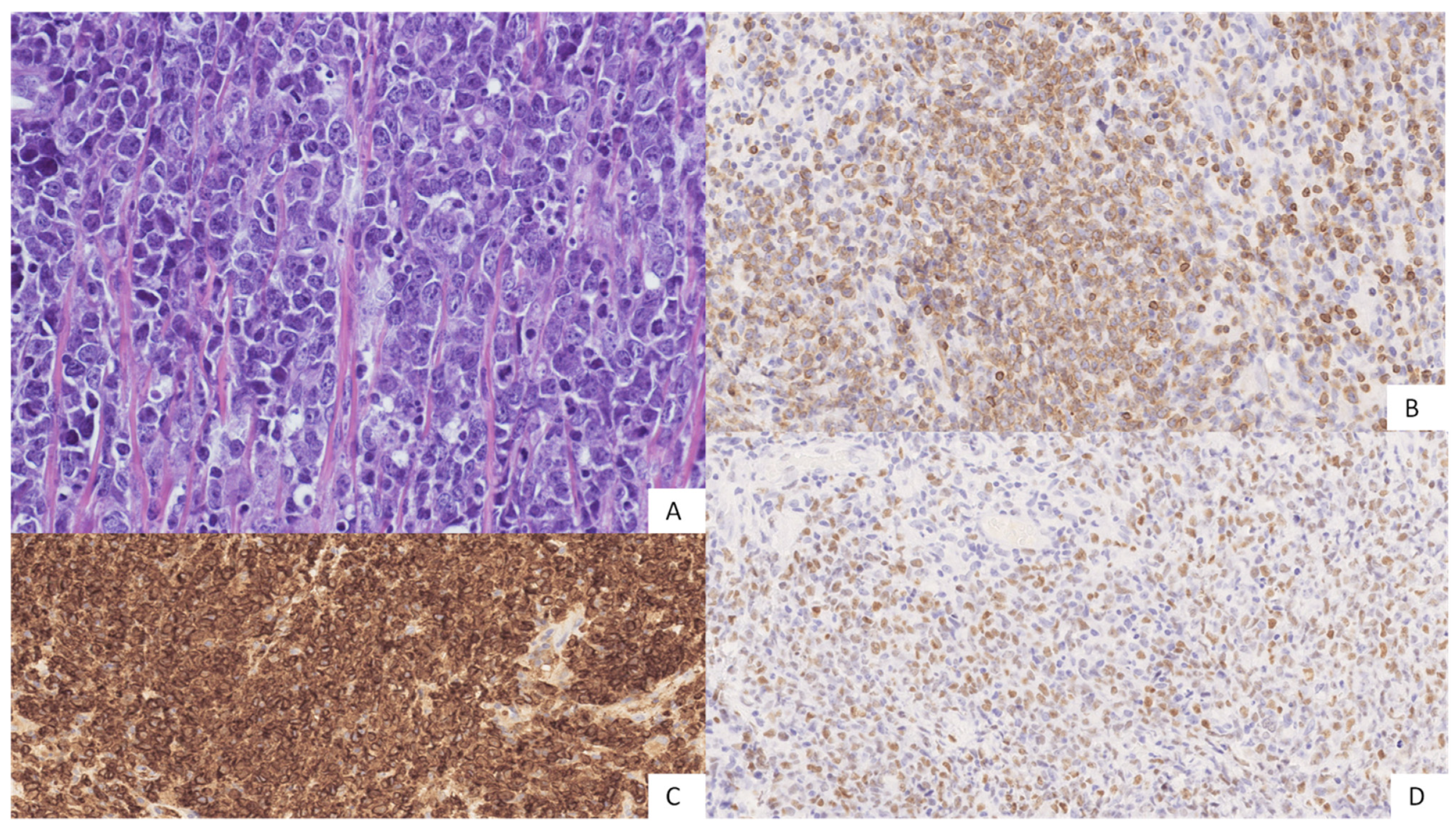
Morphologically, PCDLBCL-LT is characterized by a diffuse, non-epidermotropic, dense infiltrate with a grenz zone, which involves the entire dermis, effaces adnexal structures and often extends to the subcutaneous tissue. Overlying skin is frequently ulcerated. Cytologically, the infiltrate is usually monomorphic, consisting of round centroblastic and/or immunoblastic cells arranged in sheets (Figure 3), with very few reactive T-lymphocytes and minimal stromal reaction; occasionally pleomorphic to anaplastic cells are seen. Numerous mitoses and necrosis are usually found, while follicular structures and CD21+/CD23+ dendritic meshwork are typically lacking [86,90].
The immunophenotype of PCDLBCL-LT resembles that of the non-germinal centre-type of nodal DLBCL [91]. Neoplastic cells are positive for CD20, CD79a, bcl-2, MUM-1/IRF4 and (usually) bcl-6, but negative for CD10. FOXP1, c-MYC and cytoplasmic IgM are positive in most cases (Figure 3), whereas CD30 is usually not expressed [8,86,92]. Since bcl-2 is positive in more than 90% of cases and c-MYC is positive in over 65%, more than 60% of PCDLBCL-LT have a double-expressor (DE) phenotype [12,86]. EBV search is negative [87].
PD-L1 and CD33 may be expressed by tumour cells or by myeloid-derived suppressor cells (MDSCs), therefore, it has been suggested that they may shield the tumour against PD-1+ tumour-infiltrating lymphocytes [93]. A high proliferative index (usually >70%) is seen [8].

4.3. Molecular and Cytogenetic Features

Clonal rearrangements of IGH genes are detected in most PCDLBCLs. According to a post-germinal centre derivation, rearranged IGH genes usually carry somatic hypermutation, often with concurrent BCL-6 mutations. However, the precise definition of the cell of origin (COO) of PCDLBCL-LT is incomplete.
A study by Hoefangel et al. [85], based on gene expression analyses techniques, reported that PCDLBCL-LT has a profile similar to that of ABC-like nodal DLBCL.
On the other hand, a more recent study by Schrader et al. [94], based on Lymph2Cx algorithm, documented that the COO classification of PCLBCL-LT is more heterogeneous than expected, with only 18% of cases resulting as ABCs, 39% as germinal centre B cells (GCBs) and 43% as unclassifiable.
FISH analysis reveals translocations involving IGH, MYC, and BCL6 genes [38,92].
Recently, cases of PCDLBCL-LT with a double or triple hit status have been described [11].
In spite of bcl-2 protein overexpression, BCL-2 rearrangements are usually absent, whereas BCL-2 amplification was found in a fraction of cases. Loss of CDKN2A and CDKN2B have also been reported in more than 50% of cases. MYD88L265P mutations are the most common, being found in two thirds of patients, [8,95,96] but mutations in PIM1 and CD79B have also been seen. MYD88L265P mutations and mutations in CARD11, CD79B and TNFAIP3/A20 may indicate a constitutive activation of the NF-kB pathway [12,97,98].
Moreover, Zhou et al. [99] reported that in the MYD-88 wild-type PCDLBCL-LT, there may be a cancer-promoting mutation which activates the NF-kB pathway through different genes or activates other cancer pathways. They also found PDL1/PDL2 translocations in 40% of PCBLBCL-LT cases. Most of these genes’ mutations are commonly found in primary DLBCL of the central nervous system and in primary testicular DLBCL. Therefore, even though the mutational profile of PCDLBCL-LT seems to overlap with that of the ABC subtype of DLBCL, it is actually most similar to that of these two entities [12,99,100,101,102].
Furthermore, another study about microRNA profiling of PCBCL showed that microRNAs with a higher expression in the ABC-type of nodal DLBCL were not differentially expressed in PCDLBCL-LT, suggesting different pathogenetic mechanisms for PCDLBCL-LT cases than for nodal ones [103].

4.4. Differential Diagnosis

PCDLBCL-LT is relatively easy to diagnose once sheets of large B cells with typical morphology and immunophenotype are seen. The differential diagnosis of PCFCL with a diffuse growth pattern and large-cell cytology is the most challenging issue, which has already been discussed in the paragraph above (Table 2). Secondary skin involvement from systemic DLBCL—histologically and immunophenotypically indistinguishable from primary cutaneous forms—is frequent and can be excluded only based on clinical staging.
Predominantly blastoid or pleomorphic variants of MCL can infiltrate the skin, mimicking the histopathologic features of PCDLBCL-LT. Immunostaining for Cyclin D1 and SOX11 must be performed to exclude secondary skin involvement by MCL. Cases with Cyclin D1 expression should be tested by FISH analysis for CCND1 gene translocation [104,105].
An EBV search is required to exclude EBV+ DLBCL, which typically arises in elderly patients [8].
In cases with plasmablastic and immunoblastic differentiation, molecular and immunohistochemical tests for EBV and HHV8 are recommended. EBV and HHV8 infection associations should also be tested in any immunodeficient patient. The possibility of a precursor lymphoblastic lymphoma must be excluded by performing TdT immunostaining [104]. Some T-cell lymphomas, as mycosis fungoides, tumour stage, might show aberrant expressions of CD20 [106]. Rare, non-haematological neoplasms, such as Merkel cell carcinoma, might express B-cell markers, in particular PAX5 [104,107].
Cases with anaplastic cytology must be differentiated from CD30+/CD30-, anaplastic large T-cell lymphoma [108] and non-lymphoid, large-cell neoplasms, including melanoma, carcinoma and mesenchymal malignancies [109].

4.5. Prognosis and Treatment

PCDLBCL-LT is an aggressive disease with a five-year, disease-specific survival rate of around 50%, and many patients experience relapse despite treatment [88].
Adverse prognostic factors are the presence of multiple skin lesions (on one or both legs), involvement of both legs, inactivation of CDKN2A by gene deletion or promoter methylation, and the presence of the somatic mutation MYD88L265P [38,79,89,90,96,110].
As for the DE profile, there have been discordant observations; while Menguy et al. [91,110] observed impaired survival in cases with a DE phenotype, both Schrader et al. [92] and Lucioni et al. [11] did not correlate a DE status with a significantly worse prognosis.
Similarly, the prognostic impact of MYC rearrangements is not completely understood. While Schrader et al. [92] suggested a poorer prognosis for patients with MYC translocations, this has not been confirmed by the study by Lucioni et al. [11] that documented a poor response to treatment and a more rapid disease progression only in association with a double/triple hit status.
Standard first-line treatment is based on polychemotherapy (CHOP regimen) in combination with rituximab. However, many patients with PCDLBCL-LT are elderly and frail, making them unfit for chemotherapy [87,88,111].
When the disease is confined to the leg, another option is radiation therapy, which can be used in combination with systemic therapies or as a monotherapy for palliative purposes. For single lesions, or for lesions confined in a single area, surgery with debulking intent may also be considered [87]. Although there are no uniform recommendations for second-line treatment in case of relapse, the management of recurrences seems to be comparable to that of relapsed systemic DLBCL with an activated phenotype and, for this reason, the use of lenalidomide has been reported [89,112].

4.6. Summing Up

PCDLBCL-LT is an aggressive disease, usually arising on the leg. It is characterized by sheets of centroblasts and/or immunoblasts, with no/few admixed reactive cells. Neoplastic cells are usually positive for bcl-2, MUM-1/IRF4, bcl-6, c-MYC and IgM, but negative for CD10, and more than 60% of PCDLBCL-LT cases have a DE phenotype.
The precise definition of the COO of this lymphoma is still incomplete and its mutational profile seems to be more similar to those of primary DLBCL of the central nervous system and primary testicular DLBCL.

5. Primary Cutaneous Diffuse Large B Cell Lymphoma, Not Otherwise Specified/Other

The 2005 WHO-EORTC classification of skin tumours included a further subgroup of PCDLBCL that was named primary cutaneous diffuse large B-cell lymphoma, other (PCDLBCL, other). This group included rare cases not belonging either to the PCDLBCL-LT or to the PCFCL categories. However, since PCDLBCL, other was not a clearly defined entity, the 2018 update of the WHO-EORTC classification decided to delete it to avoid further confusion [12,113].
As of today, the debate about this entity is still ongoing. Recent articles have highlighted the presence of a group of large B-cell lymphomas, primarily arising on the skin, with peculiar morphological features and a slightly different prognosis than PCDLBCL-LT and PCFCL. This group has been called primary cutaneous B-cell lymphoma, not otherwise specified/unclassifiable (PCDLBCL-NOS/PCDLBCL-U) [10,11,73,91,114].
PCDLBCL-NOS seems to affect younger patients than PCDLBCL-LT, with a median age of 60 years at diagnosis and a male prevalence [10,11,115]. It usually develops over a longer period of time [10] and it most commonly arises as a single nodular or plaque lesion on the trunk, followed by the head and neck region, the lower limbs and finally the upper limbs [10,11,90].
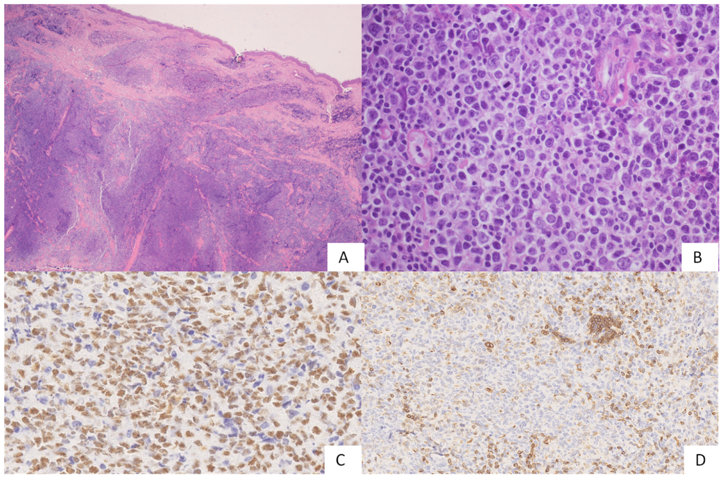
Histologically, PCDLBCL-NOS is distinguished from PCDLBCL-LT (Table 2) because it is composed of large centroblastic cells with a minority (<10%) of medium-sized cells and/or a mild to moderate reactive cellular background of small CD3+ T lymphocytes. The infiltrate most commonly presents with a diffuse growth pattern, but (rarely) vaguely nodular areas may also be seen (Figure 4). Usually, necrosis and effacement of cutaneous adnexa, along with the dendritic meshwork, are not found. PCDLBCL-NOS cells display a B cell phenotype with expressions of CD20, CD79a, bcl-6 and variable positivity for bcl2 in a minority of cases (Figure 4). PCDLBCL-NOS can be positive for c-MYC, CD10, IgM and MUM1/IRF4, but the DE status is infrequent. The proliferative index is lower than in the PCDLBCL-LT cases, with a median value of 40%. The histogenetic characterization according to the Hans algorithm showed cases with a GC profile and cases with a non-GC phenotype [10,11,73].
FISH analysis revealed BCL6 as the most frequently translocated gene, followed by MYC rearrangements; rarely, BCL2 alterations, rearrangements or an increase in gene copy number, were found. A single case of PCDLBCL-NOS with a DH status (translocations of MYC and BCL6) has been reported; the patient presented a GC histogenetic profile and bcl2 negativity by means of immunohistochemistry [11,73].
PCDLBCL-NOS seems to be less aggressive than PCDLBCL-LT. The histogenetic profile may be retained as a significant prognostic factor in this disease. In fact, there seems to be differences in survival rates between PCDLBCL-NOS GC-type and PCDLBCL-NOS non-GC-type, with the first group more similar to PCFCL and the latter presenting instead with an intermediate behaviour between PCFCL and PCDLBCL-LT [73].
Interestingly, it has been noted that BCL6 translocations have correlated with inferior survival rates in PCDLBCL-NOS [11]. Since PCDLBCL-NOS is not an entirely recognized entity, no standardized treatment protocol exists. Local radiotherapy, chemotherapy, radiotherapy in association with chemotherapy, or in a few cases, a wait-and-see approach, were used [11,73].

5.1. Diffuse Large B-Cell Lymphoma, Rare Subtypes

Primary cutaneous lymphomas with plasmablastic features are exceedingly rare. Most of these cases occur in settings of immunodeficiency (HIV-associated or iatrogenic) [86] or during the course of systemic T-cell lymphoma (such as angioimmunoblastic T-cell lymphoma). Plasmablastic lymphomas are clinically very aggressive. Histologically, they are composed of sheets of cells resembling immunoblasts, with variable degrees of plasmacytic differentiation. The lymphoma cells phenotype is consistent with terminal stages of B-cell differentiation, with negativity for CD20 and PAX5 and variable positivity for CD79a, CD38/CD138 and MUM1. Approximately 75% of cases are EBV-positive, exhibiting a latency I phenotype (EBERs expression and negativity for LMP1 and EBNA2). CD30 is frequently positive, correlating with EBV positivity, whereas, in contrast with plasma cell myeloma, CD56 is usually negative [8,116]. Plasmablastic lymphoma harbours frequent MYC rearrangements, with c-MYC protein expression by immunohistochemistry [86,116].
Intravascular B-cell lymphoma (IVBL) may present in the skin or localize to the skin during the course of the disease. IVBL is characterized by the presence of large lymphoid cells within the lumina of small- to medium-sized blood vessels, particularly capillaries and postcapillary venules [8]. This peculiar intravascular growth pattern has been attributed to a defect in homing receptors and adhesion molecules on the tumour cells (such as the lack of CD29 (b1 integrin) and CD54 (ICAM1)) [108,117]. Lymphoma cells express B-cell-associated antigens and may be positive for CD5 (38%) [8]; the majority of cases are positive for MUM1 and negative for CD10, suggesting an ABC phenotype [116] and show an overexpression of bcl-2 in the absence of BCL2 rearrangements [118].
In keeping with an ABC origin, a subset of IVBLs carry MYD88L265P and CD79B mutations [86,116]. Skin is a common site for presentation of IVBL, although most patients have a widespread, disseminated disease at the time of diagnosis. Dermatologic manifestations include a wide range of skin lesions, including erythematous patches and plaques, panniculitis-like lesions, as well as painful telangiectasias and nodular lesions with a predilection for the trunk and lower extremities [3,108].
Clinical symptoms include fever and, due to the frequent involvement of the central nervous system, focal neurological defects. Lungs, adrenal glands, thyroid, gastrointestinal system, kidneys, genitourinary tract and eyes can also be involved. IVBL is an aggressive disease, requiring systemic immunochemotherapy. However, patients presenting with a cutaneous disease have a better prognosis in most series, probably because of earlier detection and treatment [8,108,119].

5.2. EBV-Positive Mucocutaneous Ulcer

EBVMCU is a provisional entity included in the WHO Classification of Tumours of Haematopoietic and Lymphoid Tissues of 2017 [8,120]. It occurs in patients with primary or secondary immunodeficiencies, including age-related or iatrogenic immunosuppression for immune disorders, transplant recipients and HIV infection. The median age is about 66 years [121].
Clinically, EBVMCU presents as a solitary, sharply demarcated ulcer occurring mostly in the oral mucosa; other involved sites may be the skin and the gastrointestinal tract with (rarely) locoregional lymph node involvement and without systemic symptoms [12,122].
Histologically, there is a mucosal/cutaneous ulceration with the underlying presence of EBV-positive, large, atypical cells resembling immunoblasts or Hodgkin/Reed–Sternberg cells in a polymorphic background containing lymphocytes, histiocytes, plasma cells and granulocytes. Angioinvasion, necrosis and apoptotic bodies may occur. An important characteristic is the presence of a rim of CD8+ T cells at the base of the ulcer [123].
The atypical cells show expression of CD30, PAX5, MUM1/IRF4 and OCT2, with variable positivity for CD20, CD79a and BOB1. They have an ABC phenotype with negativity for CD10 and bcl6. Half of these cases express CD15 [3]. EBV positivity may be detected using an LMP1 antibody or in situ hybridization for EBV-encoded RNA (EBER) [123].
Up to 40% of cases show clonal immunoglobulin gene rearrangements and/or T-cell receptor gene (TCR) rearrangements [116,123].
Differential diagnosis includes classic Hodgkin’s lymphoma and EBV-positive DLBCL-NOS. Both of these diseases usually form masses and are more widespread (most commonly involving lymph nodes) than EBVMCU. As such, the correlation between clinical and pathological features is crucial to discriminate between these entities [122,123].
Other differential diagnoses include primary cutaneous or primary oropharyngeal anaplastic large-cell lymphoma (negativity for PAX5 and EBV) and lymphomatoid granulomatosis (almost always involving the lungs) [122].
EBVMCU usually has a benign, self-limited course with spontaneous remission or regression upon reduction of immunosuppressive medications. If a therapy is required, the use of rituximab, local radiotherapy or chemotherapy has been reported. Only rare cases experience recurrences or disease progression (mostly local spread) [3,8,12,121,123].

6. Conclusions

PCBCL is a heterogeneous group of lymphoproliferative disorders with distinct clinicopathologic features comparable with their nodal counterparts. In the last decade, we have achieved significant advancements in our understanding of the clinicopathologic features and molecular background of PCBCL with the identification of characteristic molecular alterations in different lymphoma subtypes. In addition, some novel provisional entities have been described, such as EBVMCU, further expanding the spectrum of PCBCLs. However, in spite of such advances, the histopathological diagnosis of PCBCL is still challenging, and a number of issues continue to be a matter of debate. The most compelling topics include the differential diagnosis between PCMZL, PCFCL and reactive cutaneous lymphoid infiltrates, the border between centroblast-rich PCFCL and PCDLBCL and the putative prognostic subcategorization of PCDLBCL. The hope is that, in the future, the identification of new genetic markers will be useful for refining PCBCL categories, opening up new diagnostic and therapeutic perspectives.

Funding

This research received no external funding.

Institutional Review Board Statement

Not applicable. This study is a review of current literature; it is not based on original data from patients series.

Informed Consent Statement

Not applicable.

Data Availability Statement

Not applicable.

Conflicts of Interest

The authors declare no conflict of interest.

References

  1. Kempf, W.; Zimmermann, A.-K.; Mitteldorf, C. Cutaneous lymphomas—An update 2019. Hematol. Oncol. 2019, 37 (Suppl. S1), 43–47. [Google Scholar] [CrossRef] [PubMed] [Green Version]
  2. Chen, S.T.; Barnes, J.; Duncan, L. Primary cutaneous B-cell lymphomas—Clinical and histopathologic features, differential diagnosis, and treatment. Semin. Cutan. Med. Surg. 2018, 37, 49–55. [Google Scholar] [CrossRef] [PubMed]
  3. Vitiello, P.; Sica, A.; Ronchi, A.; Caccavale, S.; Franco, R.; Argenziano, G. Primary Cutaneous B-Cell Lymphomas: An Update. Front. Oncol. 2020, 10, 651. [Google Scholar] [CrossRef] [PubMed]
  4. Hope, C.B.; Pincus, L.B. Primary Cutaneous B-cell Lymphomas. Clin. Lab. Med. 2017, 37, 547–574. [Google Scholar] [CrossRef]
  5. Grandi, V.; Violetti, S.A.; La Selva, R.; Cicchelli, S.; Delfino, C.; Fava, P.; Fierro, M.T.; Pileri, A.; Pimpinelli, N.; Quaglino, P.; et al. Primary cutaneous B-cell lymphoma: Narrative review of the literature. G. Ital. Dermatol. Venereol. 2019, 154, 466–479. [Google Scholar] [CrossRef]
  6. Wilcox, R.A. Cutaneous B-cell lymphomas: 2019 update on diagnosis, risk stratification, and management. Am. J. Hematol. 2018, 93, 1427–1430. [Google Scholar] [CrossRef] [Green Version]
  7. Goyal, A.; LeBlanc, R.E.; Carter, J.B. Cutaneous B-Cell Lymphoma. Hematol. Clin. N. Am. 2019, 33, 149–161. [Google Scholar] [CrossRef]
  8. Swerdlow, S.H.; Campo, E.; Harris, N.L.; Jaffe, E.S.; Pileri, S.A.; Stein, H.; Thiele, J. (Eds.) WHO Classification of Tumours of Haematopoietic and Lymphoid Tissues, Revised, 4th ed.; IARC: Lyon, France, 2017. [Google Scholar]
  9. Malachowski, S.J.; Sun, J.; Chen, P.-L.; Seminario-Vidal, L. Diagnosis and Management of Cutaneous B-Cell Lymphomas. Dermatol. Clin. 2019, 37, 443–454. [Google Scholar] [CrossRef]
  10. Felcht, M.; Klemke, C.-D.; Nicolay, J.P.; Weiss, C.; Assaf, C.; Wobser, M.; Schlaak, M.; Hillen, U.; Moritz, R.; Tantcheva-Poor, I.; et al. Primary cutaneous diffuse large B-cell lymphoma, NOS and leg type: Clinical, morphologic and prognostic differences. JDDG J. Dtsch. Dermatol. Ges. 2019, 17, 275–285. [Google Scholar] [CrossRef]
  11. Lucioni, M.; Pescia, C.; Bonometti, A.; Fraticelli, S.; Moltrasio, C.; Ramponi, A.; Riboni, R.; Roccio, S.; Ferrario, G.; Arcaini, L.; et al. Double expressor and double/triple hit status among primary cutaneous diffuse large B-cell lymphoma: A comparison between leg type and not otherwise specified subtypes. Hum. Pathol. 2021, 111, 1–9. [Google Scholar] [CrossRef]
  12. Willemze, R.; Cerroni, L.; Kempf, W.; Berti, E.; Facchetti, F.; Swerdlow, S.H.; Jaffe, E.S. The 2018 update of the WHO-EORTC clas-sification for primary cutaneous lymphomas. Blood 2019, 134, 1112. [Google Scholar]
  13. Hoefnagel, P.; Vermeer, M.; Jansen, P.; Heule, F.; Vader, P.C.V.V.; Sanders, C.J.G.; Gerritsen, M.J.P.; Geerts, M.L.; Meijer, C.J.L.M.; Noordijk, E.M.; et al. Primary Cutaneous Marginal Zone B-Cell Lymphoma: Clinical and therapeutic features in 50 cases. Arch. Dermatol. 2005, 141, 1139–1145. [Google Scholar] [CrossRef] [PubMed]
  14. Fink-Puches, R.; Chott, A.; Ardigo, M.; Simonitsch, I.; Ferrara, G.; Kerl, H.; Cerroni, L. The Spectrum of Cutaneous Lymphomas in Patients Less than 20 Years of Age. Pediatr. Dermatol. 2004, 21, 525–533. [Google Scholar] [CrossRef] [PubMed]
  15. Kempf, W.; Kazakov, D.; Buechner, S.A.; Graf, M.; Zettl, A.; Zimmermann, D.R.; Tinguely, M. Primary Cutaneous Marginal Zone Lymphoma in Children: A report of 3 cases and review of the literature. Am. J. Dermatopathol. 2014, 36, 661–666. [Google Scholar] [CrossRef] [PubMed] [Green Version]
  16. Goodlad, J.R.; Davidson, M.M.; Hollowood, K.; Ling, C.; MacKenzie, C.; Christie, I.; Batstone, P.J.; Ho-Yen, D.O. Primary Cutaneous B-Cell Lymphoma and Borrelia burgdorferi Infection in Patients from the Highlands of Scotland. Am. J. Surg. Pathol. 2000, 24, 1279–1285. [Google Scholar] [CrossRef]
  17. Takino, H.; Li, C.; Hu, S.; Kuo, T.-T.; Geissinger, E.; Muller-Hermelink, H.K.; Kim, B.; Swerdlow, S.H.; Inagaki, H. Primary cutaneous marginal zone B-cell lymphoma: A molecular and clinicopathological study of cases from Asia, Germany, and the United States. Mod. Pathol. 2008, 21, 1517–1526. [Google Scholar] [CrossRef] [Green Version]
  18. Suarez, F.; Lortholary, O.; Hermine, O.; Lecuit, M. Infection-associated lymphomas derived from marginal zone B cells: A model of antigen-driven lymphoproliferation. Blood 2006, 107, 3034–3044. [Google Scholar] [CrossRef] [Green Version]
  19. Arcaini, L.; Bruno, R. Hepatitis C Virus Infection and Antiviral Treatment in Marginal Zone Lymphomas. Curr. Clin. Pharmacol. 2010, 5, 74–81. [Google Scholar] [CrossRef]
  20. Arcaini, L.; Burcheri, S.; Rossi, A.; Paulli, M.; Bruno, R.; Passamonti, F.; Brusamolino, E.; Molteni, A.; Pulsoni, A.; Cox, M.; et al. Prevalence of HCV infection in nongastric marginal zone B-cell lymphoma of MALT. Ann. Oncol. 2007, 18, 346–350. [Google Scholar] [CrossRef]
  21. Arcaini, L.; Merli, M.; Volpetti, S.; Rattotti, S.; Gotti, M.; Zaja, F. Indolent B-Cell Lymphomas Associated with HCV Infection: Clinical and Virological Features and Role of Antiviral Therapy. Clin. Dev. Immunol. 2012, 2012, 638185. [Google Scholar] [CrossRef] [Green Version]
  22. Paulli, M.; Arcaini, L.; Lucioni, M.; Boveri, E.; Capello, D.; Passamonti, F.; Merli, M.; Rattotti, S.; Rossi, D.; Riboni, R.; et al. Subcutaneous ‘lipoma-like’ B-cell lymphoma associated with HCV infection: A new presentation of primary extranodal marginal zone B-cell lymphoma of MALT. Ann. Oncol. 2009, 21, 1189–1195. [Google Scholar] [CrossRef] [PubMed]
  23. Tsuji, K.; Suzuki, D.; Naito, Y.; Sato, Y.; Yoshino, T.; Iwatsuki, K. Primary cutaneous marginal zone B-cell lymphoma. Eur. J. Dermatol. 2005, 15, 480–483. [Google Scholar] [PubMed]
  24. Gibson, S.E.; Swerdlow, S.H. How I Diagnose Primary Cutaneous Marginal Zone Lymphoma. Am. J. Clin. Pathol. 2020, 154, 428–449. [Google Scholar] [CrossRef] [PubMed]
  25. Russo, I.; Fagotto, L.; Sernicola, A.; Alaibac, M. Primary Cutaneous B-Cell Lymphomas in Patients with Impaired Immunity. Front. Oncol. 2020, 10, 1296. [Google Scholar] [CrossRef] [PubMed]
  26. Cicogna, G.T.; Ferranti, M.; Alaibac, M. Diagnostic Workup of Primary Cutaneous B Cell Lymphomas: A Clinician’s Approach. Front. Oncol. 2020, 10, 988. [Google Scholar] [CrossRef]
  27. Ronchi, A.; Sica, A.; Vitiello, P.; Franco, R. Dermatological Considerations in the Diagnosis and Treatment of Marginal Zone Lymphomas. Clin. Cosmet. Investig. Dermatol. 2021, 14, 231–239. [Google Scholar] [CrossRef]
  28. Cook, J.C.; Puckett, Y. Anetoderma; StatPearls Publishing: Treasure Island, FL, USA, 2022. [Google Scholar]
  29. Hodak, E.; Feuerman, H.; Barzilai, A.; David, M.; Cerroni, L.; Feinmesser, M. Anetodermic Primary Cutaneous B-Cell Lymphoma: A unique clinicopathological presentation of lymphoma possibly associated with antiphospholipid antibodies. Arch. Dermatol. 2010, 146, 175–182. [Google Scholar] [CrossRef] [Green Version]
  30. Dalle, S.; Thomas, L.; Balme, B.; Dumontet, C.; Thieblemont, C. Primary cutaneous marginal zone lymphoma. Crit. Rev. Oncol. 2010, 74, 156–162. [Google Scholar] [CrossRef]
  31. Farhadian, J.; Terushkin, V.; Meehan, S.A.; Latkowski, J.A. Primary cutaneous marginal-zone lymphoma. Derm. Online J. 2016, 22, 13030. [Google Scholar] [CrossRef]
  32. Cho-Vega, J.H.; Vega, F.; Rassidakis, G.; Medeiros, L.J. Primary cutaneous marginal zone B-cell lymphoma. Am. J. Clin. Pathol. 2006, 125, S38–S49. [Google Scholar] [CrossRef]
  33. Kempf, W.; Kerl, H.; Kutzner, H. CD123-Positive Plasmacytoid Dendritic Cells in Primary Cutaneous Marginal Zone B-Cell Lymphoma: A Crucial Role and a New Lymphoma Paradigm. Am. J. Dermatopathol. 2010, 32, 194–196. [Google Scholar] [CrossRef]
  34. Edinger, J.T.; Kant, J.A.; Swerdlow, S.H. Cutaneous Marginal Zone Lymphomas Have Distinctive Features and Include 2 Subsets. Am. J. Surg. Pathol. 2010, 34, 1830–1841. [Google Scholar] [CrossRef]
  35. Kogame, T.; Takegami, T.; Sakai, T.; Kataoka, T.; Hirata, M.; Budair, F.M.; Ueshima, C.; Matsui, M.; Nomura, T.; Kabashima, K. Immunohistochemical analysis of class-switched subtype of primary cutaneous marginal zone lymphoma in terms of inducible skin-associated lymphoid tissue. J. Eur. Acad. Dermatol. Venereol. 2019, 33, e401–e403. [Google Scholar] [CrossRef]
  36. Van Maldegem, F.; van Dijk, R.; Wormhoudt, T.A.M.; Kluin, P.M.; Willemze, R.; Cerroni, L.; van Noesel, C.J.M.; Bende, R.J. The majority of cutaneous marginal zone B-cell lymphomas expresses class-switched immunoglobulins and develops in a T-helper type 2 inflammatory environment. Blood 2008, 112, 3355–3361. [Google Scholar] [CrossRef] [Green Version]
  37. Sun, J.R.; Nong, L.; Liu, X.Q.; Tu, P.; Wang, Y. Frequent immunoglobulin G4 expression in a common variant of primary cutaneous marginal zone B-cell lymphoma. Australas. J. Dermatol. 2017, 59, 141–145. [Google Scholar] [CrossRef]
  38. Hallermann, C.; Kaune, K.; Gesk, S.; Martin-Subero, J.; Gunawan, B.; Griesinger, F.; Vermeer, M.; Santucci, M.; Pimpinelli, N.; Willemze, R.; et al. Molecular Cytogenetic Analysis of Chromosomal Breakpoints in the IGH, MYC, BCL6, and MALT1 Gene Loci in Primary Cutaneous B-cell Lymphomas. J. Investig. Dermatol. 2004, 123, 213–219. [Google Scholar] [CrossRef] [Green Version]
  39. Li, C.; Inagaki, H.; Kuo, T.-T.; Hu, S.; Okabe, M.; Eimoto, T. Primary Cutaneous Marginal Zone B-Cell Lymphoma: A molecular and clinicopathologic study of 24 asian cases. Am. J. Surg. Pathol. 2003, 27, 1061–1069. [Google Scholar] [CrossRef]
  40. Swerdlow, S.H. Cutaneous marginal zone lymphomas. Semin. Diagn. Pathol. 2017, 34, 76–84. [Google Scholar] [CrossRef]
  41. Maurus, K.; Appenzeller, S.; Roth, S.; Kuper, J.; Rost, S.; Meierjohann, S.; Arampatzi, P.; Goebeler, M.; Rosenwald, A.; Geissinger, E.; et al. Panel Sequencing Shows Recurrent Genetic FAS Alterations in Primary Cutaneous Marginal Zone Lymphoma. J. Investig. Dermatol. 2018, 138, 1573–1581. [Google Scholar] [CrossRef] [Green Version]
  42. Baldassano, M.F.; Bailey, E.M.; Ferry, J.A.; Harris, N.L.; Duncan, L.M. Cutaneous Lymphoid Hyperplasia and Cutaneous Marginal Zone Lymphoma: Comparison of morphologic and immunophenotypic features. Am. J. Surg. Pathol. 1999, 23, 88–96. [Google Scholar] [CrossRef]
  43. Lin, P.; Molina, T.; Cook, J.R.; Swerdlow, S.H. Lymphoplasmacytic Lymphoma and Other Non–Marginal Zone Lymphomas with Plasmacytic Differentiation. Am. J. Clin. Pathol. 2011, 136, 195–210. [Google Scholar] [CrossRef]
  44. Molina, T.J.; Lin, P.; Swerdlow, S.H.; Cook, J.R. Marginal Zone Lymphomas with Plasmacytic Differentiation and Related Disorders. Am. J. Clin. Pathol. 2011, 136, 211–225. [Google Scholar] [CrossRef] [Green Version]
  45. Brenner, I.; Roth, S.; Puppe, B.; Wobser, M.; Rosenwald, A.; Geissinger, E. Primary cutaneous marginal zone lymphomas with plasmacytic differentiation show frequent IgG4 expression. Mod. Pathol. 2013, 26, 1568–1576. [Google Scholar] [CrossRef] [Green Version]
  46. Servitje, O.; Muniesa, C.; Benavente, Y.; Monsálvez, V.; Garcia-Muret, M.P.; Gallardo, F.; Domingo-Domenech, E.; Lucas, A.; Climent, F.; Rodriguez-Peralto, J.L.; et al. Primary cutaneous marginal zone B-cell lymphoma: Response to treatment and disease-free survival in a series of 137 patients. J. Am. Acad. Dermatol. 2013, 69, 357–365. [Google Scholar] [CrossRef]
  47. Prieto-Torres, L.; Manso, R.; Cieza-Díaz, D.E.; Jo, M.; Pérez, L.K.; Montenegro-Damaso, T.; Eraña, I.; Lorda, M.; Massa, D.S.; Machan, S.; et al. Large Cells with CD30 Expression and Hodgkin-like Features in Primary Cutaneous Marginal Zone B-Cell Lymphoma: A Study of 13 Cases. Am. J. Surg. Pathol. 2019, 43, 1191–1202. [Google Scholar] [CrossRef]
  48. Palmedo, G.; Hantschke, M.; Rütten, A.; Mentzel, T.; Kempf, W.; Tomasini, D.; Kutzner, H. Primary Cutaneous Marginal Zone B-cell Lymphoma May Exhibit Both the t(14;18)(q32;q21) IGH/BCL2 and the t(14;18)(q32;q21) IGH/MALT1 Translocation: An Indicator for Clonal Transformation Towards Higher-Grade B-cell Lymphoma? Am. J. Dermatopathol. 2007, 29, 231–236. [Google Scholar] [CrossRef]
  49. Boudreaux, B.W.; Patel, M.H.; Brumfiel, C.M.; Besch-Stokes, J.; DiCaudo, D.J.; Craig, F.; Rosenthal, A.C.; Rule, W.G.; Pittelkow, M.R.; Mangold, A.R. Primary cutaneous epidermotropic marginal zone B-cell lymphoma treated with total skin electron beam therapy. JAAD Case Rep. 2021, 15, 15–18. [Google Scholar] [CrossRef]
  50. Wobser, M. Therapie indolenter kutaner B-Zell-Lymphome [Treatment of indolent cutaneous B-cell lymphoma]. Hautarzt 2017, 68, 721–726. [Google Scholar] [CrossRef]
  51. Lang, C.C.V.; Ramelyte, E.; Dummer, R. Innovative Therapeutic Approaches in Primary Cutaneous B Cell Lymphoma. Front. Oncol. 2020, 10, 1163. [Google Scholar] [CrossRef]
  52. Skala, S.L.; Hristov, B.; Hristov, A.C. Primary Cutaneous Follicle Center Lymphoma. Arch. Pathol. Lab. Med. 2018, 142, 1313–1321. [Google Scholar] [CrossRef] [Green Version]
  53. Senff, N.J.; Hoefnagel, J.J.; Jansen, P.M.; Vermeer, M.; Van Baarlen, J.; Blokx, W.; Dijk, M.R.C.-V.; Geerts, M.-L.; Hebeda, K.M.; Kluin, P.M.; et al. Reclassification of 300 Primary Cutaneous B-Cell Lymphomas According to the New WHO–EORTC Classification for Cutaneous Lymphomas: Comparison with Previous Classifications and Identification of Prognostic Markers. J. Clin. Oncol. 2007, 25, 1581–1587. [Google Scholar] [CrossRef] [Green Version]
  54. Zinzani, P.L.; Quaglino, P.; Pimpinelli, N.; Berti, E.; Baliva, G.; Rupoli, S.; Martelli, M.; Alaibac, M.; Borroni, G.; Chimenti, S.; et al. Prognostic Factors in Primary Cutaneous B-Cell Lymphoma: The Italian Study Group for Cutaneous Lymphomas. J. Clin. Oncol. 2006, 24, 1376–1382. [Google Scholar] [CrossRef] [Green Version]
  55. Travaglino, A.; Varricchio, S.; Pace, M.; Russo, D.; Picardi, M.; Baldo, A.; Staibano, S.; Mascolo, M. Borrelia burgdorferi in primary cutaneous lymphomas: A systematic review and meta-analysis. JDDG: J. Dtsch. Dermatol. Ges. 2020, 18, 1379–1384. [Google Scholar] [CrossRef]
  56. Slater, D.N. Borrelia burgdorferi-associated primary cutaneous B-cell lymphoma. Histopathology 2001, 38, 73–77. [Google Scholar] [CrossRef]
  57. Petković, I.Z.; Pejcic, I.; Tiodorović, D.; Krstić, M.; Stojnev, S.; Vrbic, S. Transformation of primary cutaneous follicle centre lymphoma into primary cutaneous diffuse large B-cell lymphoma of other type. Adv. Dermatol. Allergol. 2017, 34, 625–628. [Google Scholar] [CrossRef] [Green Version]
  58. Uy, M.; Sprowl, G.; Lynch, D.T. Primary Cutaneous Follicle Center Lymphoma; StatPearls Publishing: Treasure Island, FL, USA, 2022. [Google Scholar]
  59. Berti, E.; Alessi, E.; Caputo, R.; Gianotti, R.; Delia, D.; Vezzoni, P. Reticulohistiocytoma of the dorsum. J. Am. Acad. Dermatol. 1988, 19, 259–272. [Google Scholar] [CrossRef]
  60. Fierro, M.T.; Marenco, F.; Novelli, M.; Fava, P.; Quaglino, P.; Bernengo, M.G. Long-Term Evolution of an Untreated Primary Cutaneous Follicle Center Lymphoma of the Scalp. Am. J. Dermatopathol. 2010, 32, 91–94. [Google Scholar] [CrossRef]
  61. Cerroni, L.; Arzberger, E.; Pütz, B.; Höfler, G.; Metze, D.; Sander, C.A.; Rose, C.; Wolf, P.; Rütten, A.; McNiff, J.M.; et al. Primary cutaneous follicle center cell lymphoma with follicular growth pattern. Blood 2000, 95, 3922–3928. [Google Scholar] [CrossRef]
  62. Cerroni, L.; El-Shabrawi-Caelen, L.; Fink-Puches, R.; LeBoit, P.E.; Kerl, H. Cutaneous Spindle-Cell B-Cell Lymphoma. Am. J. Dermatopathol. 2000, 22, 299–304. [Google Scholar] [CrossRef]
  63. Child, F.; Russell-Jones, R.; Woolford, A.; Calonje, E.; Photiou, A.; Orchard, G.; Whittaker, S. Absence of the t(14;18) chromosomal translocation in primary cutaneous B-cell lymphoma. Br. J. Dermatol. 2001, 144, 735–744. [Google Scholar] [CrossRef]
  64. Abdul-Wahab, A.; Tang, S.-Y.; Robson, A.; Morris, S.; Agar, N.; Wain, E.M.; Child, F.; Scarisbrick, J.; Neat, M.; Whittaker, S. Chromosomal anomalies in primary cutaneous follicle center cell lymphoma do not portend a poor prognosis. J. Am. Acad. Dermatol. 2014, 70, 1010–1020. [Google Scholar] [CrossRef]
  65. Cerroni, L.; Volkenandt, M.; Rieger, E.; Soyer, H.P.; Kerl, H. bcl-2 Protein Expression and Correlation with the Interchromoso-mal 14;18 Translocation in Cutaneous Lymphomas and Pseudolymphomas. J. Invest Dermatol. 1994, 102, 231–235. [Google Scholar] [CrossRef] [Green Version]
  66. Geelen, F.A.; Vermeer, M.; Meijer, C.J.; Van Der Putte, S.C.; Kerkhof, E.; Kluin, P.M.; Willemze, R. bcl-2 protein expression in primary cutaneous large B-cell lymphoma is site-related. J. Clin. Oncol. 1998, 16, 2080–2085. [Google Scholar] [CrossRef]
  67. Goodlad, J.R.; Krajewski, A.S.; Batstone, P.J.; McKay, P.; White, J.M.; Benton, E.C.; Kavanagh, G.M.; Lucraft, H.H. Primary Cutaneous Follicular Lymphoma. Am. J. Surg. Pathol. 2002, 26, 733–741. [Google Scholar] [CrossRef]
  68. Hallermann, C.; Kaune, K.M.; Siebert, R.; Vermeer, M.H.; Tensen, C.P.; Willemze, R.; Gunawan, B.; Bertsch, H.P.; Neumann, C. Chromosomal Aberration Patterns Differ in Subtypes of Primary Cutaneous B Cell Lymphomas. J. Investig. Dermatol. 2004, 122, 1495–1502. [Google Scholar] [CrossRef] [Green Version]
  69. Vergier, B.; Belaud-Rotureau, M.-A.; Benassy, M.-N.; Beylot-Barry, M.; Dubus, P.; Delaunay, M.; Garroste, J.-C.; Taine, L.; Merlio, J.-P. Neoplastic Cells Do Not Carry bcl2-JH Rearrangements Detected in a Subset of Primary Cutaneous Follicle Center B-cell Lymphomas. Am. J. Surg. Pathol. 2004, 28, 748–755. [Google Scholar] [CrossRef]
  70. Aguilera, N.S.I.; Tomaszewski, M.-M.; Moad, J.C.; Bauer, F.A.; Taubenberger, J.K.; Abbondanzo, S.L. Cutaneous Follicle Center Lymphoma: A Clinicopathologic Study of 19 Cases. Mod. Pathol. 2001, 14, 828–835. [Google Scholar] [CrossRef] [Green Version]
  71. Mirza, I.; Macpherson, N.; Paproski, S.; Gascoyne, R.D.; Yang, B.; Finn, W.G.; Hsi, E.D. Primary Cutaneous Follicular Lymphoma: An Assessment of Clinical, Histopathologic, Immunophenotypic, and Molecular Features. J. Clin. Oncol. 2002, 20, 647–655. [Google Scholar] [CrossRef]
  72. Pham-Ledard, A.; Cowppli-Bony, A.; Doussau, A.; Prochazkova-Carlotti, M.; Laharanne, E.; Jouary, T.; Belaud-Rotureau, M.-A.; Vergier, B.; Merlio, J.-P.; Beylot-Barry, M. Diagnostic and Prognostic Value of BCL2 Rearrangement in 53 Patients with Follicular Lymphoma Presenting as Primary Skin Lesions. Am. J. Clin. Pathol. 2015, 143, 362–373. [Google Scholar] [CrossRef] [Green Version]
  73. Lucioni, M.; Berti, E.; Arcaini, L.; Croci, G.A.; Maffi, A.; Klersy, C.; Goteri, G.; Tomasini, C.; Quaglino, P.; Riboni, R.; et al. Primary cutaneous B-cell lymphoma other than marginal zone: Clinicopathologic analysis of 161 cases: Comparison with current classification and definition of prognostic markers. Cancer Med. 2016, 5, 2740–2755. [Google Scholar] [CrossRef]
  74. Brash, D.E. UV Signature Mutations. Photochem. Photobiol. 2015, 91, 15–26. [Google Scholar] [CrossRef] [Green Version]
  75. Szablewski, V.; Ingen-Housz-Oro, S.; Baia, M.; Delfau-Larue, M.-H.; Copie-Bergman, C.; Ortonne, N. Primary Cutaneous Follicle Center Lymphomas Expressing BCL2 Protein Frequently Harbor BCL2 Gene Break and May Present 1p36 Deletion. Am. J. Surg. Pathol. 2016, 40, 127–136. [Google Scholar] [CrossRef]
  76. Gángó, A.; Bátai, B.; Varga, M.; Kapczár, D.; Papp, G.; Marschalkó, M.; Kuroli, E.; Schneider, T.; Csomor, J.; Matolcsy, A.; et al. Concomitant 1p36 deletion and TNFRSF14 mutations in primary cutaneous follicle center lymphoma frequently expressing high levels of EZH2 protein. Virchows Arch. 2018, 473, 453–462. [Google Scholar] [CrossRef]
  77. Schafernak, K.T.; Variakojis, D.; Goolsby, C.L.; Tucker, R.M.; Martínez-Escala, M.E.; Smith, F.A.; Dittman, D.; Chenn, A.; Guitart, J. Clonality Assessment of Cutaneous B-Cell Lymphoid Proliferations. Am. J. Dermatopathol. 2014, 36, 781–795. [Google Scholar] [CrossRef]
  78. Zhou, X.A.; Yang, J.; Ringbloom, K.G.; Martinez-Escala, M.E.; Stevenson, K.E.; Wenzel, A.T.; Fantini, D.; Martin, H.K.; Moy, A.P.; Morgan, E.A.; et al. Genomic landscape of cutaneous follicular lymphomas reveals 2 subgroups with clinically predictive molecular features. Blood Adv. 2021, 5, 649–661. [Google Scholar] [CrossRef]
  79. Menguy, S.; Gros, A.; Pham-Ledard, A.; Battistella, M.; Ortonne, N.; Comoz, F.; Balme, B.; Szablewski, V.; Lamant, L.; Carlotti, A.; et al. MYD88 Somatic Mutation Is a Diagnostic Criterion in Primary Cutaneous Large B-Cell Lymphoma. J. Investig. Dermatol. 2016, 136, 1741–1744. [Google Scholar] [CrossRef] [Green Version]
  80. Verdanet, E.; Dereure, O.; René, C.; Tempier, A.; Benammar-Hafidi, A.; Gallo, M.; Frouin, E.; Durand, L.; Gazagne, I.; Costes-Martineau, V.; et al. Diagnostic value of STMN1, LMO2, HGAL, AID expression and 1p36 chromosomal abnormalities in primary cutaneous B cell lymphomas. Histopathology 2017, 71, 648–660. [Google Scholar] [CrossRef]
  81. Metcalf, R.A.; Monabati, A.; Vyas, M.; Roncador, G.; Gualco, G.; Bacchi, C.E.; Younes, S.F.; Natkunam, Y.; Freud, A.G. Myeloid cell nuclear differentiation antigen is expressed in a subset of marginal zone lymphomas and is useful in the differential diagnosis with follicular lymphoma. Hum. Pathol. 2014, 45, 1730–1736. [Google Scholar] [CrossRef]
  82. Kanellis, G.; Roncador, G.; Arribas, A.; Mollejo, M.; Montes-Moreno, S.; Maestre, L.; Campos-Martin, Y.; Gonzalez, J.L.R.; Martinez-Torrecuadrada, J.L.; Sanchez-Verde, L.; et al. Identification of MNDA as a new marker for nodal marginal zone lymphoma. Leukemia 2009, 23, 1847–1857. [Google Scholar] [CrossRef] [Green Version]
  83. Battistella, M.; Beylot-Barry, M.; Bachelez, H.; Rivet, J.; Vergier, B.; Bagot, M. Primary Cutaneous Follicular Helper T-cell Lymphoma. Arch. Dermatol. 2012, 148, 832–839. [Google Scholar] [CrossRef] [Green Version]
  84. Ferrara, G.; Bevilacqua, M.; Argenziano, G. Cutaneous Spindle B-Cell Lymphoma: A Reappraisal. Am. J. Dermatopathol. 2002, 24, 526–527. [Google Scholar] [CrossRef]
  85. Hoefnagel, J.J.; Dijkman, R.; Basso, K.; Jansen, P.M.; Hallermann, C.; Willemze, R.; Tensen, C.P.; Vermeer, M. Distinct types of primary cutaneous large B-cell lymphoma identified by gene expression profiling. Blood 2005, 105, 3671–3678. [Google Scholar] [CrossRef]
  86. Sukswai, N.; Lyapichev, K.; Khoury, J.D.; Medeiros, L.J. Diffuse large B-cell lymphoma variants: An update. Pathology 2020, 52, 53–67. [Google Scholar] [CrossRef] [Green Version]
  87. Nicolay, J.P.; Wobser, M. Cutaneous B-cell lymphomas—pathogenesis, diagnostic workup, and therapy. JDDG J. Dtsch. Dermatol. Ges. 2016, 14, 1207–1224. [Google Scholar] [CrossRef]
  88. Vermeer, M.; Willemze, R. Recent advances in primary cutaneous B-cell lymphomas. Curr. Opin. Oncol. 2014, 26, 230–236. [Google Scholar] [CrossRef]
  89. Hristov, A.C.; Tejasvi, T.; Wilcox, R.A. Cutaneous B-cell lymphomas: 2021 update on diagnosis, risk-stratification, and management. Am. J. Hematol. 2020, 95, 1209–1213. [Google Scholar] [CrossRef]
  90. Kempf, W.; Denisjuk, N.; Kerl, K.; Cozzio, A.; Sander, C. Primary cutaneous B-cell lymphomas. J. Dtsch Dermatol Ges. 2012, 10, 12–23. [Google Scholar] [CrossRef]
  91. Menguy, S.; Beylot-Barry, M.; Parrens, M.; Ledard, A.; Frison, E.; Comoz, F.; Battistella, M.; Szablewski, V.; Balme, B.; Croue, A.; et al. Primary cutaneous large B-cell lymphomas: Relevance of the 2017 World Health Organization classification: Clinicopathological and molecular analyses of 64 cases. Histopathology 2019, 74, 1067–1080. [Google Scholar] [CrossRef]
  92. Schrader, A.M.R.; Jansen, P.M.; Vermeer, M.H.; Kleiverda, J.K.; Vermaat, J.S.P.; Willemze, R. High Incidence and Clinical Signifi-cance of MYC Rearrangements in Primary Cutaneous Diffuse Large B-Cell Lymphoma, Leg Type. Am. J. Surg. Pathol. 2018, 42, 1488–1494. [Google Scholar] [CrossRef] [Green Version]
  93. Mitteldorf, C.; Berisha, A.; Pfaltz, M.C.; Broekaert, S.M.C.; Schön, M.P.; Kerl, K.; Kempf, W. Tumor Microenvironment and Check-point Molecules in Primary Cutaneous Diffuse Large B-Cell Lymphoma-New Therapeutic Targets. Am. J. Surg. Pathol. 2017, 41, 998–1004. [Google Scholar] [CrossRef] [Green Version]
  94. Schrader, A.M.R.; de Groen, R.A.L.; Willemze, R.; Jansen, P.M.; Quint, K.D.; van Wezel, T.; van Eijk, R.; Ruano, D.; Tensen, C.P.; Hauben, E.; et al. Cell-of-origin classification using the Hans and Lymph2Cx algorithms in primary cutaneous large B-cell lymphomas. Virchows Arch. 2022, 480, 667–675. [Google Scholar] [CrossRef] [PubMed]
  95. Senff, N.J.; Zoutman, W.H.; Vermeer, M.H.; Assaf, C.; Berti, E.; Cerroni, L.; Espinet, B.; Cabrera, R.F.D.M.; Geerts, M.-L.; Kempf, W.; et al. Fine-Mapping Chromosomal Loss at 9p21: Correlation with Prognosis in Primary Cutaneous Diffuse Large B-Cell Lymphoma, Leg Type. J. Investig. Dermatol. 2009, 129, 1149–1155. [Google Scholar] [CrossRef] [PubMed] [Green Version]
  96. Pham-Ledard, A.; Beylot-Barry, M.; Barbe, C.; LeDuc, M.; Petrella, T.; Vergier, B.; Martinez, F.; Cappellen, D.; Merlio, J.-P.; Grange, F. High Frequency and Clinical Prognostic Value of MYD88 L265P Mutation in Primary Cutaneous Diffuse Large B-Cell Lymphoma, Leg-Type. JAMA Dermatol. 2014, 150, 1173–1179. [Google Scholar] [CrossRef] [PubMed]
  97. Pham-Ledard, A.; Prochazkova-Carlotti, M.; Andrique, L.; Cappellen, D.; Vergier, B.; Martinez, F.; Grange, F.; Petrella, T.; Beylot-Barry, M.; Merlio, J.-P. Multiple genetic alterations in primary cutaneous large B-cell lymphoma, leg type support a common lymphomagenesis with activated B-cell-like diffuse large B-cell lymphoma. Mod. Pathol. 2014, 27, 402–411. [Google Scholar] [CrossRef]
  98. Koens, L.; Zoutman, W.H.; Ngarmlertsirichai, P.; Przybylski, G.K.; Grabarczyk, P.; Vermeer, M.H.; Willemze, R.; Jansen, P.M.; Schmidt, C.A.; Tensen, C.P. Nuclear factor-κB pathway-activating gene aberrancies in primary cutaneous large B-cell lympho-ma, leg type. J. Invest. Dermatol. 2014, 134, 290–292. [Google Scholar] [CrossRef] [Green Version]
  99. Zhou, X.A.; Louissaint, A., Jr.; Wenzel, A.; Yang, J.; Martinez-Escala, M.E.; Moy, A.P.; Morgan, E.A.; Paxton, C.N.; Hong, B.; Andersen, E.F.; et al. Genomic Analyses Identify Recurrent Alterations in Immune Evasion Genes in Diffuse Large B-Cell Lymphoma, Leg Type. J. Invest. Dermatol. 2018, 138, 2365–2376. [Google Scholar] [CrossRef] [Green Version]
  100. Nayyar, N.; White, M.D.; Gill, C.M.; Lastrapes, M.; Bertalan, M.; Kaplan, A.; D’Andrea, M.R.; Bihun, I.; Kaneb, A.; Dietrich, J.; et al. MYD88 L265P mutation and CDKN2A loss are early mutational events in primary central nervous system diffuse large B-cell lymphomas. Blood Adv. 2019, 3, 375–383. [Google Scholar] [CrossRef] [Green Version]
  101. Zhou, Y.; Liu, W.; Xu, Z.; Zhu, H.; Xiao, D.; Su, W.; Zeng, R.; Feng, Y.; Duan, Y.; Zhou, J.; et al. Analysis of Genomic Alteration in Primary Central Nervous System Lymphoma and the Expression of Some Related Genes. Neoplasia 2018, 20, 1059–1069. [Google Scholar] [CrossRef]
  102. Chapuy, B.; Roemer, M.G.M.; Stewart, C.; Tan, Y.; Abo, R.P.; Zhang, L.; Dunford, A.J.; Meredith, D.M.; Thorner, A.R.; Jordanova, E.S.; et al. Targetable genetic features of primary testicular and primary central nervous system lymphomas. Blood 2016, 127, 869–881. [Google Scholar] [CrossRef] [Green Version]
  103. Koens, L.; Qin, Y.; Leung, W.Y.; Corver, W.; Jansen, P.M.; Willemze, R.; Vermeer, M.; Tensen, C. MicroRNA Profiling of Primary Cutaneous Large B-Cell Lymphomas. PLoS ONE 2013, 8, e82471. [Google Scholar] [CrossRef]
  104. Oschlies, I.; Wehkamp, U. Cutaneous B cell lymphomas: Standards in diagnostic and clinical work-up. Hints, pitfalls and recent advances. Histopathology 2022, 80, 184–195. [Google Scholar] [CrossRef] [PubMed]
  105. Wehkamp, U.; Pott, C.; Unterhalt, M.; Koch, K.; Weichenthal, M.; Klapper, W.; Oschlies, I. Skin Involvement of Mantle Cell Lymphoma May Mimic Primary Cutaneous Diffuse Large B-cell Lymphoma, Leg Type. Am. J. Surg. Pathol. 2015, 39, 1093–1101. [Google Scholar] [CrossRef] [PubMed] [Green Version]
  106. Harms, K.L.; Harms, P.; Anderson, T.; Betz, B.L.; Ross, C.W.; Fullen, D.R.; Hristov, A.C. Mycosis fungoides with CD20 expression: Report of two cases and review of the literature. J. Cutan. Pathol. 2014, 41, 494–503. [Google Scholar] [CrossRef] [Green Version]
  107. Cm, B.J.; Sahi, H.; Koljonen, V.; Böhling, T. The expression of terminal deoxynucleotidyl transferase and paired box gene 5 in Merkel cell carcinomas and its relation to the presence of Merkel cell polyomavirus DNA. J. Cutan. Pathol. 2019, 46, 26–32. [Google Scholar] [CrossRef] [Green Version]
  108. Kempf, W.; Kazakov, D.; Mitteldorf, C. Cutaneous Lymphomas. Am. J. Dermatopathol. 2014, 36, 197–210. [Google Scholar] [CrossRef] [PubMed] [Green Version]
  109. Forcucci, J.; Ralston, J.; Lazarchick, J. Diagnosing Spindle Cell Variant of Primary Cutaneous B-Cell Lymphoma: Potential Pitfalls and Solutions. Ann. Clin. Lab. Sci. 2016, 46, 209–212. [Google Scholar]
  110. Menguy, S.; Frison, E.; Prochazkova-Carlotti, M.; Dalle, S.; Dereure, O.; Boulinguez, S.; Dalac, S.; Machet, L.; Ram-Wolff, C.; Verneuil, L.; et al. Double-hit or dual expression of MYC and BCL2 in primary cutaneous large B-cell lymphomas. Mod. Pathol. 2018, 31, 1332–1342. [Google Scholar] [CrossRef] [Green Version]
  111. Hamilton, S.N.; Wai, E.S.; Tan, K.; Alexander, C.; Gascoyne, R.D.; Connors, J.M. Treatment and outcomes in patients with pri-mary cutaneous B-cell lymphoma: The BC Cancer Agency experience. Int. J. Radiat. Oncol. Biol Phys. 2013, 87, 719–725. [Google Scholar] [CrossRef]
  112. Beylot-Barry, M.; Mermin, D.; Maillard, A.; Bouabdallah, R.; Bonnet, N.; Duval-Modeste, A.-B.; Mortier, L.; Ingen-Housz-Oro, S.; Ram-Wolff, C.; Barete, S.; et al. A Single-Arm Phase II Trial of Lenalidomide in Relapsing or Refractory Primary Cutaneous Large B-Cell Lymphoma, Leg Type. J. Investig. Dermatol. 2018, 138, 1982–1989. [Google Scholar] [CrossRef] [Green Version]
  113. Willemze, R.; Jaffe, E.S.; Burg, G.; Cerroni, L.; Berti, E.; Swerdlow, S.H.; Ralfkiaer, E.; Chimenti, S.; Diaz-Perez, J.L.; Duncan, L.M.; et al. WHO-EORTC Classification for Cutaneous Lymphomas. Blood 2005, 105, 3768–3785. [Google Scholar] [CrossRef] [Green Version]
  114. Sica, A.; Vitiello, P.; Caccavale, S.; Sagnelli, C.; Calogero, A.; Dodaro, C.A.; Pastore, F.; Ciardiello, F.; Argenziano, G.; Reginelli, A.; et al. Primary cutaneous DLBCL non-GCB type: Challenges of a rare case. Open Med. 2020, 15, 119–125. [Google Scholar] [CrossRef] [PubMed] [Green Version]
  115. Kodama, K.; Massone, C.; Chott, A.; Metze, D.; Kerl, H.; Cerroni, L. Primary cutaneous large B-cell lymphomas: Clinicopathologic features, classification, and prognostic factors in a large series of patients. Blood 2005, 106, 2491–2497. [Google Scholar] [CrossRef] [PubMed]
  116. Jaffe, E.S. Navigating the cutaneous B-cell lymphomas: Avoiding the rocky shoals. Mod. Pathol. 2019, 33, 96–106. [Google Scholar] [CrossRef] [PubMed]
  117. Ponzoni, M.; Arrigoni, G.; Gould, V.E.; Del Curto, B.; Maggioni, M.; Scapinello, A.; Paolino, S.; Cassisa, A.; Patriarca, C. Lack of CD 29 (β1 integrin) and CD 54 (ICAM-1) adhesion molecules in intravascular lymphomatosis. Hum. Pathol. 2000, 31, 220–226. [Google Scholar] [CrossRef]
  118. Khalidi, H.S.; Brynes, R.K.; Browne, P.; Koo, C.H.; Battifora, H.; Medeiros, L.J. Intravascular large B-cell lymphoma: The CD5 antigen is expressed by a subset of cases. Mod. Pathol. 1998, 11, 983–988. [Google Scholar]
  119. Ferreri, A.J.M.; Campo, E.; Seymour, J.F.; Willemze, R.; Ilariucci, F.; Ambrosetti, A.; Zucca, E.; Rossi, G.; López-Guillermo, A.; Pavlovsky, M.A.; et al. Intravascular lymphoma: Clinical presentation, natural history, management and prognostic factors in a series of 38 cases, with special emphasis on the ‘cutaneous variant’. Br. J. Haematol. 2004, 127, 173–183. [Google Scholar] [CrossRef]
  120. Dojcinov, S.D.; Venkataraman, G.; Raffeld, M.; Pittaluga, S.; Jaffe, E. EBV Positive Mucocutaneous Ulcer—A Study of 26 Cases Associated with Various Sources of Immunosuppression. Am. J. Surg. Pathol. 2010, 34, 405–417. [Google Scholar] [CrossRef]
  121. Ikeda, T.; Gion, Y.; Yoshino, T.; Sato, Y. A review of EBV-positive mucocutaneous ulcers focusing on clinical and pathological aspects. J. Clin. Exp. Hematop. 2019, 59, 64–71. [Google Scholar] [CrossRef] [Green Version]
  122. Sundram, U. Cutaneous Lymphoproliferative Disorders: What’s New in the Revised 4th Edition of the World Health Organization (WHO) Classification of Lymphoid Neoplasms. Adv. Anat. Pathol. 2019, 26, 93–113. [Google Scholar] [CrossRef]
  123. Zanelli, M.; Sanguedolce, F.; Palicelli, A.; Zizzo, M.; Martino, G.; Caprera, C.; Fragliasso, V.; Soriano, A.; Valle, L.; Ricci, S.; et al. EBV-Driven Lymphoproliferative Disorders and Lymphomas of the Gastrointestinal Tract: A Spectrum of Entities with a Common Denominator (Part 1). Cancers 2021, 13, 4578. [Google Scholar] [CrossRef]
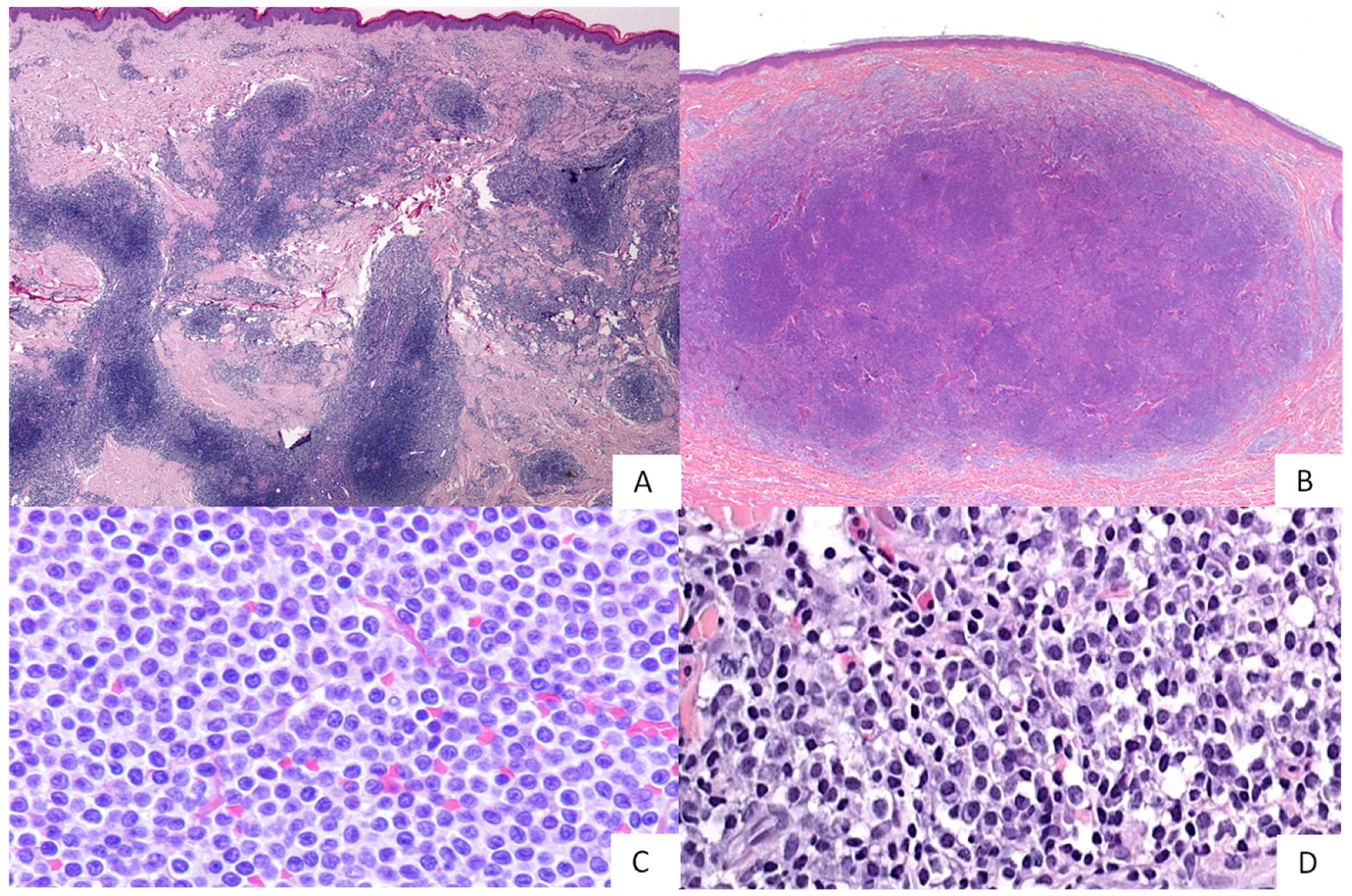
Figure 1. PCMZL. At histological evaluation, this lymphoma shows a nodular ((A), HE 2×) or a diffuse growth pattern ((B), HE 2×) and is composed of small- to medium-sized B cells, sometimes with a monocytoid appearance ((C), HE 40×) or a plasmacytic differentiation ((D), HE 40×).
Hemato 03 00023 g001
Figure 2. PCFCL. At histological evaluation, this lymphoma shows a nodular growth pattern ((A), HE 2×), sometimes nodular and vaguely diffuse ((B), 2×), composed of an admixture of centrocytes and centroblasts ((C), HE 40×), and is usually bcl6-positive ((D), 10×).
Hemato 03 00023 g002
Figure 3. PCDLBCL LT. At histological evaluation, this lymphoma is composed of sheets of large, atypical cells (centroblasts and/or immunoblasts) with numerous mitoses and no/few reactive backgrounds ((A), HE 40×). PCDLBCL-LT frequently expresses bcl2 ((B), 20×), IgM ((C), 20×), and c-MYC ((D), 20×).
Hemato 03 00023 g003
Figure 4. PCDLBCL-NOS. At histological evaluation, this lymphoma shows a diffuse growth pattern, sometimes with vaguely nodular areas ((A), HE 2×) and is composed of large centroblasts with a minority of medium-sized cells ((B), HE 40×). PCDLBCL-NOS is frequently bcl6-positive ((C), 40×) and bcl-2-negative ((D), 20×).
Hemato 03 00023 g004
Table 1. Differential diagnosis between PCFCL, PCMZL and CLH.
CharacteristicsPCFCLPCMZLCLH
Age50–60>5050
SexM > FM > FF > M
Sitehead, trunk, legarms, trunk, head, neckface (nose and cheeks), trunk, extremities
Clinical featuresplaque, nodule, tumourpapules, nodules, plaquesnodule, papules
Single/multiple lesionsusually singleoften multifocalusually single
Histology
Cells morphologycentrocytes (prevalent) and centroblastscentrocyte-like, monocytoid, lymphoplasmacytic.
Plasma cells often present in superficial dermis and at the lymphoma’s periphery
small lymphocytes
Patternnodular, nodular and diffuse, diffusenodular (more often), diffusenodular/diffuse
Skin ulcerationabsentabsentabsent
NecrosisnoNono
Adnexal effacementusually absentusually absentusually absent
Reactive T-cell CD3+ infiltratepresent, abundantpresent, abundantpresent
Dendritic meshworkpresent/absentpresent/absentpresent
ImmunophenotypeCD20+, CD79a+
bcl6+, CD10+,
bcl2- (73%)
CD20+, CD79a+, bcl2+,
CD10-, bcl6-, CD5-Cyclin-D1-, CD23- (most cases)
mixed infiltrates of B and T cells with reactive germinal centres
Ki67usually low (up to 30%)usually lowhigh in reactive germinal centres
Molecular featuresrarely translocation IGH-BCL2
up to 40% BCL2 aberrations
80–92% clonal IGH/IGK gene rearrangements,
25% translocation IGH-MALT1
80–90% polyclonal
Prognosisindolentindolentusually good
Table 2. Differential diagnosis between PCFCL, PCDLBCL NOS and PCDLBCL-LT.
CharacteristicsPCFCLPCDLBL NOSPCDLBCL-LT
Age50–606070–80
SexM>FM>FF>M
Sitehead, trunk, legtrunk, head-neck, lower limbs, upper limbsleg, trunk, head-neck, upper extremities
Clinical featuresplaque, nodule, tumournodule, plaquetumour, nodule
Single/multiple lesionsusually singlesinglesingle or multiple
Histology
Cells morphologycentrocytes (prevalent) and centroblastscentroblasts with <10% of medium sized cellscentroblast and/or immunoblast
Patternnodular, nodular and diffuse, diffusediffuse, vaguely nodulardiffuse
Skin ulcerationAbsentpresent or absentmostly present
Necrosisnorareyes
Adnexal effacementusually absentMay be presentmostly present
Reactive T-cell CD3+ infiltratepresent, abundantpresent, mild to moderatefew or absent
Dendritic meshworkpresent/absentabsentabsent
ImmunophenotypeCD20+, CD79a+, Bcl6+, CD10+, Bcl2-(73%) CD20+, CD79a+,
Bcl6+, CD10+/−, MUM1+/−, IgM+/−,
c-Myc+/−, bcl2-/+
CD20+, CD79a+, Bcl2+, MUM1+, Bcl6+, c-myc+, IgM+, CD10-
Ki67usually low (up to 30%)moderate (40%)high (>70%)
DE phenotypenoinfrequent>60% of cases
Molecular featuresrarely translocation IGH-BCL2,
up to 40% BCL2 aberrations
rearrangements of BCL6 or MYC,
rarely BCL2 alterations
IGH clonal rearrangements, translocations involving BCL6, MYC and IGH, MYD88L265P mutations
DH/TH statusnoDH status reported in literature (one case)yes
Prognosisindolentless aggressive than PCDLBCL-LT,
GC cases more similar to PCFCL,
Non-GC cases in between PCFCL and PCDLBCL-LT
aggressive
Publisher’s Note: MDPI stays neutral with regard to jurisdictional claims in published maps and institutional affiliations.