Review

A Review of Xyloglucan: Self-Aggregation, Hydrogel Formation, Mucoadhesion and Uses in Medical Devices

Institute of Advanced Chemistry of Catalonia (IQAC), Consejo Superior de Investigaciones Científicas (CSIC), Networking Research Center on Bioengineering, Biomaterials and Nanomedicine (CIBER-BBN), Jordi Girona, 18-26, 08034 Barcelona, Spain
Macromol 2022, 2(4), 562-590; https://doi.org/10.3390/macromol2040037
Submission received: 30 July 2022 / Revised: 19 November 2022 / Accepted: 21 November 2022 / Published: 6 December 2022

Abstract

:
The present paper reviews the self-aggregation, gel-forming and adsorption properties of xyloglucan (XG), and its main applications as a medical device for wound dressings, mucosal protection and ocular lubrication, as well as its uses as an excipient. XG is a branched polysaccharide composed of a central backbone of D-glucose units linked by β(1→4)-glycosidic bonds, decorated with D-xylose units through α(1→6) glycosidic bonds, and with some D-galactose units anchored to these D-xylose units via β(1→2) bonds. XG forms self-aggregates with a hierarchically ordered morphology in aqueous solutions, leading to the formation of nanofibers. Consequently, XG is a hydrogel-forming polymer able to retain large amounts of water. Inside the human digestive tract, XG is enzymatically degalactosylated, but the backbone with xylose side chains remains stable until excretion. Degalactosylated XG undergoes a fully reversible sol–gel transition, forming hydrogels between upper and lower critical temperatures. XG adsorbs on intestinal mucosa and creates a diffusion barrier that reduces permeability and also prevents bacterial infections by reducing their infiltration. Therefore, orally administered XG is considered a mucosa protectant.

Graphical Abstract

1. Introduction

1.1. Basic Features of Hydrogels

Hydrogels are hydrophilic three-dimensional (3D) polymer networks that form viscoelastic soft materials. They can swell and shrink reversibly, by absorbing or releasing very large amounts of water [1]. This viscoelastic behavior is caused by the hydration of the polymer chains, which retain a large volume fraction of water. The three-dimensional polymer networks are stabilized by crosslinks, which hold together the chains of the hydrated polymer, preserve the structural integrity of the hydrogel network and do not dissolve, even at low polymer concentrations [2]. The crosslinks that bond polymers are classified into two general categories: chemical and physical. Chemical crosslinks are covalent bonds, whereas physical crosslinks consist of weaker interactions, such as hydrogen bonds, hydrophobic interactions and chain entanglements [3,4].
Biocompatible hydrogels for biomedical applications are typically based on polysaccharides [1], which can be nonionic, such as dextran or pullulan; anionic, with carboxylic groups (e.g., alginate) [1] or sulfate groups (e.g., carrageenan, agarose, fucoidan and ulvan) [5]; or cationic, containing amino groups (e.g., chitosan) [4]. All of them can form soft viscoelastic materials that retain a very large amount of water. For this reason, they are commonly used to culture micro-organisms (e.g., agarose) and as scaffold materials for tissue engineering [1,6]. Gelling polysaccharides also attract much attention because of their therapeutic or coadjuvant effects, since they often increase the bioavailability and/or efficacy of drugs. Among neutral polysaccharides, xyloglucan (XG) has been the object of many studies because of its mucoadhesivity, and many new formulations based on XG have successfully reached the stage of clinical trials, demonstrating its beneficial effects as a mucosal protectant. XG adsorbs on mucous membranes, creating physical barriers against the diffusion of undesired species, bacteria or allergens. The main aim of the present review is to provide current knowledge on the colloidal properties of XG, as well as recent advances in its therapeutic and/or healthcare applications as a medical device.

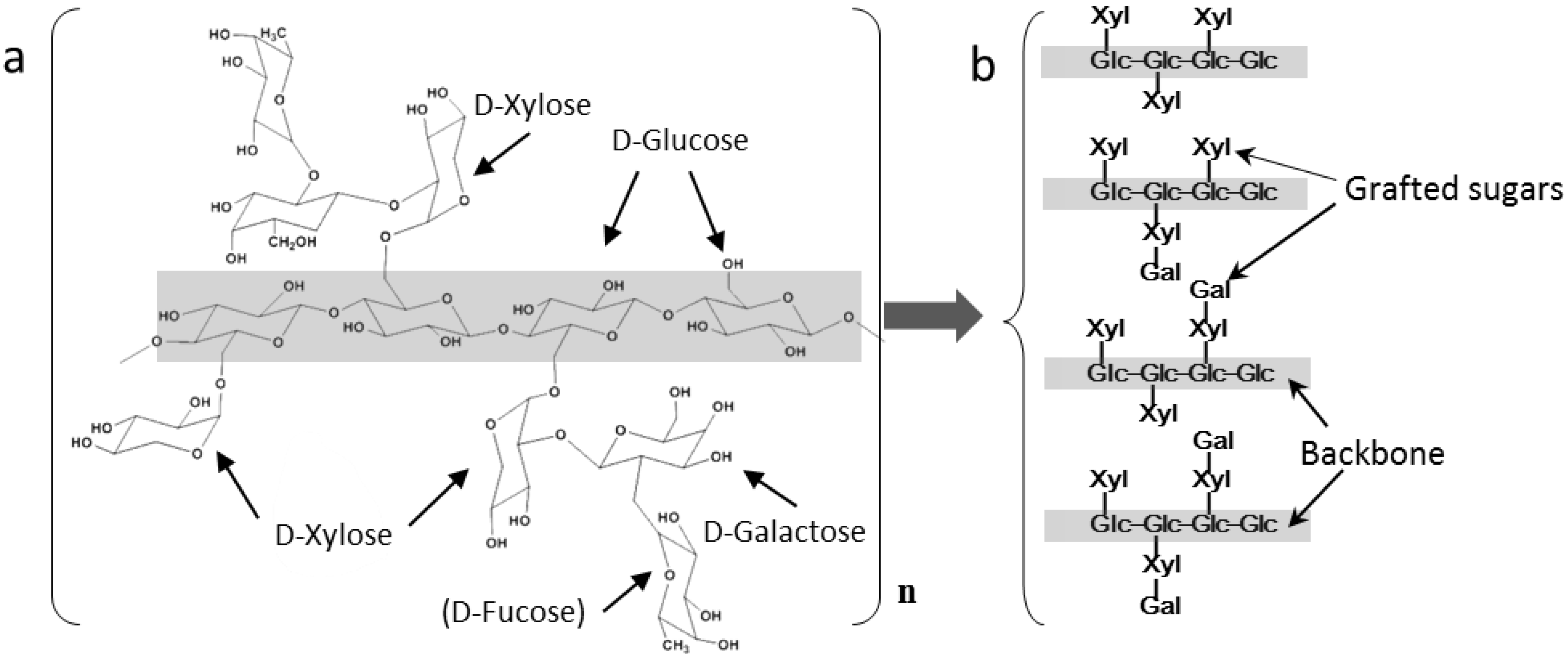
1.2. Basic Features of XG

XG is a nonionic polysaccharide that is considered to be completely biocompatible and biodegradable, and has been approved by the FDA as a food additive and used as an excipient for drug delivery. XG can be found in many edible plants, including commonly farmed species such as Lactuca sativa (lettuce) and Daucus carota (carrots), as well as many other plants, including Hymenaea courbaril (jatoba), Detarium senegalense, Detarium microcarpum, Afzelia africana and Tanacetum ptarmiciflorum. However, the most common commercial type is that extracted from the kernels of tamarind (Tamarindus indica) seeds [7]. Tamarind xyloglucan is typically produced by applying a standardized protocol that includes boiling, centrifugation and precipitation [8]. In recent years, many scientists have focused their attention on the use of XG as a carrier for drug delivery, as well as its use as a scaffold for tissue regeneration. A recent review [9] comprehensively describes the current knowledge on this subject.
The present review focuses on the colloidal, aggregation and mucoadhesive properties of XG. First the chemical structure and basic chemical properties are described, followed by a discussion of the self-aggregation properties and the ability of XG to form hydrogels and mucoadhesive films. Finally, recent advances in medical applications using XG are also reviewed.
XG is a branched polysaccharide formed by a central backbone chain that is decorated with xylose or xylose–galactose sugars that are sometimes capped with fucose ends (Figure 1a) [10,11,12,13,14]. The linear backbone constitutes D-glucose monomer units linked with β(1→4) glycosidic bonds, and therefore, its chemical structure is equivalent to that of cellulose. Typically, the length of this backbone ranges between 300 and 3000 D-glucose units [10]. Lateral D-xylose units are grafted to approximately 75% of the glucose units through α(1→6) glycosidic bonds. Moreover, some D-galactose units are anchored to these D-xylose units via β(1→2) bonds. In addition, sometimes, the galactose units are capped with D-fucose units linked to galactose with α(1→2) bonds [11]. The distributions of all these side chains have regular sequences that depend on the origin of XG. Some typical structures of XG and its main monosaccharide units (glucose, xylose and galactose, in the absence of fucose) are shown in Figure 1b.
The distribution of side sugars and their relative amounts of xylose, galactose and/or fucose depend on the vegetable species from which they are extracted. The typical monosaccharide composition of XG obtained from tamarind seeds is approximately 43–45% glucose, 35–38% xylose and 15–17% galactose, with minor amounts of arabinose and other sugars [16]. XG extracted from Hymenaea courbaril showed a slightly different composition, with 40% glucose, 34% xylose and 20% galactose [17]. XG from the seeds of nasturtium (Tropaeolum genus plants) showed a similar monosaccharide composition to that of tamarind seeds [16]. It should be noted that these differences are small and results reported in the literature can differ because of either experimental error, differences in the origin of XG or variations in extraction processes. Ieiri et al. [18] reported the relative compositions of glucose, xylose and galactose in XGs extracted from four different plants (Table 1).
The presence of impurities, mainly other saccharides, depends on the extraction and purification methods, and XG is frequently distributed with ≈95% purity. The main impurity of tamarind seed XG is a polysaccharide composed of branched (1→5)-α-L-arabinofuranan and unbranched (1→4)-β-D-galactopyranan [16]. Characterization using rheology, static light scattering and small-angle X-ray scattering has shown that XG is a polymer with a remarkable stiffness [16], which is attributed to restricted rotations around the β(1→4) bond. This stiffness is enhanced by the presence of xylose side chains that limit its conformational freedom.
XG is the most abundant hemicellulose found in dicotyledon plants, and is a major component of cell walls in these plants, with concentrations of approximately 10 wt% in their cell walls in vivo [10]. It is an important building material that provides mechanical properties to wood, since XG promotes the binding of cellulose microfibrils. Moreover, XG is present in the seeds of many dicotyledonous plants as a food reserve for germination [10]. XG is commonly found in combination with other natural polysaccharides, playing an important role in providing biomechanical strength to both plant cell walls and wood. For example, cellulose-XG-pectin complexes have been identified in biomechanical hot spots of biomass extracted from milkweeds [19]. XG is involved in the development of trunk mechanical properties and the orientation of trees in upright positions [20]. Another example of the structural role of XG in plants is in the root mucilage secretions of peas (Pisum sativum), which form a fibrous network that links root border cells [21].
XG is widely used as a thickener for low-calorie food products [11], which include flour paste, custard cream, noodles and stew, as well as being commonly used in Asian cuisine products (for example, rice cakes and dango). The technical quality and safety requirements of XG as a food additive have been standardized by the FAO/WHO Expert Committee on Food Additives (JECFA), [22]. In the human digestive tract, XG is only partially degraded by secreted enzymes, mainly β-galactosidase, which removes the galactose chain ends. The main part of the molecule (cellulose backbone with xylose side chains) remains chemically stable in the intestine since human digestive enzymes are not able to degrade it. Consequently, XG is considered a nondigestive dietary fiber that is not degraded and is not transported across intestinal membranes; thus, it generally remains in the digestive tract until excretion in feces. Nevertheless, XG may play an important role since it promotes the proliferation of certain beneficial bacteria [23,24]. Some intestinal microbiota are able to feed on XG and use it to proliferate [23]. According to in vitro fecal fermentation studies, the presence of XG induces changes in the composition of the gut microbiota [24]. For example, XG promotes the growth of Bacteroidetes, one of the dominant bacterial populations present in the human gut that plays an important role in the metabolism of rather indigestible polysaccharides present within “dietary fiber” [23]. Moreover, XG facilitates the digestion of plant polysaccharides in monogastric animals, such as swine and poultry, and it is incorporated as an additive into farm animal feed [25].
XG intake decreases the total plasma lipid concentration. In 1996, one study reported that the plasma levels of total lipids, cholesterol, triglycerides and β-lipoprotein were all reduced by approximately 15% when XG was introduced into the diet of male Wistar rats at 6 weeks of age [26]. Rats were receiving a high-fat diet, and the total lipid, cholesterol, triglyceride and liver phospholipid levels were significantly decreased after administering hydrolyzed (short-chain polysaccharides) XG. Moreover, the weight of adipose tissue in the rats was also reduced by adding hydrolyzed XG to the high-fat diet [26].
One recent article discusses current knowledge on the possible beneficial effects of XG on the gut microbiota [27]. However, many aspects of the role of XG in the gut are largely unknown. For example, the microbial community generates a wide variety of metabolites and degradation products, and knowledge of the effects of these products is scarce. In addition, most studies have focused on animal models (mainly rats), and consequently, their results cannot be extrapolated to the human digestive system. XG forms a protective barrier on mucosa membranes of the intestinal tract, but nevertheless, the precise mechanism and physiological effects are still not well understood [27].

2. Colloidal Aspects of XG

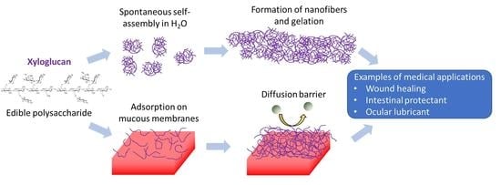
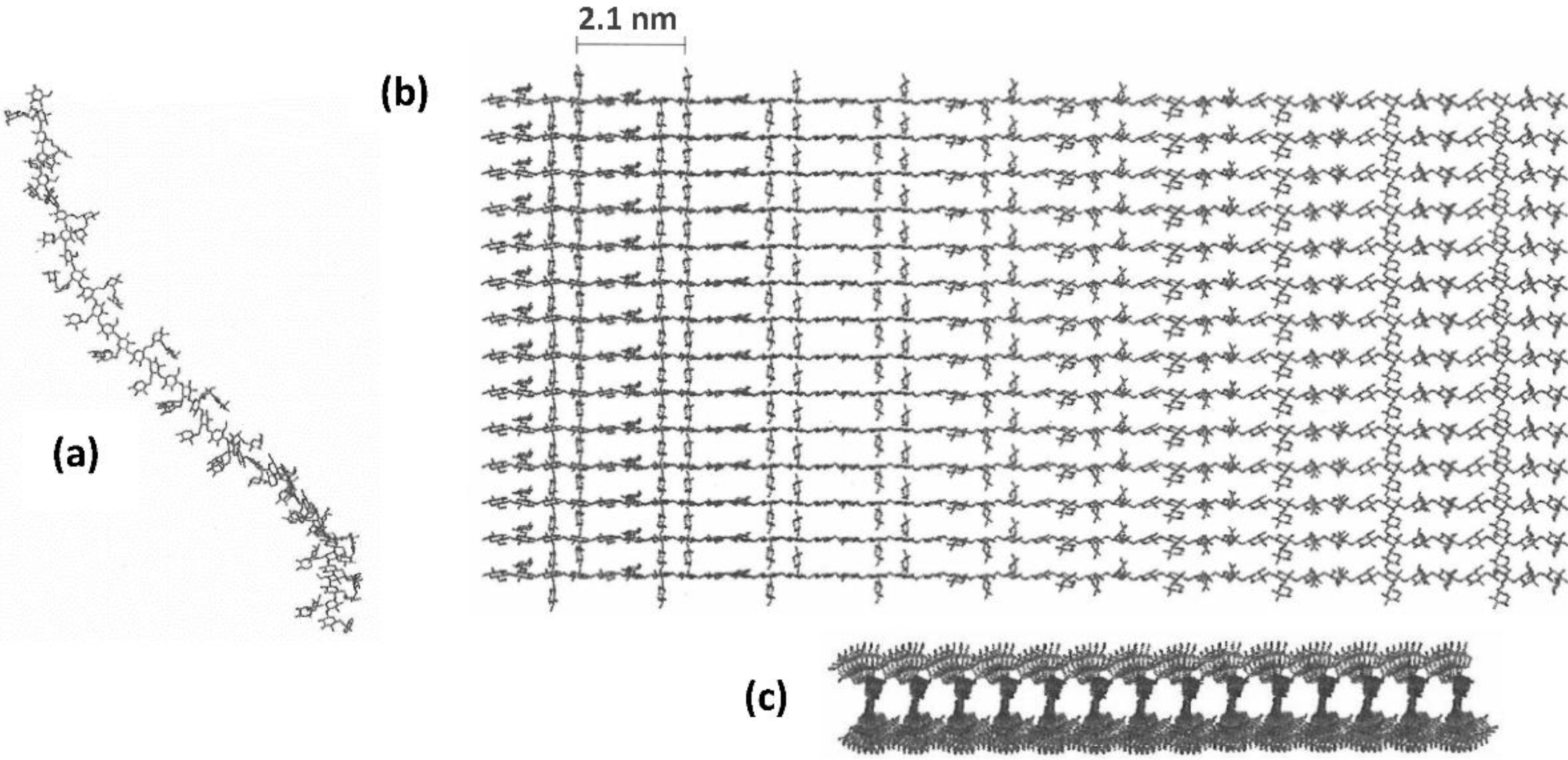
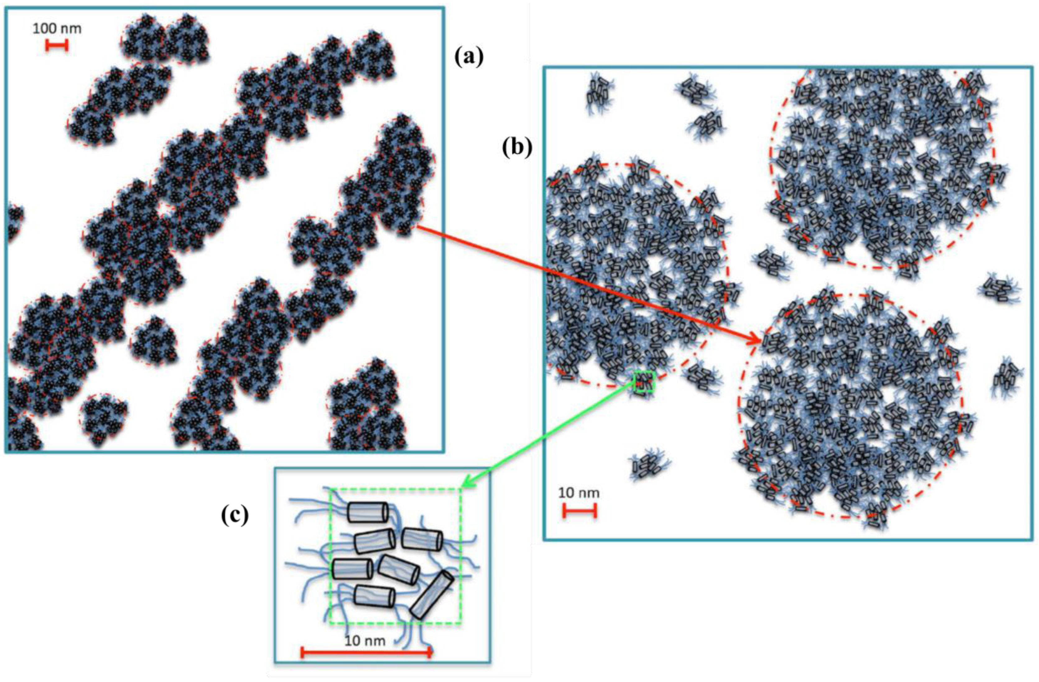
2.1. Self-Aggregation

As mentioned above, XG is a common additive in food formulations, and is used as a thickener and stabilizer. These uses are a consequence of the hydrophilic nature of the molecule, which makes it highly hydrated [28], and the ramified molecular structure that produces entanglements between molecules, leading to an increase in viscosity and viscoelastic flow behavior [11].
XG forms self-aggregates in aqueous solution. In 1993, Lang et al. studied aqueous XG solutions using static light scattering (SLS) and small-angle X-ray scattering (SAXS) [29]. The results revealed the presence of worm-like self-aggregates with rather high stiffness. These aggregates consist of lateral assemblies of polysaccharide chains. Static light scattering data showed that the stiffness is determined by the number of aggregated strands [30]. The formation of these nanofibers occurs more extensively in XG extracted from tamarind seeds than in other sources of XG, such as Detarium microcarpum [31]. These differences, depending on the origin of XG, are caused by subtle variations in the sequence of saccharide side units that either promote or hinder the tight packing of polysaccharide chains [18].
XG is a branched polysaccharide, and the formation of self-aggregates depends on the ability of the side chains to promote or hinder packing. In the case of tamarind XG, the side chains can form relatively strong intermolecular hydrogen bonds and allow packing in nanofibers and/or bundles, as documented by Yamatoya, Kajiwara and coworkers [31,32]. These authors performed a systematic study using scattering techniques and showed that individual XG chains have a twisted ribbon morphology (Figure 2a) but the supramolecular aggregates form flat layers, as depicted in the schemes shown in Figure 2b (top perspective) and Figure 2c (cross-section). This self-aggregation confirms the observed stiffness of the XG nanostructures.
Other studies performed using SAXS have also confirmed that XG aggregates preferentially have a rod-like morphology [18], which might also explain the high viscosity of aqueous XG solutions. The flexibility of XG chains seems to depend on the origin of XG, and viscosity has also been observed to vary, depending on the botanical variety from which it has been extracted.
In a more recent study, Dispenza et al. showed that XG aggregates adopt a hierarchically ordered morphology, with different morphologies observed at different size ranges [33]. They studied the morphology and structure of the self-aggregates using a combination of SAXS and small- and large-angle light scattering (SALS and LALS, respectively). These experimental techniques have confirmed that aqueous colloidal dispersions of XG form aggregated clusters with a size of hundreds of nanometers. In the case of tamarind seed xyloglucan, these clusters consist of interconnected dense aggregates with a size of ≈10 nm, which are formed via the assembly of several polymer chains. Moreover, these small aggregates form larger hierarchically organized superstructures with different morphologies at different length scales [33]. This proposed organization is displayed in Figure 3.
XG fibers were characterized using atomic force microscopy (AFM) and topographic observations on mica surfaces [34]. The specimens were prepared by dropping and drying 5 μL of 0.05 wt% XG on freshly exfoliated mica. The AFM images revealed molecular aggregates with a rod-like morphology of approximately 2.3 nm in diameter and 640 nm in length. AFM topography also revealed the presence of XG helical structures with approximately a 115 nm periodicity. These observations also showed that XG molecules aggregate and form rope-like long nanostructures, thus explaining the ability of XG to form gels [34].
Chemically modified XG also forms supramolecular aggregates. Lang et al. studied the morphology of the aggregates of carboxylated XG as a function of different derivatization degrees [30]. Carboxylation was performed via enzymatic oxidation of the galactose units, aiming to introduce negative charges and impart electrostatic repulsion. Characterization using SLS and DLS indicated the formation of polymer strands via lateral aggregation, through a similar mechanism to noncarboxylated XG. These strands might have cross-sectional diameters of approximately 8–15 nm [30].
The colloidal properties of XG and its applications in controlled drug delivery can be controlled by derivatization, as reviewed by Shukla et al. [35]. Lang et al. described the preparation of carboxylated, sulfated and alkylated derivatives [36]. The anionic derivatives showed stiff backbone chains, similarly to native XG, and the hydrophobized alkylated derivatives decreased surface and interfacial tensions. For example, XG aqueous solution had a surface tension of 61.3 mN/m whereas octyl-XG showed a surface tension of 55.6 mN/m, at 0.1 wt% concentrations [36]. The derivation of XG can be performed by combining enzymatic and chemical reactions, allowing for more selective functionalization. For example, the use of galactose oxidase [36,37] allows carboxylation only in the galactosyl ends of side chains.
Another study demonstrated that carboxymethylation of XG greatly increases water solubility, the swelling of hydrogels and viscosity [38]. The derivation of XG was also studied by Kaur et al. [39], who obtained anionic (carboxylated and sulfated) and cationic (aminated) XG derivatives that showed higher hydrophilicity, increasing water absorption. The cationic derivative, XG-NH2, had the strongest bioadhesive strength [39]. This result was attributed to electrostatic attractions between the cationic derivative and the negative charges of mucin in mucous membranes.
The critical aggregation concentration (CAC) of XG can be reduced via hydrophobization, as reported by Dilbaghi et al. [28]. Moreover, films of hydrophobized XG displayed a smoother surface in comparison to unmodified XG. Another strategy for XG modification is blending with other polymers via chemical crosslinks. One example, described by Ajovalasit et al., is the formation of XG/polyvinyl alcohol (PVA)-blended polymers via covalent crosslinking with glutaraldehyde, showing that XG-PVA films and hydrogels could be used as smart wound dressings [40]. Another example of blended polymers is the grafting of XG and polyacrylamide [41]. The resulting copolymer was shown to be a flocculant in paracetamol suspensions, promoting the aggregation of paracetamol particles.

2.2. Xyloglucan Hydrogels

XG molecules self-assemble in water, forming nanofibers composed of polymer bundles when increasing the concentration from diluted to semi-diluted. The obvious consequence is that aqueous XG solutions can produce physical hydrogels with yield stress in the absence of covalent crosslinking. This property is considered an advantage for food formulations, since hydrogels often have an aesthetic visual aspect and improve the organoleptic properties of heat-and-eat food products. XG generally forms rather viscous solutions, which is attributed to hierarchically organized superstructures. In the case of XG extracted from the tropical plant Hymenaea courbaril [17], dilute solutions with 0.5 wt% XG have a flow behavior that is approximately Newtonian at a shear rate below 10 s−1, but viscosity increases substantially when the concentration changes to 1 wt%, producing viscoelastic behavior with shear thinning (viscosity decreases with the increase in shear rate), as illustrated in Figure 4a. In the case of 1 wt% XG, the viscosity at a low shear rate is approximately 0.8 Pa.s, which is 800 times higher than the viscosity of pure water (0.001 Pa.s).
Dynamic measurements as a function of oscillation frequency showed the typical behavior of viscoelastic fluids (Figure 4b). Both the elastic modulus (G′) and the viscous modulus (G″) increase with frequency. However, G′ increases faster than G″, and thus, the materials become predominantly elastic at a high frequency. This behavior is well known and commonly observed in many polymer solutions. In conclusion, XG solutions behave as typical polymer gels.
The gelation of polysaccharide solutions, forming physical hydrogels, is explained by the well-accepted model of Flory–Stockmayer [42,43,44]. Gelation occurs at a particular concentration of a soluble polymer when the hydrated chains form an interconnected network because of crosslinking, which is induced by weak interactions that include a combination of hydrogen bonds and steric restrictions of conformational freedom. The crosslinking of hydrophilic polymers leads to the appearance of an infinitely large superstructure that retains large volumes of hydration water, shifting the macroscopic aspect from a viscous liquid to a viscoelastic fluid. The concentration at which gelation might occur (Critical Gelation Concentration—CGC) can be predicted if the conformational data (chain length, radius of gyration, etc.) of the polymer are known.
In its native form, tamarind XG does not form highly viscous gels at low concentrations. However, the removal of the galactose units substantially increases viscosity and induces the formation of hydrogels in aqueous solutions of XG at concentrations lower than 5 wt%. Consequently, the full gelation of dissolved XG is prevented by steric hindrance between galactose–xylose branches [31]. The galactose end units of XG branches are easily removed by enzymatic treatment of native XG. The β-galactosidase enzyme cuts the β(1→2) covalent bonds between the xylose and galactose units of XG side branches. This process is equivalent to that occurring in the digestive tracts of mammals. The galactose ends of XG branches are removed by the β-galactosidase enzyme that is naturally present in the digestive tract of humans, except in people with lactose intolerance. The enzyme removes galactose units under typical human gastric conditions; nevertheless the main cellulose backbone and the grafted xylitol units remain unaffected. Consequently, native XG is degalactosylated when moving through the intestine of lactose-tolerant individuals [27]. In addition, further degradation of XG might be induced by some gut microbiota. Actually, the performance of degalactosylated XG in the digestive tract seems to be the same as that of native, pristine XG. Experiments with rats have indicated that enzyme-treated XG (degalactosylated XG) virtually provides the same dietary benefits as pristine XG, improving lipid metabolism in both cases [45].
The rheological properties of enzyme-treated XG are known [46], and degalactosylated (enzyme-treated) XG undergoes a fully reversible sol–gel transition [45]. Degalactosylated XG greatly increases viscosity via heating, which is reverted to fluidification upon cooling [15,47]. The crosslinking domains are most likely formed by XG chains aligned together in the shape of flat nanoplatelets. Consequently, degalactosylated XG solutions are thermoresponsive, producing more highly viscous solutions at warm temperatures but becoming much more fluid at lower temperatures. As shown in a previous study, the removal of 35% galactose units, similar to the process that typically occurs in the human digestive tract, is sufficient to achieve thermoresponsive behavior [45].
Therefore, the gelation time and gelation temperature of degalactosylated (enzyme-modified) XG can be easily controlled by tuning the degree of galactose removal (degalactosylation). The sol–gel transition can be controlled over a wide range of temperatures, and interestingly, this transition is achieved at temperatures of approximately 37 °C [46]. An increase in the degree of galactose removal decreases the temperature of the sol–gel transition. For example, this transition is decreased from 40 to 5 °C, by increasing the removal of galactose from 35 to 58 w% [47,48,49]. Consequently, partially degalactosylated XG forms gels at body temperature, assuming that its concentration is high enough. Results with 3 wt% XG showed predominantly elastic behavior, i.e., the elastic modulus, G′, was greater than the viscous modulus, G″.
Tamatoya, Kajiwara and coworkers [31,32] studied the sol–gel transition of aqueous solutions of enzymatically treated XG and produced a phase diagram [32] (Figure 5). At low galactose removal, fluid XG solutions are observed at any temperature ranging between 0 and 130 °C. However, with a high degree of galactose removal, one hydrogel region appears, which is limited between upper and lower transition temperatures (Figure 5).
Native, pristine XG does not form thermoreversible hydrogels; however, these gels are observed when mixed with other components. For example, tamarind seed XG and sodium gellan interact synergistically since their mixtures form hydrogels, although each component alone does not form gels at the same total concentration [50]. This synergistic hydrogel formation was studied using differential scanning calorimetry (DSC), which showed the appearance of a peak when XG was mixed with gellan, indicating a strong interaction between the two polymers.
Another example of enhancing the gelling properties of native XG is the thermoreversibility exhibited in the presence of ethanol. XG from tamarind seeds shows a reversible sol–gel transition when mixed with ethanol in aqueous solutions, forming gels at temperatures below this transition. This gelation process is a different phenomenon than that observed in the case of enzymatically degraded tamarind XG, which has both lower and upper transition temperatures (Figure 5). In the case of ethanol–water solvent mixtures, crosslinking seems to occur through random interactions of XG chains caused by their poor solubility in ethanol. This gelation has been studied using time-resolved SAXS [51], revealing that the transition temperature corresponds to the dissolution point of XG aggregates that appear in presence of ethanol, which is a poor solvent for XG at low temperatures. The addition of other water-soluble alcohols, such as propylene glycol or 2-(2-ethoxyethoxy)ethanol, also results in the gelation of colloidal dispersions of native XG. This gelation has been attributed to the competition between the polymer and the alcohol for water, which causes dehydration of the polymer and increases its association and crosslinking [33].

2.3. XG Films

XG films have been prepared by casting and drying XG aqueous solutions on flat substrates. Purified tamarind XG produces colorless and transparent films with good thermomechanical properties. However, these films lack the flexibility required for many technical applications. The glass transition temperature (Tg) of XG is greater than 250 °C, indicating that it is not softened by thermal treatments. The effect of the addition of plasticizers has been studied by Berlung and coworkers [52]. Among various possible plasticizers (e.g., sorbitol, urea, glycerol and PEG), a good combination of mechanical properties (strength, toughness, Young’s modulus and ductility) was achieved with approximately 20 wt% sorbitol. The improved films also showed good thermal stability and retained transparency [52].
Thin films of native XG (i.e., less than 30 nm thick) have been formed on hydrophilic substrates. As an illustrative example, XG adsorbs on cellulose and forms thin films [53]. This adsorption was studied using atomic force microscopy, surface plasmon resonance and quartz crystal microbalance. The amount of adsorbed XG depended on the type of cellulose; the highest amount was observed for desulfated cellulose nanocrystals and the lowest was observed for amorphous regenerated cellulose. These results were attributed to the low ability of XG to penetrate across chains of amorphous cellulose, whereas it deeply penetrates into substrates composed of nanocrystalline cellulose [53].
In another example, XG with a 45% galactose removal ratio was dissolved in water and the solutions were spread over Teflon-coated surfaces and dried in an oven at 40 °C, obtaining XG homogeneous films with a controlled thickness [54]. Glycerol was added as a plasticizer, and thin films (thickness ranging from 130 to 250 nm) were obtained with quite homogeneous thickness, a smooth surface and an absence of cracks. These films were tested in vivo with rabbits for the ocular delivery of ciprofloxacin [54].
Tamarind XG has also been studied as a biocompatible polymer for packaging food products. Biopolymers are of great interest as substitutes for petroleum-based polymers in packaging applications. However, one main drawback is the high Tg, which impedes thermal processing. Another strategy to decrease the glass transition temperature without the addition of plasticizers is chemical modification. For example, Berglund, Kochumalayil and coworkers studied the use of periodate oxidation followed by reduction; this allowed for regioselective modification that opened the side carbohydrates while preserving the cellulose backbone, which remained largely intact. Using this method, the Tg of the chemically modified XG was reduced by more than 100 °C [55].
As mentioned above, XG films have high stiffness and strength, as well as good oxygen barrier properties, but these films do not resist high humidity. When in contact with water, the mechanical properties of XG films are substantially deteriorated. This drawback can be addressed by enzymatic removal of the galactose units of XG side branches. Kochumalayil et al. [12] studied the mechanical properties of degalactosylated XG films by measuring their tensile stress and dynamic mechanical thermal properties, as well as obtaining moisture sorption isotherms. They used enzymatically treated XG, and the mechanical properties of the films were improved by adding glycerol as a plasticizer. The films showed a Young’s modulus as high as 4.3 GPa, combined with an ultimate strength of 60 MPa. These properties were considered very good for an edible polymer and comparable to the mechanical properties of starch amylose. However, the water moisture content at 23 °C and 50% relative humidity was observed to be approximately 35% lower than that of starch. Regarding thermostability, XG films withstand thermal treatments up to approximately 250 °C [12]. In conclusion, degalactosylated XG may be considered a thermostable polymer for manufacturing films and is a good alternative to starch in applications such as food packaging and coatings for food containers.
Another study has confirmed that degalactosylated XG absorbs less moisture and shows much less softness in the presence of water [56]. Consequently, its mechanical properties improved under humid conditions. This modified XG provided a high Young’s modulus (4.3 GPa) measured at 92% humidity. In addition, degalactosylated XG showed lower oxygen permeability under high humidity conditions. The oxygen permeability at 80% humidity was 1.5 cm3 µm (m2 day)−1 kPa−1, which is much lower than that of native XG, at 11.5 cm3 µm (m2 day)−1 kPa−1.
The mechanical properties of pure pristine XG films are not satisfactory for biomedical applications. This drawback is often overcome by blending them with other biopolymers. In one example, XG-chitosan (XG-CH)-blended films showed good optical transparency, high tensile strength and thermostability [57]; moreover, the hydrophobicity and crystallinity were also higher in blended films. SEM observations of these films showed smooth and homogeneous film surfaces [57]. The swelling of XG-CH-blended films depended on the pH, and these films had a high degree of swelling at both neutral and acidic pH, whereas swelling was significantly lower at basic pH. This result might be attributed to the electrostatic repulsion between the protonated amino groups of chitosan, which are not charged at basic pH. Streptomycin was used as a model drug to study the use of these films for controlled release as a function of pH. XG-CH films could be appropriate for the controlled release of drugs.
Pure XG films are soluble in water, and thus, cannot be used in medical devices. Blends of XG with other hydrophilic polymers are also quite soluble in water, depending on the pH. Therefore, these films are not stable in the presence of water. One possible strategy to manufacture water-insoluble XG films is crosslinking with covalent bonds. For example, films with a high absorption of water that do not dissolve can be obtained by using glutaraldehyde (GA) as a crosslinker agent [58]. XG easily crosslinks with GA, which forms covalent bonds between adjacent XG chains. This crosslinking leads to the formation of permanent and stable hydrogels, which can be dried to form transparent films. Ajovalasit et al. [58] studied both the chemical and mechanical properties of XG/GA films, and polyvinyl alcohol (PVA) and glycerol (G) were added to improve the mechanical properties of new XG-PVA-G/GA-blended films. These crosslinked films were stable and did not dissolve in the presence of water. Interestingly, the films were able to absorb and retain a large amount of hydration water, achieving a high degree of swelling (ranging from 90 to 350%) upon the absorption of water. The crosslinked films reversibly withstood repeated cycles of drying and rehydration by absorbing water from humid air. In addition, cytotoxicity was evaluated in vitro, and the results showed that the films chemically crosslinked with GA were not cytotoxic, in either the presence or absence of PVA.
It should be noted that GA is a toxic compound [59] and it has not been approved by the FDA as a GRAS (generally recognized as safe) food additive. However, GA is commonly used as a crosslinker agent to obtain particles and films. GA is highly reactive with amino groups, and thus, it is rapidly consumed in the presence of proteins. Therefore, the concentration of free GA is often very low, below the threshold of toxicity limits, and consequently, XG films crosslinked with GA are not cytotoxic.
Another report describes the formation and properties of XG and polyacrylic acid (PA)-blended films, which were prepared via casting and drying, in the presence of glycerol that was added as a plasticizer [60]. These films were tested as mucoadhesive buccal films by measuring their bioadhesive force on porcine buccal mucosa, which was used as a model membrane. The results showed that XG was indeed a bioadhesive polymer, with adhesion forces between 0.3 and 0.9 N for films cut into square pieces of 1 cm. Moreover, an active component, rizatriptan was incorporated into the films, and ex vivo permeation assays were performed on the porcine buccal mucosa, obtaining permeations of the drug ranging from 85 to 95% in 2 h [60]. The bioadhesive activity of XG is described in more detail in the next section.

2.4. Mucoadhesivity of XG

Bioadhesion is commonly defined as the adhesion of polymers to biological substrates. In the case of mucous membranes, bioadhesion is denoted as mucoadhesion. Polymers with this property might enable improvement in the bioavailability of drugs by extending the residence time at the target mucous tissue [61]. Mucoadhesion occurs when a surface or a molecule adheres to a mucous membrane, mainly through the combination of hydrogen bonding and electrostatic attractions [62]. Mucoadhesive biomolecules have a high affinity for mucous membranes and adhere to their surface [60,62]. Certain drugs become chemically or physically anchored to mucosa, which extends their residence time on the mucous membranes of various tissues, including the gastrointestinal tract, buccal and nasal cavities, vaginal lumen or rectal mucosa [63]. Consequently, mucoadhesion is of great interest in the pharmaceutical field.
A scientific consensus has been reached on the idea that bioadhesion of a drug allows its localized delivery, promoting the entrance of this drug into the circulatory system [62]. Mucoadhesive molecules are highly hydrophilic water-soluble biopolymers that contain numerous functional groups able to interact with mucous membranes. Various different mechanisms have already been proposed to explain the phenomenon of surface adhesion, including electrostatic attractions, hydrogen bond formation, van der Waals attractions and hydration forces. However, mucoadhesion frequently occurs because of a wide number of factors, and it cannot be attributed to a single surface force [62]. The development of adhesion is often described as a two-step process [64]. In the first adsorption step, the mucoadhesive polymer diffuses to reach the mucous surface, and long-range attractive forces retain the polymer in close proximity to the surface. In the second step, various short-range interactions form between the polymer and the substrate, and the adhesion is consolidated. This two-step adhesion process occurs because some attractive forces only develop at very short distances, and thus, require the polymer to already be located close to the surface. These two steps have relative importance, depending on the site of action of the drug. For example, adsorption is a crucial stage if the galenic formulation cannot be directly applied to the target mucous tissue. However, consolidation may be the limiting step in the case of mucous membranes that are subject to dislodging stress [62].
Adhesion to mucous surfaces improves the release and permeation of active components across the mucous membranes that are targeted [63]. Therefore, mucoadhesivity enhances the therapeutic action of drugs that target the intestinal mucosa. Mucoadhesive drugs have been formulated in various preparations, including tablets for buccal delivery [65], gelling systems for ocular drug delivery [66], microgels for intravesicular administration [67] or nanoparticles targeting the gastrointestinal tract [68].
XG is mucoadhesive [9,62]. The adhesion of XG on mucous surfaces is caused by the affinity between polysaccharides and the surface of mucosa, which is rich in mucins. These proteins are members of a wide family of high-molecular-weight glycosylated proteins (glycoproteins) that are produced by epithelial cells in most animals. Mucin forms gels and constitute a major component of gel-like secretions. Mucins cover most mucous membranes in the human body and are present on the surface of most sections of the digestive tract [69], including membranes in the esophagus, stomach, small intestine and colon. The functions of mucins include lubrication, cell signaling and the formation of chemical barriers. In the digestive tract, mucins are associated with membranes and serve as receptor ligands for carbohydrate molecules [69,70]. Mucins have a morphology that resembles bottle brushes, with a central polypeptide chain that is decorated on one side with oligosaccharides. The polypeptide chain contains hydrophobic domains that allow mucin molecules to remain anchored on the lipid bilayers of membranes. The oligosaccharides (glycans) become oriented toward the exterior of the membranes, and thus, they interact with other saccharides present in the external media.
Mucoadhesion forces can be directly measured using conventional instruments for measuring surface forces, such as atomic force microscopy or surface force apparatus, as illustrated in the study by Avachat et al. [60]. Nevertheless, mucoadhesivity is a quite complex process and is difficult to evaluate. Many in vitro, in vivo and ex vivo methods have been proposed for the quantification of mucoadhesive properties, using either artificial or natural mucosa [71]. However, reproducibility and comparison of the results are quite difficult because of the lack of standardization of methods. Given the complexity of surface force measurements, mucoadhesion can also be studied using simpler techniques based on rheology [72,73]. The interactions between mucin and the tested components might be evaluated by analyzing the rheological behavior of aqueous mixtures in the presence of mucin, determining the possible synergistic increase in attractive interactions between mucin and the tested products.
Rheological synergism is defined as an increase in viscosity when two components are mixed together. In the case of aqueous mixtures of mucin and mucoadhesive molecules [72,73], the attractive forces between mucin and the tested molecule led to the appearance of gel-like viscoelastic properties, with much higher gel properties than those of mucin and the second molecule when they are observed separately. These experiments clearly demonstrated that the mucoadhesion of XG is caused by its attractive forces toward mucin glycoproteins, which are present in all mucous membranes.
Rheology is thus used as an experimental tool for the evaluation of mucoadhesivity in new formulations. In one recent example [74], the mucoadhesive properties of a formulation containing sodium chondroitin sulfate, XG and glycerol were evaluated in vitro by determining the rheological synergy of the mixture, as well as other techniques, such as the direct measurement of adhesion forces.
A recent review by N. Piqué [70] describes current knowledge of the mucoadhesivity of XG, its barrier properties on mucous membranes and its medical applications as a mucosal protector. XG prevents the adhesion of bacteria to mucous membranes. In one in vitro study, a mixture of XG, hibiscus and propolis was applied to models of intestinal and uroepithelial cells (CacoGoblet and RWPE-1, respectively), and it significantly decreased the adhesion of two pathogenic strains of E. coli on these cells, although the applied mixture did not alter the integrity of E. coli [75].
The bioadhesion of XG is increased via functionalization with thiol groups [76,77]. Thiolization enhances the mucoadhesion of a polymer 2–140-fold. Therefore, thiolized polymers (denoted thiomers) might enhance the pharmacological activity of drugs with limited permeability across mucous membranes. Formulating drugs with mucoadhesive thiomers may increase drug absorption through mucous membranes by prolonging the contact time on mucous surfaces [76]. A thiol derivative of native tamarind XG was synthesized and its mucoadhesivity was studied. Thiolization was performed using a two-step chemical process consisting of oxidation followed by conjugation with L-cysteine via an imine linkage [77]. The mucoadhesive properties were evaluated via measurements using ex vivo bioadhesion to fresh ovine intestinal mucosa. The results showed an improvement in mucoadhesivity, because the cysteine groups of thiolated XGn exhibited a stronger attachment to the intestinal mucosa. In conclusion, thiolated polymers might be worth studying, since they can provide longer residence times for drug delivery on mucous surfaces than conventional polymers with only hydroxyl functional groups.
The barrier effect of native XG was evaluated by determining the in vitro permeability of a human skin model [78]. HaCaT human keratinocytes were cultured and treated with a mixture of XG and pea proteins (PP) for 3 h. Afterward, the HaCaT cells were infected with Staphylococcus aureus. Finally, the membrane permeability of these cells was assessed by measuring the membrane integrity and the number of bacterial colonies. The application of XG and PP increased transepithelial electric resistance and reduced Lucifer yellow permeation, indicating a barrier effect [78]. Moreover, the application of XG and PP also reduced the adhesion of S. aureus.
In another study, the barrier protection exerted by XG was studied in more detail [79] using a 3D tissue model, MucilAir NasalTM, simulating nasal airways. This 3D model consists of assembled ciliated, goblet and basal cells and fully differentiated nasal epithelial cells. These cells, which form a tight and stratified nasal epithelium, are considered a good in vitro model of the nasal epithelium [80]. This tissue model was treated with 30 µL of either Rhinosectan® or controls (saline solution or budesonide) [79]. The effects of the application of the sprays were evaluated by measuring the transepithelial electric resistance; the preservation of the paracellular flux was observed using the Lucifer yellow test and the tight junction proteins were located using confocal fluorescence microscopy. Exposure to the studied spray increased the transepithelial electric resistance, indicating that the cellular tight junctions were protected, and paracellular flux was maintained in the presence of proinflammatory agents. The authors concluded that the XG-based product formed a protective physical barrier in this in vitro nasal epithelium model [79].
All these in vitro results suggest that XG contributes to preserving the integrity of skin barriers. In conclusion, XG is a nonionic branched polysaccharide that is water-soluble, forms hydrogels with viscoelastic flow behavior, and even more interestingly, readily adsorbs and adheres to mucous membranes. Consequently, XG is a promising biopolymer for medical applications in which biological barriers require reinforcement. The possible medical applications of XG are described in the next section.

3. Potential Medical Applications of XG

As discussed above, XG is an innocuous polymer obtained from natural renewable sources with gelling and mucoadhesive properties that may serve as a barrier to diffusion on the surface of mucous membranes. XG is produced at low cost and is commercially available worldwide, especially in China, India, Pakistan, Sri Lanka, Bangladesh and Bhutan. Thus, it is highly attractive for drug delivery applications. Potential pharmaceutical applications may use many possible routes of administration, including nasal, ocular, pulmonary, rectal, buccal, oral periodontal, transdermal and parenteral administration, as detailed by Kulkarni et al. [13]. Therefore, XG has been widely investigated and it is described in numerous articles on drug release and tissue engineering. The mucoadhesive properties of XG-based dosage forms allow sustained drug release to be achieved over extended periods. In addition, XG has the characteristic properties of thermoreversible gels, which form in situ when administered to the body. Therefore, XG can be used as an excipient in a wide variety of dosage forms. Because of its low cost, XG may become an increasingly important biopolymer in tissue engineering and as a carrier in drug delivery systems. Thus, it has many applications in formulations for therapeutic treatments and is also expected to receive much more attention in the future.
The present article is not intended to provide a complete or comprehensive review on the use of XG as an excipient for drug administration, since excellent reviews on this topic are available elsewhere [9,13,35,70,81]. In the present section, the main objective is to illustrate the benefits of XG as a smart gel-forming vehicle for drug release, and some selected examples are described.
One interesting case is the use of XG hydrogels as carriers for the intraperitoneal administration of mitomycin C [82]. The drug was incorporated into thermoreversible hydrogels composed of 1.5 wt% XG with 44% removal of galactose units. This partially degalactosylated XG is highly appropriate since its sol–gel transition occurs at temperatures between 22 and 27 °C depending on the xyloglucan concentration [32,83]. This property allows the formulation of fluid liquids at room temperature that form gels at 37 °C (body temperature). Mitomycin C in aqueous solutions of degalatosylated XG was injected into the peritoneal cavity of rats as fluid liquids, which soon afterward became highly viscous gels [82]. This gelation occurs precisely at the injection site and achieves controlled and sustained release of the drug for a period of approximately 6 h.
XG thermoreversible hydrogels may also serve as a delivery system for the rectal administration of drugs. Partially degalactosylated xyloglucan with 44% galactose removal was also studied, since its sol–gel transition temperature and gelling time of approximately 10 min were considered appropriate for rectal administration. The results of the in vitro release of two drugs, indomethacin and diltiazem, showed that both drugs exhibited more sustained release from the XG-based hydrogels than from commercial suppositories [83].
In in vivo assays with rabbits as animal models, XG solutions with indomethacin and diltiazem drugs were introduced as fluid liquids inside the rectal region of the animals, forming hydrogels in situ that delivered the drugs. Measurement of the plasma levels of indomethacin after rectal administration in XG-based hydrogels and commercial suppositories showed a broader peak of absorption after hydrogel administration, as well as an extended residence time. Moreover, the histological examination of rectal tissue did not show any damage. Morphological studies of rectal mucosa after the administration of the hydrogels showed no evidence of tissue damage. Therefore, these in vivo tests indicated that the applied XG hydrogel is safe for rectal administration [83], suggesting the potential of degalactosylated XG hydrogels as thermosensitive vehicles for rectal drug delivery.
In another example of in situ gelation, a mixture of XG, the block copolymer surfactant Pluronic F127 and poly(ethylene glycol) 6000 was studied as a thermoreversible gel formulation for the intranasal delivery of zolmitriptan or ketorolac, two anti-inflammatory drugs used for the acute treatment of migraine in adults [84]. The composition of the mixture was optimized to form gels at body temperature, extending the residence time of the two drugs, and thus, increasing their bioavailability. The histopathological characterization of the nasal mucosa indicated that the product was safe for nasal administration.
In a recent retrospective clinical trial of a treatment for ulcerative colitis, the probiotic bacteria Bifidobacterium animalis subsp. lactis was administered in an intracolonic single dose with XG. This case study focused on 10 patients who had been suffering from ulcerative colitis and had not responded well to previous treatments [85]. Seven patients were taking 5-acetylsalicylic acid and azathioprine and three patients were receiving vedolizumab. The administered dose contained 200 billion colony-forming units and 4 g of XG, which were dispersed in 250 mL of saline solution. The response to the intracolonic treatment was evaluated via colonoscopic examination six weeks after administration. After administration, all patients continued receiving the previous therapy, either with 5-acetylsalicylic acid and azathioprine or with vedolizumab. The intracolonic administration of B. animalis and XG was effective at inducing mucosal healing and the remission of ulcerative colitis symptoms [85]. Nevertheless, this clinical trial still cannot be considered fully conclusive since additional studies with a much larger number of patients are required.
The aforementioned examples illustrate the advantages of hydrogels as smart drug-delivery vehicles. Thermoreversible hydrogels are specifically indicated since they can be applied as fluid liquids, and in situ gelation is induced by a small increase in temperature. In situ gelation increases the bioavailability of drugs with poor absorption or fast removal, and many studies have already indicated that XG hydrogels have great potential as carriers for drug delivery. Nevertheless, most of the published research on XG hydrogels for biomedical applications has only been performed at the preclinical level. Safety data are often not available, and in many cases, performance in a real clinical environment has not been evaluated [13,81]. Therefore, more clinical studies are needed to confirm the potential advantages of XG hydrogels.
The present review focuses mainly on the physical chemistry of XG and its medical applications, including XG-based materials for wound dressing, mucosal protection and ocular lubrication.

3.1. Material for Wound Dressings

Wound dressings should perform several functions, enhancing natural healing processes. They are expected to protect the damaged area from external infection by preventing contact with bacteria, as well as protecting the wound from abrasion and impact [86]. Moreover, wound dressings should absorb wound exudates, stimulate the proliferation of fibroblasts and the migration of keratinocytes, and finally, promote re-epithelialization [87]. Drugs, such as antibiotics, can also be incorporated into dressings for release in a controlled manner. Many different biocompatible polymers have been used for the manufacture of wound dressings. They include synthetic polymers such as polymethacrylate, polyvinylpyrrolidone or polyvinyl alcohol, and natural edible polymers such as chitosan, hyaluronic acid or gelatin [87,88]. Recently, other natural biopolymers such as alginate or XG have also been extensively studied, since their fibers might be appropriate for the manufacture of wound dressings [58,89].
In the case of XG extracted from tamarind seed kernels, in vitro scratch tests indicated the potential for healing [8,90]. These tests were conducted on human fibroblast cells over 24-h periods, showing good evolution of these cells compared to control samples (nontreated) and positive controls (in contact with fetal bovine serum) [8]. In another in vitro study, soft hydrogel films composed of XG/polyvinyl alcohol mixtures, which were crosslinked with glutaraldehyde and softened with glycerol [58], were evaluated as dressings for wound healing [90]. The results showed good biocompatibility of the films, while immunogenic response was not detected. The films constituted a good barrier against bacterial infiltration, and triggered a reasonable coagulation cascade to activate wound healing. The authors concluded that this XG-based material was a good candidate for further in vivo studies of wound dressing performance [90].
The biocompatibility of wound dressings is very important, but a high exudate absorption ability is also required. In dressings composed of XG/polyvinyl alcohol mixtures (XG-PVA films), crosslinked with glutaraldehyde (GA) and softened with glycerol [58], [90], the capacity for the absorption of fetal bovine serum proteins was evaluated via staining with Coomassie blue [91]. The absorption of the proteins on the dressings was observed as intense blue staining. In addition, the ability of the dressing to retain proteins was also evaluated, showing that approximately 50% of proteins were retained in the films after washing with PBS [91]. It was found that these XG-PVA wound dressings are able to swell, allowing for the absorption of body fluids by as much as approximately four times the initial weight of the dressings [40,58]. Because of crosslinking with GA, the films did not lose their integrity and remained stable even at high loads of absorbed body fluids. The degree of swelling was monitored by measuring both electric conductivity and permittivity, and detecting changes in the arrangement of chains upon swelling.
The potential uses of XG-based materials in dressings for wound healing have been included in a recent review on thermosensitive hydrogels for local application on wounds [47]. In vitro studies have already indicated that XG is a potentially appropriate dressing material for wound healing. In a recent study [92], film dressings composed of XG–sucrose hybrids were prepared by mixing the two biopolymers in aqueous solutions followed by casting and drying at 37 °C. The wound healing rates were evaluated by observing the reduction in wound areas in 7-week-old male Wistar rats with wounds induced by frostbite. The results showed that faster healing was produced when XG–sucrose sheets were applied (Figure 6). The authors concluded that XG–sucrose hybrid films might be appropriate for the treatment of pressure ulcers and other wounds.
Another potential use or gel-forming biopolymers is in the prevention of peritoneal adhesion, after pelvic or abdominal surgery. Intra-abdominal adhesions are often a postoperative complication of surgery that involves manipulation of the lower digestive tract [93]. Prevention is often mediated by the intraoperative placement of mechanical anti-adhesive barriers. One common example is the use of hyaluronic acid (HA)–PBS hydrogels. HA-PBS materials are applied before dissection, aiming to protect peritoneal surfaces from surgical trauma. The application of HA-PBS reduces surgical damage in animal models, which has led to reduced postsurgical peritoneal adhesion. In clinical studies, HA-PBS hydrogels reduce both the incidence and severity of peritoneal adhesion [93,94].
Thermoreversible degalactosylated XG has also been tested as a possible physical barrier to prevent peritoneal adhesion. Degalactosylated XG (4 wt%) in water forms a low-viscosity solution in water at low temperature, but it rapidly forms a viscous hydrogel if heated to 37 °C. The initial XG low-viscosity solution is easily injected, and after a relatively short period of time, it gels. Assays were performed in a rat model and the results showed that thermoreversible XG was easily introduced into rat bowels and effectively prevented postoperative adhesion [95].

3.2. Mucosal Protectant

XG is widely accepted as a mucosal protectant since it imparts protective barrier properties to mucous membranes [70,96,97,98]. In a recent review, XG was listed as the most promising new mucoprotectant, along with gelatin tannate [98]. Intestinal membranes control the absorption and permeability of nutrients while preventing the entrance of toxins and pathogens. Defects and/or diseases in such membranes result in gastrointestinal disorders. XG has the ability to adsorb on mucous membranes and decrease their permeability because it acts as a barrier preventing diffusion across membranes. Consequently, XG restores the normal functions of intestinal barriers, which is particularly interesting in the case of diarrhea events caused by the presence of pathogens. Currently, several pharmacological approaches are used to reduce intestinal permeability [98]. They include aminosalicylates, corticoids and TNF-α factor, which reduce inflammation; probiotics that promote corrections in the intestinal flora; and mucoprotectants that produce protective layers on the epithelium.
The protective effect of XG in association with agar was tested on animal models (rats) of bacterial gastroenteritis and urinary tract infections. These infections were induced by oral administration of Salmonella enterica and Enterococcus hirae for three days. In this test, two days before inducing bacterial gastroenteritis, an oral formulation of XG (10 mg/kg) and agar (5 mg/kg) was preventively administered to the rats for 7 days. Twenty-four hours after the preventive treatment, the rats were sacrificed, and samples from their urinary tracts and intestines were extracted. The results revealed the protective barrier properties of the combination of XG and agar. This preventive treatment reduced tight junction permeability and infiltration due to bacterial infections [96]. Moreover, this treatment decreased the number of bacterial colonies observed in the urinary tract, indicating that elimination in feces was enhanced. In conclusion, the protective barrier properties of XG and agar allowed for a reduction in gastroenteritis disorders and urinary tract infections in rats [96]. Therefore, the administration of XG may represent a therapeutic tool for the treatment of uncomplicated urinary tract infections (UTIs), as recently reviewed by L. Rodríguez-Mañas [97], who focused on UTIs in elderly patients.
XG also plays a role in the treatment of functional bloating [99], defined as recurrent high abdominal pressure. Bloating is an unpleasant situation that might be considered a diagnosed disease, with symptoms that cannot be attributed to irritable bowel syndrome or other gastrointestinal disorders. A product composed of xyloglucan and two heat-killed bacteria (Lactobacillus reuteri and Bifidobacterium brevis) was evaluated in a clinical trial with adult patients. This study was performed as a double-blind randomized trial, which included female and male patients aged between 18 and 65 years who had been previously diagnosed with functional bloating. Treatment with oral administration of the XG-based product was compared to more conventional treatment with oral administration of simethicone, in a double-blind manner. The XG-based and simethicone products were orally administered three times per day for 20 consecutive days. The results were evaluated as a function of time (Days 0, 2, 10 and 20), with the final follow-up at Day 30. The efficacy was evaluated by quantifying abdominal symptoms (distension, pain and flatulence) in combination with hydrogen breath tests [99]. The two treatments produced similar reductions in hydrogen levels in breath. At Day 20, all patients exhibited lower production of hydrogen in their digestive tracts, and no differences were observed between the two treatments. However, the results also showed that the XG-based product was able to relieve abdominal symptoms, and this effect was more intense for the XG-product than for simethicone treatment. In conclusion, the XG-based product seems to be effective at relieving symptoms of functional bloating in patients older than 18 years.
In one study with children suffering from infantile colic [100], an XG product that contained heat-killed Lactobacillus reuteri and Bifidobacterium brevis was administered to 46 pediatric patients aged 3–16 weeks, and the results were compared to those obtained with the administration of lactase, which is the current first-line treatment for infantile colic. The objective was to evaluate the efficacy and safety of the XG-based product. The duration of crying episodes was significantly shorter in infants who received the XG-based product than in those who received lactase [100]. In this clinical trial, the number of patients was small and only lactase was used as a control, and consequently, further evaluation in larger studies is required. However, more recently, it has been reported that administration of the XG product is not well tolerated by young babies, and oral administration of this product (XG plus Tyndallized Lactobacillus reuteri and Bifidobacterium brevis) is not recommended in very young infants [101].
In the aforementioned studies, XG was administered in products that also included heat-killed (Tyndallized) bacteria. The health benefit of the uptake of nonviable bacteria is a topic that is the subject to debate. According to the joint reports of the Food and Agriculture Organization (FAO) and the World Health Organization (WHO), a probiotic is defined as “live microorganisms which when administered in adequate amounts confer a health benefit on the host” [102,103]. According to this definition, Tyndallized bacteria are not probiotic, and the term “Tyndallized probiotic” should be avoided since it might be misleading for nonexpert readers. Obviously, heat-killed bacteria do not proliferate, and thus, the administration of these bacteria frequently does not significantly alter the composition of intestinal flora. However, oral administration of Tyndallized bacteria provides some health benefits [103]. Dead bacteria and their metabolites may induce biological responses that are often attributed to immunomodulatory effects. These beneficial effects of Tyndallized bacteria have been confirmed in in vitro assays, as well as in vivo in animal models and clinical trials [103]. This topic is not described in more detail here, since it is not within the scope of the present review.
In the case of acute gastroenteritis, astringent, anti-inflammatory and antibacterial polymers are often administered in combination with oral rehydration, aiming to reduce unpleasant diarrhea or loose stools [104]. One example is gelatin tannate, which has recently been reported to be effective in improving stool consistency in children suffering diarrhea events [105]. XG can also be used in these patients, as reported by Condratovici et al. [106], who performed a randomized, open-label, parallel-group clinical trial that included pediatric patients (aged between 3 months and 12 years). The main objective was to compare the conventional treatment of acute gastroenteritis (diarrhea) with the administration of oral rehydration solution (ORS) to another treatment in which XG was combined with ORS at a 1:1 ratio (ORS-XG). The diarrheal symptoms of 36 patients were assessed as a function of time. Patients treated with ORS-XG had a better evolution of symptoms than patients who were treated with ORS alone. After 6 h of treatment, ORS-XG produced a significantly greater decrease in the number of stools, and this effect continued to be observed over time (Days 3 and 5). In addition, a reduction in the average number of nausea and vomiting events was also reported. Treatment with XG in combination with ORS was well tolerated and safe. In conclusion, XG is considered an efficacious treatment to be administered in combination with an oral rehydration solution for the treatment of acute gastroenteritis in children, leading to a rapid reduction in diarrheal symptoms (Condratovici et al., [106]). Some of the results of this study are shown in Figure 7, where the administration of XG (Xilaplus) combined with oral rehydration solution (ORS) decreases faster the percentage of patients with liquid stools (Figure 7a), as well as the percentage of patients who report abdominal pain (Figure 7b). The results of this clinical trial represent the safety and efficacy of orally administered XG. However, the number of patients was small, and further double-blind and randomized tests with a large number of patients are required [106].
Another clinical trial was recently performed [107] with the objective of evaluating the efficacy of XG and agar for reducing acute diarrhea in children and comparing that treatment to a placebo. In both cases, both XG+agar and the placebo, the treatment was applied while maintaining oral administration of the rehydration solution (ORS). This clinical trial was performed in a double-blind and randomized manner. Children with acute gastroenteritis received ORS+XG+agar (50 patients) or ORS+placebo (also 50 patients). ORS+XG+agar more rapidly reduced the number of type 6–7 stools (Bristol scale) than ORS+placebo. In addition, ORS+XG+agar improved other symptoms (vomiting and flatulence) to a greater extent than ORS+placebo. Therefore, the authors concluded that ORS+XG+agar was effective at reducing symptoms of acute diarrhea in children, and it was a safe treatment [107].
The observed rapid reduction in diarrhea symptoms is attributed to the mucoadhesion of XG, which forms a protective barrier on mucous membranes. These aspects are discussed in a review published by Piqué et al. [70]. XG possesses a “mucin-like” molecular structure, which produces adhesion to mucous membranes. As a consequence of this mucoadhesion, XG might function as a “physical barrier” to reduce the adherence of bacteria and their proliferation in the gastrointestinal mucosa. This action would not be pharmacological, since XG is not able to pass mucous membranes, and thus, it is not absorbed. One should be aware of the differences between “adsorption” (laying on an interface) and “absorption” (penetrating across the interface). XG interacts with mucous membranes in a way in which it is “adsorbed” but not “absorbed”, and therefore, it can be classified as a medical device. The scientific evidence that supports this model of action is discussed by N. Piqué [70]. Some studies are highlighted here to provide a brief summary.
First, in vitro studies were performed by N. Piqué and coauthors [108], to evaluate whether XG might be useful as a physical barrier against bacterial adhesion to mucous tissues. The in vitro tests were conducted using Caco-2 and CacoGobletTM cells, which were treated with either Utipro (a medical device formulation containing XG) or untreated (controls for comparison). Adherence and intracellular invasion by E. coli were evaluated. The XG formulation was not cytotoxic, protected cell tight junctions and prevented intracellular invasion by E. coli [108]. Consequently, the XG medical device could certainly form a protective barrier. However, these studies only described the initial in vitro experiments, and more specific research using in vivo models and performing clinical trials are required [70,108].
Clinical trials were performed [109,110] to evaluate whether a mucosal protectant that contained a combination of XG, pea protein, tannins and xylo-oligosaccharides (commercial name: Gelsectan®) would exert a beneficial effect on patients with irritable bowel syndrome (IBS) by protecting the intestinal mucosa and enhancing its barrier properties [109]. IBS is a gastrointestinal disorder that produces irregular movement in bowels, resulting in cyclic diarrhea and/or constipation, as well as abdominal pain. Frequently, it is not a serious disease; however, it negatively affects the quality of life of patients [111]. Unfortunately, no definite cure is available for IBS, and current treatments are often based on dietary control, the administration of fiber-rich products and the avoidance of gluten. IBS is a common disorder, as more than 10% of people suffer from it to a certain degree in developed countries.
In a clinical trial, 60 patients with IBS were randomly selected for double-blind administration of either a mucosal protectant or a placebo for 28 days. Afterward, patients were crossed over and received the other product for another 28-day period. Finally, patients were followed until Day 166 of the starting trial [109]. After 28 days of receiving the initial treatment, a significantly larger number of patients with product administration reported improvements in the quality of their stools (presenting 3−4 Bristol scale stools), compared to patients who received the placebo (87% versus 0%; with p = 0.0019). Moreover, on Day 56, a significantly larger ratio of patients who abandoned the placebo and were shifted to receive the mixture of mucosa protectors reported normal stools than patients who were still receiving the placebo (93% versus 23%; with p = 0.0001). A summary of these results is shown in Figure 8. Interestingly, adverse events related to this clinical trial were not reported. In conclusion, a formulation based on XG proved to be effective at reducing symptoms that affected patients with irritable bowel syndrome.
The efficacy of XG in the treatment of ulcerative colitis has also been studied in both animal tests and clinical trials. In one test with an animal model, XG was orally administered at either 100 or 300 mg/kg/day to mice with ulcerative colitis for 7 days. The results showed that this treatment reduced inflammation and promoted healing, attenuating colitis [112]. However, more detailed studies are required. XG not only exerts a barrier effect, but also influences microbiota. XG dietary fiber is fermented in the colon, releasing short-chain fatty acids (e.g., acetic, propionic and butyric acids), which, in turn, contribute more to modifying or altering the microbiota. Therefore, the precise role of XG should be elucidated.
It should be noted that the XG concentrations reached after oral administration are much lower than those required to produce microgels, which are typically around 0.5–1.0 wt%. Consequently, XG hydrogels with viscoelastic properties are not formed in the gastrointestinal tract. The precise mechanism of the “barrier effect” exerted by XG is unknown. Moreover, the effects of XG on the gastrointestinal tract are complex and not completely understood. XG also influences the microbiota, in addition to modifying the adsorption of micro-organisms on the mucosa. XG is labeled as “mucosal protectant”, but its precise mode of action is still to be elucidated.
Since XG is bioadhesive on mucous membranes [70], its potential ability to prevent urinary tract infections (UTIs) has been studied. In vitro assays indicated that the combination of XG, propolis and hibiscus forms a bioprotective barrier on cultures of intestinal (CacoGoblet) and uroepithelial (RWPE-1) human cells, preventing the adhesion of pathogenic strains of E. coli on these cells [75]. Later, a prospective clinical trial was performed with a product classified as a medical device formulated with XG (100 mg) and extracts of Hibiscus sabdariffa (100 mg) and propolis (100 mg) [113]. The study was conducted with 61 female patients aged 23 to 53 years who were affected by recurrent UTIs (rUTIs). These patients were followed for 6 months. During this interval, 54 patients did not suffer UTIs, whereas 7 patients (11.4%) reported UTIs and required treatments with antibiotic administration. The product containing XG, hibiscus and propolis improved the quality of life of female patients affected by rUTIs. Interestingly, the treatment seemed to reduce the need for antibiotics [113], and consequently, this treatment affected the intestinal microbiota to a lesser extent.
The precise mechanism was not studied, but the beneficial effect was proposed to be related to the mucosal protection exerted by the three components (XG, hibiscus and propolis). Hibiscus and propolis are systemically absorbed after oral delivery, whereas XG is not adsorbed. Consequently, hibiscus and propolis might exert a beneficial effect at the urinary level, while XG might provide a protective barrier against bacteria in the intestinal tract [113]. More recently, the reported data have been re-evaluated, confirming that the product that combines XG, hibiscus and propolis has superior effectivity in adult women with uncomplicated cystitis [114].
In another clinical study, a product containing XG, agar, hibiscus and propolis (also classified as a medical device) was tested for the treatment of uncomplicated UTIs in adults, as described by Costache et al. [115]. This study was designed as a double-blind, randomized and multicenter clinical trial. Either the product containing XG and agar or the placebo was administered in combination with ciprofloxacin, an antimicrobial agent. The product containing XG and agar was administered to 20 patients, and the placebo was administered to the other 20 patients. The results showed that the product containing XG and agar reduced the number of positive urocultures, which were defined as having a bacterial count greater than 103 CFU/mL. The percentage of positives was reduced from 100% to approximately 0% in 11 days. Recurrence occurred in three patients, 15%, on Day 76. Consistently, the results obtained with the placebo were approximately 45% positive urocultures on Day 11. Moreover, recurrence was observed in 14 patients, 70%, on Day 11. Therefore, the authors concluded that XG in combination with agar was a safe mixture that was well tolerated and reduced both bacteriological and symptomatic parameters in adults with uncomplicated UTIs [115]. XG improves the functions of nasal ciliary cells. An in vitro test was performed on a tissue model consisting of MucilAirTM nasal cells, to which an XG-based product, Rhinosectan®, was applied [116]. The results showed that the application of the product did not impair nasal cell ciliary movements, enhanced mucociliary clearance ability, improved phagocytosis and subsequently reduced mucin secretion. These results were also attributed to a positive effect of the XG-based product on the regulation of nasal secretions, although more detailed studies are required to fully understand the precise mechanism underlying its function. The data on mucociliary clearance are shown in Figure 9.
A randomized double-blind clinical study was performed on patients who suffered from rhinosinusitis [117]. The application of Rhinosectan® nasal spray reduced the main symptoms of sinusitis and decreased its severity compared to a classical nasal saline solution. The XG sprayed product relieved more rhinosinusitis symptoms than a physiological saline spray, and moreover, the XG product was well tolerated [117]. This effect is also attributed to a protective physical barrier produced by the adsorption of XG on nasal mucosa [70], which might impart optimal viscoelastic rheological properties to the mucus. As mentioned above, XG-based formulations produce a protective layer on mucous tissues that increases the transepithelial electric resistance, indicating that the cellular tight junctions are protected; moreover, this protective layer preserves the paracellular flux in the presence of proinflammatory agents [79]. This protective barrier also reduces the contact of nasal cells with allergens and other triggering factors.
The authors concluded that the XG-based spray created a protective physical barrier on nasal epithelial cells in vitro, suggesting that the spray might be helpful in managing nasal respiratory diseases, such as rhinosinusitis or rhinitis. However, the real scenario is probably more complex, since XG concentrations are typically too small to form a continuous adsorption layer. It should be remarked that XG also regulates nasal secretions and might enhance mucociliary clearance ability. Therefore, the final outcome might be the balance of various contributing effects. This is a very intriguing topic that is not fully understood, and deserves to be the focus of future scientific studies.

3.3. Ocular Lubricant

In addition, because of its water-retention properties, as well as its appropriate viscosity, XG is used as an excipient for ocular lubrication. XG may be an appropriate component in formulations for the relief of symptoms related to dry eye disease (DED). It is a multifactorial disease that is mainly caused by a loss of homeostasis in the tear film [118]. It is characterized by a deficient lipid monolayer on tear liquid, often as a consequence of meibomian gland dysfunction, which results in an excessive evaporation rate of the aqueous film on the eyes. This process leads to discomfort and eye irritation, and therefore, it is recognized as a disease by the Tear Film and Ocular Surface Society (TFOS). Specialized information about DED is available in the proceedings of the Second (2017) International Dry Eye Workshop (DEWS II) [118,119,120].
Traditionally, drops of ocular lubricants and artificial tears, both of which are over-the-counter products, have been used as the first-line treatment for DED [121], which often only provides short-term, temporary relief. Currently, DED symptoms are managed by the ocular application of aqueous solutions, which frequently contain eye lubricants in combination with lipids. The lubricant component replaces the tear aqueous film and the presence of lipids allows the creation of a protective monolayer, which reduces evaporation and consequently temporary reduces discomfort associated with DED. The lubricants might be glycerol or sodium hyaluronate, whereas common lipids present in over-the-counter products are phospholipids or lipoic acid choline ester.
In a recent clinical trial, lipid-based and non-lipid formulations were compared for the treatment of DED. Ninety-nine patients were recruited for a multicenter, double-masked, randomized and parallel group clinical trial in which either lipid-based nano-emulsions or nonlipid aqueous solutions were administered at least four times a day for 6 months [122]. The two formulations (lipid-based and nonlipid) provided satisfactory treatment of DED. However, some differences in the performance of lipid and nonlipid formulations were observed. The lipid-based product provided the most significant improvements in lipid-deficient patients, whereas the nonlipid product resulted in similar improvements in patients without lipid insufficiency. Therefore, the lipid-based product might allow broader and more sustained relief in all types of DED.
XG has been shown to impart lubricant properties to tears. One study showed that a 0.5 wt% XG solution has an eye lubrication effect similar to that of a 0.2 wt% hyaluronic acid solution [123]; another study focused on the combination of these two lubricants, XG and hyaluronic acid [124]. The combination of the two lubricant components seemed to exert a synergistic effect, since the performance of the mixture was superior to that of the single components. Moreover, a recent study [125] also showed that a mixture of the eye lubricants XG and hyaluronic acid improves the treatment outcome compared to the use of XG alone. Researchers have hypothesized that noncovalent interactions of XG with other polymers induce a synergistic effect. The combination of hyaluronic acid and XG effectively manages DED symptoms, and the results indicated effectiveness similar to hyaluronic acids in the presence of liposomes and crocin [125].
As XG has the potential to be used in artificial tear formulations, one important application is in ocular drug delivery. XG, as well as other biocompatible polysaccharides, has many advantages, including its natural origin, low cost and nontoxicity. In addition, XG also has the ability to adsorb on mucous membranes, which can be useful for sustained and longer drug release. Therefore, XG is a component that may be included as an excipient for ophthalmic products, for example, in the treatment of glaucoma and/or excessive intraocular pressure (IOP) [126]. Long-term high IOP is one of the major causes of glaucoma, and its treatment often includes the ocular administration of timolol maleate, a beta-blocker [127].
XG has been proposed as an excipient for the ocular administration of timolol maleate, by incorporating this active component into XG-based soft hydrogels [128]. The rheological properties and the therapeutic effects of timolol in a 2 wt% XG solution were compared to those obtained with two reference formulations. These in vivo studies were performed using Dutch-belted rabbits, administering formulations only once and measuring IOP as a function of time for a period of 24 h. It was observed that XG solutions provided high timolol concentrations in the eye without major systemic absorption, which was an advantage, as timolol is known to produce side effects. The XG-based product resulted in a greater reduction in IOP, between 2 and 3 mmHg, during a period as long as 19 h after administration, without inducing side effects.

4. Concluding Remarks

Xyloglucan (XG) is a natural polysaccharide extracted from plants. It is a branched macromolecule that consists of a linear backbone of cellulose, on which short side chains are grafted. XG is a highly polar and water-soluble polymer with a flexible chain, and thus, it is able to retain a large amount of hydration water. Moreover, it self-assembles in aqueous solution and organizes itself into hierarchically organized structures that consist of chain aggregates in the nanometer range and microfibers at the micrometer scale. Consequently, aqueous solutions of XG have high viscosity, with pseudoplastic flow and viscoelastic behavior.
Because of this structure, XG is only partially degraded in the digestive tract of humans, until excreted in feces. Therefore, it is a nondigestive dietary fiber that is unable to be transported across intestinal membranes. It has the interesting physical property of adsorption on hydrophilic mucous membranes. XG has a strong affinity for these membranes, which have a surface rich in hydrated nonionic polysaccharides. Therefore, XG is mucoadhesive and tends to form adsorption layers on the surface of mucous membranes, which include gastric, intestinal, nasal and other internal mucosa. The adsorption of XG on the mucous surface slows down diffusion across these membranes and decreases the retention of possible pathogenic micro-organisms. This process is often denoted in the literature as a “barrier effect”, since it is a consequence of physical adsorption, instead of pharmacological effects. Experimental results provide clear evidence of the benefits of the oral administration of XG. In the case of gastrointestinal disorders, XG reduces the duration and intensity of symptoms; for example, it can be administered for the treatment of several digestive conditions, such as acute gastroenteritis, functional bloating, irritable bowel syndrome and ulcerative colitis. XG may be administered as a food supplement, in combination with oral rehydration solutions and/or probiotics. Beneficial effects on other internal membranes, such as nasal or urinary mucosa, have also been observed. Many examples are available in the literature, and evidence is supported by randomly performed, double-blind clinical trials that document certain benefits compared to placebos.

Funding

This work was funded by Noventure S.L., the Spanish Ministry of Science and Innovation (project CTQ2017-84998-P) and the Catalan Government (grant 2017SGR1778). Public sponsorship included support of the European Regional Development Fund (Fondo Europeo de Desarrollo Regional, FEDER).

Data Availability Statement

The data presented in this study are available on request from the corresponding author.

Acknowledgments

J. Esquena-Moret acknowledges the R&D UB-CSIC Associated Unit “Grupo de Nanotecnología Farmacéutica”. Moreover, J. Esquena is grateful to Félix Berrocal and Lina Ciranna from Noventure S.L. for engaging in useful discussions. In addition, J. Esquena is also very grateful to C. Rodríguez-Abreu from the Institute of Advanced Chemistry of Catalonia (IQAC-CSIC) and M.J. Garcia-Celma from the University of Barcelona (UB) for their long-standing collaborations and support, including many useful discussions. S. Vílchez and J. Miras, members of the Group of Surface Chemistry of IQAC-CSIC, are also acknowledged for their kind assistance and strong support. B. Zhao is acknowledged for drawing the chemical structure of XG.

Conflicts of Interest

The writing of an expert report on which the present review is partially based was financed by Noventure S.L.

In Memoriam

This article is dedicated to the memory of Carme Moret Orriols, who passed away on 11 March 2021.

References

  1. Yang, Q.; Peng, J.; Xiao, H.; Xu, X.; Qian, Z. Polysaccharide hydrogels: Functionalization, construction and served as scaffold for tissue engineering. Carbohydr. Polym. 2021, 278, 118952. [Google Scholar] [CrossRef]
  2. Gulrez, S.K.; Al-Assaf, S.; Phillips, G.O. Hydrogels: Methods of Preparation, Characterization and Applications. In Progress in Molecular and Environmental Bioengineering, from Analysis to Modelling and Technology Applications; Carpi, A., Ed.; IntechOpen: London, UK, 2011. [Google Scholar]
  3. Miras, J.; Liu, C.; Blomberg, E.; Thormann, E.; Vílchez, S.; Esquena, J. pH-Responsive Chitosan Nanofilms Crosslinked with Genipin. Colloids Surf. A 2021, 616, 126229. [Google Scholar] [CrossRef]
  4. Vílchez, S.; Samitier, V.; Porras, M.; Esquena, J.; Erra, P. Chitosan Hydrogels Covalently Crosslinked with a Natural Reagent. Tenside Surfactants Deterg. 2009, 46, 13–17. [Google Scholar] [CrossRef]
  5. Muthukumar, J.; Chidambaram, R.; Sukumaran, S. Sulfated polysaccharides and its commercial applications in food industries—A review. J. Food Sci. Technol. 2020, 58, 2453–2466. [Google Scholar] [CrossRef]
  6. Filho, G.P.C.; Lima, M.E.G.B.; Rocha, H.A.D.O.; Moreira, S.M.G. Role of sulfated polysaccharides from seaweeds in bone regeneration: A systematic review. Carbohydr. Polym. 2022, 284, 119204. [Google Scholar] [CrossRef] [PubMed]
  7. Bashier, A.; Sharma, P.K.; Warsi, M.H. Extraction and characterization of xyloglucan (tamarind seed polysaccharide) as pharmaceutical excipient. Pharmacogn. J. 2016, 20, 17–19. [Google Scholar]
  8. Babu, P.R.; Mathai, M.T.; Julius, A.; Madhu, V. Dual property of tamarind seed polysaccharide aid wound healing. Int. J. Adv. Sci. An Technol. 2019, 28, 1130–1141. [Google Scholar]
  9. Dutta, P.; Giri, S.; Giri, T.K. Xyloglucan as green renewable biopolymer used in drug delivery and tissue engineering. Int. J. Biol. Macromol. 2020, 160, 55–68. [Google Scholar] [CrossRef]
  10. Fry, S.C. The Structure and Functions of Xyloglucan. J. Exp. Bot. 1989, 40, 1–11. [Google Scholar] [CrossRef]
  11. Nishinari, K.; Takemasa, M.; Yamatoya, K.; Shirakawa, M. Xyloglucan. In Handbook of Hydrocolloids; Williams, P.A., Phillips, G.O., Eds.; Woodhead Publishing Series in Food Science; Technology and Nutrition: Amsterdam, The Netherlands, 2009; pp. 535–566. [Google Scholar]
  12. Kochumalayil, J.; Sehaqui, H.; Zhou, Q.; Berglund, L.A. Tamarind seed xyloglucan—A thermostable high-performance biopolymer from non-food feedstock. J. Mater. Chem. 2010, 20, 4321–4327. [Google Scholar] [CrossRef]
  13. Kulkarni, A.D.; Joshi, A.A.; Patil, C.L.; Amale, P.D.; Patel, H.M.; Surana, S.J.; Belgamwar, V.S.; Chaudhari, K.S.; Pardeshi, C.V. Xyloglucan: A functional biomacromolecule for drug delivery applications. Int. J. Biol. Macromol. 2017, 104, 799–812. [Google Scholar] [CrossRef] [PubMed]
  14. Teleman, A. Hemicelluloses and pectins. In Wood Chemistry and Wood Biotechnology; Ek, M., Gellerstedt, G., Henriksson, G., Eds.; De Gruyter: Berlin, Germany, 2009; pp. 101–120. [Google Scholar]
  15. Shirakawa, M.; Yamatoya, K.; Nishinari, K. Tailoring of xyloglucan properties using an enzyme. Food Hydrocoll. 1998, 12, 25–28. [Google Scholar] [CrossRef]
  16. Gidley, M.J.; Lillford, P.J.; Rowlands, D.W.; Lang, P.; Dentini, M.; Crescenzi, V.; Edwards, M.; Fanutti, C.; Reid, J.G. Structure and solution properties of tamarind-seed polysaccharide. Carbohydr. Res. 1991, 214, 299–314. [Google Scholar] [CrossRef] [PubMed]
  17. Arruda, I.R.; Albuquerque, P.B.; Santos, G.R.; Silva, A.G.; Mourão, P.A.; Correia, M.T.; Vicente, A.A.; Carneiro-Da-Cunha, M.G. Structure and rheological properties of a xyloglucan extracted from Hymenaea courbaril var. courbaril seeds. Int. J. Biol. Macromol. 2015, 73, 31–38. [Google Scholar] [CrossRef] [Green Version]
  18. Ieiri, D.; Kawamura, T.; Mimura, M.; Urakawa, H.; Yuguchi, Y.; Kajiwara, K. Solution Structure for Xyloglucans Extracted from Various Seeds. Sen’i Gakkaishi 2003, 59, 93–98. [Google Scholar] [CrossRef] [Green Version]
  19. Singh, M.; Pahal, V.; Ahuja, D. Isolation and characterization of microfibrillated cellulose and nanofibrillated cellulose with “biomechanical hotspots”. Carbohydr. Polym. 2020, 234, 115827. [Google Scholar] [CrossRef] [PubMed]
  20. Mellerowicz, E.J.; Immerzeel, P.; Hayashi, T. Xyloglucan: The Molecular Muscle of Trees. Ann. Bot. 2008, 102, 659–665. [Google Scholar] [CrossRef] [PubMed] [Green Version]
  21. Ropitaux, M.; Bernard, S.; Follet-Gueye, M.L.; Vicré, M.; Boulogne, I.; Driouich, A. Xyloglucan and cellulose form molecular cross-bridges connecting root border cells in pea (Pisum sativum). Plant Physiol. Biochem. 2019, 139, 191–196. [Google Scholar] [CrossRef] [PubMed]
  22. Rao, P.S.; Krishna, S. Tamarind seed polysaccharide. Curr. Sci. 1947, 16, 256. [Google Scholar] [PubMed]
  23. Tauzin, A.S.; Kwiatkowski, K.J.; Orlovsky, N.I.; Smith, C.J.; Creagh, A.L.; Haynes, C.A.; Wawrzak, Z.; Brumer, H.; Koropatkin, N.M. Molecular Dissection of Xyloglucan Recognition in a Prominent Human Gut Symbiont. mBio 2016, 7, e02134-15. [Google Scholar] [CrossRef] [PubMed] [Green Version]
  24. Cantu-Jungles, T.M.; Nascimento, G.E.D.; Zhang, X.; Iacomini, M.; Cordeiro, L.M.; Hamaker, B.R. Soluble xyloglucan generates bigger bacterial community shifts than pectic polymers during in vitro fecal fermentation. Carbohydr. Polym. 2018, 206, 389–395. [Google Scholar] [CrossRef]
  25. Baker, J.; Duarte, M.; Holanda, D.; Kim, S. Friend or Foe? Impacts of Dietary Xylans, Xylooligosaccharides, and Xylanases on Intestinal Health and Growth Performance of Monogastric Animals. Animals 2021, 11, 609. [Google Scholar] [CrossRef]
  26. Yamatoya, K.; Shirakawa, M.; Kuwano, K.; Suzuki, J.; Mitamura, T. Effects of hydrolyzed xyloglucan on lipid metabolism in rats. Food Hydrocoll. 1996, 10, 369–372. [Google Scholar] [CrossRef]
  27. Chen, H.; Jiang, X.; Li, S.; Qin, W.; Huang, Z.; Luo, Y.; Li, H.; Wu, D.; Zhang, Q.; Zhao, Y.; et al. Possible beneficial effects of xyloglucan from its degradation by gut microbiota. Trends Food Sci. Technol. 2020, 97, 65–75. [Google Scholar] [CrossRef]
  28. Dilbaghi, N.; Ahuja, M.; Bernela, M.; Kumar, S.; Bhardwaj, P.; Kaur, H. Synthesis and characterization of novel amphiphilic tamarind seed xyloglucan-octenyl succinic anhydride conjugate. J. Polym. Res. 2020, 27, 236. [Google Scholar] [CrossRef]
  29. Lang, P.; Kajiwara, K. Investigations of the architecture of tamarind seed polysaccharide in aqueous solution by different scattering techniques. J. Biomater. Sci. Polym. Ed. 1993, 4, 517–518. [Google Scholar] [CrossRef]
  30. Lang, P.; Kajiwara, K.; Burchard, W. Investigations on the solution architecture of carboxylated tamarind seed polysaccharide by static and dynamic light scattering. Macromolecules 1993, 26, 3992–3998. [Google Scholar] [CrossRef]
  31. Urakawa, H.; Mimura, M.; Kajiwara, K. Diversity and Versatility of Plant Seed Xyloglucan. Trends Glycosci. Glycotechnol. 2002, 14, 355–376. [Google Scholar] [CrossRef] [Green Version]
  32. Yamanaka, S.; Yuguchi, Y.; Urakawa, H.; Kajiwara, K.; Shirakawa, M.; Yamatoya, K. Gelation of Enzymatically Degraded Xyloglucan Extracted from Tamarind Seed. Sen’i Gakkaishi 1999, 55, 528–532. [Google Scholar] [CrossRef] [Green Version]
  33. Dispenza, C.; Todaro, S.; Sabatino, M.A.; Martino, D.C.; Martorana, V.; Biagio, P.L.S.; Maffei, P.; Bulone, D. Multi-scale structural analysis of xyloglucan colloidal dispersions and hydro-alcoholic gels. Cellulose 2020, 27, 3025–3035. [Google Scholar] [CrossRef]
  34. Kozioł, A.; Cybulska, J.; Pieczywek, P.M.; Zdunek, A. Evaluation of Structure and Assembly of Xyloglucan from Tamarind Seed (Tamarindus indica L.) with Atomic Force Microscopy. Food Biophys. 2015, 10, 396–402. [Google Scholar] [CrossRef] [PubMed] [Green Version]
  35. Shukla, A.K.; Bishnoi, R.S.; Kumar, M.; Fenin, V.; Jain, C.P. Applications of Tamarind seeds Polysaccharide-based copolymers in Controlled Drug. Asian J. Pharm. Pharmacol. 2018, 4, 23–30. [Google Scholar] [CrossRef]
  36. Lang, P.; Masci, G.; Dentini, M.; Crescenzi, V.; Cooke, D.; Gidley, M.; Fanutti, C.; Reid, J. Tamarind seed polysaccharide: Preparation, characterisation and solution properties of carboxylated, sulphated and alkylaminated derivatives. Carbohydr. Polym. 1992, 17, 185–198. [Google Scholar] [CrossRef]
  37. Parikka, K.; Leppänen, A.-S.; Xu, C.; Pitkänen, L.; Eronen, P.; Österberg, M.; Brumer, H.; Willför, S.; Tenkanen, M. Functional and Anionic Cellulose-Interacting Polymers by Selective Chemo-Enzymatic Carboxylation of Galactose-Containing Polysaccharides. Biomacromolecules 2012, 13, 2418–2428. [Google Scholar] [CrossRef] [PubMed]
  38. Mali, K.; Dhawale, S.C.; Dias, R.J. Extraction, Characterization and Functionalization of Tamarind Gum. Res. J. Pharm. Technol. 2019, 12, 1745. [Google Scholar] [CrossRef]
  39. Kaur, G.; Mahajan, M.; Bassi, P. Derivatized Polysaccharides: Preparation, Characterization, and Application as Bioadhesive Polymer for Drug Delivery. Int. J. Polym. Mater. Polym. Biomater. 2013, 62, 475–481. [Google Scholar] [CrossRef]
  40. Ajovalasit, A.; Caccami, M.C.; Amendola, S.; Sabatino, M.A.; Alotta, G.; Zingales, M.; Giacomazza, D.; Occhiuzzi, C.; Marrocco, G.; Dispenza, C. Development and characterization of xyloglucan-poly(vinyl alcohol) hydrogel membrane for Wireless Smart wound dressings. Eur. Polym. J. 2018, 106, 214–222. [Google Scholar] [CrossRef]
  41. Nandi, G.; Changder, A.; Ghosh, L.K. Graft-copolymer of polyacrylamide-tamarind seed gum: Synthesis, characterization and evaluation of flocculating potential in peroral paracetamol suspension. Carbohydr. Polym. 2019, 215, 213–225. [Google Scholar] [CrossRef]
  42. Flory, P.J. Molecular Size Distribution in Three Dimensional Polymers. I. Gelation. J. Am. Chem. Soc. 1941, 63, 3083–3090. [Google Scholar] [CrossRef]
  43. Flory, P.J. Molecular Size Distribution in Three Dimensional Polymers. II. Trifunctional Branching Units. J. Am. Chem. Soc. 1941, 63, 3091–3096. [Google Scholar] [CrossRef]
  44. Stockmayer, W.H. Theory of Molecular Size Distribution and Gel Formation in Branched Polymers II. General Cross Linking. J. Chem. Phys. 1944, 12, 125–131. [Google Scholar] [CrossRef]
  45. Yamatoya, K.; Yamazaki, K.; Shirakawa, M.; Baba, O. Effects of xyloglucan with the partial removal of galactose on plasma lipid concentration. J. Funct. Foods 2011, 3, 275–279. [Google Scholar] [CrossRef]
  46. Nisbet, D.; Crompton, K.; Hamilton, S.; Shirakawa, S.; Prankerd, R.; Finkelstein, D.; Horne, M.; Forsythe, J. Morphology and gelation of thermosensitive xyloglucan hydrogels. Biophys. Chem. 2006, 121, 14–20. [Google Scholar] [CrossRef]
  47. Darge, H.F.; Andrgie, A.T.; Tsai, H.-C.; Lai, J.-Y. Polysaccharide and polypeptide based injectable thermo-sensitive hydrogels for local biomedical applications. Int. J. Biol. Macromol. 2019, 133, 545–563. [Google Scholar] [CrossRef]
  48. Brun-Graeppi, A.K.; Richard, C.; Bessodes, M.; Scherman, D.; Narita, T.; Ducouret, G.; Merten, O.W. Study on the sol-gel transition of xyloglucan hydrogels. Carbohydr. Polym. 2010, 80, 555–562. [Google Scholar] [CrossRef]
  49. Moneo-Sánchez, M.; Alonso-Chico, A.; Knox, J.P.; Dopico, B.; Labrador, E.; Martín, I. β-(1,4)-Galactan remodelling in Arabidopsis cell walls affects the xyloglucan structure during elongation. Planta 2018, 249, 351–362. [Google Scholar] [CrossRef] [Green Version]
  50. Nitta, Y.; Kim, B.S.; Nishinari, K.; Shirakawa, M.; Yamatoya, K.; Oomoto, T.; Asai, I. Synergistic gel formation of xyloglucan/gellan mixtures as studied by rheology, DSC, and circular dichroism. Biomacromolecules 2003, 4, 1654–1660. [Google Scholar] [CrossRef]
  51. Yamanaka, S.; Yuguchi, Y.; Urakawa, H.; Kajiwara, K.; Shirakawa, M.; Yamatoya, K. Gelation of tamarind seed polysaccharide xyloglucan in the presence of ethanol. Food Hydrocoll. 2000, 14, 125–128. [Google Scholar] [CrossRef]
  52. Bergström, E.M.; Salmen, L.; Kochumalayil, J.; Berglund, L. Plasticized xyloglucan for improved toughness—Thermal and mechanical behaviour. Carbohydr. Polym. 2012, 87, 2532–2537. [Google Scholar] [CrossRef]
  53. Kittle, J.D.; Qian, C.; Edgar, E.; Roman, M.; Esker, A. Adsorption of Xyloglucan onto Thin Films of Cellulose Nanocrystals and Amorphous Cellulose: Film Thickness Effects. ACS Omega 2018, 3, 14004–14012. [Google Scholar] [CrossRef]
  54. Mahajan, H.S.; Deshmukh, S.R. Development and evaluation of gel-forming ocular films based on xyloglucan. Carbohydr. Polym. 2015, 122, 243–247. [Google Scholar] [CrossRef] [PubMed]
  55. Kochumalayil, J.J.; Zhou, Q.; Kasai, W.; Berglund, L.A. Regioselective modification of a xyloglucan hemicellulose for high-performance biopolymer barrier films. Carbohydr. Polym. 2013, 93, 466–472. [Google Scholar] [CrossRef] [PubMed]
  56. Kochumalayil, J.J.; Berglund, L.A. Water-soluble hemicelluloses for high humidity applications—Enzymatic modification of xyloglucan for mechanical and oxygen barrier properties. Green Chem. 2013, 16, 1904–1910. [Google Scholar] [CrossRef]
  57. Simi, C.K.; Abraham, T.E. Biodegradable biocompatible xyloglucan films for various applications. Colloid Polym. Sci. 2009, 288, 297–306. [Google Scholar] [CrossRef]
  58. Ajovalasit, A.; Sabatino, M.A.; Todaro, S.; Alessi, S.; Giacomazza, D.; Picone, P.; Di Carlo, M.; Dispenza, C. Xyloglucan-based hydrogel films for wound dressing: Structure-property relationships. Carbohydr. Polym. 2018, 179, 262–272. [Google Scholar] [CrossRef] [PubMed]
  59. Takigawa, T.; Endo, Y. Effects of Glutaraldehyde Exposure on Human Health. J. Occup. Health 2006, 48, 75–87. [Google Scholar] [CrossRef]
  60. Avachat, A.M.; Gujar, K.N.; Wagh, K.V. Development and evaluation of tamarind seed xyloglucan-based mucoadhesive buccal films of rizatriptan benzoate. Carbohydr. Polym. 2013, 91, 537–542. [Google Scholar] [CrossRef]
  61. Roy, S.; Pal, K.; Anis, A.; Pramanik, K.; Prabhakar, B. Polymers in Mucoadhesive Drug-Delivery Systems: A Brief Note. Des. Monomers Polym. 2009, 12, 483–495. [Google Scholar] [CrossRef] [Green Version]
  62. Smart, J.D. The basics and underlying mechanisms of mucoadhesion. Adv. Drug Deliv. Rev. 2005, 57, 1556–1568. [Google Scholar] [CrossRef]
  63. Cook, M.T.; Khutoryanskiy, V.V. Mucoadhesion and mucosa-mimetic materials—A mini-review. Int. J. Pharm. 2015, 495, 991–998. [Google Scholar] [CrossRef] [Green Version]
  64. Wu, S. Formation of adhesive bond. In Polymer Interface and Adhesion; Marcel Dekker Inc.: New York, NY, USA, 1982; pp. 359–447. [Google Scholar]
  65. Nafee, N.A.; Ismail, F.A.; Boraie, N.A.; Mortada, L.M. Mucoadhesive Delivery Systems. I. Evaluation of Mucoadhesive Polymers for Buccal Tablet Formulation. Drug Dev. Ind. Pharm. 2004, 30, 985–993. [Google Scholar] [CrossRef] [PubMed]
  66. Ludwig, A. The use of mucoadhesive polymers in ocular drug delivery. Adv. Drug Deliv. Rev. 2005, 57, 1595–1639. [Google Scholar] [CrossRef] [PubMed]
  67. Cook, M.T.; Schmidt, S.A.; Lee, E.; Samprasit, W.; Opanasopit, P.; Khutoryanskiy, V.V. Synthesis of mucoadhesive thiol-bearing microgels from 2-(acetylthio)ethylacrylate and 2-hydroxyethylmethacrylate: Novel drug delivery systems for chemotherapeutic agents to the bladder. J. Mater. Chem. B 2015, 3, 6599–6604. [Google Scholar] [CrossRef] [Green Version]
  68. Sakuma, S.; Sudo, R.; Suzuki, N.; Kikuchi, H.; Akashi, M.; Hayashi, M. Mucoadhesion of polystyrene nanoparticles having surface hydrophilic polymeric chains in the gastrointestinal tractle. Int. J. Pharm. 1999, 177, 161–172. [Google Scholar] [CrossRef]
  69. Pelaseyed, T.; Hansson, G.C. Membrane mucins of the intestine at a glance. J. Cell Sci. 2020, 133, 240929. [Google Scholar] [CrossRef] [Green Version]
  70. Piqué, N.; Gómez-Guillén, M.D.C.; Montero, M.P. Xyloglucan, a Plant Polymer with Barrier Protective Properties over the Mucous Membranes: An Overview. Int. J. Mol. Sci. 2018, 19, 673. [Google Scholar] [CrossRef] [Green Version]
  71. da Silva, J.B.; Ferreira, S.B.S.; de Freitas, O.; Bruschi, M.L. A critical review about methodologies for the analysis of mucoadhesive properties of drug delivery systems. Drug Dev. Ind. Pharm. 2017, 43, 1053–1070. [Google Scholar] [CrossRef]
  72. Hassan, E.E.; Gallo, J.M. A Simple Rheological Method for the in Vitro Assessment of Mucin-Polymer Bioadhesive Bond Strength. Pharm. Res. 1990, 7, 491–495. [Google Scholar] [CrossRef]
  73. Caramella, C.; Bonferoni, M.C.; Rossi, S.; Ferrari, F. Rheological and tensile tests for the assessment of polymer-mucin interactions. Eur. J. Pharm. Biopharm. 1994, 40, 213–217. [Google Scholar]
  74. Pecora, T.M.; Ragazzo, B.; Bertin, W.; Ragonese, A.; Mascagni, M.; Maffei, P.; Pignatello, R. Rheological behavior of a new mucoadhesive oral formulation based on sodium chondroitin sulfate, xyloglucan and glycerol. J. Funct. Biomater. 2021, 12, 28. [Google Scholar] [CrossRef]
  75. Fraile, B.; Alcover, J.; Royuela, M.; Rodríguez, D.; Chaves, C.; Palacios, R.; Piqué, N. Xyloglucan, hibiscus and propolis for the prevention of urinary tract infections: Results of in vitro studies. Future Microbiol. 2017, 12, 721–731. [Google Scholar] [CrossRef] [PubMed]
  76. Hanif, M.; Zaman, M.; Qureshi, S. Thiomers: A Blessing to Evaluating Era of Pharmaceuticals. Int. J. Polym. Sci. 2015, 2015, 146329. [Google Scholar] [CrossRef] [Green Version]
  77. Madgulkar, A.R.; Bhalekar, M.R.; Asgaonkar, K.D.; Dikpati, A.A. Synthesis and characterization of a novel mucoadhesive derivative of xyloglucan. Carbohydr. Polym. 2016, 135, 356–362. [Google Scholar] [CrossRef] [PubMed]
  78. Campolo, M.; Lanza, M.; Filippone, A.; Paterniti, I.; Casili, G.; Scuderi, S.A.; Ardizzone, A.; Cuzzocrea, S.; Esposito, E. Evaluation of a Product Containing Xyloglucan and Pea Protein on Skin Barrier Permeability. Ski. Pharmacol. Physiol. 2020, 33, 231–236. [Google Scholar] [CrossRef]
  79. De Servi, B.; Ranzini, F.; Piqué, N. Protective barrier properties of Rhinosectan® spray (containing xyloglucan) on an organotypic 3D airway tissue model (MucilAir): Results of an in vitro study. Allergy, Asthma Clin. Immunol. 2017, 13, 1–8. [Google Scholar] [CrossRef]
  80. Mercier, C.; Jacqueroux, E.; He, Z.; Hodin, S.; Constant, S.; Perek, N.; Boudard, D.; Delavenne, X. Pharmacological characterization of the 3D MucilAir™ nasal model. Eur. J. Pharm. Biopharm. 2019, 139, 186–196. [Google Scholar] [CrossRef]
  81. Pardeshi, C.V.; Kulkarni, A.D.; Belgamwar, V.S.; Surana, S.J. Xyloglucan for drug delivery applications. In Fundamental Biomaterials: Polymers; Woodhead Publishing: Amsterdam, The Netherlands, 2018; pp. 143–169. [Google Scholar] [CrossRef]
  82. Suisha, F.; Kawasaki, N.; Miyazaki, S.; Shirakawa, M.; Yamatoya, K.; Sasaki, M.; Attwood, D. Xyloglucan gels as sustained release vehicles for the intraperitoneal administration of mitomycin C. Int. J. Pharm. 1998, 172, 27–32. [Google Scholar] [CrossRef]
  83. Miyazaki, S.; Suisha, F.; Kawasaki, N.; Shirakawa, M.; Yamatoya, K.; Attwood, D. Thermally reversible xyloglucan gels as vehicles for rectal drug delivery. J. Control. Release 1998, 56, 75–83. [Google Scholar] [CrossRef]
  84. Kumar, A.; Garg, T.; Sarma, G.S.; Rath, G.; Goyal, A.K. Optimization of combinational intranasal drug delivery system for the management of migraine by using statistical design. Eur. J. Pharm. Sci. 2015, 70, 140–151. [Google Scholar] [CrossRef]
  85. Bozkurt, H.S.; Kara, B. A new treatment for ulcerative colitis: Intracolonic Bifidobacterium and xyloglucan application. Eur. J. Inflamm. 2020, 18, 2058739220942626. [Google Scholar] [CrossRef]
  86. Bullock, A.; Pickavance, P.; Haddow, D.; Rimmer, S.; MacNeil, S. Development of a calcium-chelating hydrogel for treatment of superficial burns and scalds. Regen. Med. 2010, 5, 55–64. [Google Scholar] [CrossRef]
  87. Kamoun, E.A.; Kenawy, E.-R.S.; Chen, X. A review on polymeric hydrogel membranes for wound dressing applications: PVA-based hydrogel dressings. J. Adv. Res. 2017, 8, 217–233. [Google Scholar] [CrossRef]
  88. Balakrishnan, B.; Mohanty, M.; Umashankar, P.; Jayakrishnan, A. Evaluation of an in situ forming hydrogel wound dressing based on oxidized alginate and gelatin. Biomaterials 2005, 26, 6335–6342. [Google Scholar] [CrossRef] [PubMed]
  89. Hrynyk, M.; Martins-Green, M.; Barron, A.E.; Neufeld, R.J. Alginate-PEG Sponge Architecture and Role in the Design of Insulin Release Dressings. Biomacromolecules 2012, 13, 1478–1485. [Google Scholar] [CrossRef] [PubMed]
  90. Picone, P.; Sabatino, M.A.; Ajovalasit, A.; Giacomazza, D.; Dispenza, C.; Di Carlo, M. Biocompatibility, hemocompatibility and antimicrobial properties of xyloglucan-based hydrogel film for wound healing application. Int. J. Biol. Macromol. 2018, 121, 784–795. [Google Scholar] [CrossRef]
  91. Picone, P.; Sabatino, M.A.; Ajovalasit, A.; Brancato, O.R.; Giacomazza, D.; Dispenza, C.; Di Carlo, M. Data concerning the protein absorption and retention properties of xyloglucan-based hydrogel film. Data Brief 2018, 21, 1950–1953. [Google Scholar] [CrossRef]
  92. Hirose, K.; Sasatsu, M.; Toraishi, T.; Onishi, H. Novel xyloglucan sheet for the treatment of deep wounds: Preparation, physicochemical characteristics, and in vivo healing effects. Biol. Pharm. Bull. 2019, 42, 1409–1414. [Google Scholar] [CrossRef] [PubMed] [Green Version]
  93. Rocca, A.; Aprea, G.; Surfaro, G.; Amato, M.; Giuliani, A.; Paccone, M.; Salzano, A.; Russo, A.; Tafuri, D.; Amato, B. Prevention and treatment of peritoneal adhesions in patients affected by vascular diseases following surgery: A review of the literature. Open Med. 2016, 11, 106–114. [Google Scholar] [CrossRef] [PubMed]
  94. Şahin, H.; Toman, H.; Kiraz, H.A.; Şimşek, T.; Erbaş, M.; Özkul, F.; Arık, M.K.; Hancı, V. Effects of sugammadex on the prevention of postoperative peritoneal adhesions. Kaohsiung J. Med. Sci. 2015, 31, 463–467. [Google Scholar] [CrossRef] [Green Version]
  95. Zhang, E.; Li, J.; Zhou, Y.; Che, P.; Ren, B.; Qin, Z.; Ma, L.; Cui, J.; Sun, H.; Yao, F. Biodegradable and injectable thermoreversible xyloglucan based hydrogel for prevention of postoperative adhesion. Acta Biomater. 2017, 55, 420–433. [Google Scholar] [CrossRef]
  96. Esposito, E.; Campolo, M.; Casili, G.; Lanza, M.; Franco, D.; Filippone, A.; Peritore, A.F.; Cuzzocrea, S. Protective Effects of Xyloglucan in Association with the Polysaccharide Gelose in an Experimental Model of Gastroenteritis and Urinary Tract Infections. Int. J. Mol. Sci. 2018, 19, 1844. [Google Scholar] [CrossRef] [PubMed] [Green Version]
  97. Rodríguez-Mañas, L. Urinary tract infections in the elderly: A review of disease characteristics and current treatment options. Drugs Context 2020, 9, 2020-4-13. [Google Scholar] [CrossRef] [PubMed]
  98. Eutamene, H.; Beaufrand, C.; Harkat, C.; Theodorou, V. The role of mucoprotectants in the management of gastrointestinal disorders. Expert Rev. Gastroenterol. Hepatol. 2017, 12, 83–90. [Google Scholar] [CrossRef] [PubMed]
  99. Burta, O.; Iacobescu, C.; Mateescu, R.B.; Nicolaie, T.; Tiuca, N.; Pop, C.S. Efficacy and safety of APT036 versus simethicone in the treatment of functional bloating: A multicentre, randomised, double-blind, parallel group, clinical study. Transl. Gastroenterol. Hepatol. 2018, 3, 72. [Google Scholar] [CrossRef]
  100. Vandenplas, Y.; Bacarea, A.; Marusteri, M.; Bacarea, V.; Constantin, M.; Manolache, M. Efficacy and safety of APT198K for the treatment of infantile colic: A pilot study. J. Comp. Eff. Res. 2017, 6, 137–144. [Google Scholar] [CrossRef]
  101. Spanish Agency for Medicines and Medical Devices. Retirada del Mercado de la Suspensión Oral de uso Pediátrico Aprotecol, Fabricada por Noventure S.L. 2018. Available online: https://www.aemps.gob.es/informa/notasInformativas/productosSanitarios/seguridad/2018/docs/NI-PS_10-2018-Aprotecol-Noventure.pdf (accessed on 6 August 2022).
  102. Hill, C.; Guarner, F.; Reid, G.; Gibson, G.R.; Merenstein, D.J.; Pot, B.; Morelli, L.; Canani, R.B.; Flint, H.J.; Salminen, S.; et al. Expert consensus document: The International Scientific Association for Probiotics and Prebiotics consensus statement on the scope and appropriate use of the term probiotic. Nat. Rev. Gastroenterol. Hepatol. 2014, 11, 506–514. [Google Scholar] [CrossRef] [Green Version]
  103. Piqué, N.; Berlanga, M.; Miñana-Galbis, D. Health Benefits of Heat-Killed (Tyndallized) Probiotics: An Overview. Int. J. Mol. Sci. 2019, 20, 2534. [Google Scholar] [CrossRef] [PubMed] [Green Version]
  104. Donovan, E.J. A diagnostic approach to chronic diarrhea. Dis. Colon Rectum 1969, 12, 364–370. [Google Scholar] [CrossRef]
  105. Serban, E.D.; Manolache, M. Gelatin tannate versus other antidiarrheal medication in children with acute gastroenteritis: A retrospective, observational study. J. Comp. Eff. Res. 2019, 8, 187–194. [Google Scholar] [CrossRef] [Green Version]
  106. Condratovici, C.P.; Bacarea, V.; Piqué, N. Xyloglucan for the treatment of acute gastroenteritis in children: Results of a randomized, controlled, clinical trial. Gastroenterol. Res. Pract. 2016, 2016, 6874207. [Google Scholar] [CrossRef] [Green Version]
  107. Santos, J.; Musta, V.; Luca, C.M.; Belei, O.A.; Cambrea, S.C. Randomized, placebo-controlled trial of xyloglucan and gelose for the treatment of acute diarrhea in children. Expert Rev. Gastroenterol. Hepatol. 2020, 15, 325–331. [Google Scholar] [CrossRef]
  108. de Servi, B.; Ranzini, F.; Piqué, N. Effect of Utipro® (containing gelatin-xyloglucan) against Escherichia coli invasion of intestinal epithelial cells: Results of an in vitro study. Futur. Microbiol. 2016, 11, 651–658. [Google Scholar] [CrossRef]
  109. Trifan, A.; Burta, O.; Tiuca, N.; Petrisor, D.C.; Lenghel, A.; Santos, J. Efficacy and safety of Gelsectan for diarrhoea-predominant irritable bowel syndrome: A randomised, crossover clinical trial. United Eur. Gastroenterol. J. 2019, 7, 1093–1101. [Google Scholar] [CrossRef] [Green Version]
  110. Colomier, E.; Algera, J.; Melchior, C. Pharmacological Therapies and Their Clinical Targets in Irritable Bowel Syndrome with Diarrhea. Front. Pharmacol. 2021, 11, 629026. [Google Scholar] [CrossRef] [PubMed]
  111. Poortmans, P.; Kindt, S. Diagnostic approach to chronic diarrhoea and recent insights in treatment of functional diarrhoea including irritable bowel syndrome. Acta Gastro-Enterol. Belg. 2020, 83, 461–474. [Google Scholar]
  112. Periasamy, S.; Lin, C.-H.; Nagarajan, B.; Sankaranarayanan, N.V.; Desai, U.R.; Liu, M.-Y. Mucoadhesive role of tamarind xyloglucan on inflammation attenuates ulcerative colitis. J. Funct. Foods 2018, 47, 1–10. [Google Scholar] [CrossRef] [PubMed]
  113. Cai, T.; Tamanini, I.; Cocci, A.; Di Maida, F.; Caciagli, P.; Migno, S.; Mereu, L.; Tateo, S.; Malossini, G.; Palmieri, A.; et al. Xyloglucan, hibiscus and propolis to reduce symptoms and antibiotics use in recurrent UTIs: A prospective study. Futur. Microbiol. 2019, 14, 1013–1021. [Google Scholar] [CrossRef]
  114. Cai, T.; Anceschi, U.; Tamanini, I.; Migno, S.; Rizzo, M.; Liguori, G.; Garcia-Larrosa, A.; Palmieri, A.; Verze, P.; Mirone, V.; et al. Xyloglucan, Hibiscus and Propolis in the Management of Uncomplicated Lower Urinary Tract Infections: A Systematic Review and Meta-Analysis. Antibiotics 2021, 11, 14. [Google Scholar] [CrossRef]
  115. Costache, R.C.; Novac, B.; Bardan, T.R.; Agapie, D.N.; Edu, A. Xyloglucan + Gelose Combination versus Placebo as Adjuvant Therapy to First-Line Antimicrobials for Uncomplicated Urinary Tract Infection in Adults. Urol. Int. 2019, 102, 468–475. [Google Scholar] [CrossRef]
  116. Piqué, N.; De Servi, B. Rhinosectan® spray (containing xyloglucan) on the ciliary function of the nasal respiratory epithelium; results of an in vitro study. Allergy, Asthma Clin. Immunol. 2018, 14, 41. [Google Scholar] [CrossRef]
  117. Allegrini, A.; Pavone, D.; Carluccio, F. A randomized controlled trial comparing a xyloglucan-based nasal spray with saline in adults with symptoms of rhinosinusitis. Curr. Med. Res. Opin. 2017, 34, 377–385. [Google Scholar] [CrossRef] [PubMed]
  118. Craig, J.P.; Nichols, K.K.; Akpek, E.K.; Caffery, B.; Dua, H.S.; Joo, C.-K.; Liu, Z.; Nelson, J.D.; Nichols, J.J.; Tsubota, K.; et al. TFOS DEWS II Definition and Classification Report. Ocul. Surf. 2017, 15, 276–283. [Google Scholar] [CrossRef] [PubMed]
  119. Nelson, J.D.; Craig, J.P.; Akpek, E.K.; Azar, D.T.; Belmonte, C.; Bron, A.J.; Clayton, J.A.; Dogru, M.; Dua, H.S.; Foulks, G.N.; et al. TFOS DEWS II Introduction. Ocul. Surf. 2017, 15, 269–275. [Google Scholar] [CrossRef]
  120. Craig, J.P.; Nelson, J.D.; Azar, D.T.; Belmonte, C.; Bron, A.J.; Chauhan, S.K.; de Paiva, C.S.; Gomes, J.A.; Hammitt, K.M.; Jones, L.; et al. TFOS DEWS II Report Executive Summary. Ocul. Surf. 2017, 15, 802–812. [Google Scholar] [CrossRef]
  121. Silbert, J.A.; Bitton, E.; Bhagat, K. Advances in Diagnosis and Management of Dry Eye Disease. Adv. Ophthalmol. Optom. 2019, 4, 13–38. [Google Scholar] [CrossRef]
  122. Craig, J.P.; Muntz, A.; Wang, M.T.; Luensmann, D.; Tan, J.; Huarte, S.T.; Xue, A.L.; Jones, L.; Willcox, M.D.; Wolffsohn, J.S. Developing evidence-based guidance for the treatment of dry eye disease with artificial tear supplements: A six-month multicentre, double-masked randomised controlled trial. Ocul. Surf. 2021, 20, 62–69. [Google Scholar] [CrossRef] [PubMed]
  123. Rolando, M.; Valente, C. Establishing the tolerability and performance of tamarind seed polysaccharide (TSP) in treating dry eye syndrome: Results of a clinical study. BMC Ophthalmol. 2007, 7, 5. [Google Scholar] [CrossRef] [Green Version]
  124. Barabino, S.; Rolando, M.; Nardi, M.; Bonini, S.; Aragona, P.; Traverso, C.E. The Effect of an Artificial Tear Combining Hyaluronic Acid and Tamarind Seeds Polysaccharide in Patients with Moderate Dry Eye Syndrome: A New Treatment for Dry Eye. Eur. J. Ophthalmol. 2014, 24, 173–178. [Google Scholar] [CrossRef]
  125. Sánchez-González, J.M.; De-Hita-Cantalejo, C.; Sánchez-González, M.C. Crosslinked hyaluronic acid with liposomes and crocin for management symptoms of dry eye disease caused by moderate meibomian gland dysfunction. Int. J. Ophthalmol. 2020, 13, 1368–1373. [Google Scholar] [CrossRef]
  126. Rahić, O.; Tucak, A.; Omerović, N.; Sirbubalo, M.; Hindija, L.; Hadžiabdić, J.; Vranić, E. Novel Drug Delivery Systems Fighting Glaucoma: Formulation Obstacles and Solutions. Pharmaceutics 2020, 13, 28. [Google Scholar] [CrossRef]
  127. Radcliffe, N. The impact of timolol maleate on the ocular tolerability of fixed-combination glaucoma therapies. Clin. Ophthalmol. 2014, 8, 2541–2549. [Google Scholar] [CrossRef] [PubMed] [Green Version]
  128. Burgalassi, S.; Chetoni, P.; Panichi, L.; Boldrini, E.; Saettone, M. Xyloglucan as a Novel Vehicle for Timolol: Pharmacokinetics and Pressure Lowering Activity in Rabbits. J. Ocul. Pharmacol. Ther. 2000, 16, 497–509. [Google Scholar] [CrossRef] [PubMed]
Figure 1. Chemical structure of XG, repeated n times, indicating the central backbone and the grafted saccharides (a); four possible sequences of repeating units (b) depending on the origin of XG (Glc: glucose; Xyl: xylose; Gal: galactose). D-fucose may not be present. (Adapted from Shirakawa et al. [15] with permission from Elsevier.)
Macromol 02 00037 g001
Figure 2. (a) Flat-ribbon morphology of a single XG chain in aqueous solution; (b) flat lateral self-aggregates of XG chains, simulated by Urakawa et al.; and (c) cross-section of flat self-aggregates. (Reproduced from Reference [31], with permission from Trends in Glycoscience and Glycotechnology.)
Macromol 02 00037 g002
Figure 3. Scheme of the hierarchically organized self-assembly of XG in water, as deduced from SALS, LALS and SAXS determinations of 3 and 4 wt% XG dispersions at 25 °C. Large fiber-like aggregates (a) consist of small-scale aggregates (b) that are formed by the self-assembly of polymer chains (c). (Reproduced from [33] with permission from Springer Nature B.V.)
Macromol 02 00037 g003
Figure 4. Viscosity (a) and viscoelastic properties (G′ and G″ elastic and viscous moduli, respectively) (b) of two XG aqueous solutions, at 0.5 and 1.0 wt% concentrations. (Adapted from [17], with permission from Elsevier.)
Macromol 02 00037 g004
Figure 5. Temperature of the sol–gel transition of degalactosylated XG. Filled squares and empty squares indicate the lower and the upper transition temperatures, respectively. (Reproduced from Reference [32], with permission from Trends in Glycoscience and Glycotechnology.)
Macromol 02 00037 g005
Figure 6. Wound healing processes in Wistar rats. XG–sucrose films resulted in faster healing when compared to a XG–sucrose hydrogel and a commercial polyurethane film. The materials were applied on wounds and covered with three sheets of gauze. The control was gauze alone. (Results reproduced from [92] with permission from the authors and The Pharmaceutical Society of Japan; copyright 2019.)
Macromol 02 00037 g006
Figure 7. Evolution of some clinical symptoms of gastroenteritis during the 5-day period, comparing the treatment with oral rehydration solution (ORS) and treatment with XG and ORS (Xilaplus + ORS). (a) Percentage of patients with diarrhea (stools at 6/7 on the Bristol scale); (b) percentage of patients with abdominal pain. (Adapted from Condratovici et al. [106], with permission from the Hindawi Editorial Office.)
Macromol 02 00037 g007
Figure 8. Evolution of (a) the percentage of patients who evacuated normal stools (3–4 on the Bristol scale) and (b) the number of patients who reported abdominal pain, as a function of time. The mucosal protectant (formulation with XG, pea proteins and tannins (PPT), and xylo-oligosaccharides (XOS)) was compared to a placebo. In both cases, the treatment was administered for 28 days. Afterward, the treatments were exchanged and patients received the other product for another period of 28 days. Treatments were stopped on Day 56, and patients were followed for an additional 60 days until Day 116. (Adapted from Trifan et al., 2019, [109], with permission from SAGE Publishing.)
Macromol 02 00037 g008
Figure 9. Mucociliary clearance of a nasal epithelium tissue model, after exposure to a pure saline solution used as a control, or a diluted (1/10 dilution ratio) and a concentrated (pure) Rhinosectan® sample. Two exposure times were tested: 15 or 60 min. The clearance was quantified as the velocity of movement of polystyrene microparticles with a size of 30 μm. Asterisks indicate p values (Student T test): ** p < 0.01, * p < 0.05 comparing to saline solutions. (Figure adapted from Reference [116], with permission of BMC, editor of the journal Allergy, Asthma and Clinical Immunology).
Macromol 02 00037 g009
Table 1. Glucose/xylose/galactose molar ratios. (Adapted from [18], with permission from The Society of Fiber Science and Technology, Japan.)
D-GlucoseD-XyloseD-Galactose
Afzelia africana10.800.41
Detarium microcarpum10.810.37
Hymenaea courbaril10.730.25
Tamarindus indica10.750.38
Publisher’s Note: MDPI stays neutral with regard to jurisdictional claims in published maps and institutional affiliations.

