Systematic Review

The Emerging Role of Artificial Intelligence in Dermatology: A Systematic Review of Its Clinical Applications

by
Ernesto Martínez-Vargas
1,
Jeaustin Mora-Jiménez
1,
Sebastian Arguedas-Chacón
1,
Josephine Hernández-López
2 and
Esteban Zavaleta-Monestel
3,*
1
Pharmacy Department, Hospital Clínica Bíblica, San Jose 1307-1000, Costa Rica
2
Hospital Clínica Bíblica, San Jose 1307-1000, Costa Rica
3
Health Research Unit, Hospital Clínica Bíblica, San Jose 1307-1000, Costa Rica
*
Author to whom correspondence should be addressed.
Submission received: 31 March 2025 / Revised: 2 May 2025 / Accepted: 15 May 2025 / Published: 21 May 2025
(This article belongs to the Collection Artificial Intelligence in Dermatology)

Abstract

:
Background: Artificial intelligence (AI) has emerged as a transformative tool in modern medicine, particularly in dermatology, where it supports the diagnosis and management of various skin diseases, including skin cancer. Through machine learning and deep learning techniques, AI enables accurate analysis of clinical and dermoscopic images, improving early detection and clinical outcomes. Objective: This systematic review aimed to evaluate the clinical applications of AI in dermatology, focusing on its impact on diagnostic accuracy, workflow efficiency, and access to specialized care. Methods: The review was conducted according to PRISMA guidelines. Peer-reviewed studies published between January 2020 and March 2025 in English or Spanish were included if they evaluated AI-based tools for dermatological diagnosis, classification, or treatment. Animal studies, editorials, non-peer-reviewed articles, and studies with an unclear methodology were excluded. A comprehensive search was performed in PubMed, Scopus, IEEE Xplore, and Google Scholar between December 2024 and March 2025. The risk of bias was assessed qualitatively, using a tailored framework based on study design, dataset transparency, and clinical applicability. Results: A total of 29 studies met the inclusion criteria. AI tools demonstrated high performance in melanoma detection, achieving up to 90% accuracy and 85% sensitivity. In clinical settings, AI support reduced mismanagement of malignant lesions from 58.8% to 4.1% and avoided 27% of unnecessary procedures in benign cases. Additional tools such as convolutional neural networks and imaging systems like FotoFinder also showed promising results. Limitations: Limitations of the evidence include the heterogeneity of AI models, lack of external validation, and a moderate-to-high risk of bias. Conclusions: AI has demonstrated robust clinical potential in dermatology, particularly in cancer detection and workflow optimization. However, further studies are required to address challenges such as algorithmic bias, data privacy, and regulatory oversight. Funding and registration: This review received no external funding and was not registered in a systematic review registry.

1. Introduction

Medicine has benefited from the rise of artificial intelligence (AI) technologies. Specifically in the specialty of dermatology, these new technologies have focused on improving the accuracy and efficiency of diagnoses, especially in the detection and treatment of skin cancer.
Currently, skin cancer is the most common cancer worldwide, and melanoma is the deadliest form of this cancer. In the United States, in 2024, melanoma was attributed to approximately 90% of deaths associated with skin cancer, which translates into approximately 8000 deaths [1]. Melanoma is presented as a health problem that affects everyone, and, like all skin cancer, it has a better prognosis when detected in the early stages of its development, so its early detection is vital [2,3].
Typically, dermatologists use a dermatoscope, a specialized magnifying lens that improves diagnostic ability by showing skin details that are not easily visible to the naked eye. However, the limited access for dermatologists and the time it takes for specialists to analyze the images are still problems to be considered [2,3]. Today, AI offers innovative solutions that allow accurate detection of diseases in short times, which contributes to optimizing the flow of medical care and its quality, facilitating the timely start of the correct treatments and, consequently, improving prognosis and survival rates [4].
AI can complement advanced techniques such as telereflectance confocal microscopy, through image analysis algorithms that improve the interpretation of results, optimize pattern detection, and increase diagnostic accuracy. Likewise, the use of deep learning neural networks has allowed the development of computer-aided diagnostic systems, which analyze clinical and dermoscopic images with a high efficiency [3].
In this context, systems such as FotoFinder have emerged as key tools in the integration of AI in dermatology, combining digital dermoscopy with image analysis algorithms to optimize the detection and monitoring of skin lesions. FotoFinder enables automated capture and analysis of skin images, making it easier to identify subtle changes in lesions over time [5,6].
The use of this type of tool demonstrates the potential of AI in dermatology, not only as a support in the early detection of melanoma and other pathologies, but also as a resource to optimize medical care, reduce diagnosis times, and improve clinical outcomes [6].
In the future, advances in AI are expected to continue to refine these technologies, integrate more sophisticated predictive models, improve the personalization of diagnoses, and facilitate greater accessibility to dermatological care globally. With the continued development of more accurate algorithms, AI has the potential to transform the management of skin diseases, promoting a more efficient, preventive, and equitable approach to healthcare [7].
Although there are multiple publications on artificial intelligence (AI) in dermatology, many focus on technical aspects without systematically evaluating its actual clinical impact. This review aims to fill that gap by synthesizing recent evidence on validated AI applications in clinical settings, analyzing their diagnostic accuracy, their usefulness in optimizing care times, and their potential for expanding access to dermatologic care. In doing so, we aim to provide a useful and up-to-date guide for dermatologists, researchers, and decision-makers interested in implementing these technologies in clinical practice.

2. Methods

The present analysis corresponds to a systematic review that aims to evaluate and synthesize the scientific evidence on the application of artificial intelligence in dermatology, with a particular focus on its use for the diagnosis, classification, and treatment of skin diseases. The methodology used follows the guidelines established by PRISMA to ensure rigor in the selection and analysis of the available literature.

2.1. Protocol and Registration

Although a structured protocol was developed to guide this systematic review, it was not prospectively registered in PROSPERO or any other public registry. The absence of prospective registration is acknowledged as a limitation in terms of transparency; however, all methodological decisions were defined prior to data extraction and adhered to throughout the review process.

2.2. Inclusion and Exclusion Criteria

For the selection of studies, specific inclusion criteria were defined to ensure the review focused on the most relevant and original contributions. Only primary research articles evaluating the use of artificial intelligence in dermatology were included, specifically those describing the application of machine learning, deep learning, neural networks, and other AI-based techniques for the diagnosis and treatment of dermatological conditions. Systematic reviews, narrative reviews, and opinion papers were excluded to maintain the focus on original empirical evidence. To ensure the timeliness and relevance of the findings, the search was restricted to studies published between 1 January 2020 and 31 January 2025, in either English or Spanish.
On the other hand, studies that were not directly related to the application of artificial intelligence in dermatology were excluded. Research in animal or experimental models that lacked clear clinical applicability was also ruled out. We excluded opinion articles, editorial comments, letters to editors, and studies without peer review, as well as those with an unclear methodology or with inconclusive results.

2.3. Search Strategy

The search strategy included a combination of key terms and Boolean operators [AND, OR] to ensure the inclusion of relevant studies. The following keywords were used: “Artificial Intelligence”, “Machine Learning”, “Deep Learning”, “Neural Networks”, “Dermatology”, “Skin Diseases”, “Skin Cancer”, and “Melanoma”. In the Table 1, Searches were performed in PubMed, Scopus, IEEE Xplore, and Google Scholar. A MeSH-based search strategy was applied in PubMed to refine and limit the results to indexed terms related to artificial intelligence and dermatological conditions. Filters were used to include only studies published between 2020 and 2025, in English or Spanish, and to select full-text articles, clinical trials, observational studies, and systematic reviews. This structured approach aimed to ensure comprehensive coverage of the clinical applications of AI in dermatology. This search was conducted from December 2024 to March 2025.

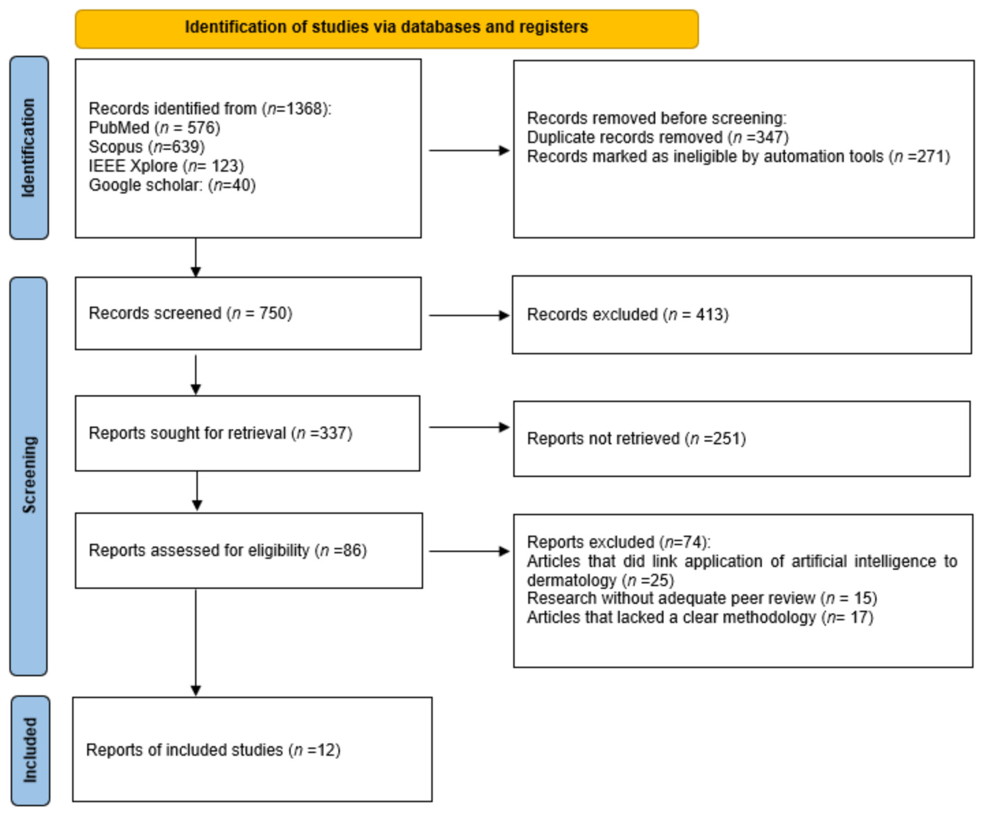
2.4. Study Selection Process

The selection process was carried out in several stages. First, duplicate items were removed. Subsequently, an initial review of titles and abstracts was carried out to rule out those studies that did not meet the inclusion criteria. The articles that passed this first phase were evaluated in their entirety to confirm their relevance and applicability within the review.
Two researchers independently reviewed the titles and abstracts of each reference obtained in the searches to collect and assess the full texts of relevant studies that may meet the inclusion criteria. References from relevant original articles and reviews were also manually searched. Any discrepancies between the researchers were resolved by a third researcher through a process of analysis and discussion.

2.5. Data Collection Process and Data Items

A narrative synthesis was conducted to summarize the findings of the studies included. Studies were grouped based on the clinical application of artificial intelligence (e.g., melanoma detection, inflammatory skin diseases, cosmetic dermatology), type of AI technology used (e.g., convolutional neural networks, deep learning), and study design. Key outcomes such as diagnostic performance metrics (sensitivity, specificity, accuracy), impact on clinical decision-making, and the use of AI-enabled devices were compared across studies. Tables were used to present structured summaries of the evidence included. Finally, the information was extracted from the selected articles and compiled into a standardized Excel document.

2.6. Certainty of Evidence and Reporting Bias

Given the heterogeneity of the included studies and the predominance of observational and experimental designs, a formal assessment of publication bias was not feasible. However, selective reporting was considered by comparing reported outcomes with stated objectives in each study. The certainty of evidence was not evaluated using GRADE due to the exploratory and narrative nature of this review; however, the strength of conclusions was moderated based on study design limitations, potential biases, and reproducibility of findings.

2.7. Risk of Bias Assessment

Due to the heterogeneity of included studies, a formal ROBINS-I evaluation was applied only to observational studies. For experimental and AI-based research, a tailored quality assessment was conducted focusing on dataset transparency, external validation, clinical applicability, and outcome reporting. Studies were categorized as having low, moderate, or high risk of bias based on these domains. Two reviewers independently assessed each study and resolved disagreements through discussion.
Additionally, only one of the included studies reported interrater agreement metrics, specifically using Fleiss’ kappa to assess concordance among human evaluators. None of the studies included in this review reported kappa values or similar agreement statistics comparing the AI system’s performance directly with dermatologists. This lack of standardized agreement metrics limits the ability to quantify the consistency and clinical alignment between human and AI-based decision-making.

3. Results

3.1. Included Studies

A total of 1368 records were identified through database searching, of which 618 were removed prior to screening due to duplication or ineligibility. After title and abstract screening of 750 records, 337 full-text reports were sought for retrieval. Of these, 251 could not be retrieved, and 86 full-text articles were assessed for eligibility. Ultimately, 12 studies met the inclusion criteria and were included in the systematic review. The full study selection process is illustrated in the PRISMA flow diagram (Figure 1).

3.2. Analysis and Discussion

Once data extraction was completed, a comparative analysis of the results obtained in the different studies was conducted. Table 2 explores the type of report, pathology, application, software used, and main findings. Emerging trends in the use of artificial intelligence in dermatology, the most commonly used technologies, and advances in the diagnosis and treatment of skin diseases are presented.
In addition, recurrent limitations were identified in the literature, including challenges related to the interpretation of AI models, the lack of validation in clinical settings, and difficulties in integrating these tools into routine medical practice.
The analysis also made it possible to point out knowledge gaps and opportunities for future research, which helped to contextualize the current state of evidence and suggest possible directions for further studies.

4. Discussion

Currently, there are computer systems capable of mimicking human cognitive functions, known as artificial intelligence (AI). These technologies have found various applications in the field of medicine, particularly in dermatology, where they are mainly used for the detection of melanomas, the type of skin cancer with the highest mortality rate. Through the analysis of clinical images, AI contributes to improving diagnostic accuracy and facilitating the early identification of suspicious lesions [17].
This process is based on machine learning, a branch of AI (Figure 2) that allows systems to learn from large volumes of data and make predictions without the need for explicit programming, thus optimizing pattern recognition in images (in this case, dermatological images). In turn, deep learning, a subcategory of machine learning, uses deep neural networks to mimic human learning and improve accuracy in complex tasks. These artificial neural networks are inspired by the structure and functioning of the human brain, organized into multiple layers of artificial neurons that hierarchically process information. Thanks to this approach, deep learning has demonstrated a great ability to analyze medical images, allowing subtle patterns to be detected that may go unnoticed in traditional evaluations. Both machine learning and deep learning are the foundations of AI [7,17].

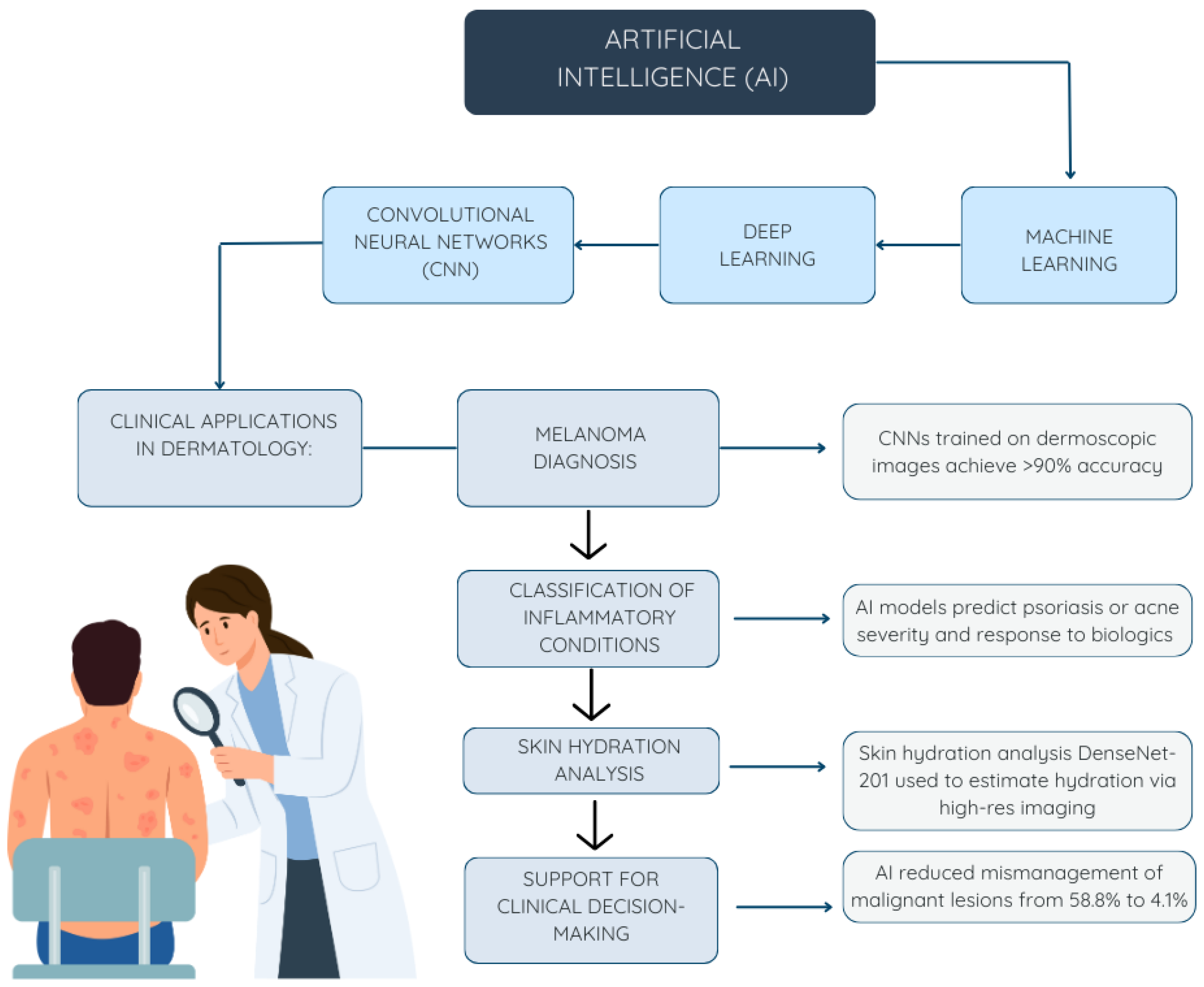
4.1. AI in Dermatology

In dermatology, the most useful aspect of AI is to use images to primarily analyze potential skin cancers, ulcers, and psoriasis [18]. Although traditional skin quality assessments are based on manual observation, these can be subjective and difficult to quantify, so the use of AI based on image recognition can help professionals improve the efficiency and accuracy of their diagnoses [19].

4.1.1. Identification of Malignancies

Neoplasm detection is the most widely documented application of the use of AI in dermatology. These technologies make it possible to differentiate between benign nevi and melanomas by analyzing detailed images of skin lesions at the pixel level. Through deep learning algorithms, the images are decomposed for individualized analysis, optimizing the extraction of key features that facilitate an accurate classification of malignancies, offering significant advantages over traditional diagnostic methods, offering an average accuracy of 90% and a sensitivity of 85%, which highlights the potential of AI for this type of complex tasks [11].
A multi-center study conducted in 2022 evaluated the diagnostic accuracy of 88 dermatologists with different levels of clinical experience (less than 2 years, between 2 and 5 years, and more than 5 years) by comparing their performance with and without the support of AI technologies, specifically the FotoFinder system when evaluating 30 different clinical cases. The results showed that, in all experience groups, the use of AI was associated with a significant increase in the accuracy of the diagnosis of malignant lesions, as illustrated in Figure 3 [13].
The use of AI in this context contributes substantially to early diagnosis, reducing waiting times and enabling the implementation of targeted and personalized treatments, which can translate into an increase in the survival rate in patients with skin cancer [17,20]. In addition, these technologies function as support tools in clinical decision-making, assisting professionals in the identification and characterization of suspicious lesions, which increases diagnostic confidence and promotes more efficient and accurate care [4,12].

4.1.2. Inflammatory Skin Diseases

As in the detection of melanomas, AI has been used for the identification of inflammatory skin conditions such as psoriasis, dermatitis, and acne, through the analysis and classification of dermatological images. In addition, deep learning techniques have been applied not only for diagnosis but also to predict the effectiveness of biological therapies and optimize ongoing treatments. To achieve this, relevant clinical parameters such as previous treatment history, patient demographics, and the presence of comorbidities are integrated, allowing for more personalized and efficient care [17].
Likewise, these AI techniques have proven to be effective in the determination and identification of pharmacological targets, in the repurposing of drugs with the potential to be effective in dermatology, and in the detection of biomarkers of inflammatory skin diseases [7,17].

4.1.3. Uses in Cosmetic Dermatology

The main use that has been given to artificial intelligence in this field of medicine is the detection of melanomas or other skin cancers; however, it has been possible to use deep learning algorithms that, through capacitive contact images and high-resolution ultrasounds, allow the determination with an accuracy of 83.8% of the water content in various facial areas. such as the cheeks, chin, tear duct, forehead, lips, neck, and nose [19]. In this case, the technology used is the DenseNet-201, which is a deep convolutional neural network that allows image classification, object detection, and segmentation, and whose applications in medicine beyond dermatology include radiology and oncology [20,21].
This machine learning model was evaluated using high-resolution MRI imaging, and the results showed that artificial intelligence could measure skin hydration with an accuracy remarkably like manual assessments performed by dermatologists. This allows for a non-invasive method, capable of determining the level of skin hydration in a way comparable to the human assessment, with the additional advantage of saving time and providing an objective parameter to evaluate the effectiveness of cosmetic treatments [19,22].

4.2. Emerging Technologies

The development of AI is not limited to software, but also a large amount of hardware has been developed, such as the FotoFinder, Canfield Vectra WBS360 and Antera 3D, which have been applied in several hospitals around the world and have proven to have great reliability by offering more intuitive and versatile examination methods than the traditional devices used in dermatology.
FotoFinder specializes in the early detection of skin cancer, using total body mapping and digital dermoscopy to identify and track changes in lesions, supported by artificial intelligence algorithms that estimate the risk of malignancy. The Canfield Vectra WB360, on the other hand, offers full-body 3D capture with 92 cameras that allow lesions to be evaluated from multiple angles, detect new formations, and accurately document the evolution of the skin, being ideal for clinical studies and esthetic dermatology. In contrast, Antera 3D focuses on localized areas, offering a microscopic 3D analysis of skin texture, pigmentation, and vascularization, useful both in esthetics and in the monitoring of inflammatory pathologies. Comparatively, FotoFinder is ideal for oncology monitoring, Vectra WB360 excels in global three-dimensional analysis, and Antera 3D excels in detailed, quantitative analysis of specific areas of the skin [9,14,16,23,24].
In the Table 3, among the technologies analyzed, only FotoFinder is certified as a Class IIa medical device under Regulation (EU) 2017/745 (MDR), supporting its clinical use and commercial distribution across the European Union [25]. However, its implementation is not limited to Europe; it is currently in use in other regions as well.
The European MDR requires comprehensive technical documentation, detailed clinical evaluation reports, and oversight from notified bodies for device approval. In contrast, the U.S. Food and Drug Administration (FDA) follows a risk-based framework that includes the 510(k) process for substantially equivalent devices and Premarket Approval (PMA) for higher-risk innovations. Historically, the European system placed greater emphasis on post-market surveillance than on pre-market clinical data, though the 2021 MDR update significantly tightened pre-approval requirements [2].
These regulatory differences directly affect the pace at which AI tools in dermatology are approved and adopted globally. For instance, an algorithm approved by the FDA through the 510(k) pathway may require additional clinical validation under the EU MDR. This fragmented regulatory landscape presents critical ethical challenges, including disparities in access, variations in clinical oversight, and the risk of prematurely deploying insufficiently validated technologies in less regulated settings [2].

4.3. Application of AI in Real Cases

Between 2014 and 2019, a comprehensive study was conducted in which 562 skin lesions were analyzed that dermatologists classified as “unclear,” i.e., difficult to diagnose with certainty. The aim was to assess how AI can influence clinical decision-making, especially to prevent mistreatment [15].
Initially, dermatologists opted for clinical follow-up in 57.3% of these cases and treatment or excision in 42.5%. However, when contrasting these decisions with definitive diagnoses, a high margin of error was observed: 58.8% of truly malignant lesions were incorrectly managed, either with follow-up or without action, while 43.9% of benign lesions were treated or removed unnecessarily. Malignant lesions included cases of lentigo maligna, lentigo maligna melanoma, invasive melanoma, nodular melanoma, superficial melanoma, pigmented basal cell carcinoma, and actinic keratosis, while benign lesions corresponded to melanocytic nevus, lentigo solar, and seborrheic keratosis [15].
Subsequently, the use of the Moleanalyzer Pro v3.8.0 AI software (FotoFinder Systems GmbH, Bad Birnbach, Bavaria, Germany) was introduced, which analyzed a single dermoscopic image per case using convolutional neural networks (CNN). By following their recommendations, the rate of mismanagement in malignant lesions would have been drastically reduced from 58.8% to 4.1%, preventing 94% skin cancers from being underestimated. In benign lesions, AI would have made it possible to reduce unnecessary excisions from 43.9% to 31.7%, thus reducing unjustified surgical interventions [15].
These findings consolidate how the use of AI in dermatology substantially improves diagnostic accuracy and favors safer and more efficient management of difficult-to-evaluate lesions, in addition to increasing the early detection of skin cancer and avoiding unnecessary invasive treatments in benign lesions.

4.4. Possible Bioethical Problems

The integration of AI in dermatology poses significant ethical challenges that may limit its safe and equitable clinical application and its acceptance by both medical staff and patients in general.
One of the main risks is the possibility of misdiagnosis derived from algorithmic biases, which usually originate from the underrepresentation of some population groups, which compromises diagnostic accuracy in these populations [26].
However, a representative example is the fact that most open access repositories have a notable lack of images of women compared to those of men, which generates a gender imbalance that could negatively influence the training of artificial intelligence systems and their diagnostic capacity. However, a prospective, blinded, multi-center study conducted in 19 French dermatology centers between 2019 and 2020, in which 1447 lesions suspected of melanoma were analyzed, showed that, despite this disparity in training data, the convolutional neural network achieved similar diagnostic performance in both genders. Specifically, the AI achieved a specificity of 98.7% in men and 96.9% in women, and a sensitivity of 87.0% and 87.1%, respectively, indicating that the model was equally accurate in diagnosing skin lesions in both men and women [8].
On the other hand, the security and privacy of dermatological data are critical areas, given that AI systems require large volumes of sensitive information, whose protection against unauthorized access must be guaranteed through robust regulatory frameworks, mainly associated with medical devices or, in some cases, a total absence of specific regulations for AI, a situation that mainly affects low-development countries, including Latin America, where the legal framework is poorly defined or absent [27,28].
Today, the importance of maintaining human medical intervention as a key element in ensuring acceptance, accountability, and transparency is underlined. Addressing these ethical challenges through data diversification, advanced safety protocols, and governance frameworks is essential for responsible AI implementation, not only in dermatology but in all medical areas [29].

5. Conclusions

Artificial intelligence (AI) is rapidly consolidating itself as a valuable and practical resource for dermatologists seeking to enhance diagnostic accuracy, reduce variability in clinical decisions, and improve access to quality care. Its integration into dermatological practice has already demonstrated substantial benefits across multiple domains. From the early detection of malignant skin lesions, where AI systems have matched or exceeded the accuracy of dermatologists, to the optimization of treatment decisions in benign and inflammatory conditions, AI has contributed to more efficient and personalized care. In the context of inflammatory skin diseases, machine learning models have enabled more accurate classification and better prediction of treatment responses, advancing the goals of precision medicine. In cosmetic dermatology, AI tools now offer objective assessments of skin parameters such as hydration or texture, using non-invasive and scalable techniques. The development of emerging technologies that combine advanced imaging with AI-driven analysis is further expanding the possibilities for real-time diagnosis, longitudinal monitoring, and workflow efficiency. These advancements reflect the growing clinical maturity of AI in dermatology and reinforce its role as a meaningful aid in both diagnostic and therapeutic decision-making, with tangible benefits for patients and healthcare systems alike.

Author Contributions

Research: E.Z.-M., E.M.-V.; methodology: E.M.-V., E.Z.-M.; project management: E.Z.-M.; supervision: E.Z.-M., S.A.-C.; verification: E.Z.-M., S.A.-C., J.M.-J., J.H.-L.; writing—original draft: E.M.-V.; writing—review and editing: E.M.-V. All authors have read and agreed to the published version of the manuscript.

Funding

This research received no external funding.

Institutional Review Board Statement

Not applicable.

Informed Consent Statement

Not applicable.

Data Availability Statement

Data supporting the results of this study are available upon reasonable request from the corresponding author. Due to privacy and ethical restrictions, data are not publicly available. However, aggregated summary data are included in the manuscript and

Conflicts of Interest

The authors declare no conflicts of interest.

Abbreviations

The following abbreviations are used in this manuscript:
AIArtificial intelligence
CNNConvolutional Neural Network

References

  1. Fateeva, A.; Eddy, K.; Chen, S. Current State of Melanoma Therapy and Next Steps: Battling Therapeutic Resistance. Cancers 2024, 16, 1571. [Google Scholar] [CrossRef] [PubMed]
  2. Liopyris, K.; Gregoriou, S.; Dias, J.; Stratigos, A.J. Artificial Intelligence in Dermatology: Challenges and Perspectives. Dermatol. Ther. 2022, 12, 2637–2651. [Google Scholar] [CrossRef]
  3. Aksoy, S.; Demircioglu, P.; Bogrekci, I. Advanced Artificial Intelligence Techniques for Comprehensive Dermatological Image Analysis and Diagnosis. Dermato 2024, 4, 173–186. [Google Scholar] [CrossRef]
  4. Giavina-Bianchi, M.; de Sousa, R.M.; Paciello, V.Z.d.A.; Vitor, W.G.; Okita, A.L.; Prôa, R.; Severino, G.L.D.S.; Schinaid, A.A.; Espírito Santo, R.; Machado, B.S. Implementation of Artificial Intelligence Algorithms for Melanoma Screening in a Primary Care Setting. PLoS ONE 2021, 16, e0257006. [Google Scholar] [CrossRef]
  5. Cerminara, S.E.; Cheng, P.; Kostner, L.; Huber, S.; Kunz, M.; Maul, J.-T.; Böhm, J.S.; Dettwiler, C.F.; Geser, A.; Jakopović, C.; et al. Diagnostic Performance of Augmented Intelligence with 2D and 3D Total Body Photography and Convolutional Neural Networks in a High-Risk Population for Melanoma under Real-World Conditions: A New Era of Skin Cancer Screening? Eur. J. Cancer 2023, 190, 112954. [Google Scholar] [CrossRef] [PubMed]
  6. Madarkar, M.S.; Koti, V.R. FotoFinder Dermoscopy Analysis and Histopathological Correlation in Primary Localized Cutaneous Amyloidosis. Dermatol. Pract. Concept. 2021, 11, e2021057. [Google Scholar] [CrossRef]
  7. Li, Z.; Koban, K.C.; Schenck, T.L.; Giunta, R.E.; Li, Q.; Sun, Y. Artificial Intelligence in Dermatology Image Analysis: Current Developments and Future Trends. J. Clin. Med. 2022, 11, 6826. [Google Scholar] [CrossRef] [PubMed]
  8. Sies, K.; Winkler, J.K.; Fink, C.; Bardehle, F.; Toberer, F.; Buhl, T.; Enk, A.; Blum, A.; Stolz, W.; Rosenberger, A.; et al. Does Sex Matter? Analysis of Sex-Related Differences in the Diagnostic Performance of a Market-Approved Convolutional Neural Network for Skin Cancer Detection. Eur. J. Cancer 2022, 164, 88–94. [Google Scholar] [CrossRef]
  9. Winkler, J.K.; Kommoss, K.S.; Vollmer, A.S.; Blum, A.; Stolz, W.; Kränke, T.; Hofmann-Wellenhof, R.; Enk, A.; Toberer, F.; Haenssle, H.A. Computerizing the First Step of the Two-Step Algorithm in Dermoscopy: A Convolutional Neural Network for Differentiating Melanocytic from Non-Melanocytic Skin Lesions. Eur. J. Cancer 2024, 210, 114297. [Google Scholar] [CrossRef]
  10. Veeramani, N.; Jayaraman, P. A Promising AI Based Super Resolution Image Reconstruction Technique for Early Diagnosis of Skin Cancer. Sci. Rep. 2025, 15, 5084. [Google Scholar] [CrossRef]
  11. Pham, T.-C.; Luong, C.-M.; Hoang, V.-D.; Doucet, A. AI Outperformed Every Dermatologist in Dermoscopic Melanoma Diagnosis, Using an Optimized Deep-CNN Architecture with Custom Mini-Batch Logic and Loss Function. Sci. Rep. 2021, 11, 17485. [Google Scholar] [CrossRef] [PubMed]
  12. Useini, V.; Tanadini-Lang, S.; Lohmeyer, Q.; Meboldt, M.; Andratschke, N.; Braun, R.P.; Barranco García, J. Automatized Self-Supervised Learning for Skin Lesion Screening. Sci. Rep. 2024, 14, 12697. [Google Scholar] [CrossRef]
  13. Winkler, J.K.; Blum, A.; Kommoss, K.; Enk, A.; Toberer, F.; Rosenberger, A.; Haenssle, H.A. Assessment of Diagnostic Performance of Dermatologists Cooperating with a Convolutional Neural Network in a Prospective Clinical Study: Human with Machine. JAMA Dermatol. 2023, 159, 621–627. [Google Scholar] [CrossRef]
  14. Crawford, M.E.; Kamali, K.; Dorey, R.A.; MacIntyre, O.C.; Cleminson, K.; MacGillivary, M.L.; Green, P.J.; Langley, R.G.; Purdy, K.S.; DeCoste, R.C.; et al. Using Artificial Intelligence as a Melanoma Screening Tool in Self-Referred Patients. J. Cutan. Med. Surg. 2024, 28, 37–43. [Google Scholar] [CrossRef]
  15. Kommoss, K.S.; Winkler, J.K.; Mueller-Christmann, C.; Bardehle, F.; Toberer, F.; Stolz, W.; Kraenke, T.; Hofmann-Wellenhof, R.; Blum, A.; Enk, A.; et al. Observational Study Investigating the Level of Support from a Convolutional Neural Network in Face and Scalp Lesions Deemed Diagnostically ‘Unclear’ by Dermatologists. Eur. J. Cancer 2023, 185, 53–60. [Google Scholar] [CrossRef]
  16. Anqi, S.; Xiukun, S.; Ai’e, X. Quantitative Evaluation of Sensitive Skin by ANTERA 3D® Combined with GPSkin Barrier®. Ski. Res. Technol. 2022, 28, 840–845. [Google Scholar] [CrossRef] [PubMed]
  17. Omiye, J.A.; Gui, H.; Daneshjou, R.; Cai, Z.R.; Muralidharan, V. Principles, Applications, and Future of Artificial Intelligence in Dermatology. Front. Med. 2023, 10, 1278232. [Google Scholar] [CrossRef]
  18. Koka, S.S.-A.; Burkhart, C.G. Inteligencia artificial en dermatología: Usos actuales, deficiencias y posibles oportunidades para una mayor implementación en el diagnóstico y la atención. Open Dermatol. J. 2023, 17, e187437222304140. [Google Scholar] [CrossRef]
  19. Kania, B.; Montecinos, K.; Goldberg, D.J. Artificial Intelligence in Cosmetic Dermatology. J. Cosmet. Dermatol. 2024, 23, 3305–3311. [Google Scholar] [CrossRef]
  20. Salim, F.; Saeed, F.; Basurra, S.; Qasem, S.N.; Al-Hadhrami, T. DenseNet-201 and Xception Pre-Trained Deep Learning Models for Fruit Recognition. Electronics 2023, 12, 3132. [Google Scholar] [CrossRef]
  21. Paluri, K.V.; Gupta, N.; Mishra, A.K.; Gupta, A.; Nain, G. An Explainable AI-Based Automated Acne Diagnosis Using Transfer Learning and DenseNet121. In Proceedings of the 2024 10th International Conference on Advanced Computing and Communication Systems (ICACCS), Coimbatore, India, 14–15 March 2024; pp. 1086–1091. [Google Scholar] [CrossRef]
  22. Vatiwutipong, P.; Vachmanus, S.; Noraset, T.; Tuarob, S. Artificial Intelligence in Cosmetic Dermatology: A Systematic Literature Review. IEEE Access 2023, 11, 71407–71425. [Google Scholar] [CrossRef]
  23. Morihisa, Y.; Rikimaru-Nishi, Y.; Ohmaru, Y.; Ino, K.; Rikimaru, H.; Kiyokawa, K. Scientific Validation of Clinical Visual Scales and Antera 3DTM Consistency with Derived Measurements in the Assessment of Infantile Haemangioma after Laser Therapy. J. Plast. Reconstr. Aesthetic Surg. 2024, 91, 47–55. [Google Scholar] [CrossRef]
  24. Horsham, C.; O’Hara, M.; Sanjida, S.; Ma, S.; Jayasinghe, D.; Green, A.C.; Schaider, H.; Aitken, J.F.; Sturm, R.A.; Prow, T.; et al. The Experience of 3D Total-Body Photography to Monitor Nevi: Results From an Australian General Population-Based Cohort Study. JMIR Dermatol. 2022, 5, e37034. [Google Scholar] [CrossRef] [PubMed]
  25. Humans.txt MDR Certificate for FotoFinder. Available online: https://www.fotofinder.de/es/compania/news/mdr-certificate-for-fotofinder (accessed on 30 April 2025).
  26. Ning, Y.; Teixayavong, S.; Shang, Y.; Savulescu, J.; Nagaraj, V.; Miao, D.; Mertens, M.; Ting, D.S.W.; Ong, J.C.L.; Liu, M.; et al. Generative Artificial Intelligence and Ethical Considerations in Health Care: A Scoping Review and Ethics Checklist. Lancet Digit. Health 2024, 6, e848–e856. [Google Scholar] [CrossRef]
  27. Patil, R.S.; Kulkarni, S.B.; Gaikwad, V.L. Artificial Intelligence in Pharmaceutical Regulatory Affairs. Drug Discov. Today 2023, 28, 103700. [Google Scholar] [CrossRef] [PubMed]
  28. Pantanowitz, L.; Hanna, M.; Pantanowitz, J.; Lennerz, J.; Henricks, W.H.; Shen, P.; Quinn, B.; Bennet, S.; Rashidi, H.H. Regulatory Aspects of Artificial Intelligence and Machine Learning. Mod. Pathol. 2024, 37, 100609. [Google Scholar] [CrossRef]
  29. Alowais, S.A.; Alghamdi, S.S.; Alsuhebany, N.; Alqahtani, T.; Alshaya, A.I.; Almohareb, S.N.; Aldairem, A.; Alrashed, M.; Bin Saleh, K.; Badreldin, H.A.; et al. Revolutionizing Healthcare: The Role of Artificial Intelligence in Clinical Practice. BMC Med. Educ. 2023, 23, 689. [Google Scholar] [CrossRef]
Figure 1. Selection process of studies included in systematic review.
Dermato 05 00009 g001
Figure 2. Hierarchical relationship between artificial intelligence (AI), machine learning, deep learning, and convolutional neural networks (CNNs), and their clinical applications in dermatology.
Dermato 05 00009 g002
Figure 3. Impact of AI support on diagnostic accuracy according to dermatologists’ experience.
Dermato 05 00009 g003
Table 1. Databases chosen, keywords, search strategy, and filters used in the initial process of collecting articles.
DatabaseKeywordsSearch StrategyFilters AppliedNumber of Possible Items Selected
PubMedArtificial intelligence, machine learning, Deep learning, neural networks, dermatology, skin desease, skin cancer, melanoma(“Artificial Intelligence” [MeSH] OR “Machine Learning” [MeSH] OR “Deep Learning” OR “Neural Networks, Computer” [MeSH])
AND (“Dermatology” [MeSH] OR “Skin Diseases” [MeSH] OR “Skin Neoplasms” [MeSH] OR “Skin Cancer”)
Full texts available. Clinical trials, observational studies, systematic reviews. Published between 2020–2025.576
ScopusArtificial intelligence, machine learning, Deep learning, neural networks, dermatology, skin desease, skin cancer, melanoma“Artificial intelligence” OR “machine learning” OR “deep learning” OR “neural networks”)
AND (“dermatology” OR “skin diseases” OR “skin cancer” OR “cutaneous”)
Original articles and reviews, published in Spanish or English between 2020–2025.639
IEEE XploreArtificial intelligence, machine learning, Deep learning, neural networks, dermatology, skin desease, skin cancer, melanoma(“All Metadata”:”artificial intelligence” OR “machine learning” OR “deep learning” OR “neural networks”)
AND (“All Metadata”:”dermatology” OR “skin diseases” OR “skin cancer”)
Journals published in English between 2020–2025.132
Google ScholarArtificial intelligence, machine learning, Deep learning, neural networks, dermatology, skin desease, skin cancer, melanoma“Artificial Intelligence” OR “Machine Learning” OR “Deep Learning” OR “Neural Networks”
AND “Dermatology” OR “Skin Diseases” OR “Skin Cancer”
Peer-reviewed or indexed scientific articles published in Spanish or English between 2020–2025.40
Table 2. Articles included in the systematic review.
PaperReport TypePathology/ApplicationSofware UsedMain Findings
Sies et al. (2022) [8]Retrospective, non-interventional observational studyPatients at high risk for melanomaMoleanalyzer Pro (FotoFinder Systems GmbH, Bad Birnbach, Bavaria, Germany), a market-approved convolutional neural network (CNN) developed by FotoFinder SystemsDespite gender imbalance in the training data (60% males vs. 40% females), the evaluated CNN showed no significant differences in sensitivity, specificity, or AUC when stratified by sex. It achieved high accuracy for skin cancer classification regardless of gender.
Cerminara et al. (2023) [5].Prospective, observational, multi-center studySkin cancer detection and dermoscopic screeningVectra WB360 (v4.7.1) for 3D total body photography-FotoFinder ATBM Master (v3.3.1.0) for 2D total body photography-Medicam 1000 and VISIOMED D200evo for manual lesion photographyThe 3D CNN showed higher sensitivity and consistency than the 2D approach for melanoma detection, with performance comparable to dermatologists. However, both systems overestimated nevus counts compared to manual counting.
Winkler et al. (2024) [9]Cross-sectional studyClassification of melanocytic vs. non-melanocytic lesionsSPSS version 29 for statistical analysis and a modified neural network architectureThe CNN achieved a 91% accuracy (AUC 0.981) in distinguishing melanocytic from non-melanocytic lesions, outperforming the majority of dermatologists. External validation was conducted on over 1100 lesions.
Giavina-Bianchi et al. (2021) [4]Retrospective, single-center observational studyMelanoma screening and malignant skin lesion triage in primary careEfficientNetB6 ensemble model with synthetic data generated via StyleGAN2, trained using ISIC2019 and PH2 datasetsThe AI-assisted computer-aided diagnosis (CAD) tool reached a precision of 89.3% using dermoscopic images and 84.7% with clinical images, providing reliable management recommendations for the detection of skin cancer in primary care settings.
Veeramani and Jayaraman (2025) [10]Experimental study with technical validationImage reconstruction of intermediate skin lesions (melanoma)MELIIGAN, a CNN-based reconstruction system using residual attentionMELIIGAN improved the reconstruction quality of skin lesion images, achieving PSNR > 40 and SSIM > 0.94. It enhanced fine detail visibility and reduced classification errors in intermediate melanoma lesions.
Pham et al. (2021) [11]Experimental computational studyMelanoma diagnosis using dermoscopic imagesDenseNet169 CNN was implemented in Python 3.8 using TensorFlow 2.10.0 (Google LLC, Mountain View, CA, USA) and PyTorch 1.12.1 (Meta Platforms, Inc., Menlo Park, CA, USA)The AI system outperformed 157 dermatologists (MCass-D), reaching a sensitivity of 90% and specificity of 93.8%, with performance superior to the current state of the art.
Useini et al. (2023) [12]Prospective, single-center observational studyAI-based full-body detection tool with self-supervised learningDeep learning framework PyTorch, combined with the Lightly library for self-supervised learning modelsThe total-body screening tool achieved 82% precision in detecting the 10 most suspicious lesions and reached an average sensitivity of 95% for expert-confirmed cases, improving diagnostic agreement and supporting broader generalization in clinical practice.
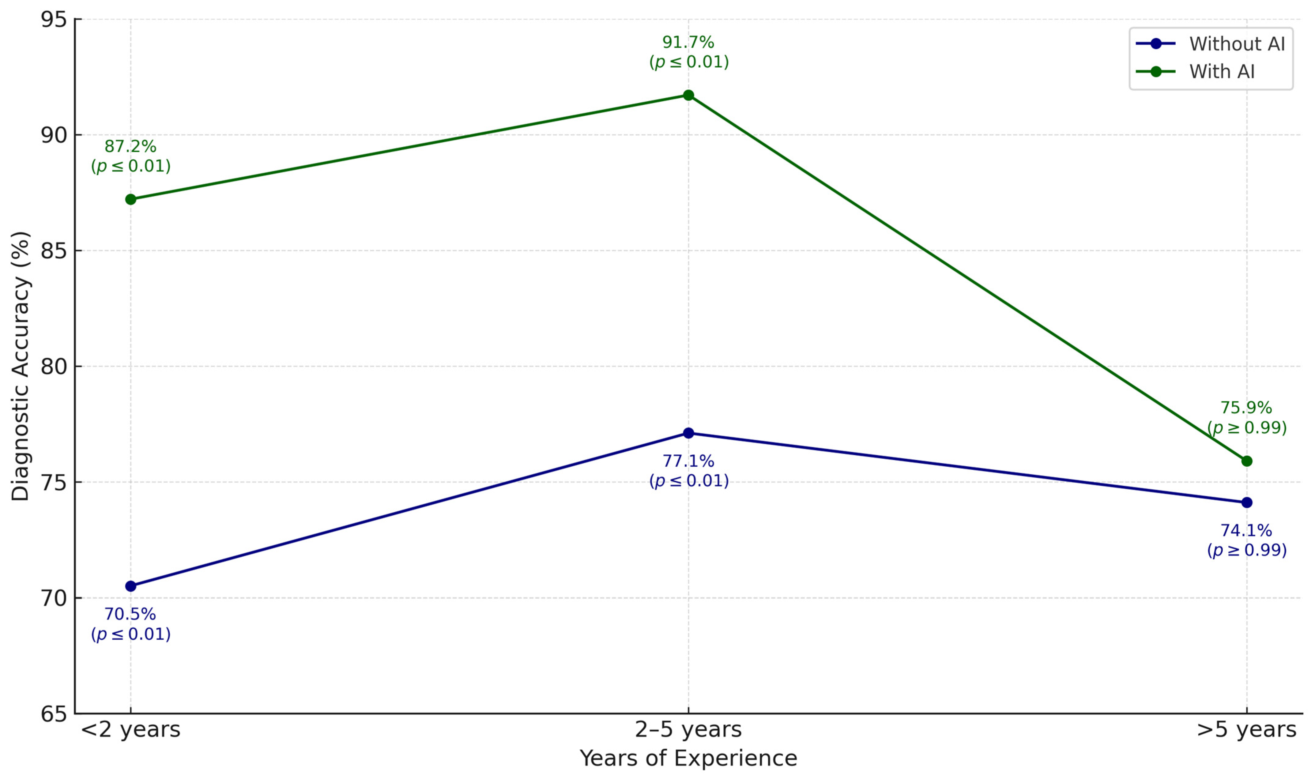
Winkler et al. (2023) [13]Prospective, multi-center clinical studyDiagnosis of melanocytic lesionsCommercial CNN (Moleanalyzer Pro, FotoFinder Systems)The cooperation between dermatologists and CNN significantly improved their sensitivity, specificity, and diagnostic accuracy, while also reducing unnecessary excisions. The benefit was most evident among professionals with less clinical experience.
Hull et al. (2023) [14].Prospective, single-center observational studyScreening for malignant skin lesionsCommercial CNN (Moleanalyzer Pro, FotoFinder Systems)The AI system demonstrated sensitivity and specificity comparable to expert dermatologists in detecting malignant skin lesions, although it missed 6 in situ melanomas. It is proposed as a useful tool for populations with limited access to dermatology specialists.
Kommoss et al. (2023) [15].Multi-center, retrospective observational studyDiagnosis of facial and scalp skin lesions with unclear clinical presentationCNN (Moleanalyzer Pro, FotoFinder Systems)The AI model reduced incorrect clinical management decisions in diagnostically unclear cases, lowering the error rate for malignant lesions from 58.8% to 4.1% and for benign lesions from 43.9% to 31.7%. The model demonstrated superior performance compared to dermatologists, even when they had full access to patient clinical data.
Anqi et al. (2022) [16].Observational clinical studyObjective evaluation of sensitive skin and sensitive skin syndromeANTERA 3D® (Miravex Ltd., Dublin, Ireland) combined with GPSkin Barrier® (GPOWER Inc., Seoul, Republic of Korea)In patients with sensitive skin, ANTERA 3D® detected significant increases in hemoglobin texture and affected area, while GPSkin Barrier® revealed higher transepidermal water loss (TEWL) and lower skin hydration. The combined use of both tools provided objective and quantifiable measures to support the clinical characterization of sensitive skin.
Table 3. International regulatory status of advanced dermatological imaging technologies.
SystemFDA ApprovalCE Approval (MDR)Countries Where It Is ApprovedType of Approval
FotoFinderNoYesEuropean Union countriesClass IIa medical device under Regulation (EU) 2017/745 (MDR) [23].
Canfield Vectra WBS360NoUnavailableUnavailableNo public information is available on its specific regulatory approval.
Antera 3DUnavailableUnavailableUnavailableNo public information is available on its regulatory approval status.
Disclaimer/Publisher’s Note: The statements, opinions and data contained in all publications are solely those of the individual author(s) and contributor(s) and not of MDPI and/or the editor(s). MDPI and/or the editor(s) disclaim responsibility for any injury to people or property resulting from any ideas, methods, instructions or products referred to in the content.

