Case Report

Stimulator of InterferoN Genes (STING)-Associated Vasculopathy with Onset in Infancy Syndrome (SAVI) Associated with Disseminated Molluscum Contagiosum Under Baricitinib Treatment

1
Dortmund Hospital gGmbH, University Witten/Herdecke, 44137 Dortmund, Germany
2
Department of Dermatology, Christian Hospital Unna, 59423 Unna, Germany
3
Department of Dermatology, Ruhr-University Bochum, 44791 Bochum, Germany
*
Author to whom correspondence should be addressed.
Submission received: 28 December 2024 / Revised: 8 March 2025 / Accepted: 10 March 2025 / Published: 2 April 2025
(This article belongs to the Special Issue What Is Your Diagnosis?—Case Report Collection)

Abstract

:
Background/objectives: Stimulator of Interferon Genes (STING)-associated vasculopathy with onset in infancy (SAVI) is a rare autoinflammatory disorder caused by gain-of-function mutations in the TMEM173 gene. These mutations result in chronic activation of the STING pathway and excessive type I interferon production, leading to systemic inflammation, vascular abnormalities, interstitial lung disease, and skin ulcerations. Janus kinase (JAK) inhibitors, including baricitinib, have shown promise in mitigating systemic and organ-specific manifestations. However, these inhibitors broadly suppress immune pathways, potentially increasing vulnerability to infections. Case presentation: This case report describes a 21-year-old woman with SAVI (due to a heterozygous TMEM173 mutation) who developed disseminated molluscum contagiosum (MC) while receiving baricitinib therapy. Laboratory results revealed lymphopenia, low CD4/CD8 ratio, and impaired immune cell activity, suggesting compromised antiviral immunity. Discussion: Despite SAVI’s association with excessive type I interferon signaling, this chronic hyperactivation may cause immune dysregulation, exhausting T cells and natural killer cells vital for viral defense. Furthermore, baricitinib suppresses interferon signaling via the JAK-STAT pathway, reducing inflammatory damage in SAVI but also impairing antiviral responses. Moreover, MC viruses evade host immune defenses by antagonizing STING and TANK-binding kinase 1-mediated interferon activation, further contributing to infection risk. This report is the first to document MC in a SAVI patient and highlights the rare complication of disseminated MC due to impaired type I interferon signaling and immune suppression from baricitinib therapy. This case underscores the need for vigilance regarding viral infections in SAVI patients treated with JAK inhibitors.

1. Introduction

Stimulator of Interferon Genes (STING)-associated vasculopathy with onset in infancy syndrome (SAVI) is a rare autoinflammatory disorder caused by gain-of-function mutations in the TMEM173 gene. These mutations result in excessive activation of the STING signaling pathway, leading to chronic overproduction of type I interferons. The clinical presentation includes severe systemic inflammation, vascular changes, interstitial lung disease, and skin ulcerations, which can manifest as early as infancy [1,2,3]. The disease is characterized not only by its high morbidity but also by the lack of specific and long-term effective therapies. The treatment of SAVI syndrome poses a significant challenge due to its underlying pathophysiology. Conventional immunosuppressive therapies, such as corticosteroids or methotrexate, are often insufficient to control symptoms. In recent years, Janus kinase (JAK) inhibitors, such as baricitinib, have shown promising results. These medications act by disrupting cytokine signaling, including type I interferons, thereby modulating the excessive immune response. Early reports have demonstrated significant improvements in both systemic and organ-specific symptoms in SAVI syndrome patients treated with JAK inhibitors. However, these commonly used inhibitors of the JAK-STAT pathway exert broad effects on multiple signaling pathways leading to more general immunosuppression beyond type I interferon signaling [4,5,6,7,8].
The molluscum contagiosum (MC) virus is an unclassified member of the Poxviridae family, encompassing types I to IV DNA viruses. The MC virus causes a benign, cutaneous-limited infection that occurs only in humans. Risk factors for MC infection include a compromised immune system (e.g., patients with HIV, organ transplant, or chemotherapy), frequent attendance at MC “hot spots” (e.g., day care centers, schools, or pools), skin-to-skin contact with an infected individual (sexual or nonsexual), and atopic dermatitis. Clinically, MC presents as firm rounded papules, pink or skin-colored, with a shiny, often umbilicated surface. The duration of the lesions is variable, but in most cases, they are self-limited in a period of 6–9 months [9,10]. JAK inhibitors potentially cause infections, particularly herpes virus-associated conditions; however, the occurrence of MC has only rarely been reported in the literature [1,2,3,4,5,6,7,8,9,10,11,12,13,14,15,16,17,18]. The present case report highlights for the first time the occurrence of disseminated long-term molluscum contagiosum (MC) in an adult patient with SAVI who was under treatment with baricitinib.

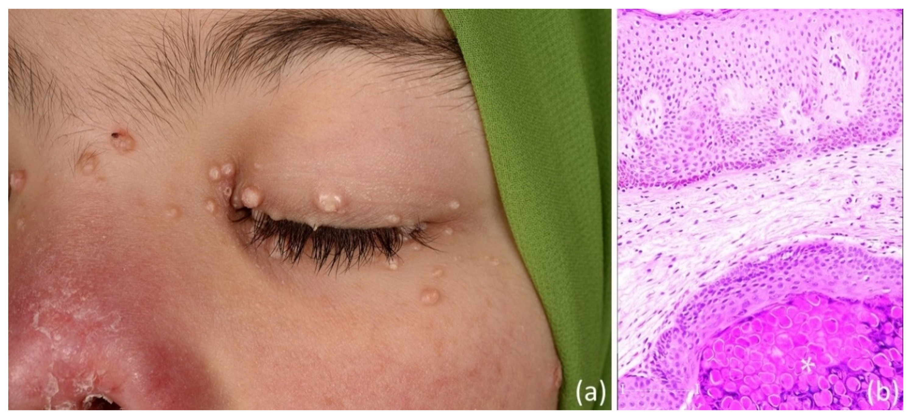
2. Case Presentation

A 21-year-old Turkish female attended our department with a 2-year history of disseminated papules on her face, neck and upper trunk. At the age of 4 years, the patient was diagnosed with SAVI (heterozygous mutation p.N154S in the TMEM173 gene). Her case was first reported in 2014 by Liu et al. [1]. Over the years, her clinical symptoms with respect to SAVI included characteristic pathologies as follows: anemia, elevated CRP, gammaglobulinemia, leukopenia, lymphopenia, thrombocytosis, acral violaceous plaques and nodules on the face and earlobes, oral ulcers, painful ulcers, eschars, livedo reticularis, amputation of fingers and toes (3rd year of life), nasal septum perforation, and mild interstitial lung disease. She also had a history for recurrent infections, such as otitis externa and erysipelas. Her previous treatment included systemic prednisolone, methotrexate, cyclophosphamide, etanercept, anakinra, and tocilizumab. At the age of 17 years, she was put on oral baricitinib therapy, where her daily dosage was 4 mg in the morning and 3 mg in the evening.
At clinical presentation, she showed disseminated skin-colored, partly umbilicated papules (2 to 6 mm diameter) on her face, neck and upper trunk that were consistent with MC (Figure 1a). Laboratory pathologies included erythrocytes 3.34 Mio/µL (4.2–5.4), hemoglobin 9.3 g/dL (12–16), CRP 11.6 mg/L (<5), leukocytes 3970/µL (4600–9500), lymphocytes 840/µL (1000–4050), T helper lymhocytes 214/µL (410–1590), natural killer cells 58/µL (90–660), and low CD4/CD8 ratio of 0.89 (0.8–2.0). HIV and hepatitis serology were negative. Serum interferon-ү-induced protein (IP-10) was only slightly elevated with 1173 pg/mL (<1092). After explaining the added complications of secondary infection and delayed wound healing to the immunocompromised patient, we performed curettage of MC under general anesthesia. Following treatment, the clinical diagnosis was confirmed on histopathology (Figure 1b).

3. Discussion

At first glance, it seems unusual for a patient with SAVI, characterized by massive overproduction of type I interferons, to develop disseminated molluscum contagiosum. Type I interferons are central to antiviral immunity, particularly against DNA and RNA viruses, by promoting the expression of antiviral proteins, enhancing immune cell responses, and limiting viral replication. The overactivation of this pathway in SAVI would theoretically provide enhanced antiviral protection. However, there are several possible explanations for the observation in the present case. While SAVI is associated with excessive type I interferon production, this chronic hyperactivation can lead to immune dysregulation and impaired overall immune functionality [1,2,3,4]. Chronic type I interferon signaling in SAVI creates an environment of persistent inflammation that eventually exhausts the immune system. This occurs because continuous interferon exposure leads to sustained expression of interferon-stimulated genes, the upregulation of inhibitory receptors (e.g., PD-1, LAG-3, and TIM-3) and negative feedback regulators such as suppressors of cytokine signaling proteins, and metabolic reprogramming. Together, these changes drive T cells and other immune cells into an exhausted state, undermining their ability to effectively respond to pathogens. Indeed, SAVI-induced immune impairment is similar to chronic viral infections (e.g., HIV, chronic HBV, and COVID-19-associated immune dysregulation) [19]. While both SAVI and chronic viral infections can result in a state of immune exhaustion through chronic activation and sustained interferon signaling, the origins of this dysfunction are distinct—genetic mutations in SAVI versus persistent viral antigens in chronic infections—which ultimately influences their clinical management and treatment strategies. Whereas the common thread in SAVI and other interferonopathies is the elevated type I interferon activity, the genetic origins, cytokine milieu, clinical manifestations, and specific immune cell alterations help to distinguish the immunological profiles of these conditions [4].
Hence, chronic type I interferon activation in SAVI alone may have caused immune exhaustion, making the present patient more vulnerable to MC. Moreover, the present patient had impaired cellular immune capacity as indicated by decreased circulating T helper and natural killer cells. Together, the impaired immune functionality observed in SAVI patients may result in viral infections, independently of treatment induced JAK-STAT pathway inhibition. Indeed, the JAK inhibitor baricitinib blocks the signaling of cytokines that rely on the JAK-STAT pathway, including type I interferons. By dampening this pathway, baricitinib reduces the inflammatory and autoinflammatory damage caused by SAVI but also suppresses the antiviral activity mediated by interferon signaling. This suppression could potentially predispose the patient to viral infections, such as MC, particularly if the medication significantly reduces the immune system’s ability to respond to viruses [4,5,6,7,8]. The baseline infection risk in SAVI patients is likely lower than that seen in patients whose interferon signaling is further compromised by JAK therapy. We have to stress, however, that infection risk comparisons between JAK-treated and non-treated SAVI patients are difficult due to SAVI’s rarity [1,2,3,4,5,6,7,8].
In the present case, an assessment of serum IP-10 did not indicate drastic T cell-mediated immune activation, likely due to JAK inhibition. However, the decrease in interferon-stimulating genes and IP-10 has not been consistently observed under treatment of SAVI using JAK inhibitors such as baricitinib [2,7]. Moreover, all poxviruses, including the MC virus, have an array of proteins to counter host detection and activation of the interferon responses. Reiss et al. [11] have recently highlighted the ability of the MC-160 protein to antagonize STING-mediated immune responses as well as block ubiquitination of TANK-binding kinase 1 (TBK1). Cyclic GMP-AMP synthase/STING are expressed in keratinocytes and function to sense the presence of dsDNA during MC infection [11]. Hence, their model indicates that MC-160 likely alters the TBK1 signaling complex to decrease interferon-β activation at the molecular intersection of the cyclic GMP-AMP synthase/STING and mitochondrial antiviral signaling pathways [11]. Hence, the aforementioned MC-induced immune evasion mechanisms in combination with SAVI as well as JAK-induced immune dysregulation might have contributed to the disseminated presentation in the present patient.
Among patients receiving JAK, herpes zoster is the most commonly reported viral infection. This heightened risk arises primarily because JAK can dampen interferon signaling and other cytokine pathways crucial for controlling viral replication, thereby facilitating the reactivation of the latent varicella-zoster virus. Beyond herpes zoster, reactivation or new infections with other herpes viruses (e.g., herpes simplex) are sometimes noted, although less frequently. Viral respiratory infections (such as influenza) may also be slightly more common, but the most notable and consistent signal remains the increased incidence in herpes zoster [18]. Interestingly, Isufi et al. [12] recently identified in a large meta-analysis of randomized placebo-controlled trials an increased risk of overall infections during treatment with JAK inhibitors compared to placebo in phase 2 and phase 3 trials across all indications. Notably, subanalyses indicated potentially increased risks for both dermatologic and non-dermatologic conditions (e.g., respiratory tract infections), but only the results for dermatologic indications were statistically significant. An increased risk of herpes zoster virus infections in dermatologic patients was observed [12,13,18]. Isufi et al. [12] did not find significantly increased or decreased risk of infections according to the mode of action of JAK inhibitors. In the world literature, we identified only four case reports on the occurrence of MC infection under the use of JAK inhibitors (upadacitinib, ruxolitinib, and baricitinib) [14,15,16,17]. All patients were between 31- and 74-years-old. MC occurred between 2 and 12 weeks after starting JAK treatment. The latter were administered for treatment of atopic dermatitis, polycythemia vera, and rheumatoid arthritis. Several of these patients discontinued JAK inhibitors and experienced resolution of MC.

4. Conclusions

To date, no cases of MC have been reported in SAVI patients in the literature. The development of disseminated MC in SAVI patients is likely due to reduced type I interferon signaling from baricitinib therapy combined with impaired cellular immunity. Therefore, MC appears to be a very rare, albeit possible and fortunately harmless, complication in patients with SAVI.

Author Contributions

Conceptualization, T.G.; data interpretation, T.G.; investigation, Y.D., L.S. and T.G.; writing—original draft preparation, T.G.; visualization, T.G., Y.D. and L.S. All authors have read and agreed to the published version of the manuscript.

Funding

This research received no external funding.

Institutional Review Board Statement

This case study was conducted in accordance with the Declaration of Helsinki, and the protocol was approved by the Ethics Committee of Ruhr-University Bochum (#4222-12, date of the approval: 30 November 2015).

Informed Consent Statement

Informed consent was obtained from the patients presented.

Data Availability Statement

Data are contained within the article.

Conflicts of Interest

The authors declare no conflicts of interest.

References

  1. Liu, Y.; Jesus, A.A.; Marrero, B.; Yang, D.; Ramsey, S.E.; Sanchez, G.A.M.; Tenbrock, K.; Wittkowski, H.; Jones, O.Y.; Kuehn, H.S.; et al. Activated STING in a vascular and pulmonary syndrome. N. Engl. J. Med. 2014, 371, 507–518. [Google Scholar] [PubMed]
  2. Volpi, S.; Insalaco, A.; Caorsi, R.; Santori, E.; Messia, V.; Sacco, O.; Terheggen-Lagro, S.; Cardinale, F.; Scarselli, A.; Pastorino, C.; et al. Efficacy and Adverse Events During Janus Kinase Inhibitor Treatment of SAVI Syndrome. J. Clin. Immunol. 2019, 39, 476–485. [Google Scholar] [PubMed]
  3. Dai, Y.; Liu, X.; Zhao, Z.; He, J.; Yin, Q. Stimulator of Interferon Genes-Associated Vasculopathy with Onset in Infancy: A Systematic Review of Case Reports. Front. Pediatr. 2020, 8, 577918. [Google Scholar]
  4. Mendonça, L.O.; Frémond, M.L. Interferonopathies: From concept to clinical practice. Best Pract. Res. Clin. Rheumatol. 2024, 38, 101975. [Google Scholar] [PubMed]
  5. Kretzschmar, G.; Páez, L.P.; Tan, Z.; Wang, J.; Gonzalez, L.; Mugabo, C.H.; Johnsson, A.; Chen, Y.; Mikeš, J.; Lakshmikanth, T.; et al. Normalized Interferon Signatures and Clinical Improvements by IFNAR1 Blocking Antibody (Anifrolumab) in Patients with Type I Interferonopathies. J. Clin. Immunol. 2024, 45, 31. [Google Scholar] [CrossRef] [PubMed]
  6. Frémond, M.L.; Rodero, M.P.; Jeremiah, N.; Belot, A.; Jeziorski, E.; Duffy, D.; Bessis, D.; Cros, G.; Rice, G.I.; Charbit, B.; et al. Efficacy of the Janus kinase 1/2 inhibitor ruxolitinib in the treatment of vasculopathy associated with TMEM173-activating mutations in 3 children. J. Allergy Clin. Immunol. 2016, 138, 1752–1755. [Google Scholar] [PubMed]
  7. Sanchez, G.A.M.; Reinhardt, A.; Ramsey, S.; Wittkowski, H.; Hashkes, P.J.; Berkun, Y.; Schalm, S.; Murias, S.; Dare, J.A.; Brown, D.; et al. JAK1/2 inhibition with baricitinib in the treatment of autoinflammatory interferonopathies. J. Clin. Investig. 2018, 128, 3041–3052. [Google Scholar] [PubMed]
  8. Sadjadian, P.; Wille, K.; Griesshammer, M. Ruxolitinib-Associated Infections in Polycythemia Vera: Review of the Literature, Clinical Significance, and Recommendations. Cancers 2020, 12, 3132. [Google Scholar] [CrossRef] [PubMed]
  9. Hebert, A.A.; Bhatia, N.; Del Rosso, J.Q. Molluscum Contagiosum: Epidemiology, Considerations, Treatment Options, and Therapeutic Gaps. J. Clin. Aesthet. Dermatol. 2023, 16 (Suppl. S1), S4–S11. [Google Scholar] [PubMed]
  10. Meza-Romero, R.; Navarrete-Dechent, C.; Downey, C. Molluscum contagiosum: An update and review of new perspectives in etiology, diagnosis, and treatment. Clin. Cosmet. Investig. Dermatol. 2019, 12, 373–381. [Google Scholar] [PubMed]
  11. Reiss, B.T.; Bouza, L.; Thomas, S.; Suarez, C.D.; Hill, E.R.; Nichols, D.B. The MC160 protein of the molluscum contagiosum virus dampens cGAS/STING-induced interferon-β activation. Exp. Mol. Pathol. 2023, 134, 104876. [Google Scholar] [CrossRef] [PubMed]
  12. Isufi, D.; Jensen, M.B.; Loft, N.; Skov, L.; Elberling, J.; Alinaghi, F. Risk of infections during treatment with oral Janus kinase inhibitors in randomized placebo-controlled trials: A systematic review and meta-analysis. JAAD Int. 2024, 18, 106–116. [Google Scholar] [PubMed]
  13. Abu Rached, N.; Gambichler, T.; Ocker, L.; Schultheis, B.; Susok, L.; Schmidt, W.; Bechara, F.G. Upadacitinib treatment associated with varicella zoster infection complicated by haemophagocytic lymphohistiocytosis in a patient with severe hidradenitis suppurativa. J. Eur. Acad. Dermatol. Venereol. 2024, 38, e139–e141. [Google Scholar] [CrossRef] [PubMed]
  14. Wong, G.N.; Lee, S.; Foley, P. A case of widespread molluscum contagiosum caused by baricitinib, a Janus kinase inhibitor. Australas. J. Dermatol. 2019, 60, e334–e335. [Google Scholar] [PubMed]
  15. De Luca, E.; Gori, N.; Chiricozzi, A.; Di Stefani, A.; Peris, K. Periocular molluscum contagiosum in an atopic dermatitis patient treated with upadacitinib. J. Dermatolog Treat. 2022, 33, 3068–3069. [Google Scholar] [PubMed]
  16. Kawano, N.; Shiratori, T.; Kawada, A. Case of molluscum contagiosum of an aged female in association with the use of upadacitinib. J. Dermatol. 2022, 49, e171–e172. [Google Scholar] [PubMed]
  17. Kinoshita, M.; Ogawa, Y.; Kawamura, T.; Kirito, K.; Shimada, S. Case of disseminated molluscum contagiosum caused by ruxolitinib, a Janus kinase 1 and 2 inhibitor. J. Dermatol. 2016, 43, 1387–1388. [Google Scholar] [PubMed]
  18. Lamberg, O.; Pandher, K.; Troost, J.P.; Lim, H.W. Long-term adverse event risks of oral JAK inhibitors versus immunomodulators: A literature review. Arch. Dermatol. Res. 2024, 317, 109. [Google Scholar] [CrossRef] [PubMed]
  19. Gambichler, T.; Reuther, J.; Scheel, C.H.; Becker, J.C. On the use of immune checkpoint inhibitors in patients with viral infections including COVID-19. J. Immunother. Cancer 2020, 8, e001145, Erratum in J. Immunother. Cancer 2021, 9, 1. [Google Scholar] [CrossRef] [PubMed]
Figure 1. Showing multiple skin-colored papules on the face of a female with Stimulator of Interferon Genes (STING)-associated vasculopathy with onset in infancy syndrome (a). Histopathology (b) confirmed MC by characteristic Henderson–Patterson bodies (*, bar: 50 µm).
Dermato 05 00006 g001
Disclaimer/Publisher’s Note: The statements, opinions and data contained in all publications are solely those of the individual author(s) and contributor(s) and not of MDPI and/or the editor(s). MDPI and/or the editor(s) disclaim responsibility for any injury to people or property resulting from any ideas, methods, instructions or products referred to in the content.

