Article

Combining Vascular Targeting Agents with Radiation: An Effective Anti-Tumor Treatment but Associated with Radiation-Induced Systemic Toxicity

1
Experimental Clinical Oncology-Department Oncology, Aarhus University Hospital, 8200 Aarhus, Denmark
2
Danish Centre for Particle Therapy, Aarhus University Hospital, 8200 Aarhus, Denmark
*
Author to whom correspondence should be addressed.
These authors contributed equally to this work.
Radiation 2024, 4(4), 325-335; https://doi.org/10.3390/radiation4040024
Submission received: 12 August 2024 / Revised: 4 October 2024 / Accepted: 21 October 2024 / Published: 25 October 2024

Simple Summary

Radiation is an effective cancer therapy but combining it with drug-based treatments that specifically target the tumor vascular supply can enhance response. Our aim was to investigate the efficacy of this combination in a pre-clinical tumor model and how it might affect systemic toxicity. The results showed that the anti-tumor benefit was associated with a local radiation-induced inflammatory response inducing systemic toxicity. This study indicates the issues that need consideration when combining vascular targeting agents and radiation in cancer therapy.

Abstract

This study investigated the effect of combining radiation with an angiogenesis inhibitor and vascular disrupting agent on tumor response and systemic toxicity. CDF1 mice with 200 mm3 foot implanted C3H mammary carcinomas were treated with TNP-470 (100 mg/kg every second day for 2 weeks; s.c.) and combretastatin A-4 phosphate (CA4P; 1 × 250 mg/kg, i.p.). Radiation (230-kV X-rays) was locally administered to tumors of restrained non-anesthetized mice. Response was tumor growth delay and change in mouse body weight. Radiation induced changes in serum levels of 10 cytokines up to 72-h after irradiation were measured using a Luminex assay. The results showed that TNP-470 (100 mg/kg × 7) or CA4P (250 mg/kg × 1) significantly (Student’s t-test; p < 0.05) inhibited tumor growth; the greatest effect when these two drugs were combined. TNP-470 and CA4P, alone or together, also significantly enhanced tumor response to radiation. No systemic toxicity occurred with drugs administered alone or in combination, but toxicity was observed when TNP-470 was combined with radiation. Serum cytokine levels only showed a significant transient increase in IL-6 1-h after irradiating. In conclusion, combining different acting vascular targeting agents with radiation increased anti-tumor activity. However, this benefit may sometimes be associated with a radiation-induced inflammatory response increasing systemic toxicity.

1. Introduction

Tumor cells, like normal cells, require an adequate supply of oxygen and nutrients to maintain viability and to allow the tumor to grow. Initially these factors come from the host normal vascular supply. However, as tumors continue to grow, they reach a size in which the tumor cells exceed the diffusion distance of oxygen and other essential nutrients [1]. Further growth and development is only possible if tumors form their own functional vascular system from the host vascular supply by the process of angiogenesis [2]. The significance of the tumor neo-vasculature makes it an excellent therapeutic target and two key approaches have evolved [3,4]. The first prevents further development of the tumor vascular network by inhibiting the angiogenesis process. These angiogenesis inhibitors (AIs) primarily target angiogenic growth factors, especially vascular endothelial growth factor (VEGF), the major growth factor in tumor neo-vascularization [5,6]. Monoclonal antibodies and inhibitors of endothelial cell receptor-associated tyrosine kinase activity can also inhibit growth factors [7,8,9]. One can also target the various physical steps in angiogenesis, including basement membrane degradation, endothelial cell migration and proliferation, and tube formation [7,8,9]. Some agents are multifactorial inhibitors of angiogenesis, and this is true for TNP-470, a synthetic analog of fumagillin [10]. It can inhibit endothelial cell migration and proliferation [11,12]. It can also affect vascular hyperpermeability [12], which is a characteristic feature of pathological angiogenesis that is mediated via VEGF expression [13,14]. As a result, TNP-470 has been shown to inhibit tumor growth in several pre-clinical studies [15]. Although TNP-470 never became established clinically, its multifunctional activity makes it an excellent agent for pre-clinical studies.
A second approach targets the already established tumor vessels and tries to inhibit vascular function by damaging the vessels. This group of vascular targeting agents are collectively referred to as vascular disrupting agents (VDAs). They include physical treatments like hyperthermia or photodynamic therapy [16]. Various chemotherapeutic drugs, such as Vinca alkaloids and arsenic trioxide, can be considered as VDAs, as can various biological response modifiers like tumor necrosis factor and interleukins, and certain ligand-based approaches that use antibodies, peptides, or growth factors that selectively bind to tumor vessels [16,17]. More commonly, VDAs involve small molecule drugs of which the leading compounds are those that depolymerize tubulin [4,15]. Of these latter compounds, combretastatin A-4 phosphate (CA4P) is the one that has been most extensively studied both pre-clinically [15] and clinically [4].
When used at non-toxic doses, the effects of either AIs or VDAs on tumor growth inhibition are not sufficient to induce tumor control [15]. Anti-tumor activity can be increased by combining AIs and VDAs [4,15,18], but tumor control is still not achieved. This suggests that the full potential of any vascular targeting agent (VTA) can only be achieved if VTAs are combined with more conventional chemotherapy or radiotherapy [15,19]. Moreover, since AIs and VDAs induce vascular effects via different mechanisms, it has been suggested that combining AIs and VDAs could be an effective method to improve response to conventional therapies [4].
The aim of this study was to investigate the combination of an AI and VDA with radiation. We chose radiation as our standard therapy rather than chemotherapy because the mechanism of action is well known (i.e., the induction of DNA strand breaks) [20,21] and numerous pre-clinical studies have demonstrated the benefit of combining AIs or VDAs with radiation [15]. For the AI we used TNP470, while for the VDA it was CA4P. Experiments were performed using a mouse implanted C3H mammary carcinoma model that has been well documented to respond to VTAs applied alone or combined with radiation [22,23]. Furthermore, in order to determine the therapeutic potential of such a tri-modality therapy, the possible effects of this combination on systemic toxicity were also studied.

2. Materials and Methods

2.1. Animal and Tumor Model

C3H mammary carcinomas, grown in the right rear foot of 10–14-week-old female CDF1 mice, were used in all experiments. Details of its derivation and maintenance have been described previously [24]. Experimental tumors were produced from large flank tumors. These were dissected under sterile conditions, macroscopically viable tumor tissue minced with a pair of scissors and 5–10 μL of this material injected into the right rear foot of 10–14 weeks old female CDF1 mice. When foot tumors reached approximately 200 mm3 in size, as determined from the formula D1 × D2 × D3 × π/6, where the D values represent the three orthogonal diameters, experiments were started. This was approximately 2–3 weeks after inoculation. Attempts were made to randomize the tumor bearing mice into the different treatment groups. However, since tumors grew at different rates, they did not achieve the 200 mm3 starting volume on the same day. Thus, to ensure that tumors starting treatment on the same day were distributed among the different treatment groups, some selection was necessary.

2.2. Drug Preparation

Drugs were prepared prior to each experiment and kept cold and protected from light. CA4P was supplied by OXiGENE (Waltham, MA, USA) and dissolved in 0.9% sterile saline. It was given as a single intraperitoneal (i.p.) injection of 0.02 mL/g mouse body weight. TNP-470 (O-chloroacetyl carbamoyl fumagillol) was supplied by Takeda Chemical Industries, Ltd. (Osaka, Japan), and suspended in 99.9% ethanol and dissolved in a 5% Arabic gum-saline solution. It was injected subcutaneous (s.c.) every second day for a two-week period (days 1, 3, 5, 7, 9, 11, 13). The injection volume each time being 0.01 mL/g mouse body weight. The CA4P and TNP-470 doses used were based on previous studies [25,26] and considered optimal.

2.3. Radiation Treatment

Radiation was given using a conventional therapeutic X-ray machine (230 kV, 15 mA, 2-mm Al filter, 1.1-mm Cu half-value layer, dose rate, 2.3 Gy/min) as previously described [27]. An integrating chamber was used for dosimetry. Tumors in the right rear foot of mice were locally irradiated by retraining non-anesthetized animals in specially constructed Lucite jigs, with the tumor-bearing legs exposed and loosely attached to the jig with tape. The remainder of the mouse was shielded by 1 cm of lead. To ensure homogeneity of the radiation dose, tumors were immersed in a water bath set at 25 °C with about 5 cm of water between the X-ray source and the tumor.

2.4. Treatment Response

Tumor response to drug and radiation treatment was assessed using a tumor growth delay assay [22,27]. This involved measuring tumor volume on a daily basis from the start of treatment, and calculating the tumor growth time (time to reach 3 times the tumor volume at the start of treatment). Mouse body weight was measured daily as an indicator of treatment-induced systemic toxicity.

2.5. Cytokine Assays

Individual blood samples (100 μL) were taken from the sub-orbital sinus of mice either in untreated animals or at 1, 3, 6, 24 or 72 h after irradiation. Only one blood sample was taken per animal and mice were euthanized by cervical dislocation immediately after obtaining the blood sample. These samples were then centrifuged (1000 g; 10 min; with brake), the serum removed, and stored at −80 °C until assay. A panel of cytokines, including IL-1b, IL-2, IL-6, IL-10, IL-12P40, IL-12P70, IL-13, IFN-gamma, GM-CSF and TNF-α, were measured using a BioPlex mouse cytokine immunoassay (Bio-Rad) and a Procarta mouse cytokine 10plex immunoassay according to the manufacturer’s instructions. This broad spectrum of cytokines was selected because they were available in commercial kits and had been previously evaluated in pre-clinical studies in our laboratory. Samples were run using Luminex 100 with Bioplex 200 system and Bioplex Manager software (v 6.1) and STarStation 2.0 software, respectively. The minimum level of detection was 1 pg/mL.

2.6. Data and Statistical Analysis

All results are shown as mean values ± 1 standard error (S.E.) Statistical analysis of the data were performed using the Student’s t-test after testing for variance homogeneity using an F-test. The selected level of significance was p < 0.05.

3. Results

The growth of this C3H mammary carcinoma in control animals or under the different treatment conditions is illustrated in Figure 1. Control tumors grew with a doubling time of around 3 days and a mean (±1 S.E.) tumor growth time (TGT3) of 4.4 days (±0.3). This tumor growth time increased to 5.5 days (±0.4), 5.5 days (±0.7), and 6.0 days (±0.6) following treatment with CA4P, TNP-470, and CA4P with TNP-470, respectively. However, only in the CA4P treated groups were these increases significant.
When tumors were irradiated there was a clear dose-response relationship (Figure 2). No significant effect on tumor growth was observed with a 5 Gy dose, but at higher radiation doses a linear increase in response was observed; the calculated slope value being 1.90 (correlation coefficient of 0.9995 for the regression line). This slope value increased when treating mice with CA4P, TNP-470, or the combination of VTAs; the respective slope values being 3.20 (with CA4P; correlation coefficient of 0.9608), 7.80 (with TNP-470; correlation coefficient of 0.9727) and 9.96 (with CA4P+TNP-470; correlation coefficient of 0.9479).
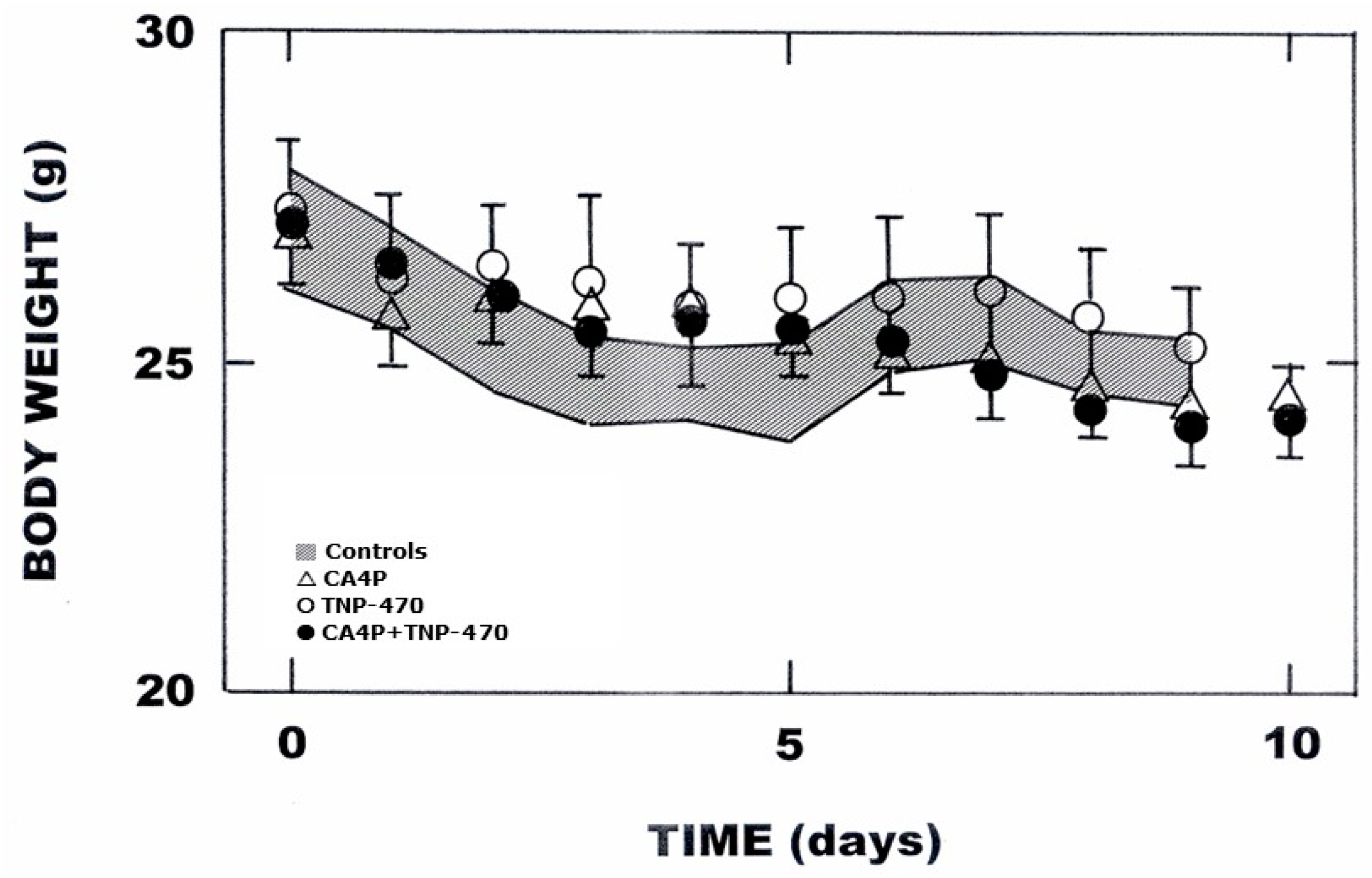
The possible influence of the different treatment schedules on systemic toxicity is illustrated in Figure 3 and Figure 4. Untreated tumor bearing mice showed a gradual decline in body weight over the 9-day period of tumor growth (Figure 3). Injecting CA4P, TNP-470, or the combination of CA4P and TNP-470 did not influence this weight loss.
A slight decrease in body weight was also found for mice in which their tumors were irradiated with 10 Gy (Figure 4), the maximum dose used in the combination studies in Figure 2. This was not altered when CA4P and radiation were combined. However, systemic toxicity increased when radiation was combined with TNP-470 or the combination of CA4P with TNP-470. This drop in body weight was significantly different from radiation alone from day 7 onwards, reaching a 21–22% decrease in body weight 15 days after treatment started. This nadir level was transient and body weight began to recover two days later, returning to normal values around 23–25 days in both groups of animals; this recovery period is probably related to the TNP-470 treatment stopping after 13 days.
In an attempt to understand how local tumor irradiation might affect systemic toxicity of TNP-470, a cytokine assay was performed and the results summarized in Table 1.
Measurements were made from 1 to 72 h after irradiating with 10 Gy and the results obtained were highly variable. Both increases and decreases were found with various cytokines at the different time points, but the only statistically significant changes were a decrease at 3-h with IL-1b; an increase at 1-h with IL-6 but this had significantly decreased by 3-h; and a significant decrease in IL-12p70 at 1, 24, and 72 h after irradiation.

4. Discussion

Targeting tumor vasculature as a therapeutic approach has been extensively studied and numerous VTAs have reached clinical evaluation [4]. Although AIs and VDAs are often grouped together as one single entity they are in fact two distinct approaches targeting different aspects of the tumor neo-vasculature [3]; AIs inhibit the angiogenesis process while VDAs disrupt the already established vasculature. In the current study, the AI we used was TNP-470, an agent that has been shown to inhibit various aspects of the angiogenesis process [11,12]. The VDA used was CA4P, a microtubule depolymerizing agent that induces endothelial cell death [28]. Using either of these VTAs we were able to delay the growth of this C3H mammary carcinoma model (Figure 1). The drug doses and schedules used for TNP-470 and CA4P actually resulted in identical effects on tumor growth. Combining TNP-470 and CA4P together gave a tumor growth inhibitory effect that would be expected from a simple additive response of each agent alone. Similar results have been reported in other studies in which AIs and VDAs have been combined [29,30,31,32,33], supporting the argument that AIs and VDAs work on different vascular targets and that such a combination is complimentary.
It is also clear from the data of Figure 1 that when AIs and VDAs are administered alone or even in combination, tumor growth is delayed, but it is never completely inhibited. Again, this has been seen in other studies [30,31,32,33], although one study did report 100% tumor regression when TNP-470 and 5,6-dimethylxanthenone-4-acetic acid (DMXAA) were combined [29]. However, the dose of DMXAA was high and alone induced 67% tumor regression; in our studies such a high dose was shown to be toxic with 100% lethality within 24-h after administration [25]. The general failure to completely prevent tumor growth or induce tumor control with VTAs suggests that for the full potential to be achieved, VTAs should be combined with other therapies. This has been extensively investigated (for review see [15]) with the two most common combination approaches studied being with chemotherapy and radiation. In this C3H mammary carcinoma model, radiation inhibited tumor growth in a dose-dependent fashion (Figure 2). This radiation response was enhanced by both TNP-470 and CA4P, with the TNP-470 effect being superior to that seen with CA4P. Our previous studies with CA4P and radiation in the C3H mammary carcinoma found a schedule dependent effect on radiation response [34]. When CA4P was administered within a few hours after irradiating there was a significant enhancement of the anti-tumor response. However, this was lost if CA4P was injected prior to the radiation treatment. This was probably the result of the CA4P-induced vascular shut-down making some tumor cells hypoxic at the time of irradiation and thus radiation resistant. Additional studies in this C3H mammary carcinoma demonstrated the ability of CA4P to induce hypoxia [34,35]. This schedule dependency between radiation and VDAs appears to be a general observation [34]. The tumor cells affected by VDAs typically are those in the central tumor region, with those cells in the periphery remaining untouched [36,37,38,39]. This is because cells in the periphery actually receive their oxygen and nutritional supply from nearby normal tissue blood vessels, and such normal tissue vessels are generally not affected by VDA treatment [36]. Tumor cells in this peripheral region are also likely to be better oxygenated than those in the more central regions and thus more radiation sensitive. This would suggest that the effect of combining VDAs with radiation would simply result in an additive response. However, the slope values for the radiation and radiation with CA4P curves in Figure 2 were not the same (1.90 for radiation alone and 3.20 for radiation+CA4P) and this 1.7-fold increase in the slope ratio by giving CA4P does suggest a greater than additive effect, although it is unclear as to the mechanism responsible.
With TNP-470 the enhancement of radiation response was substantial and clearly greater than an additive response; the slope value obtained from the data of Figure 2 for radiation+TNP-470 was 7.80, some 4.1 times greater than the 1.90 obtained with radiation alone. Several other studies have investigated the combination of TNP-470 with radiation [26,40,41], reporting greater anti-tumor activity with the combination compared to each agent alone, but it was unclear as to whether the effect was additive or synergistic. There are several possible explanations as to why the combination of radiation and TNP-470 could result in a greater than additive response. It has been proposed that AIs can “normalize” tumor vessels [42]. This is the process by which the primitive and chaotic abnormal tumor vasculature is basically “pruned”, thus becoming more “normal”, allowing for more efficient delivery of therapeutics, nutrients, and oxygen. The latter effect would improve radiation response. However, such effects on tumor oxygenation are only transient [43]. Moreover, normalization is not a universal phenomenon with many more studies reporting a decrease in oxygenation after treatment with AIs [15,44]. This apparent contradictory effect is also seen with TNP-470 in which both an increase [26] and decrease [45] in hypoxia were reported. Tumor oxygenation status is only relevant at the time of irradiation [46] and since in our study, TNP-470 was actually administered for a two-week period after irradiating any effect on oxygenation status does not explain the enhanced radiation response. An alternative mechanism could be “target interaction” whereby the induced radiation damage sensitizes the tumor to the AI. This has been shown in vivo in which FaDu head and neck tumor xenografts, normally unresponsive to the tyrosine kinase inhibitor PTK787/ZK 222584, become sensitive to the anti-tumor activity of this drug by implanting tumors in a site that had previously been irradiated [47]. The greatest anti-tumor effect in our C3H mammary carcinoma study was found when radiation, CA4P and TNP-470 were all combined with the slope value being 9.96. Although the ratio of the slope values for radiation alone and that for radiation + both VTAs resulted in a value of 5.2, this was no greater than simply adding the slope ratios for radiation with CA4P (slope ratio of 1.7) or TNP-470 (slope ratio of 4.1), suggesting an additive effect of CA4P and TNP-470 on radiation response.
Improving tumor response to treatment with VTAs and radiation, even if just an additive effect is obtained, will only be of benefit if such combinations do not enhance normal tissue toxicity. Neither CA4P nor TNP-470 had any influence on systemic toxicity (Figure 3). No effect was observed when these VTAs were combined. Side effect issues generally are not reported in pre-clinical studies with VTAs even though side effects are seen in patients undergoing VTA therapy [48,49]. However, some pre-clinical studies have investigated VTA-induced changes in body weight. Similar to our findings, others have reported no effect of CA4P on body weight [50,51]. For TNP-470, conflicting results have been reported, with both a decrease in body weight [52,53] and no effect [54] as we observed; these effects on body weight have generally been attributed to ataxia resulting from an impairment of food intake [52,53].
When the VTAs were combined with radiation there were absolutely no effects on body weight loss with CA4P alone (Figure 4). However, significant decreases were seen in the groups given TNP-470. Body weight actually continued to drop throughout the two-week treatment period, but once treatment with TNP-470 stopped body weight recovered relatively quickly. In an effort to further understand the reasons for this effect we measured serum levels of various cytokines. It is established that radiation can induce the expression of inflammation-related cytokines (for review see [55]). Many of these cytokines are those investigated in our current study and shown in Table 1. However, despite measuring the serum levels of 10 different cytokines up to 72-h after irradiating, the only significant increase was for IL-6 at 1-h. Other studies have also reported a radiation-induced increase in IL-6 expression in both normal and tumor cells [56,57,58,59]. It is difficult to understand why our IL-6 effect was so transient or why no significant increases were seen with any of the other cytokines. Increased levels were observed with certain cytokines at specific time points (i.e., IL-1b at 24 and 72-h, IL-6 at 72-h, IL-10 at 1 and 3-h, IFN-γ at 3-h), but as a consequence of variability in the results obtained, the errors were large and thus the mean changes not significantly different to controls. Such large variability has been reported by us previously [60]. Only one radiation dose (10 Gy) was used in our assessment, but such a dose is considered optimal for inducing pro-inflammatory responses [61]. It was also the dose used in the toxicity studies (Figure 4). Measurements of the various cytokines were made early after irradiating and up to 72-h later. Although one study has suggested that substantial changes in cytokine levels occurs around 1–2 weeks after irradiating [62], other studies show significant increases within 24-h [56,57,58,59]. Furthermore, the decreases in body weight shown in Figure 4 begin to occur within the first few days after irradiating, suggesting that making our measurements within the 72-h post-irradiation period was appropriate. Clearly, the increase in systemic toxicity to TNP-470, following local tumor radiation, has to be the consequence of a radiation-induced inflammatory response. However, whether this is simply the result of the transient significant increase in IL-6 or by some other unknown mechanism remains unclear. A more comprehensive investigation, with the focus on trying to elucidate the reasons for the induced systemic toxicity, is obviously warranted. This could involve a number of different parameters, such as measurements of blood cell numbers, estimates of various enzyme levels, and histological assessment of possible damage in a range of normal tissues.

5. Conclusions

VTAs are currently in clinical evaluation as anti-cancer therapies, especially when used to augment conventional cancer treatments such as radiation. Since AIs and VDAs have different mechanisms of action, their full therapeutic potential is likely when they are used in combination with radiotherapy. The anti-tumor effectiveness of such a combination was seen in the current study. However, there was also a significant increase in systemic toxicity, which negates any benefit. Whether similar trends are seen with other combinations of VTAs and radiation is not known, but is a critical issue that should be investigated prior to any clinical application.

Author Contributions

Conceptualization, M.N., R.M., E.E. and M.R.H.; methodology and analysis, M.N., R.M., L.B., B.S.S. and M.R.H.; writing—original draft preparation, M.N. and R.M.; review and editing, M.N., R.M. and M.R.H.; funding acquisition, M.R.H. All authors have read and agreed to the published version of the manuscript.

Funding

This research was funded by grants from the Danish Cancer Society (Grant number R40-A2022-11-S2) and the Danish Council for Independent Research: Medical Sciences. (Grant number DFF—4004-00362).

Institutional Review Board Statement

All animal studies were conducted according to the animal welfare policy of Aarhus University (http://dyrefaciliteter.au.dk, accessed on 10 January 2022), and with the Danish Animal Experiments Inspectorate’s approval (Animal license number 2016-15-0201-01024).

Data Availability Statement

The data presented in this study are available on request from the corresponding author. The data are not publicly available because the results from these experiments and all other animal experiments at our institute are stored in a single data depository. Access is, therefore, limited to only qualified, relevant personnel.

Acknowledgments

The authors would like to thank Dorthe Grand, Pia Schjerbeck, Birthe Hermansen and Inger Marie Horsman for excellent technical assistance.

Conflicts of Interest

All authors declare no conflict of interest. The funders had no role in the design of the study; in the collection, analyses, or interpretation of data; in the writing of the manuscript, or in the decision to publish the results.

References

  1. Brem, S.; Brem, H.; Folkman, J.; Finkelstein, D.; Patz, A. Prolonged tumor dormancy by prevention of neovascularization in the vitreous. Cancer Res. 1976, 36, 2807–2812. [Google Scholar] [PubMed]
  2. Folkman, J. How is blood vessel growth regulated in normal and neoplastic tissue? Cancer Res. 1986, 46, 467–473. [Google Scholar] [PubMed]
  3. Siemann, D.W.; Bibby, M.C.; Dark, G.; Dicker, A.; Eskens, F.A.L.M.; Horsman, M.R.; Marmé, D.; LoRusso, P.M. Differentiation and definition of vascular-targeted therapies. Clin. Cancer Res. 2005, 11, 416–420. [Google Scholar] [CrossRef] [PubMed]
  4. Siemann, D.W.; Chaplin, D.J.; Horsman, M.R. Realizing the potential of vascular targeted therapy: The rationale for combining vascular disrupting agents and anti-angiogenic agents to treat cancer. Cancer Investig. 2017, 35, 519–534. [Google Scholar] [CrossRef]
  5. Ferrara, N.; Gerber, H.P.; LeCouter, J. The biology of VEGF and its receptors. Nat. Med. 2003, 9, 669–676. [Google Scholar] [CrossRef] [PubMed]
  6. Pugh, C.W.; Ratcliffe, P.J. Regulation of angiogenesis by hypoxia: Role of the HIF system. Nat. Med. 2003, 9, 677–684. [Google Scholar] [CrossRef] [PubMed]
  7. Marmé, D. The impact of anti-angiogenic agents on cancer therapy. J. Cancer Res. Clin. Oncol. 2003, 129, 607–620. [Google Scholar] [CrossRef]
  8. Eskens, F.A.L.M. Angiogenesis inhibitors in clinical development; where are we now and where are we going? Br. J. Cancer 2004, 90, 1–7. [Google Scholar] [CrossRef]
  9. Ferrara, N.; Kerbel, R.S. Angiogenesis as a therapeutic target. Nature 2005, 438, 967–974. [Google Scholar] [CrossRef]
  10. Ingber, D.; Fujita, T.; Kishimoto, S.; Sudo, K.; Kanamaru, T.; Brem, H.; Folkman, J. Synthetic analogues of fumagillin that inhibit angiogenesis and suppress tumour growth. Nature 1990, 348, 555–557. [Google Scholar] [CrossRef]
  11. Abe, J.; Zhou, W.; Takuwa, N.; Taguchi, J.; Kurokawa, K.; Kumada, M.; Takuwa, Y. A fumagillin derivative angiogenesis inhibitor, AGM-1470, inhibits activation of cyclin-dependent kinases and phosphorylation of retinoblastoma gene product but not protein tyrosyl phosphorylation or protooncogene expression in vascular endothelial cells. Cancer Res. 1994, 54, 3407–3412. [Google Scholar] [PubMed]
  12. Satchi-Fainaro, R.; Mamluk, R.; Wang, L.; Short, S.M.; Nagy, J.A.; Feng, D.; Dvorak, A.M.; Dvorak, H.F.; Puder, M.; Mukhopadhyay, D.; et al. Inhibition of vessel permeability by TNP-470 and its polymer conjugate, carplostatin. Cancer Cell 2005, 7, 251–261. [Google Scholar] [CrossRef] [PubMed]
  13. Carmeliet, P.; Collen, D. Molecular basis of angiogenesis. Role of VEGF and VE-cadherin. Ann. N. Y. Acad. Sci. 2000, 902, 249–262. [Google Scholar] [CrossRef] [PubMed]
  14. Matsumoto, T.; Claesson-Welsh, L. VEGF receptor signal transduction. Sci. STKE 2001, 2001, re21. [Google Scholar] [CrossRef]
  15. Horsman, M.R.; Siemann, D.W. Pathophysiologic effects of vascular-targeting agents and the implications for combination with conventional therapies. Cancer Res. 2006, 66, 11520–11539. [Google Scholar] [CrossRef]
  16. Denekamp, J.; Hill, S. Angiogenic attack as a therapeutic strategy for cancer. Radiother. Oncol. 1991, 20, 103–112. [Google Scholar] [CrossRef]
  17. Thorpe, P.E. Vascular targeting agents as cancer therapeutics. Clin. Cancer Res. 2004, 10, 415–427. [Google Scholar] [CrossRef]
  18. Siemann, D.W.; Chaplin, D.J.; Walicke, P.A. A review and update of the current status of the vascular-disabling agent combretastatin-A4 phosphate (CA4P). Exp. Opin. Investig. Drugs 2009, 18, 189–197. [Google Scholar] [CrossRef]
  19. Siemann, D.W.; Horsman, M.R. Vascular targeted therapies in oncology. Cell Tissue Res. 2009, 335, 241–248. [Google Scholar] [CrossRef]
  20. Vens, C.; Koritzinsky, M.; Wouters, B.G. Irradiation-induced damage and the DNA damage response. In Basic Clinical Radiobiology, 5th ed.; Joiner, M.C., van der Kogel, A.J., Eds.; CRC Press: Boca Raton, FL, USA, 2019; pp. 9–20. [Google Scholar]
  21. Zeman, E.M. The biological basis of radiation oncology. In Clinical Radiation Oncology, 5th ed.; Tepper, J.E., Foote, R.L., Michalski, J.M., Eds.; Elsevier: Philadelphia, PA, USA, 2021; pp. 2–38. [Google Scholar]
  22. Horsman, M.R.; Murata, R. Combination of vascular targeting agents with thermal and radiation therapy. Int. J. Radiat. Oncol. Biol. Phys. 2002, 54, 1518–1523. [Google Scholar] [CrossRef]
  23. Wittenborn, T.R.; Horsman, M.R. Targeting tumour hypoxia to improve outcome of stereotactic radiotherapy. Acta Oncol. 2015, 54, 1385–1392. [Google Scholar] [CrossRef] [PubMed]
  24. Overgaard, J. Simultaneous and sequential hyperthermia and radiation treatment of an experimental tumor and its surrounding normal tissue in vivo. Int. J. Radiat. Oncol. Biol. Phys. 1980, 6, 1507–1517. [Google Scholar] [CrossRef] [PubMed]
  25. Breidahl, T.; Nielsen, F.U.; Stodkilde-Jorgensen, H.; Maxwell, R.J.; Horsman, M.R. The effects of the vascular disrupting agents combretastatin A-4 disodium phosphate, 5,6-dimethylxanthenone-4-acetic acid and ZD6126 in a murine tumour: A comparative assessment using MRI and MRS. Acta Oncol. 2006, 45, 306–316. [Google Scholar] [CrossRef] [PubMed]
  26. Murata, R.; Nishimura, Y.; Hiraoka, M. An antiangiogenic agent (TNP-470) inhibited reoxygenation during fractionated radiotherapy of murine mammray carcinoma. Int. J. Radiat. Oncol. Biol. Phys. 1997, 37, 1107–1113. [Google Scholar] [CrossRef]
  27. Horsman, M.R.; Chaplin, D.J.; Overgaard, J. Combination of nicotinamide and hyperthermia to eliminate radio-resistant chronically and acutely hypoxic tumour cells. Cancer Res. 1990, 50, 7430–7436. [Google Scholar]
  28. Tozer, G.M.; Kanthou, C.; Baguley, B.C. Disrupting tumour blood vessels. Nat. Rev. Cancer 2005, 5, 423–435. [Google Scholar] [CrossRef]
  29. Ching, L.M.; Xu, Z.F.; Gummer, B.H.; Palmer, B.D.; Joseph, W.R.; Baguley, B.C. Effect of thalidomide on tumour necrosis factor production and anti-tumour activity induced by 5,6-dimethylxanthenone-4-acetic acid. Br. J. Cancer 1995, 72, 339–343. [Google Scholar] [CrossRef]
  30. Mysliwski, A.; Bigda, J.; Koszalka, P.; Szmit, E. Synergistic effect of the angiogenesis inhibitor TNP-470 and tumor necrosis factor (TNF) on Bomirski Ab melanoma in hamsters. Anticancer. Res. 2000, 20, 4643–4647. [Google Scholar]
  31. Siemann, D.W.; Shi, W. Efficacy of combined antiangiogenic and vascular disrupting agents in treatment of solid tumors. Int. J. Radiat. Oncol. Biol. Phys. 2004, 60, 1233–1240. [Google Scholar] [CrossRef]
  32. Siemann, D.W.; Shi, W. Dual targeting of tumor vasculature: Combining avastin and vascular disrupting agents (CA4P or Oxi4503). Anticancer. Res. 2008, 28, 2027–2031. [Google Scholar]
  33. Landuyt, W.; Ahmed, B.; Nuyts, S.; Theys, J.; Op De Beeck, M.; Runders, A.; Anné, J.; van Oosterom, A.; Van Der Bogaert, W.; Lambin, P. In vivo antitumor effect of vascular targeting combined with either ionizing radiation or anti-angiogenesis treatment. Int. J. Radiat. Oncol. Biol. Phys. 2001, 49, 443–450. [Google Scholar] [CrossRef] [PubMed]
  34. Iversen, A.B.; Busk, M.; Horsman, M.R. Induction of hypoxia by vascular disrupting agents and the significance for their combination with radiation therapy. Acta Oncol. 2013, 52, 1320–1326. [Google Scholar] [CrossRef] [PubMed]
  35. Iversen, A.B.; Busk, M.; Bertelsen, L.B.; Laustsen, C.; Munk, O.L.; Nielsen, T.; Wittenborn, T.R.; Bussink, J.; Lok, J.; Stødkilde-Jørgensen, H.; et al. The potential of hyperpolarized 13C magnetic resonance spectroscopy to monitor the effect of combretastatin based vascular disrupting agents. Acta Oncol. 2017, 56, 1626–1633. [Google Scholar] [CrossRef]
  36. Dark, G.D.; Hill, S.A.; Prise, V.E.; Tozer, G.M.; Pettit, G.R.; Chaplin, D.J. Combretastatin A-4, an agent that displays potent and selective toxicity towrads tumor vasculature. Cancer Res. 1997, 57, 1829–1834. [Google Scholar] [PubMed]
  37. Lash, C.J.; Li, A.E.; Rutland, M.; Baguley, B.C.; Zwi, L.J.; Wilson, W.R. Enhancement of the anti-tumour effects of the antivascular agent 5,6-dimethylxanthenone-4-acetic acid (DMXAA) by the combination with 5-hydroxytryptamine and bioreductive drugs. Br. J. Cancer 1998, 78, 439–445. [Google Scholar] [CrossRef]
  38. Li, L.; Rojiani, A.; Siemann, D.W. Targeting the tumor vasculature with combretastatin A-4 disodium phosphate: Effects on radiation therapy. Int. J. Radiat. Oncol. Biol. Phys. 1998, 42, 899–903. [Google Scholar] [CrossRef]
  39. Siemann, D.W.; Rojiani, A.M. Enhancement of radiation therapy by the novel vascular targeting agent ZD6126. Int. J. Radiat. Oncol. Biol. Phys. 2002, 53, 164–171. [Google Scholar] [CrossRef]
  40. Teicher, B.A.; Holden, S.A.; Ara, G.; Alvarez Sotomayor, E.; Huang, Z.D.; Chen, Y.-N.; Brem, H. Potentiation of cytotoxic cancer therapies by TNP-470 alone and with other anti-angiogenic agents. Int. J. Cancer 1994, 57, 920–925. [Google Scholar] [CrossRef]
  41. Lund, E.L.; Bastholm, L.; Kristjansen, P.E.G. Therapeutic synergy of TNP-470 and ionizing radiation: Effects on tumor growth, vessel morphology, and angiogenesis in human glioblastoma multiforme xenografts. Clin. Cancer Res. 2000, 6, 971–978. [Google Scholar]
  42. Jain, R.K. Normalizing tumor vasculature with anti-angiogenic therapy: A new paradigm for combination therapy. Nat. Med. 2001, 7, 987–989. [Google Scholar] [CrossRef]
  43. Winkler, F.; Kozin, S.V.; Tong, R.; Chae, S.-S.; Booth, M.F.; Garkavtsev, I.; Xu, L.; Hicklin, D.J.; Fukumura, D.; di Tomaso, E.; et al. Kinetics of vascular normalization by VEGFR2 blockade governs brain tumor response to radiation: Role of oxygenation, angiopoietin-1, and matrix metalloproteinases. Cancer Cell 2004, 6, 553–563. [Google Scholar] [CrossRef] [PubMed]
  44. Siemann, D.W.; Dai, Y.; Horsman, M.R. Hypoxia, metastasis, and antiangiogenic therapies. In Hypoxia and Cancer: Biological Implications and Therapeutic Opportunities; Melillo, G., Ed.; Springer: New York, NY, USA, 2014; pp. 205–227. [Google Scholar]
  45. Teicher, B.A.; Holden, S.A.; Ara, G.; Dupuis, N.P.; Liu, F.; Yuan, J.; Ikebe, M.; Kakeji, Y. Influence of an anti-angiogenic treatment on 9L gliosarcoma: Oxygenation and response to cytotoxic therapy. Int. J. Cancer 1995, 61, 732–737. [Google Scholar] [CrossRef] [PubMed]
  46. Michael, B.D.; Adams, G.E.; Hewitt, H.B.; Jones, W.B.; Watts, M.E. A post effect of oxygen in irradiated bacteria: A submillisecond fast mixing study. Radiat. Res. 1973, 54, 239–251. [Google Scholar] [CrossRef] [PubMed]
  47. Zips, D.; Eicheler, W.; Geyer, P.; Hessel, F.; Dörfler, A.; Thames, H.D.; Haberey, M.; Baumann, M. Enhanced susceptibility of irradiated tumor vessels to vascular endothelial growth factor receptor tyrosine kinase inhibition. Cancer Res. 2005, 65, 5374–5379. [Google Scholar] [CrossRef]
  48. Eskens, F.A.L.M.; Verweij, J. The clinical toxicity profile of vascular endothelial growth factor (VEGF) and vascular endothelial growth factor receptor (VEGFR) targeting angiogenesis inhibitors; a review. Eur. J. Cancer 2006, 42, 3127–3139. [Google Scholar] [CrossRef]
  49. Subbiah, I.M.; Lenihan, D.J.; Tsimberidou, A.M. Cardiovascular toxicity profiles of vascular-disrupting agents. Oncologist 2011, 16, 1120–1130. [Google Scholar] [CrossRef]
  50. Zhang, Y.-L.; Li, B.-Y.; Yang, R.; Xia, L.-Y.; Fan, A.-L.; Chu, Y.-C.; Wang, L.-J.; Wang, Z.-C.; Jiang, A.-Q.; Zhu, H.-L. A class of novel tubulin polymerization inhibitors exert effective anti-tumor activity via mitotic catastrophe. Eur. J. Med. Chem. 2019, 163, 896–910. [Google Scholar] [CrossRef]
  51. Tang, Z.; Xiong, D.; Song, J.; Ye, M.; Liu, J.; Wang, Z.; Zhang, L.; Xiao, X. Antitumor drug combretastatin-A4 phosphate aggravates the symptoms of dextran sulfate sodium-induced ulcerative colitis in mice. Front. Pharmacol. 2020, 11, 339. [Google Scholar] [CrossRef]
  52. Drevs, J.; Hofmann, I.; Hugenschmidt, H.; Wittig, C.; Majar, H.; Müller, M.; Wood, J.; Martiny-Baron, G.; Unger, C.; Marmé, D. Effects of PTK787/ZK 222584, a specific inhibitor of vascular endothelial growth factor receptor tyrosine kinases, on primary tumor, metastasis, vessel density, and blood flow in a murine renal cell carcinoma model. Cancer Res. 2000, 60, 4819–4824. [Google Scholar]
  53. Cernaianu, G.; Frank, S.; Erbstösser, K.; Leonhardt, S.; Cross, M.; McIvor, Z.; Scholz, G.; Dansranjavin, T.; Celik, I.; Tannapfel, A.; et al. TNP-470 fails to block the onset of angiogenesis and early tumor establishment in an intravital minimal disease model. Int. J. Color. Dis. 2006, 21, 143–154. [Google Scholar] [CrossRef]
  54. Emoto, M.; Ishiguro, M.; Iwasaki, H.; Kikuchi, M.; Kawarabayashi, T. Effect of angiogenesis inhibitor TNP-470 on the growth, blood flow, and microvessel density in xenografts of human uterine carcinosarcoma in nude mice. Gynecol. Oncol. 2003, 89, 88–94. [Google Scholar] [CrossRef] [PubMed]
  55. Schaue, D.; Kachikwu, E.L.; McBride, W.H. Cytokines in radiobiological responses: A review. Radiat. Res. 2012, 178, 505–523. [Google Scholar] [CrossRef] [PubMed]
  56. Brach, M.A.; Gruss, H.J.; Kaisho, T.; Asano, Y.; Hirano, T.; Herrmann, F. Ionizing radiation induces expression of interleukin 6 by human fibroblasts involving activation of nuclear factor-kappa B. J. Biol. Chem. 1993, 268, 8466–8472. [Google Scholar] [CrossRef] [PubMed]
  57. Beetz, A.; Messer, G.; Oppel, T.; van Beuningen, D.; Peter, R.U.; Kind, P. Induction of interleukin 6 by ionizing radiation in a human epithelial cell line: Control by corticosteroids. Int. J. Radiat. Biol. 1997, 72, 33–43. [Google Scholar] [CrossRef]
  58. Hong, A.; Zhang, M.; Leigh, B.; Stevens, G. Induction of interleukin-6 and oncostatin M by radiation in Kaposi’s sarcoma cell lines. Int. J. Radiat. Oncol. Biol. Phys. 2001, 50, 533–540. [Google Scholar] [CrossRef]
  59. Baselet, B.; Belmans, N.; Coninx, E.; Lowe, D.; Janssen, A.; Michaux, A.; Tabury, K.; Raj, K.; Quintens, R.; Benotmane, M.A.; et al. Functional gene analysis reveals cell cycle changes and infammation in endothelial cells irradiated with a single X-ray dose. Front. Pharmacol. 2017, 8, 213. [Google Scholar] [CrossRef]
  60. Brøndum, L.; Sørensen, B.S.; Eriksen, J.G.; Mortensen, L.S.; Lønbro, S.; Overgaard, J.; Alsner, A. An evaluation of multiplex bead-based analysis of cytokines and soluble proteins in archived lithium heparin plasma, EDTA plasma and serum samples. Scand. J. Clin. Lab. Investig. 2016, 76, 601–611. [Google Scholar] [CrossRef]
  61. Schaue, D.; McBride, W.H. Links between innate immunity and normal tissue radiobiology. Radiat. Res. 2010, 173, 406–417. [Google Scholar] [CrossRef]
  62. Rubin, P.; Johnston, C.J.; Williams, J.P.; McDonald, S.; Finkelstein, J.N. A perpetual cascade of cytokines postirradiation leads to pulmonary fibrosis. Int. J. Radiat. Oncol. Biol. Phys. 1995, 33, 99–109. [Google Scholar] [CrossRef]
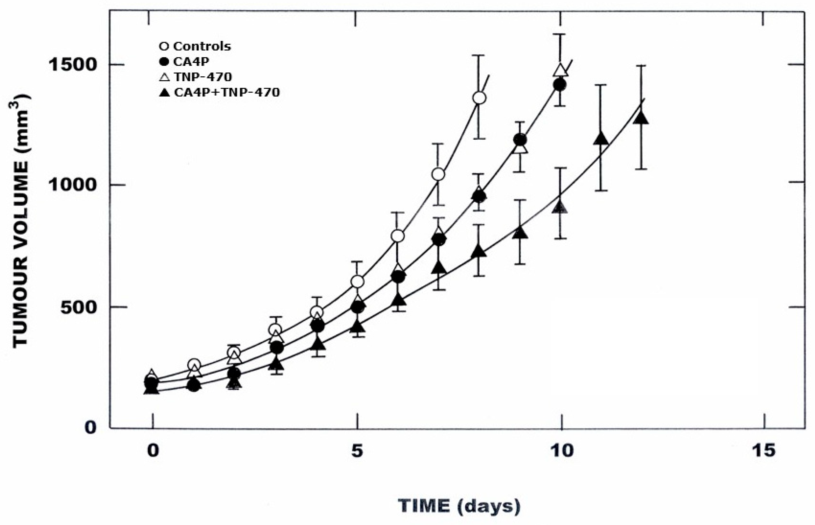
Figure 1. The effect of VTA treatment on the growth of a C3H mammary carcinoma. Mice bearing 200 mm3 foot tumors were given either no drug treatments (); CA4P (⬤; 1 × 250 mg/kg; i.p.); TNP470 (; 6 × 100 mg/kg; s.c.); or CA4DP + TNP-470 (▲). Changes in tumor volume from the start of treatment are shown as means (±1 S.E.) for at least 7 mice/group; lines drawn by eye.
Radiation 04 00024 g001
Figure 2. The effect of combining radiation ± VTA treatment on the growth of a C3H mammary carcinoma. Mice bearing 200 mm3 foot tumors were given either radiation alone (); radiation + CA4P (⬤; 1 × 250 mg/kg; i.p.); radiation + TNP-470 (; 7 × 100 mg/kg; s.c.); or radiation + CA4P + TNP-470 (▲). Results show means (±1 S.E.) of the time for tumors to reach 3 × treatment volume for at least 8 mice/group; lines drawn following regression analysis using individual tumor growth time values.
Radiation 04 00024 g002
Figure 3. The effect of VTA treatment on body weight of CDF1 mice. Animals with 200 mm3 foot implanted C3H mammary carcinomas were given either no treatment (shaded area); CA4P (; 1 × 250 mg/kg; i.p.); TNP-470 (; 5 × 100 mg/kg; s.c.); or CA4P + TNP-470 (⬤). Changes in body weight from the start of treatment are shown as means (±1 S.E.) for at least 7 mice/group.
Radiation 04 00024 g003
Figure 4. The effect of radiation ± VTA treatment on body weight of CDF1 mice. Animals with 200 mm3 foot implanted C3H mammary carcinomas were given either radiation alone (shaded area; 10 Gy), radiation + CA4P (; 1 × 250 mg/kg; i.p.); radiation + TNP-470 (; 7 × 100 mg/kg; s.c.); or radiation + CA4P + TNP-470 (⬤). Changes in body weight from the start of treatment are shown as means (±1 S.E.) for at least 8 mice/group.
Radiation 04 00024 g004
Table 1. Relative effect of irradiation on serum cytokine levels *.
IL-1bIL-2IL-6IL-10IL-12 p40IL-12 p70IL-13IFN-γGM-CSFTNF-α
Controls1.00 **
(0.25)
1.00
(0.22)
1.00
(0.09)
1.00
(0.41)
1.00
(0.05)
1.00
(0.22)
1.00
(0.17)
1.00
(0.60)
1.00
(0.08)
1.00
(0.15)
1 h after RT0.58
(0.24)
0.57
(0.20)
1.50
(0.14)
2.27
(1.43)
0.83
(0.06)
0.55
(0.08)
0.74
(0.07)
0.45
(0.18)
0.79
(0.07)
0.60
(0.12)
3 h after RT0.40
(0.11)
0.86
(0.51)
0.76
(0.05)
1.41
(1.11)
1.09
(0.05)
0.47
(0.14)
0.88
(0.08)
1.44
(1.40)
0.83
(0.10)
0.77
(0.13)
6 h after RT0.60
(0.10)
0.48
(0.13)
0.90
(0.10)
1.13
(0.36)
0.98
(0.05)
0.71
(0.21)
0.89
(0.15)
0.63
(0.49)
0.92
(0.08)
0.82
(0.12)
24 h after RT1.40
(0.65)
0.76
(0.34)
0.73
(0.10)
0.87
(0.44)
0.83
(0.06)
0.56
(0.12)
0.76
(0.10)
0.67
(0.32)
1.01
(0.10)
0.97
(0.14)
72 h after RT1.38
(0.72)
0.48
(0.15)
2.29
(1.68)
0.52
(0.18)
0.79
(0.06)
1.14
(0.32)
0.75
(0.10)
0.67
(0.44)
0.98
(0.10)
1.05
(0.15)
* Animals with 200 mm3 foot implanted C3H mammary carcinomas had their tumors locally irradiated with 10 Gy (RT). Blood samples were taken at the indicated times after irradiating and serum levels of the various cytokines measured. ** Results show relative means (±I S.E.) for an average of 14 mice/group. Indicates those values significantly different from controls (Student’s t-test; p < 0.05).
Disclaimer/Publisher’s Note: The statements, opinions and data contained in all publications are solely those of the individual author(s) and contributor(s) and not of MDPI and/or the editor(s). MDPI and/or the editor(s) disclaim responsibility for any injury to people or property resulting from any ideas, methods, instructions or products referred to in the content.