Communication

Lymphoscintigraphic Indications in the Diagnosis, Management and Prevention of Secondary Lymphedema

International University of Rome (UNINT), High Education, 00147 Rome, Italy
Radiation 2023, 3(1), 40-45; https://doi.org/10.3390/radiation3010004
Submission received: 14 November 2022 / Revised: 15 December 2022 / Accepted: 13 January 2023 / Published: 15 February 2023
(This article belongs to the Section Radiation in Medical Imaging)

Abstract

:

Simple Summary

Diagnosis of lymphedema is not easy, because the pathology can also be confused with other clinical manifestations (for example, edema due to venous insufficiency). To confirm the diagnosis, lymphoscintigraphy is confirmed as the “gold standard” procedure for the diagnosis of lymphedema, particularly in secondary type. The radiation dose level places it in the category of low radiation risk for cancer.

Abstract

Secondary lymphedema is caused by damage to the lymphatic system, often following an oncological tumor removal intervention, or even by an accident. The diagnosis of lymphedema is not easy, because the disease can also be confused with other clinical manifestations (for example, venous insufficiency edema), though an experienced Lymphologist is usually able to diagnose it with good accuracy. To confirm the diagnosis, it is often necessary to resort to specialist imaging tests for an anatomo-functional definition of the pathology. Among these, lymphoscintigraphy is confirmed as the “gold standard” procedure for the diagnosis of lymphedema. Lymphoscintigraphy has been included in the Italian Guidelines by the Ministry of Health.

1. Introduction

Lymphedema is a common and chronic condition resulting from an impairment of lymphatic drainage caused by congenital abnormalities, lymph injury or infection [1]. Chronic lymphedema is a progressive disease that significantly affects patients’ quality of life [2,3].
As is known, secondary lymphedema is caused by damage to the lymphatic system, often following an oncological tumor removal intervention, or even by an accident, which has severed or damaged the lymphatic vessels and/or lymph nodes.
It is estimated that about 20% of women treated for breast cancer will develop upper limb lymphedema. This percentage rises to 40% in cancer survivors after gynecological cancers (cervix, uterus and ovaries) and prostate cancers, and to 67% in patients undergoing inguinal lymph node removal, such as for the treatment of melanoma [4]. Lymphatic stasis may not manifest immediately, but also not long after surgery.
Lymphedema can also be caused by scarring of the lymphatic vessels due to repeated infections.
Even patients in a state of severe obesity can experience lymphedema, since excess fat, when full-bodied and of high volume, ‘crushes’ the lymphatic vessels and obstructs them, thus causing acquired lymphatic insufficiency [5].
We are well aware that lymphedema is essentially a hydraulic problem, caused by the stasis of the lymph that is not properly drained [6].
From the semiotic point of view, in secondary lymphedema the skin is healthy but appears swollen. Digital pressure does not leave a significant impression, as in the case of edema due to venous insufficiency. The limbs may appear swollen and considerably increased in size.

2. Lymphoscintigraphy

The diagnosis of lymphedema is not easy, because the disease can also be confused with other clinical manifestations (for example, venous insufficiency edema), but an experienced Lymphologist is usually able to diagnose it with good accuracy.
However, to confirm the diagnosis, it is often necessary to resort to specialist imaging tests for an anatomo-functional definition of the pathology.
Among these, lymphoscintigraphy, first introduced in 1953 by Sherman and Ter-Pogossian [7], is confirmed as the “gold standard” procedure for the diagnosis of lymphedema. The main indication of lymphoscintigraphy is the early detection of the impairment of the lymphatic system in patients with clinical suspicion of primary and secondary lymphedema [8]. It is demonstrated that in lymphoscintigraphy-confirmed lymphedema, only 17% had positive clinical signs, demonstrating the diagnostic precocity of lymphoscintigraphy [9].
The lymphoscintigraphy procedure only requires a subcutaneous injection of a small amount of radiotracer, and is quite simple and safe. Following subcutaneous administration, the radiotracer is picked up by the lymphatic vessels and lymphatic images are acquired by a gamma camera [10].
This test is not only able to refute or not the doctor’s diagnosis, but it can also visually report the status of the lymphatic system thanks to a precise and function-based imaging technique. By means of lymphoscintigraphy it is possible to see exactly the superficial and deep lymphatic passages, to assess their draining efficiency and also to see the points where the normal flow of the lymph is interrupted (Figure 1).
It is demonstrated that the vast majority of patients had deep lymphatic vessel abnormalities [11]; it also happens, as in the case of Figure 1, that the superficial lymphatic outflow is slowed down and that with muscle activation due to walking, a good deep outflow is activated up to the inguinal and lumboaortic lymph nodes.
In patients with post-traumatic lymphatic edema (57 cases) a reduced visualization of the lymph nodes of the limb root was observed in 24.2% of cases, with the simultaneous presence of “dermal back flow” corresponding to the affected anatomical area. In these cases, the prognosis for complete recovery is less favorable [12].
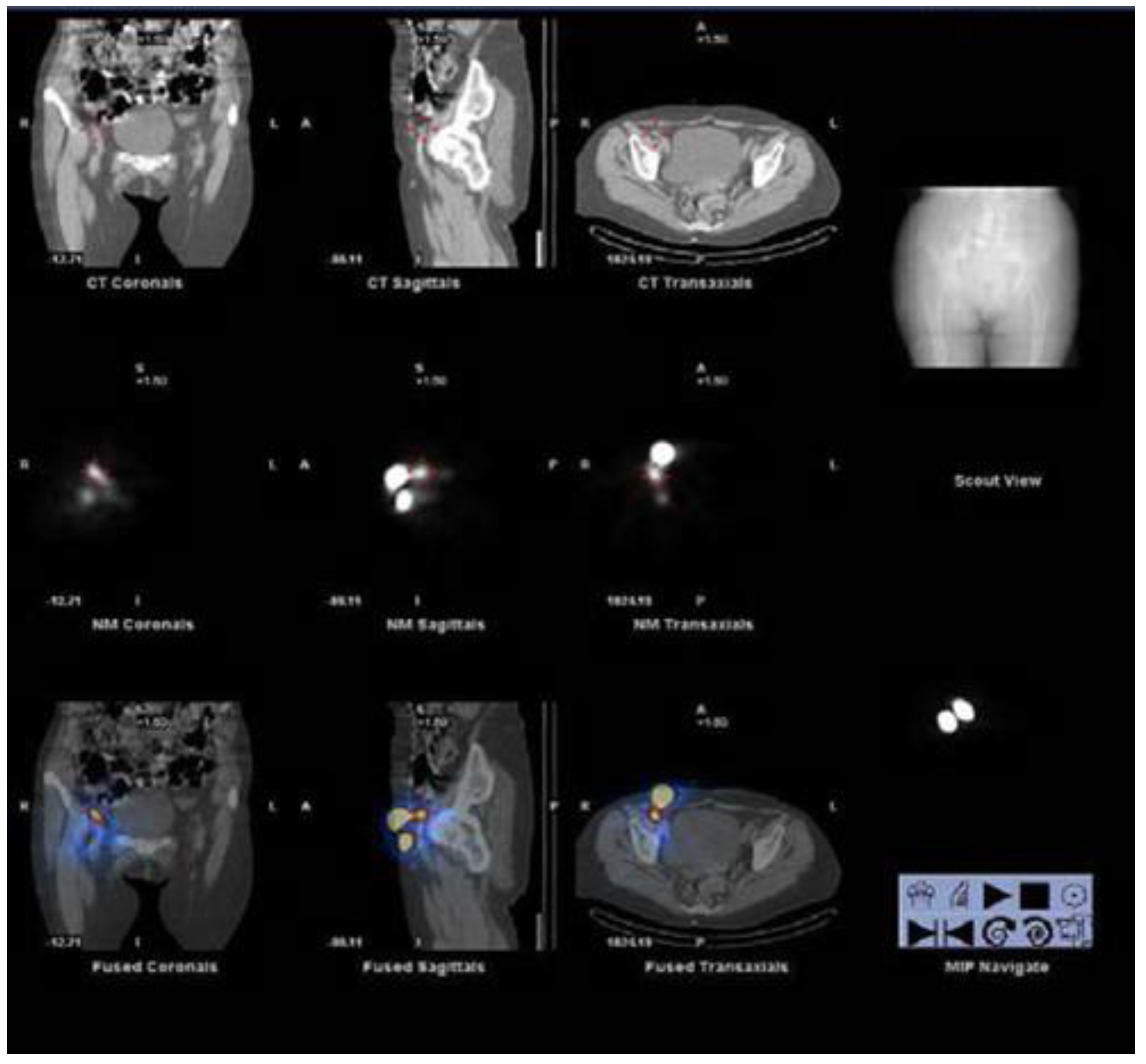
Facial lymphoscintigraphic investigation after the injection of 99mTc-labeled colloids at the forehead, between the eyebrows, seems to represent a simple and valuable way to assess facial lymphatic drainage and to establish the diagnosis of facial lymphedema [9]. SPECT/CT acquisition in patients with post-traumatic edema, but also in other cases, is also useful to demonstrate deep lymphatic drainages not visible on planar imaging [13] (Figure 2). SPECT/CT offers the possibility to check the distribution of lymphatic flow on sagittal, coronal and transverse sections and to observe the functional data of lymphatic outflow and lymph node distribution, comparing them to the anatomical CT images acquired on the same sections. To conduct this, it is necessary to use hybrid equipment capable of acquiring functional nuclear medicine images and anatomical CT at the same time [14].
In one study, it was found that total lymphatic obstruction was present in 56% of unilateral gynecological cancer-related lymphedema and that the stages of lymphoscintigraphy were statistically correlated with episodes of cellulite, circumferential difference and CT volume difference [15].
Lymphoscintigraphy is therefore an essential examination to establish the exact origin of the lymphatic block; this can be useful (often, indispensable) to establish the right therapy for the patient, and to agree together on a long-term maintenance path [16,17,18].
Some authors suggest that the deep circulation should also be studied with lymphoscintigraphy in patients with unilateral or bilateral lower limb lymphedema (LLE) (or sensation of LLE) without morphological or functional signs of lymphatic insufficiency, but whose history and clinical examination are indicative of the presence of lymphedema [19].
Lymphoscintigraphy has been shown to be an effective and objective method of evaluating response to treatment, as regards microsurgical therapy [20], but also for lymphatic drainage and physical therapies [21,22,23] in patients with secondary lymphedema.
Lymphoscintigraphy can also be used for predicting response to treatment before its beginning [24].
Among patients who have undergone an aortocoronary bypass [25], the onset of early or late but persistent lymphedema after partial saphenous phlebectomy appears in a few cases, but is easily diagnosed with lymphoscintigraphy. In 71% of the cases in which the surgery has not changed the normal lymphatic flow in the operated limb, it could still be possible to avoid iatrogenic lymphedema in predisposed subjects selected on lymphoscintigraphy before surgery [12].
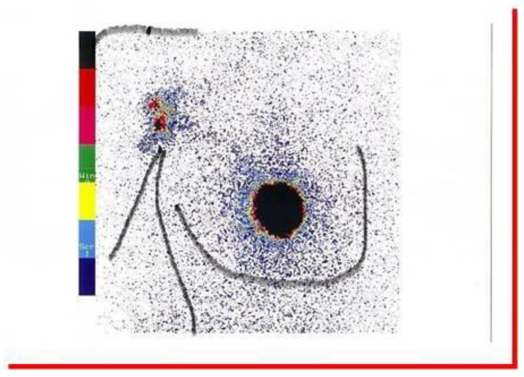
Sentinel lymph node biopsy, the procedure used to selectively remove lymph nodes during surgery in cancer patients, such as breast cancer or some types of melanoma, can greatly reduce the risk of developing lymphedema. The lymphoscintigraphy detection of the sentinel lymph node is a further diagnostic/prognostic element that is added to the indications of lymphoscintigraphy in secondary lymphedema [14,26,27] (Figure 3).
Moreover, the radiation dose level places lymphoscintigraphy, even if in SPECT/CT mode, in the category of low-risk radiation investigations for the occurrence of any malignancy. One case of cancer is equivalent to 105 SPECT/CT lymphoscintigraphy procedures. Furthermore, this dose is lower than in previously published studies, suggesting that patients were well-protected during the entire procedure [28].

3. Conclusions

In favor of lymphoscintigraphy we can mention the simplicity of the procedure, and the minimal invasiveness that makes it accessible even in pediatric age. In addition, its easy repeatability is useful for monitoring lymphedematous pathology and the therapeutic path.
Negative factors consist of the non-presence in all health facilities of a nuclear medicine service, but its execution on an outpatient basis allows the patient to go to the closest nuclear medicine center in the area. About the use of radioactive isotopes, the scientific literature is comforting in this sense since, as mentioned above, the radiation dose level places lymphoscintigraphy, even if in SPECT/CT mode, in the category of low-risk radiation investigations.
Compared to MRI, lymphoscintigraphy offers more or less the same diagnostic potential. But however sophisticated the MRI technique can be made, it can never replace the functional aspect shown by the nuclear medicine technique [29].
A further impetus to the development of nuclear medicine lies in the conception, design and construction of ever more efficient hybrid machines and/or the design and synthesis of new hybrid radiopharmaceuticals, in order to provide more information with only one exam [30].
In conclusion, the possibility offered to nuclear medicine to be a primary protagonist in the development of the diagnosis and therapy of complex pathologies, including a series of rare diseases, must be considered [31]. In this field, lymphoscintigraphy proves to be a useful, if not indispensable, diagnostic tool for the prevention of secondary lymphedema.
It is so useful that lymphoscintigraphy has been included in the Italian Guidelines by the Ministry of Health [32]. These guidelines state that: “In view of an appropriate management of patients with lymphedematous disease, the following aspects are fundamental: …Ability to work directly or with connected centers all the diagnostic tests needed to define the clinical picture (Lymphangioscintigraphy, high-resolution echography ultrasound, CT)”.

Funding

This research received no external funding.

Conflicts of Interest

The author declares no conflict of interest.

References

  1. Szuba, A.; Shin, W.S.; Strauss, H.W.; Rockson, S. The third circulation: Radionuclide lymphoscintigraphy in the evaluation of lymphedema. J. Nucl. Med. 2003, 44, 43–57. [Google Scholar] [PubMed]
  2. Taghian, N.R.; Miller, C.L.; Jammallo, L.S.; O’Toole, J.; Skolny, M.N. Lymphedema following breast cancer treatment and impact on quality of life: A review. Crit. Rev. OncolHematol. 2014, 92, 227–234. [Google Scholar] [CrossRef]
  3. Finnane, A.; Hayes, S.C.; Obermair, A.; Monika, J. Quality of life of women with lowerlimb lymphedema following gynecological cancer. Expert Rev. Pharmacoecon. Outcomes Res. 2011, 11, 287–297. [Google Scholar] [CrossRef] [PubMed] [Green Version]
  4. Barreto Spandonari, V.M. Valutazione Della Qualità di Vita dei Pazienti Dopo Intervento di Microchirurgia Linfatica per il Trattamento del Linfedema; 2021. Available online: unire.unige.it (accessed on 18 October 2022).
  5. Yoshida, S.; Koshima, I.; Imai, H.; Uchiki, T.; Sasaki, A.; Fujioka, Y.; Nagamatsu, S.; Yokota, K.; Yamashita, S. Lymphovenous anastomosis for morbidly obese patients with lymphedema. Plast. Reconstr. Surg. Glob Open 2020, 8, e2860. [Google Scholar] [CrossRef]
  6. Garza, R.; Skoracki, R.; Hock, K.; Povoski, S.P. A comprehensive overview on the surgical management of secondary lymphedema of the upper and lower extremities related to prior oncologic therapies. BMC Cancer 2017, 17, 1–18. [Google Scholar] [CrossRef] [PubMed] [Green Version]
  7. Sherman, A.I.; Ter-Pogossian, M. Lymphnode concentration of radioactive colloidal gold following interstitial injection. Cancer 1953, 6, 1238–1240. [Google Scholar] [CrossRef] [PubMed]
  8. Mango, L.; Mangano, A.M.; Semprebene, A. Lymphoscintigraphy: Preventive, Diagnostic and Prognostic Value. ARC J. Radiol. Med. Imaging 2020, 4, 15–18. [Google Scholar]
  9. Jayaraj, A.; Raju, S.; May, C.; Pace, N. The diagnostic unreliability of classic physical signs of lymphedema. J. Vasc. Surgery Venous Lymphat. Disord. 2019, 7, 890–897. [Google Scholar] [CrossRef]
  10. Hou, G.; Jiang, Y.; Jing, H.; Xu, W.; Xu, K.F.; Chen, L.; Li, F.; Cheng, W. Usefulness of 99mTc-ASC lymphoscintigraphy and SPECT/CT in the evaluation of rare lymphatic disorders: Gorham-Stout disease, lymphangioma, and lymphangioleiomyomatosis. Medicine 2020, 99, e22414. [Google Scholar] [CrossRef]
  11. Campisi, C.C.; Ryan, M.; Villa, G.; Di Summa, P.; Cherubino, M.; Boccardo, F.; Campisi, C. Rationale for study of the deep subfascial lymphatic vessels during lymphoscintigraphy for the diagnosis of peripheral lymphedema. Clin. Nucl. Med. 2019, 44, 91–98. [Google Scholar] [CrossRef]
  12. Michelini, S.; Failla, A.; Moneta, G.; Mango, L.; Iannacoli, M. Iatrogenic secondary lymphedema: A reality to be avoided. Eur. J. Lymphol. 2002, 9, 99. [Google Scholar]
  13. Bourgeois, P.; Peters, E.; Van Mieghem, A.; Vrancken, A.; Giacalone, G.; Zeltzer, A. Edemas of the face and lymphoscintigraphic examination. Sci. Rep. 2021, 11, 1–7. [Google Scholar] [CrossRef] [PubMed]
  14. Mangano, A.M.; Addabbo, W.; Semprebene, A.; Ventroni, G.; Mango, L. Search Sentinel Lymph Node in Melanoma: SPECT/CT Added Value. A Case Rep. J. Cancer Res Forecast. 2018, 1, 1001. [Google Scholar]
  15. Pappalardo, M.; Lin, C.; Ho, O.A.; Kuo, C.F.; Lin, C.Y.; Cheng, M.H. Staging and clinical correlations of lymphoscintigraphy for unilateral gynecological cancer–related lymphedema. J. Surg. Oncol. 2020, 121, 422–434. [Google Scholar] [CrossRef] [PubMed]
  16. Yuan, Z.; Luo, Q.; Zhu, J.; Lu, H.; Zhu, R. The role of radionuclide lymphoscintigraphy in extremity lymphedema. Ann. Nucl. Med. 2006, 20, 341–344. [Google Scholar] [CrossRef] [PubMed]
  17. Raghuram, A.C.; Yu, R.P.; Sung, C.; Huang, S.; Wong, A.K. The Diagnostic Approach to Lymphedema: A Review of Current Modalities and Future Developments. Curr. Breast Cancer Rep. 2019, 11, 365–372. [Google Scholar] [CrossRef]
  18. Mango, L.; Ventroni, G.; Michelini, S. Lymphoscintigraphy and clinic. Res. Rev. Insights 2017, 1, 1–5. [Google Scholar] [CrossRef] [Green Version]
  19. Barbieux, R.; Roman, M.M.; Rivière, F.; Leduc, O.; Leduc, A.; Bourgeois, P.; Provyn, S. Scintigraphic investigations of the deep and superficial lymphatic systems in the evaluation of lower limb edema. Sci. Rep. 2019, 9, 1–9. [Google Scholar] [CrossRef] [Green Version]
  20. Gloviczki, P.; Fisher, J.; Hollier, L.H.; Pairolero, P.C.; Schirger, A.; Wahner, H.W. Microsurgical lymphovenous anastomosis for treatment of lymphedema: A critical review. J. Vasc. Surg. 1988, 7, 647–652. [Google Scholar] [CrossRef] [Green Version]
  21. Kafejian-Haddad, A.; Perez, J.; Castiglioni, M.; Miranda, J.F.; de Figueiredo, L. Lymphscintigraphic evaluation of manual lymphatic drainage for lower extremity lymphedema. Lymphology 2006, 39, 1. [Google Scholar]
  22. de Godoy, J.; Batigalia, F.; Godoy, M.F. Preliminary evaluation of a new more simplified physiotherapy technique for lymphatic drainage. Lymphology 2002, 35, 91. [Google Scholar] [PubMed]
  23. Hwang, J.; Kwon, J.; Lee, K.; Choi, J.; Kim, B.; Lee, B.; Kim, D.I. Changes in lymphatic function after complex physical therapy for lymphedema. Lymphology 1999, 32, 15. [Google Scholar]
  24. Szuba, A.; Strauss, W.; Sirsikar, S.; Rockso, S. Quantitative radionuclide lymphoscintigraphy predicts outcome of manual lymphatic therapy in breast cancer-related lymphedema of the upper extremity. Nucl. Med. Commun. 2002, 23, 1171. [Google Scholar] [CrossRef]
  25. Michelini, S.; Failla, A.; Moneta, G.; Mango, L. Iatrogenic post-saphenectony lymphedemas following aorto-coronaric by-pass. Eur. J. Lymphol. 2005, 15, 18. [Google Scholar]
  26. Morton, D.L.; Wen, D.-R.; Wong, J.H.; Economou, J.S.; Cagle, L.A.; Storm, F.K.; Foshag, L.J.; Cochran, A.J. Technical Details of Intraoperative Lymphatic Mapping for Early Stage Melanoma. Arch. Surg. 1992, 127, 392–399. [Google Scholar] [CrossRef] [PubMed]
  27. Giuliano, A.E.; Kirgan, D.M.; Guenther, J.M.; Morton, D.L. Lymphatic Mapping and Sentinel Lymphadenectomy for Breast Cancer. Ann. Surg. 1994, 220, 391–401. [Google Scholar] [CrossRef]
  28. Almujally, A.; Sulieman, A.; Salah, H.; Alanazi, B.; Calliada, F. Patient Dosimetry in SPECT/CT Lymphoscintigraphy Examinations. J. Res. Med. Dent. Sci. 2020, 8, 97–100. [Google Scholar]
  29. Kim, G.; Smith, M.P.; Donohoe, K.J.; Johnson, A.R.; Singhal, D.; Tsai, L.L. MRI staging of upper extremity secondary lymphedema: Correlation with clinical measurements. Eur. Radiol. 2020, 30, 4686–4694. [Google Scholar] [CrossRef]
  30. Mango, L. Nuclear Medicine in the Third Millennium. J. Nucle. Med. Clinic Imag. 2019, 1, 1. [Google Scholar]
  31. Weber, W.A.; Czernin, J.; Anderson, C.J.; Badawi, R.D.; Barthel, H.; Bengel, F.; Bodei, L.; Buvat, I.; DiCarli, M.; Graham, M.M.; et al. The Future of Nuclear Medicine, Molecular Imaging, and Theranostics. J. Nucl. Med. 2020, 61, 263S–272S. [Google Scholar] [CrossRef] [PubMed]
  32. Michelini, S.; Cestari, M.; Ricci, M.; Leone, A.; Galluccio, A.; Cardone, M. talian Guidelines on Lymphedema: New public regulations 2017. J. Theor. Appl. Vasc. Res. 2017, 1, 119–123. [Google Scholar] [CrossRef]
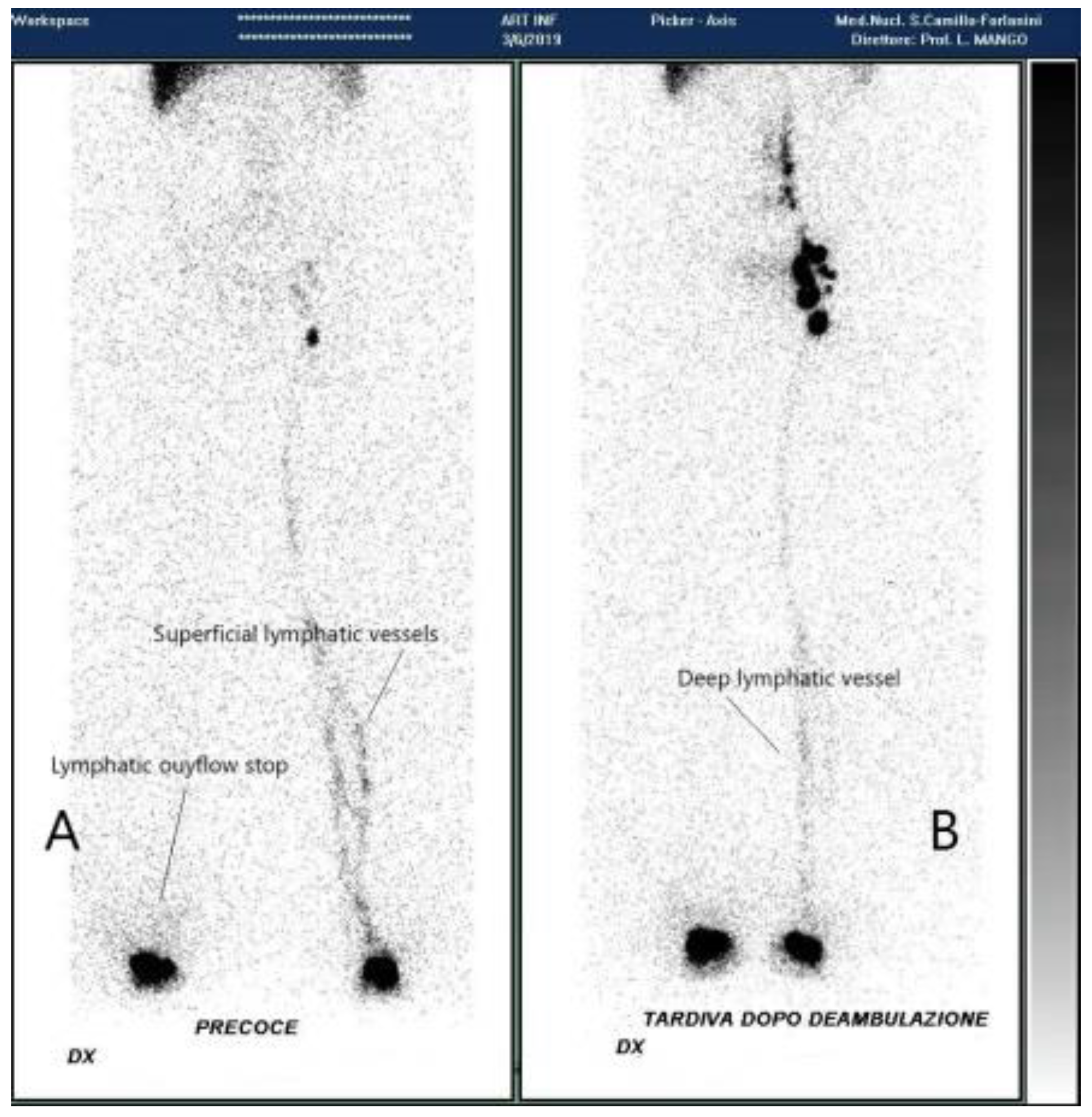
Figure 1. Lymphoscintigraphy of the limbs: (A) at time of injection (B) after 20 min walk.
Radiation 03 00004 g001
Figure 2. SPET/CT lymphoscintigraphy of the pelvis.
Radiation 03 00004 g002
Figure 3. Right Anterior Oblique 45° sentinel node lymphoscintigraphy.
Radiation 03 00004 g003
Disclaimer/Publisher’s Note: The statements, opinions and data contained in all publications are solely those of the individual author(s) and contributor(s) and not of MDPI and/or the editor(s). MDPI and/or the editor(s) disclaim responsibility for any injury to people or property resulting from any ideas, methods, instructions or products referred to in the content.