Article

Seroprevalence of Antibodies to Brucella spp. and Neospora caninum in Cattle from Delta Region of Egypt: Correlation of Seropositivity with Abortion History

1
Division of Internal Medicine, Department of Animal Medicine, Faculty of Veterinary Medicine, South Valley University, Qena 83523, Egypt
2
Department of Infectious Disease, Faculty of Veterinary Medicine, Arish University, Arish 45516, Egypt
3
Department of Clinical Microbiology and Immunology, Faculty of Medicine, King Abdulaziz University, Jeddah 21589, Saudi Arabia
4
Special Infectious Agents Unit, King Fahd Medical Research Center, King Abdulaziz University, Jeddah 22252, Saudi Arabia
5
The National Institute of Oceanography and Fisheries (NIOF), Aswan 81521, Egypt
6
Department of Pathology and Laboratory Diagnosis, College of Veterinary Medicine, Qassim University, Buraydah 51452, Saudi Arabia
7
Department of Veterinary Preventive Medicine, College of Veterinary Medicine, Qassim University, Buraydah 51452, Saudi Arabia
8
Agricultural Research Center (ARC), Animal Health Research Institute-Mansoura Provincial Lab (AHRI-Mansoura), P.O. Box 264-Giza, Cairo 12618, Egypt
9
Department of Theriogenology, Faculty of Veterinary Medicine, Damanhour University, Damanhour 22511, Egypt
10
Institute of Parasitology, Department of Infectious Diseases and Pathobiology, Vetsuisse-Faculty, University of Bern, Länggassstrasse 122, CH-3012 Bern, Switzerland
11
Friedrich-Loeffler Institut, Institute of Bacterial Infections and Zoonosis, Naumburger Str. 96a, 07743 Jena, Germany
12
Department of Bacteriology, Immunology, and Mycology, Faculty of Veterinary Medicine, Benha University, Toukh 13736, Moshtohor, Egypt
*
Author to whom correspondence should be addressed.
Immuno 2024, 4(4), 374-384; https://doi.org/10.3390/immuno4040024
Submission received: 26 August 2024 / Revised: 8 October 2024 / Accepted: 11 October 2024 / Published: 13 October 2024

Abstract

Bovine brucellosis and neosporosis are reported as potential abortifacient infections in cattle worldwide. Brucellosis is additionally a zoonotic bacterial infection caused by numerous Brucella species. Meanwhile, neosporosis is a protozoan parasitic disease that is implicated in causing high economic losses in the cattle industry. Herein, we attempted to investigate the seroprevalence of specific antibodies to Brucella spp. and Neospora caninum using commercially available ELISAs. In addition, we conducted risk factor analysis and estimated the correlation of seropositivity of both pathogens with the recorded abortions in the tested herds. Serum samples from cattle (n = 460) collected from various governorates in the Delta region, northern Egypt, were targeted in this study. Overall, a seroprevalence of 5.4%, 33.3%, and 1.3% was revealed for Brucella spp., N. caninum, and mixed seropositivity, respectively. The location (Kafr El Sheikh vs. Dakahlia vs. Al-Qalyubiya vs. Damietta governorates) and a history of abortion (yes vs. no vs. unknown) were analyzed as risk factors of infection. Kafr El Sheikh governorate (57.7%, p = < 0.0001) and a history of abortion (54.1%, p = < 0.0001) were considered risk factors for Brucella spp. seropositivity compared to the reference factors Al-Qalyubiya (1.1%) and unknown abortion history (0.6%). In the case of N. caninum, the location was also considered a risk factor because the seropositive rates were significantly higher in Damietta (51%, p = 0.001) and Dakahlia (33.4%, p = 0.026) compared to Kafr El Sheikh (11.3%, set as a reference). Conversely to Brucella, animals without a history of abortion exhibited a higher seropositive rate for N. caninum (47.6%, p = 0.009) compared to those with a history of abortion (21.6%, set as reference). For further investigations into the association between abortion and the obtained seropositive rates, we also analyzed the reactivity by comparing samples of animals with, without, and unknown history of abortion. We detected high seroreactivity for Brucella spp. in samples collected from animals with a history of abortion, as demonstrated in the recorded antibody levels and correlation coefficient (Pearson r = 0.919). Based on our data, despite the higher seroprevalence of N. caninum compared to that of Brucella species, Brucella spp. might be the primary cause of abortion in our tested cattle population.

1. Introduction

Brucella (B.) spp. and Neospora caninum (N. caninum) are intracellular abortifacient microbial agents with a global presence. These pathogens can infect a variety of warm-blooded hosts, which imposes substantial threats to both veterinary and public health (in the case of Brucella spp.) due to their economic and potential zoonotic significance, especially in regions that have intensive livestock industry, such as Egypt [1,2,3].
Brucella spp. are small aerobic, non-spore-forming, non-motile Gram-negative coccobacilli that are capable of surviving inside macrophages and other leukocytes, allowing them to evade the host’s humoral and cellular immunity [4]. The genus Brucella is divided into 13 species based on differences in pathogenicity and host preference. Among these, B. melitensis, B. suis, and B. abortus pose the greatest risks to both humans and farm animals. Multiple animal species, particularly sheep, goats, cattle, buffaloes, and pigs, have their reproductive organs colonized by these pathogens, which can lead to infertility and miscarriages [4,5].
Bovine brucellosis is caused primarily by B. abortus, while in some areas where cattle and sheep or goats live closely together, the infection can also be caused by B. melitensis. On rare occasions, B. suis might lead to chronic infection in the cattle’s mammary gland, but no cases of abortion or animal transmission have been reported yet. Brucellae are typically transmitted through direct or indirect exposure to the infected animals or infected materials, as well as through consumption of contaminated water, pasture, or feed [6]. Humans may become infected by consuming contaminated raw milk or unpasteurized dairy products, as well as by handling infected animals or coming in contact with their secretions [7].
Neospora caninum, a protozoan parasite, was first isolated from dogs, the definitive host, in 1984. Other wild canids such as coyote, grey wolf, and dingo were also identified as definitive hosts for N. caninum. Nowadays, it is well recognized globally to infect several farm and wild animals that serve as intermediate hosts, such as sheep, goats, cattle, buffaloes, and camels [8,9]. Cattle and other animals can become horizontally infected with N. caninum by consuming feed contaminated with oocysts from infected dog feces or by coming into contact with contaminated water or soil. However, the main mode of transmission is thought to be vertical, passing from the dam to the calf through the placenta [10]. While the zoonotic potential of Brucella spp. has been well documented, the presence of clinical neosporosis in humans remains uncertain. However, few studies have identified the presence of either antibodies [11,12] or parasite DNA [13] in human samples.
Egypt has one of Africa’s largest cattle resources, highlighting the importance of managing infectious diseases effectively to maintain the livestock industry’s stability and productivity. Among other contributory factors, the country’s substantial livestock resources and economic benefits are misaligned due to prevalent infections. Bovine brucellosis [14,15] and neosporosis [16] are among these infections and have been reported in several regions of the country. These infections lead to significant economic losses, which are related to the premature culling of infected animals, reduced reproductive efficiency, diminished milk yield, and the shortened lifespan of infected cattle [10,17]. Financial losses have been estimated for abortions in cattle induced by Brucella spp. and N. caninum from the world and Egypt [16,18,19]. Thus, our understanding of bovine brucellosis and neosporosis and the related risk factors can lead to the development of effective strategies to mitigate their impact on animal production systems and to minimize the risk of zoonotic transmission to humans.
We previously revealed the high seropositivity of small ruminants in Egypt to Brucella species (8.6%), N. caninum (11.9%), and Toxoplasma gondii (46.1%) [1]. In addition, another study demonstrated the high exposure of cattle to T. gondii (59.4%), N. caninum (13.2%), and Coxiella burnetii (4.7%) [20]. However, this latter study examined a smaller number of cattle (n = 106) and focused on different regions (southern Egypt) than our present study. Although dairy cattle are susceptible to both brucellosis [15] and neosporosis [16], antibodies to Brucella spp. and N. caninum have not been tested simultaneously in dairy cattle from Egypt. To our knowledge, this is the first serological study aimed at estimating the prevalence of these infections in cattle across the Delta region, northern Egypt, especially in relation to infection, co-infection, and association with abortion history.

2. Materials and Methods

2.1. Ethical Approval

This study was performed according to standard procedures identified by the Research Bio-Ethics Standards at Arish University, North Sinai, Egypt. This study was approved by the Research Ethical Committee at Arish University ARU/Vet.02. A team of proficient veterinarians and staff collected the blood samples after consulting with authorities and animal owners.

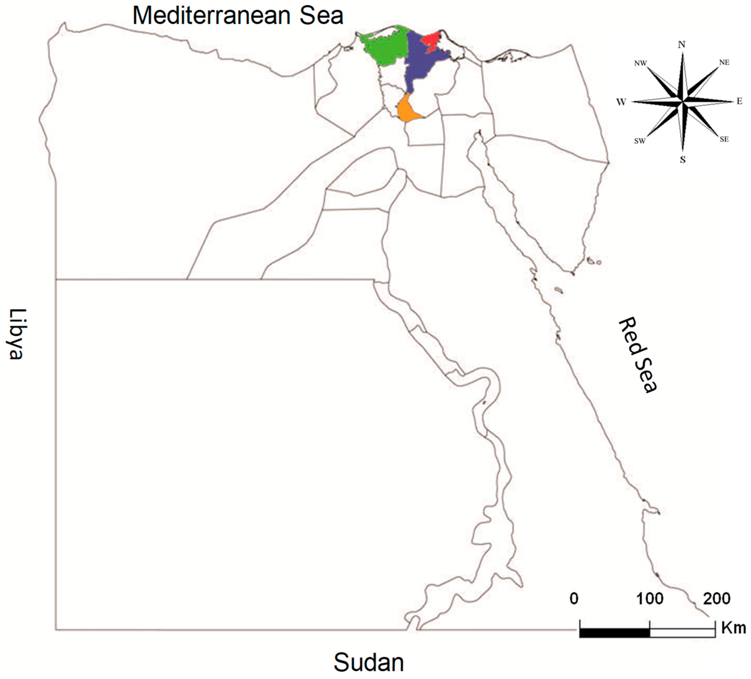
2.2. Description of the Animals and Regions in This Study

Serum samples (n = 460) of cows from various governorates in the Delta region of Egypt were collected for this study during the period of March 2015–January 2016. Location (Kafr El Sheikh vs. Dakahlia vs. Al-Qalyubia vs. Damietta governorates) and history of abortion (yes vs. no vs. unknown) were analyzed as risk factors of infection. Blood samples were randomly collected from cows that experienced abortion (Kafr El Sheikh), from aborting, non-aborting, and unknown cows (Dakahlia), as well as those where a history of abortion was excluded (Damietta) or unknown (Al-Qalyubia) (Table 1). Sera collected from animals with a history of abortion were confined mostly to medium- and large-sized farms (50 to 200 heads/farm) and collected during the first few months after abortions occurred. All animals included in this study were cattle cows, 2–5 years old, of different breeds. Availability of samples and owner cooperation determined the numbers and groups of tested animals in this study. None of the sampled animals was vaccinated against Brucella infection, as confirmed by animal owners or the farm staff (Table 1 and Figure 1).
Blood samples were collected via puncture of the jugular vein using glass tubes without anticoagulant. Sera were separated from these blood samples and were transferred on ice to our laboratory at South Valley University and stored at −20 °C until use in ELISA testing.

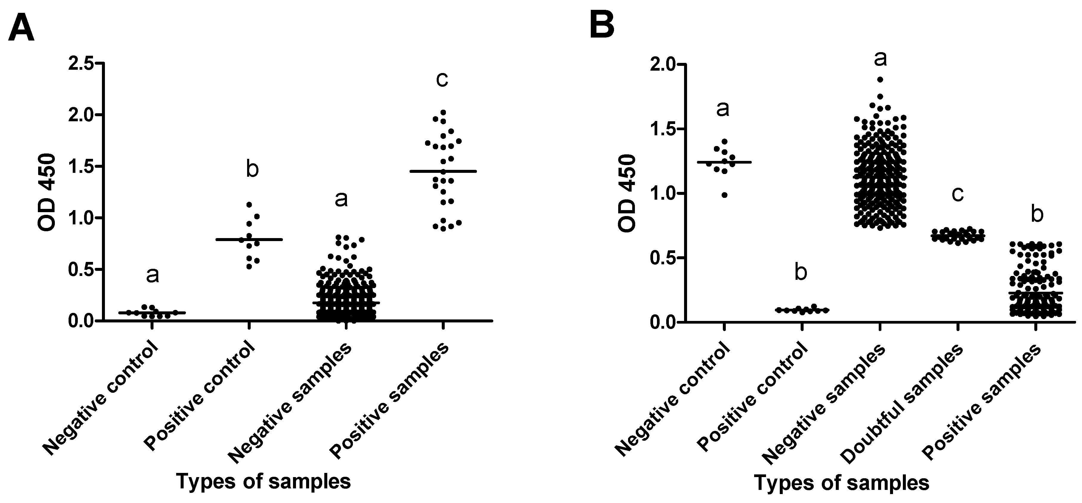
2.3. Serological Diagnosis of Brucella Species and Neospora caninum

Concerning Brucella species, serum samples were analyzed with the indirect multi-species ELISA for brucellosis (ID.vet, Grabels, France). As a preliminary step, serum samples and controls were diluted 1:20 and tested. For N. caninum, serum samples were analyzed using a competitive multi-species ELISA for neosporosis (ID.vet, Grabels, France). Serum samples and controls were diluted 1:2.
All procedures related to the experiment, calculation, and interpretation were performed according to the manufacturer’s instructions.
The ODs of all ELISA results were read at 450 nm and measured with an Infinite® F50/Robotic ELISA reader (Tecan Group Ltd., Männedorf, Switzerland).

2.4. Statistical Analysis

The p-values and odds ratio were also calculated with GraphPad Prism version 5 (GraphPad Software Inc., La Jolla, CA, USA). The results were considered significant when the p-value was <0.05 or highly significant when the p-value was <0.0001.

3. Results

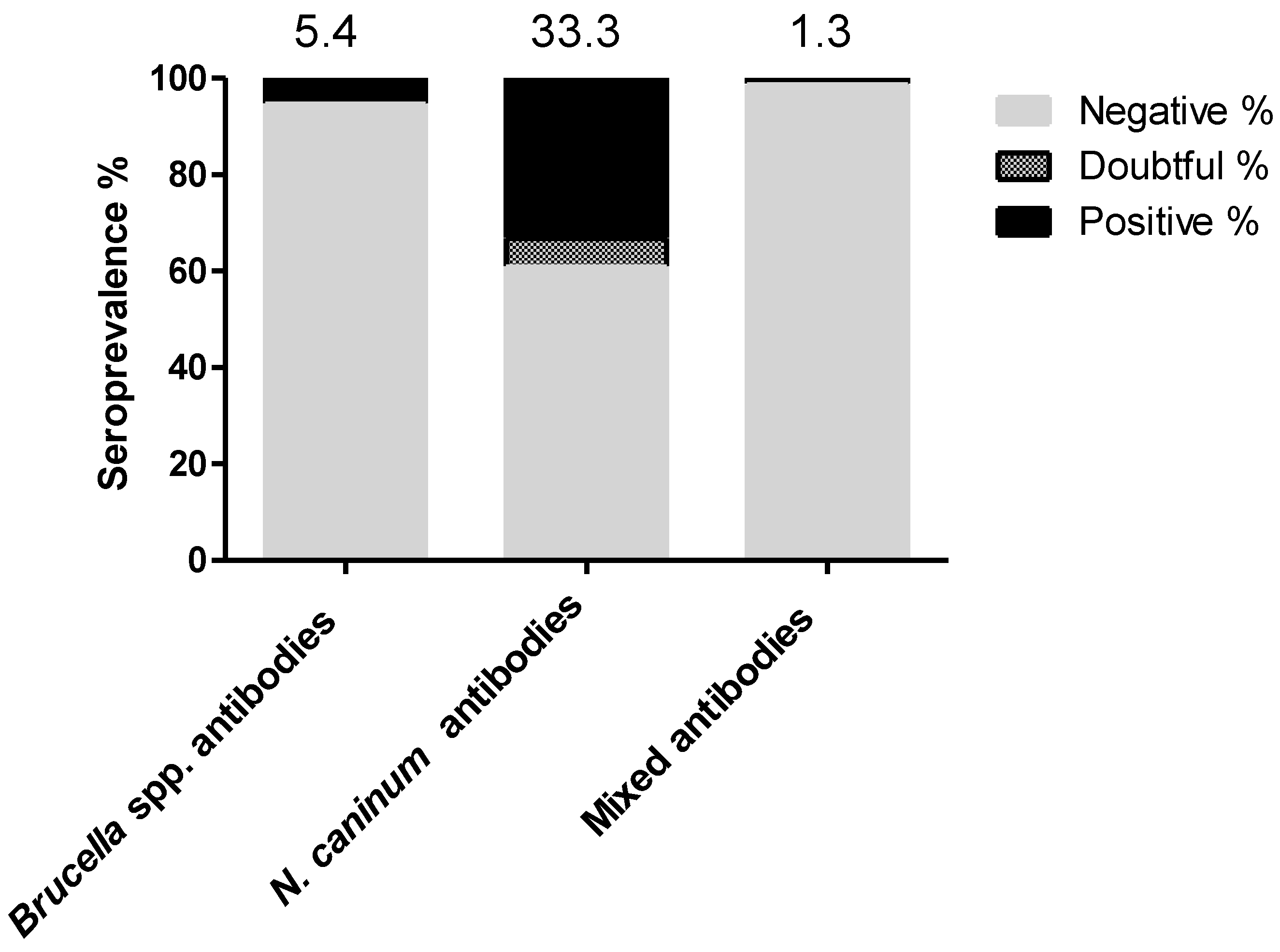
3.1. Seroprevalence Rates and Risk Factors of Brucella Species and Neospora caninum Antibodies in Tested Cows

The overall seroprevalence revealed that the seropositive rates for Brucella spp., N. caninum, and mixed antibodies among the tested cows were 5.4% (25/460; 95% confidence interval 3.6–8), 33.3% (153/460; 95% CI 29–37.8), and 1.3% (6/460; 95% CI 0.5–3), respectively (Figure 2). Doubtful results were only recorded for N. caninum antibodies (5.9%, 27/460; 95% CI 4–8.5), and they were treated as “negative” in the risk factor analysis.
The location (Kafr El Sheikh vs. Dakahlia vs. Al-Qalyubia vs. Damietta governorates) and a history of abortion (yes vs. no vs. unknown) were analyzed as risk factors of infection. Regarding Brucella, Kafr El Sheikh governorate (57.7%, p = < 0.0001) and a history of abortion (54.1%, p = < 0.0001) were considered risk factors compared to other reference factors, such as Al-Qalyubia (1.1%) and unknown abortion history (0.6%) (Table 2).
In the case of N. caninum, location was also considered a risk factor because the seropositive rates were significantly higher in Damiatta (51%, p = 0.001) and Dakahlia (33.4%, p = 0.026) compared to Kafr El Sheikh (11.3%, set as a reference). The animals without a history of abortion exhibited a higher seropositive rate for N. caninum (47.6%, p = 0.009) than those with a history of abortion (21.6%, set as reference) (Table 3).

3.2. Dynamicity of Antibody Levels and Relationship with Abortion History in Tested Cows

Firstly, we estimated the antibody levels across all test groups using ELISAs. Significant differences were observed between the negative and positive samples, both for controls and field samples, in Brucella species and N. caninum assays (p = < 0.05) (Figure 3). Additionally, for N. caninum ELISA testing, the antibody levels for the doubtful samples were markedly different from those of the negative or positive field samples, as well as the control samples (p = < 0.05) (Figure 3).
Then, we investigated the antibody levels of tested groups based on the available information on abortion history. For Brucella species, higher seroreactivity was noticed in the sera of animals that experienced abortion compared to those without or of unknown abortion history (p = < 0.05) (Figure 4A). In the case of N. caninum, however, a significantly lower seroreactivity was recorded in aborting cows compared to those without or of unknown abortion history (p = < 0.05) (Figure 4B).
Furthermore, a strong correlation (Pearson r = 0.919) between the antibody levels and abortion history was detected for Brucella spp. (Figure 5A). For N. caninum, only a moderate association (Pearson r = 0.668) was found (Figure 5B).

4. Discussion

Brucella spp. and N. caninum are notorious abortifacient infectious agents in cattle in different countries, including Egypt [2,8,14,21,22]. Both pathogens are implicated in the drastic economic losses in the cattle industry because of abortion, stillbirth, fetal abnormalities, the culling of infected animals, and loss of milk yield [6,8]. A comparative study on the seroprevalence of Brucella spp. and N. caninum and possible linkage with abortion history in cattle has not yet been conducted in Egypt. However, in our previous study, a similar approach was taken in Egypt for the serological prevalence of Brucella spp., T. gondii, and N. caninum in sheep and goats [1].
In the current study, a high seroprevalence rate for N. caninum (33.3%) was detected among the tested cattle cows in the Delta region of Egypt. The seroprevalence for Brucella spp. was lower (5.4%), but positively associated with the abortion history of the cows. Regarding the seroprevalence of Brucella spp. in cattle, our results were similar to those reported by Samaha et al. (2008) [21] in Al-Qalyubia (5.43%), one of our tested governorates, in the Delta of Egypt and Menofia: 6.43% using the Rose Bengal test (RBT). While our positive rate was lower than those detected in cow sera collected from various Egyptian regions (45.8%) [23] and lower than the rate in a group of cows suspected to be infected with brucellosis (77.2%) in the Menofia farms [24] using the RBT. Consistently, our data were higher than those recorded by Hegazy et al. (2011) [25], who reported 0.79% in cattle sera collected from Upper Egypt and 2.16% in cattle sera collected from various regions of Egypt [26]. However, previous data on the prevalences of brucellosis in farm animals and humans in Egypt, including different serological tests and molecular approaches, were already comprehensively reported in numerous reviews [14,21,27].
In the case of N. caninum, our recorded seroprevalence in cattle (33.3%) was similar to those detected by Gerges et al. (2018) (29%) [28] and El-Mohamady et al. (2022) (30.17%) [29] in cattle from various Egyptian regions where the history of abortion was available. Consistently, our detected seropositive rate was higher than those previously detected by Ibrahim et al. (2009) [30] in cattle from Sharika governorate (20.43%), by Ibrahim et al. (2021) [31] in cattle from Menoufia (14.89%), and by Fereig et al. (2016) [32] in cattle from Qena and Sohag, in southern Egypt (18.9%). However, another study detected a higher seroprevalence rate of 68% for N. caninum when the buffaloes at the Cairo slaughterhouse were tested [33]. This variability might be attributed to the differences in collection time, place, and animals, as well as the diagnostic tests used.
In addition, we analyzed the location and abortion history as influencing factors for both Brucella species and N. caninum seropositivity. Regarding Brucella species, the samples from Kafr El Sheikh showed a higher seropositivity compared to other tested locations in Egypt. This might be because these samples originated exclusively from aborting cows. Noteworthy, although the location was regarded as a risk factor for N. caninum seropositivity, the positive rate from regions without a history of abortion (Damietta; 51%) was higher than that from regions with a history of abortion (Kafr El Sheikh; 11.5%).
Furthermore, we conducted several analysis methods to validate our results and to detect the correlation between the seropositivity of Brucella species and N. caninum with the abortion history in the tested samples (Figure 3, Figure 4 and Figure 5). First, we performed a comparison between all the test sample groups and the control samples using the ELISA OD values to validate our results using field samples. The values of our positive test samples were significantly higher than those of the negative samples or negative control and were comparable to the positive controls provided by the manufacturer. These tendencies were observed for both the Brucella spp. and N. caninum results. Second, we compared the dynamicity of the antibody levels of Brucella spp. and N. caninum among different sample groups, including the confirmed existence or absence of abortion history, as well as unknown abortion history, in the collected samples. In the case of Brucella spp., a significantly higher OD value representing positive samples was observed among the samples with confirmed abortion history compared to those without or with unknown abortion history. On the contrary, a significantly higher OD value representing negative samples was observed among the samples with a confirmed abortion history compared to those without or with unknown abortion history in the case of N. caninum. Third, we analyzed the correlation coefficients using Pearson’s correlation coefficient among the OD values of the three groups (with or without an unknown abortion history). The analysis revealed that there is a strong correlation (|r| = 0.919) among the seropositivity to Brucella spp. but moderately correlated with N. caninum (|r| = 0.668) and with the confirmed abortion history. This observation was recorded in our previous similar report conducted on sheep and goats [1]. In that study, we found that the abortion history was correlated primarily with the high seropositivity to Brucella spp. but not with T. gondii or N. caninum.
In view of the lack of available data, it was not easy to compare our findings with those from similar approaches. Regardless of the comparative study, both Brucella (B. abortus and B. melitensis) and N. caninum were implicated as major causes of abortion in cattle. Meanwhile, some epidemiological studies conducted a similar approach of testing samples from cattle with or without an abortion history or in a sample population with an abortion history only, to identify independent causative agents. In this regard, many studies reported a high seropositive rate of Brucella spp. in samples from aborting cows and other ruminants in Egypt [1,15,18,34]. Similarly, for N. caninum, high seroprevalence was recorded in samples from cattle [28,29] and camels from Egyptian regions [35] with a history of abortion or reproductive problems.
Our previous and current study sought to compare a group of well-known abortifacient agents in sheep and goats [1] and cattle (this study). This approach would be valuable in spotlighting the real cause of abortion in farm animals in Egypt. Few studies have correlated the causative agent with abortion occurrence using potent serological tests, as has been reported for N. caninum [36] and Brucella spp. [14]. Serology-based detection is advantageous because it is a non-invasive method that can be used on live animals before the occurrence of abortion, which will greatly assist in the control of abortion among farm animals. This could be achieved by testing and quarantining the seropositive animals before use in breeding or before being aborted.
In addition to our previous study [1], the current study revealed Brucella spp. as the major possible cause of abortion among cattle, sheep, and goats in Egypt and highlighted the difficulty of eradicating it due to inefficient surveillance and control programs [21]. Nevertheless, this finding does not exclude N. caninum as a possible cause of abortion in Egyptian farm animals, particularly with the detection of a high seropositive rate. Further studies with a similar approach, comparing various clinically relevant pathogens and different sample groups, are required to better assess the real cause of abortion among farm animals in Egypt.

5. Conclusions

This study provides valuable information on the seroprevalence of two major abortifacient agents in cattle in Egypt, namely Brucella and N. caninum. We demonstrated a high seroprevalence among cattle from the Delta region of Egypt for N. caninum (33.3%) and a lower one for Brucella spp. (5.4%). Furthermore, the seropositivity to Brucella spp. was positively associated with a history of abortion in the sampled cows. The data provide valuable input for developing more potent policies for controlling brucellosis and neosporosis in cattle. Bovine brucellosis should also be addressed because of its possible hazards to public health and the economy.

Author Contributions

Conceptualization and design, R.M.F.; experiments, R.M.F., A.M.M., M.Z.A., M.A.O., M.E.-D., H.I.E., K.S. and G.W.; formal analysis, investigation, R.M.F.; resources and shared materials, R.M.F., A.M.M., A.S.A., M.Z.A., M.A.O., A.M.A., M.E.-D., H.I.E., K.S., C.F.F. and G.W.; writing—original draft, R.M.F., A.M.M., A.S.A., C.F.F. and G.W.; writing—review and editing, R.M.F., A.M.M., A.S.A., M.Z.A., M.A.O., A.M.A., M.E.-D., H.I.E., K.S., C.F.F. and G.W.; project administration and funding acquisition, R.M.F. All authors have read and agreed to the published version of the manuscript.

Funding

This research received no external funding.

Institutional Review Board Statement

This research was approved by the Research BioEthics Standards at Arish University, North Sinai, Egypt, and the Research Ethical Committee at Arish University ARU/Vet.02.

Informed Consent Statement

Not applicable.

Data Availability Statement

All data generated and analyzed during this study are included in this published article. Raw data supporting the findings of this study are available from the corresponding author on request.

Acknowledgments

We would like to thank our colleagues at the Department of Animal Medicine, Faculty of Veterinary Medicine, South Valley University, Qena, for their cooperation and technical assistance.

Conflicts of Interest

The authors declare no conflicts of interest.

References

  1. Fereig, R.M.; Wareth, G.; Abdelbaky, H.H.; Mazeed, A.M.; El-Diasty, M.; Abdelkhalek, A.; Mahmoud, H.Y.; Ali, A.O.; El-Tayeb, A.; Alsayeqh, A.F.; et al. Seroprevalence of specific antibodies to Toxoplasma gondii, Neospora caninum, and Brucella spp. in sheep and goats in Egypt. Animals 2022, 12, 3327. [Google Scholar] [CrossRef] [PubMed]
  2. Nayeri, T.; Moosazadeh, M.; Sarvi, S.; Daryani, A. Neospora caninum infection in aborting bovines and lost fetuses: A systematic review and meta-analysis. PLoS ONE 2022, 17, e0268903. [Google Scholar] [CrossRef] [PubMed]
  3. Ntivuguruzwa, J.B.; Kolo, F.B.; Mwikarago, E.I.; Van Heerden, H. Characterization of Brucella spp. and other abortigenic pathogens from aborted tissues of cattle and goats in Rwanda. Vet. Med. Sci. 2022, 8, 1655–1663. [Google Scholar] [CrossRef]
  4. Yang, J.; Wang, Y.; Hou, Y.; Sun, M.; Xia, T.; Wu, X. Evasion of host defense by Brucella. Cell Insight 2023, 3, 100143. [Google Scholar] [CrossRef]
  5. Alamian, S.; Amiry, K.; Bahreinipour, A.; Etemadi, A.; Tebianian, M.; Mehrabadi, M.H.F.; Dadar, M. Brucella species circulating in rural and periurban dairy cattle farms: A comparative study in an endemic area. Trop. Anim. Health Prod. 2021, 53, 200. [Google Scholar] [CrossRef] [PubMed]
  6. Mitiku, W.; Desa, G. Review of bovine brucellosis and its public health significance. Healthcare Rev. 2020, 1, 16–33. [Google Scholar] [CrossRef]
  7. Hosein, H.I.; Hamdy, M.E.; Zaitoun, A.; Menshawy, A.M.; Rouby, S.R.; Madkour, B.; Mazeed, A.M.; Abdel-Ra’ouf, A. Brucella Prevalent Strains Circulating in Egypt during 2020-2021: Bacteriological and Molecular Study. J. Vet. Med. Res. 2021, 28, 12–20. [Google Scholar] [CrossRef]
  8. Dubey, J.P.; Lindsay, D.S. A review of Neospora caninum and neosporosis. Vet. Parasitol. 1996, 67, 1–59. [Google Scholar] [CrossRef]
  9. VanLeeuwen, J.; Muraya, J.; Gitau, G.; Makau, D.; Crane, B.; McKenna, S.; Wichtel, J. Seroprevalence and risk factors of Neospora caninum and Bovine Viral Diarrhoea Virus in smallholder dairy cattle in Kenya. East Afr. J. Sci. Technol. Innov. 2021, 3, 2021. [Google Scholar] [CrossRef]
  10. Waap, H.; Bärwald, A.; Nunes, T.; Schares, G. Seroprevalence and Risk Factors for Toxoplasma gondii and Neospora caninum in Cattle in Portugal. Animals 2022, 12, 2080. [Google Scholar] [CrossRef]
  11. Tranas, J.; Heinzen, R.A.; Weiss, L.M.; McAllister, M.M. Serological evidence of human infection with the protozoan Neospora caninum. Clin. Diagn. Lab. Immunol. 1999, 6, 765–767. [Google Scholar] [CrossRef] [PubMed]
  12. Duarte, P.O.; Csordas, B.G.; Oshiro, L.M.; Higa, L.d.O.S.; Zimmermann, N.P.; Martins, K.R.; Barros, J.C.; Andreotti, R. Serological evaluation of Neospora caninum in pregnant women treated at referral center for prenatal screening in Mato Grosso do Sul, Brazil. Rev. Bras. Parasitol. Vet. 2020, 29, e010820. [Google Scholar] [CrossRef]
  13. Duarte, P.O.; Oshiro, L.M.; Zimmermann, N.P.; Csordas, B.G.; Dourado, D.M.; Barros, J.C.; Andreotti, R. Serological and molecular detection of Neospora caninum and Toxoplasma gondii in human umbilical cord blood and placental tissue samples. Sci. Rep. 2020, 10, 9043. [Google Scholar] [CrossRef] [PubMed]
  14. Wareth, G.; Hikal, A.; Refai, M.; Melzer, F.; Roesler, U.; Neubauer, H. Animal brucellosis in Egypt. J. Infect. Dev. Ctries. 2014, 8, 1365–1373. [Google Scholar] [CrossRef]
  15. Selim, A.; Attia, K.; Ramadan, E.; Hafez, Y.M.; Salman, A. Seroprevalence and molecular characterization of Brucella species in naturally infected cattle and sheep. Prev. Vet. Med. 2019, 171, 104756. [Google Scholar] [CrossRef]
  16. Selim, A.; Alshammari, A.; Gattan, H.S.; Marzok, M.; Salem, M.; Al-Jabr, O.A. Neospora caninum infection in dairy cattle in Egypt: A serosurvey and associated risk factors. Sci. Rep. 2023, 13, 15489. [Google Scholar] [CrossRef]
  17. Ran, X.; Cheng, J.; Wang, M.; Chen, X.; Wang, H.; Ge, Y.; Ni, H.; Zhang, X.X.; Wen, X. Brucellosis seroprevalence in dairy cattle in China during 2008–2018: A systematic review and meta-analysis. Acta Trop. 2019, 189, 117–123. [Google Scholar] [CrossRef] [PubMed]
  18. El-Diasty, M.; Salah, K.; El-Hofy, F.I.; Abd El Tawab, A.A.; Soliman, E.A. Investigation of an outbreak of brucellosis in a mixed dairy farm and evaluation of a test and slaughter strategy to release the herd out of the quarantine. Ger. J. Vet. Res. 2022, 2, 1–9. [Google Scholar] [CrossRef]
  19. Metwally, S.; Hamada, R.; Sobhy, K.; Frey, C.F.; Fereig, R.M. Seroprevalence and risk factors analysis of Neospora caninum and Toxoplasma gondii in cattle of Beheira, Egypt. Front. Vet. Sci. 2023, 10, 1122092. [Google Scholar] [CrossRef]
  20. Farag, S.I.; Cano-Terriza, D.; Gonzálvez, M.; Salman, D.; Aref, N.-E.M.; Mubaraki, M.A.; Jiménez-Martín, D.; García-Bocanegra, I.; Elmahallawy, E.K. Serosurvey of selected reproductive pathogens in domestic ruminants from Upper Egypt. Front. Vet. Sci. 2023, 10, 1267640. [Google Scholar] [CrossRef]
  21. Samaha, H.; Al-Rowaily, M.; Khoudair, R.M.; Ashour, H.M. Multicenter study of brucellosis in Egypt. Emerg. Infect. Dis. 2008, 14, 1916–1918. [Google Scholar] [CrossRef] [PubMed]
  22. Akinyemi, K.O.; Ajoseh, S.O.; Anjorin, A.; Salami, W.S.; Lawal, A.O.; Bassiouny, M.; Neubauer, H.; Wareth, G. A systematic scoping review of microbial pathogens in ruminants with or without a history of abortions in Nigeria. Ger. J. Vet. Res. 2023, 3, 34–51. [Google Scholar] [CrossRef]
  23. Ibrahim, A.K.; AbdelAll, A.A.; Amin, A.S. Long-term diagnostic studies for detection of Brucella spp. in milk samples. Glob. Vet. 2012, 8, 54–61. [Google Scholar]
  24. Amin, M.; Ahmed, S.; Zaki, H.; Ismail, R. Serological and molecular studies on the diagnosis of bovine brucellosis. Nat. Sci. 2012, 10, 68–76. [Google Scholar]
  25. Hegazy, Y.M.; Molina-Flores, B.; Shafik, H.; Ridler, A.L.; Guitian, F.J. Ruminant brucellosis in Upper Egypt (2005–2008). Prev. Vet. Med. 2011, 101, 173–181. [Google Scholar] [CrossRef] [PubMed]
  26. Kaoud, H.; Zaki, M.; El-Dahshan, A.; Nasr, S. Epidemiology of brucellosis among farm animals. Nat. Sci. 2010, 8, 190–197. [Google Scholar]
  27. Musallam, I.I.; Abo-Shehada, M.N.; Hegazy, Y.M.; Holt, H.R.; Guitian, F.J. Systematic review of brucellosis in the Middle East: Disease frequency in ruminants and humans and risk factors for human infection. Epidemiol. Infect. 2016, 144, 671–685. [Google Scholar] [CrossRef]
  28. Gerges, A.A.; Mona, S.; Fathi, A.; Mohamed, H.; Abou-gazia, K.H.A.; Mohamed, H.F. Detection of Neospora caninum and Coxiella burnetii antibodies in milk and serum of infected dairy cattle. Assiut Vet. Med. J. 2018, 64, 49–57. [Google Scholar]
  29. El-Mohamady, R.S.; Gerges, A.M.; Abd-Elhafeiz, Y.G.M. Investigation of the association between bovine viral diarrhea virus and Neospora caninum as a cause of abortion in cattle. J. Applied Vet. Sci. 2022, 7, 11–17. [Google Scholar] [CrossRef]
  30. Ibrahim, H.M.; Huang, P.; Salem, T.A.; Talaat, R.M.; Nasr, M.I.; Xuan, X.; Nishikawa, Y. Prevalence of Neospora caninum and Toxoplasma gondii antibodies in Northern Egypt. Am. J. Trop. Med. Hyg. 2009, 80, 263–267. [Google Scholar] [CrossRef]
  31. Ibrahim, H.M.; Abdel-Rahman, A.A.; Bishr, N.M. Seroprevalence of Neospora caninum and Toxoplasma gondii IgG and IgM antibodies among buffaloes and cattle from Menoufia Province, Egypt. J. Parasit. Dis. 2021, 45, 952–958. [Google Scholar] [CrossRef] [PubMed]
  32. Fereig, R.M.; AbouLaila, M.R.; Mohamed, S.G.; Mahmoud, H.Y.; Ali, A.O.; Ali, A.F.; Hilali, M.; Zaid, A.; Mohamed, A.E.A.; Nishikawa, Y. Serological detection and epidemiology of Neospora caninum and Cryptosporidium parvum antibodies in cattle in southern Egypt. Acta Trop. 2016, 162, 206–211. [Google Scholar] [CrossRef] [PubMed]
  33. Dubey, J.P.; Romand, S.; Hilali, M.; Kwok, O.C.H.; Thulliez, P. Seroprevalence of antibodies to Neospora caninum and Toxoplasma gondii in water buffaloes (Bubalus bubalis) from Egypt. Int. J. Parasitol. 1998, 28, 527–529. [Google Scholar] [CrossRef] [PubMed]
  34. El-Diasty, M.; El-Said, R.; Abdelkhalek, A. Seroprevalence and molecular diagnosis of sheep brucellosis in Dakahlia governorate, Egypt. Ger. J. Vet. Res. 2021, 1, 34–39. [Google Scholar] [CrossRef]
  35. Selim, A.; Abdelhady, A. Neosporosis among Egyptian camels and its associated risk factors. Trop. Anim. Health Prod. 2020, 52, 3381–3385. [Google Scholar] [CrossRef]
  36. Abdelbaky, H.H.; Nishimura, M.; Shimoda, N.; Hiasa, J.; Fereig, R.M.; Tokimitsu, H.; Inokuma, H.; Nishikawa, Y. Evaluation of Neospora caninum serodiagnostic antigens for bovine neosporosis. Parasitol Int. 2020, 75, 102045. [Google Scholar] [CrossRef]
Figure 1. Map of Egypt showing the collection sites of cattle sera from the Delta region of Egypt. The green-colored area refers to Kafr El Sheikh, the blue-colored area refers to the Dakahlia governorate, the red-colored area refers to the Damietta governorate, the orange-colored area refers to the Al-Qalyubia governorate.
Immuno 04 00024 g001
Figure 2. Seroprevalence of Brucella spp., Neospora caninum, and mixed infections antibodies in tested cattle. ELISAs were used for testing the serum samples of cattle (n = 460) for the detection of specific antibodies against selected pathogens. Values above the bars refer to the estimated seroprevalence rates.
Immuno 04 00024 g002
Figure 3. Evaluation of seroreactivity levels among positive and negative samples against Brucella spp. and N. caninum. Antibody levels indicated in ELISA optical density (OD) of positive and negative test samples and control negative and positive samples provided in the kit were compared. (A) OD among field and control samples tested by Brucella ELISA kit. (B) OD among field and control samples tested by N. caninum ELISA kit. The different letters above the bars in the graphs indicate the statistically significant differences among groups (one-way ANOVA with Tukey–Kramer post hoc analysis, p < 0.05). Samples were identified as negative or positive based on % of inhibition of the manufacturer’s instructions for each kit. Neospora ELISA is a competitive ELISA, in which high OD values mean negative serum samples.
Immuno 04 00024 g003
Figure 4. Comparison of antibody levels against Brucella spp. and N. caninum in relation to the abortion history. Antibody levels indicated in ELISA optical density (OD) of three sample groups categorized based on availability of abortion history were compared. (A) OD among samples tested by Brucella ELISA kit. (B) OD among samples tested by N. caninum ELISA kit. As this is a competitive ELISA, higher OD values mean lower Ab levels. The different letters above the bars in the graphs indicate the statistically significant differences between groups (one-way ANOVA with Tukey–Kramer post hoc analysis, p < 0.05). Yes refers to samples with known abortion history (n = 37), no refers to samples whose abortion history was not recorded (n = 84), and unknown refers to undetermined abortion history among the tested samples (n = 339). Neospora ELISA is a competitive ELISA, in which high OD values mean negative serum samples.
Immuno 04 00024 g004
Figure 5. Correlation between antibody levels and abortion history in Brucella species and Neospora caninum. Scatter graphs show the correlation between the antibody levels indicated in the mean value of ELISA optical density (OD) and the abortion history. (A) The correlation was tested for Brucella species. (B) The correlation was tested for N. caninum. As this is a competitive ELISA, higher OD values mean lower Ab levels. Group 1 = undetermined abortion history (n = 339), Group 2 = no abortion history (n = 84), and Group 3 = samples with known abortion history (n = 37). The black points refer to the mean value of all samples within the specified groups, according to the abortion history. The equation represents the approximation formula. The broken line represents the calculated line of best fit. Correlation coefficients were calculated using Pearson’s correlation coefficient: |r| = 0.70, strong correlation; 0.5 < |r| < 0.7, moderately strong correlation; and |r| = 0.3–0.5 weak-to-moderate correlation. Neospora ELISA is a competitive ELISA, in which high OD values mean negative serum samples.
Immuno 04 00024 g005
Table 1. Details of collected samples.
GovernorateAbortion History *Total
YesNoUnknown
Kafr El Sheikh260026
Dakahlia1133246290
Al-Qalyubia009393
Damietta051051
Total3784339460
* Refers to abortion history in which three categories were identified. Yes refers to cows with confirmed abortion history, no refers to cows with confirmed no history of abortion, and unknown refers to a group of cows for which abortion history was not confirmed by the owners or not registered in the documents.
Table 2. Seroprevalence of Brucella in relation to place and abortion history.
Analyzed FactorNo. of TestedNo. of Negative
(%; 95% CI)
No. of Positive
(%; 95% CI)
OR (95% CI) *p-Value *
Governorate
Kafr El Sheikh2611 (42.3; 24–62.8)15 (57.7; 37.2–76)125.5(15.1–1044)<0.0001
Dakahlia290284 (97.9; 95.4–99.2)6 (2.1; 0.8–4.7)1.9 (0.2–16.4)1.000
Al-Qalyubia 9392 (98.9; 93.3–99.9)1 (1.1; 0.1–6.7)Ref Ref
Damietta5148 (94.1; 82.8–98.5)3 (5.9; 1.5–17.2)5.7 (0.6–56.8)0.127
Total460435 (94.6; 92–96.4)25 (5.4; 3.6–8)--
Abortion history
Yes3717 (45.9; 29.9–62.9)20 (54.1; 37.1–70.1)198.2 (42.8–918.6) <0.0001
No8481 (96.4; 89.2–99.1)3 (3.6; 0.9–10.8)6.2 (1–38)0.055
Unknown339337 (99.4; 97.7–99.9)2 (0.6; 0.1–2.4)RefRef
Total460435 (94.6; 92–96.4)25 (5.4; 3.6–8)--
* The odds ratio at 95% confidence interval and the p-value were calculated by GraphPad Prism version 5. The result is significant at p < 0.05, as calculated by Fisher’s exact test. Ref: value used as a reference.
Table 3. Seroprevalence of Neospora caninum in relation to place and abortion history.
Analyzed FactorNo. of TestedNo. of Negative
(%; 95% CI)
No. of Positive
(%; 95% CI)
OR (95% CI) *p-Value *
Governorate
Kafr El Sheikh2623 (88.5; 68.7–97)3 (11.5; 3–31.3)Ref Ref
Dakahlia290193 (66.6; 60.8–72)97(33.4; 28.1–39.2)3.8 (1.1–13.2)0.026
Al-Qalyubia 9366 (71; 60.5–79.7)27 (29; 20.3–39.5)3.1 (0.9–11.3)0.079
Damietta5125 (49; 34.9–63.2)26 (51; 36.8–65.1)8 (2.1–29.9)0.001
Total460307 (66.7; 62.2–71)153 (33.3; 29–37.8)--
Abortion history
Yes3729 (78.4; 61.3–89.6)8 (21.6; 10.4–38.7)Ref Ref
No8444 (52.4; 41.3–63.3)40 (47.6; 36.7–58.7)3.3 (1.3–8)0.009
Unknown339234 (69; 63.8–73.9)105 (31; 26.1–36.3)1.6 (0.7–3.7)0.264
Total460307 (66.7; 62.2–71)153 (33.3; 29–37.8)--
* The odds ratio at 95% confidence interval and the p-value were calculated by GraphPad Prism version 5. The result is significant at p < 0.05, as calculated by Fisher’s exact test. Ref: value used as a reference.
Disclaimer/Publisher’s Note: The statements, opinions and data contained in all publications are solely those of the individual author(s) and contributor(s) and not of MDPI and/or the editor(s). MDPI and/or the editor(s) disclaim responsibility for any injury to people or property resulting from any ideas, methods, instructions or products referred to in the content.

