Article

CDX-585, a Bispecific Antibody with Dual Targeting of ILT4 and PD-1 Checkpoint Pathways

by
Michael B. Murphy
1,*,
Laura Vitale
1,
Shukai Xia
2,
Zeyu Peng
2,
Thomas O’Neill
1,
Jay Lillquist
1,
Anna Wasiuk
1,
Jeff Weidlick
1,
Jenifer Widger
1,
Laura Mills-Chen
1,
Andrea Crocker
1,
Colleen Patterson
1,
James Boyer
1,
April R. Baronas
1,
Mingjiu Chen
2,
Hugh M. Davis
2,
Mark Ma
2,
Joel Goldstein
1,
Lawrence J. Thomas
1,
Diego Alvarado
1,
Henry C. Marsh
1 and
Tibor Keler
1
add Show full author list remove Hide full author list
1
Celldex Therapeutics, Inc., Hampton, NJ 08827, USA
2
Biosion, Inc., Nanjing 210061, China
*
Author to whom correspondence should be addressed.
Immuno 2023, 3(3), 273-288; https://doi.org/10.3390/immuno3030018
Submission received: 31 May 2023 / Revised: 12 June 2023 / Accepted: 5 July 2023 / Published: 7 July 2023
(This article belongs to the Section Cancer Immunology and Immunotherapy)

Abstract

:
Immunoglobulin-like transcript 4 (ILT4) is an immunosuppressive molecule predominantly expressed on myeloid cells. Recent studies combining ILT4 suppression with programmed cell death 1 (PD-1)/programmed cell death ligand 1 (PD-L1) blockade have shown promising signs of activity in immune checkpoint inhibitor refractory patients. We theorized that coupling ILT4 and PD-1/PD-L1 blockade in a bispecific antibody (bsAb) may provide greater immune activating properties than combining the individual mAbs due to enhanced bridging of APCs to T cells. To test this approach, we developed CDX-585, a tetravalent ILT4xPD-1 IgG1-scFv bsAb from novel PD-1 and ILT-4 mAbs. CDX-585 is a potent antagonist of both PD-1 and ILT4. CDX-585 promotes M1 macrophage polarization and enhances pro-inflammatory cytokine secretion in response to lipopolysaccharide or CD40 agonist mAb treatment. In mixed lymphocyte reaction (MLR) assays, CDX-585 is more potent than the combination of parental antibodies. In a humanized NCG mouse SK-MEL-5 tumor model, CDX-585 exhibits greater antitumor activity than the combination of parental mAbs. A pilot study of CDX-585 in cynomolgus macaques confirmed a mAb-like pharmacokinetic profile without noted toxicities. These studies demonstrate that CDX-585 effectively combines ILT4 and the PD-1 blockade into one molecule that is more potent than the combination of the parental antibodies, providing the rationale to advance this bsAb into clinical studies.

1. Introduction

Immune checkpoint inhibitors (CPI), targeting the programmed cell death (PD-1)–programmed cell death ligand 1 (PD-L1) axis and cytotoxic T-lymphocyte-associated protein 4 (CTLA-4) have demonstrated significant clinical activity in various solid tumor types [1,2,3,4,5,6,7]. However, many patients fail to respond or eventually become refractory to T cell-directed checkpoint therapy. Immunosuppression from innate immune cells has been recognized as an important mechanism, leading to T cell silencing and tumor progression [8,9]. Innate mechanisms leading to tumor escape include interruption of myeloid cell development, decreasing antigen presentation, secretion of immunosuppressive cytokines, and the upregulation of checkpoint molecules [10]. Myeloid-derived suppressor cells (MDSC) serve as another important immunosuppressive mechanism [11], and their presence in the tumor microenvironment (TME) is associated with poor outcomes in cancer [12,13,14]. MDSCs have been shown to dampen T cell activation and their cytotoxic effector functions [15,16]. Additionally, tumor-associated macrophages (TAMs) promote tumor progression and metastasis by producing growth factors and proteolytic enzymes [17,18]. The functions of these immunosuppressive myeloid cells (MDSCs and TAMs) make them obvious targets for combination therapies. To further improve patient outcomes, novel approaches, such as combining a classical CPI with a myeloid CPI to drive more efficient T cell activation, merit further investigation.
Immunoglobulin-like transcript (ILT) receptors, also known as leukocyte Ig-like receptors (LIRs), comprise a family of 11 type 1 transmembrane receptors that contain extracellular Ig-like domains. ILT receptors are categorized as activating (LILRA1-6) or inhibitory (LILRB1-5) receptors that modulate the activity of both innate and adaptive immune cells [19,20,21]. The inhibitory receptors possess long cytoplasmic tails, containing immunoreceptor tyrosine inhibitory motifs (ITIM), whereas the activating receptors harbor short cytoplasmic tails that partner with immunoreceptor tyrosine activating motif (ITAM)-containing FcRγ [22]. ILT receptors are expressed on a wide range of immune cells, including dendritic cells (DCs), macrophages, B cells, T cells, neutrophils, and eosinophils, as well as non-immune cells, such as osteoclasts, neurons, and endothelial cells [23]. Inhibitory ILT family members have restrictive expression patterns, for instance, ILT2 is expressed on monocytes, DCs, B cells, NK cells, and T cells [20], while ILT4 is almost exclusively expressed by cells of the myelomonocytic lineage [24,25,26]. In the TME, ILT4 is expressed by TAMs and MDSCs, resulting in myeloid cell polarization towards immunosuppressive phenotypes, which is directly associated with tumor progression and metastasis [11,27]. Furthermore, expression of ILT4 in several different tumor types is associated with poor clinical outcomes [28,29].
Activation of the ITIM-bearing ILT4 receptor by its cognate ligands HLA-G and HLA Class I acts as an innate immune cell checkpoint, resulting in the recruitment of SHP-1/2 phosphatases, leading to the suppression of myeloid cell activation and decreased antigen presentation [30,31]. Similarly, ILT4 antagonism in the TME shifts immunosuppressive macrophages towards a M1 pro-inflammatory state, promoting T cell activation [32]. Tumor-derived ILT4 has been implicated in T cell senescence and subsequent tumor growth [33], while antagonism of ILT4 has been shown to provide antitumor effects [32]. Additionally, genetic elimination of the sole mouse ortholog, paired immunoglobulin-like receptor B (PIR-B), results in a reduced tumor burden [34]. Thus, ILT4 activation has been postulated as a resistance mechanism for the PD-1/PD-L1 blockade, and therapeutic inhibition of ILT4 in combination with PD-1 blockade has demonstrated early signs of activity in CPI-refractory patients [35].
Dual inhibition of receptors that suppress both the myeloid and T cell compartments is a promising strategy to improve outcomes for patients undergoing resistance to checkpoint inhibition and supportive early clinical data have recently been reported [35]. The rationale for combining PD-1 and the ILT4 blockade compelled us to engineer these activities into a single molecule. Here, we describe CDX-585, a novel bispecific antibody (bsAb) that simultaneously blocks ILT4 and PD-1 binding to their respective ligands. We report the generation, characterization, and functional activities of CDX-585 in both in vitro and in vivo preclinical models.

2. Materials and Methods

2.1. Development and Characterization of Bispecific Antibodies

Antibodies to ILT4 and PD-1 were generated by immunization of BALB/c mice with the extracellular domains of recombinant human ILT4 or PD-1. Splenocytes were used for hybridoma preparation by standard polyethylene glycol fusion techniques. The variable heavy and light chain regions of selected antibodies were cloned into a human IgG1κ expression vector, expressed in ExpiCHO cells (Invitrogen) and further characterized. The anti-PD-1 antibody, E1A9, and the anti-ILT4 antibody, 7B1, were humanized and selected for further development.
For the bsAb, an expression vector encoded the full-length humanized anti-PD-1 mAb E1A9 IgG1κ heavy and light chains and the single chain variable fragment (scFv) of humanized anti-ILT4 7B1 mAb genetically linked in the VL–VH orientation to the C-terminus of the E1A9 mAb heavy chain. Cysteine residues were introduced, one in the 7B1 VL and one in the 7B1 VH, to stabilize the scFv. These vectors were transfected into HD-BIOP3 cells, and proteins were purified by protein A and size exclusion chromatography. All purified mAbs and bsAbs contained <0.5 endotoxin units/mg.

2.2. Affinity Determination Using Bio-Layer Interferometry

The mAbs or bsAbs were captured on anti-human Fc capture biosensors (ForteBio, Fremont, CA, USA). Binding was determined by exposing the loaded biosensor to human PD-1-HIS or human ILT4-HIS. Affinity measurements were determined using twofold serial dilutions of analyte, ranging from 50 to 0.195 nM. The association and dissociation curves were fitted to a 1:1 binding model using the data analysis software according to the manufacturer’s guidelines.

2.3. ELISA Assays

The anti-human IgG Fc-specific polyclonal antibody (Jackson ImmunoResearch, West Grove, PA, USA) was purchased, while the extracellular domains of recombinant human ILT4, recombinant human PD-1, and soluble PD-1-mouse Fc fusion were made in-house. For ELISA, plates were coated with purified extracellular domains of target proteins overnight, and, the following day, plates were washed and blocked. Following blocking, plates were exposed to increasing concentrations of antibodies, and binding was detected using a horse radish peroxidase (HRP)-labeled goat anti-human IgG (Fc-specific) antibody and developed with 3,3,5,5-tetramethylbenzidine substrate.

2.4. Flow Cytometry

HEK-293 cells transfected with human ILT4 or PD-1 were incubated with increasing concentrations of mAbs for 20 min, and the bound antibodies were detected with a phycoerythrin (PE)-labeled goat anti-human IgG Fc-specific secondary antibody (Jackson ImmunoResearch). For bsAb binding, HEK-293 cells transfected with human ILT4 were incubated with bsAb dilutions before adding human PD-1-mouse Fc, which was detected with a PE-labeled goat anti-mouse IgG (Fc specific) antibody (Jackson ImmunoResearch). Cell-associated fluorescence was determined by analysis using a FACSCanto II™ instrument (BD Biosciences, Franklin Lakes, NJ, USA). To assess the effect of mAbs or bsAbs on ligand binding, ILT4 expressing HEK293 cells were briefly pre-incubated with the mAbs, bsAbs, or controls to provide them with an advantage to bind ILT4 in the absence of ligand, followed by the addition of fluorescently-labeled HLA-G or HLA-A2 tetramer (Fred Hutchinson, Seattle, WA, USA) and analyzed on a FACSCanto II™ instrument.

2.5. Cell-Based PD-1 Signaling Assay

The effect of the bsAb on the blockade of PD-1 signaling was performed per the manufacturer’s instructions. In short, PD-L1 expressing cells were added to a 96-well tissue culture plate and incubated at 37 °C/5% CO2 for 16 h prior to the addition of diluted antibodies and PD-1 effector cells. The PD-L1 expressing cells, antibodies, and PD-1 effector cells are incubated at 37 °C/5% CO2 for 6 h, followed by addition of Bio-Glo reagent and quantifiated on a PerkinElmer Victor X luminometer. When co-cultured with blocking antibodies, the PD-1–PD-L1 interaction is interrupted, and the subsequent T cell receptor (TCR) activation induces luminescence via the activation of a nuclear factor of activated T cell (NFAT) pathway.

2.6. Immune Cell Activation Assays

For macrophage polarization assays, human monocytes were differentiated for 6 days with 100 ng/mL M-CSF (R&D Systems, Minneapolis, MN, USA) in the presence of antibodies (6.7 nM). After differentiation, cells were activated with lipopolysaccharide (LPS) (10 ng/mL) overnight to promote a pro-inflammatory environment. Supernatant was harvested and analyzed for TNF-α and IL-10 production by ELISA (R&D Systems). The cells were stained for CD274 expression and analysis by flow cytometry.
For TNF-α induction assays, monocyte-derived macrophages (cultured with 100 ng/mL M-CSF) or DCs (cultured with 100 ng/mL GM-CSF and 10 ng/mL IL-4) were differentiated for 6 days prior to inclusion in activation assays. Following differentiation, monocyte-derived macrophages or DCs were incubated overnight with increasing concentrations of antibodies (starting at 33 nM) and LPS (10 ng/mL) or CDX-1140 (5 µg/mL). In all assays, human isotype controls were included to assess non-specific effects. The supernatant was harvested and analyzed for TNF-α production by ELISA (R&D Systems).

2.7. Human T Cell Stimulation

Ninety-six well tissue culture plates were prepared by adding 1 μg/mL anti-CD3 mAb (OKT3 eBioscience), and they were coated overnight at 4 °C. After washing the wells with PBS, 1 × 10⁵ peripheral blood mononuclear cells (PBMC) were added to each well in media and incubated overnight at 37 °C and 5% CO2, followed by the addition of test antibodies for 3 days at 37 °C and 5% CO2, and then supernatants were harvested and analyzed for IL-2 or IFN-γ production by ELISA (R&D Systems).

2.8. Mixed Lymphocyte Reaction

Human PBMCs were isolated from buffy coats using Ficoll separation, and CD4+ cells were further isolated using magnetic bead separation (Miltenyi, Gaithersburg, MD, USA). Monocyte-derived DCs were generated from PBMCs by adhering to plastic and then cultured for 7 days in RPMI medium containing 10% FBS, 10 ng/mL IL-4 plus 100 ng/mL GM-CSF. Cells were harvested and confirmed to be 80% DCs by expression of CD11c. The CD4+ cells and DCs from allogeneic donors were co-incubated at a 10:1 ratio in the presence of LPS (10 ng/mL) or CDX-1140 (0.5 µg/mL), with mAb or bsAb, for 4 days. Supernatants were harvested and analyzed for IL-2 or IFN-γ production by ELISA (R&D Systems).

2.9. Mouse Xenograft Tumor Model

For the xenograft melanoma model, thirty female CD34+ cord blood-engrafted humanized NCG mice (Charles River Laboratories, Wilmington, MA, USA) were implanted subcutaneously with 2.0 × 10⁷ SK-MEL-5 tumor cells (American Type Culture Collection), followed by weekly treatment with human IgG1 (25 mg/kg), anti-ILT4 mAb 7B1 (18.75 mg/kg), anti-PD-1 mAb E1A9 (18.75 mg/kg), a combination of 7B1 (18.75 mg/kg) and E1A9 (18.75 mg/kg), or CDX-585 (25 mg/kg) for five weeks. Tumors were measured by caliper three times per week, and mice were euthanized according to pre-defined endpoint criteria. All studies were performed in accordance with IACUC-approved protocols, consistent with “The Guide for the Care and Use of Laboratory Animals (8th Edition, The National Academies Press, Washington, DC, USA). Study code 08-2020, approved, August 2020”.

2.10. Non-Good Laboratory Practice (GLP) Pilot Non-Human Primate Study

Three cynomolgus monkeys (one male, two females) received a single 10 mg/kg slow bolus intravenous (i.v.) injection (2–3 min) of CDX-585, via a cephalic catheter. Animals were followed for 21 days. Evaluations included clinical signs, body temperature, clinical pathology parameters (hematology, coagulation, clinical chemistry, and urinalysis), and toxicokinetic parameters. Body weights were recorded once prior to bsAb administration and weekly thereafter. This was designed as a survival study with no planned necropsy. Blood samples were assayed for circulating levels of cytokines/chemokines using custom plates (MesoScale Discovery, Rockville, MD, USA). All studies were performed in accordance with IACUC-approved protocols, consistent with “The Guide for the Care and Use of Laboratory Animals (8th Edition, The National Academies Press) and the U.S. Animal Welfare Act. Study Code CE2101, approved May 2021”.

2.11. Statistical Analysis

Statistical significance was evaluated using two-way ANOVA or the paired Student’s t-test, as appropriate.

3. Results

3.1. Characterization of Novel ILT4 and PD-1 Antibodies

Monoclonal antibodies against ILT4 or PD-1 were generated by immunizing mice with recombinant human ILT4 or PD-1 proteins and standard hybridoma technology. The ILT4 mAb designated 7B1 was humanized and selected for the development of the bsAb CDX-585, based on its functional characteristics. As shown in Figure 1, 7B1 is specific for the ILT4 receptor and does not bind other ILT family members with significant similarity to ILT4 (Figure 1a). Additionally, the 7B1 mAb bound well to naturally expressed ILT4 on human myeloid cells, including monocytes, macrophages, and DCs (Supplementary Figure S1). Importantly, 7B1 blocked binding of the tetramerized HLA-G ligand to ILT4-expressing cells with low nanomolar potency (Figure 1b), blocked HLA-A2 binding (Figure 1c), and promoted TNF-α secretion from monocyte-derived macrophages primed with LPS, similar to Merck’s anti-ILT4 mAb 1E1 (G4), which we believe to be MK-4830 (US20180298096A1) (Figure 1d). These data indicate that 7B1 can block ILT4 binding to its cognate ligands, resulting in an enhanced pro-inflammatory phenotype.
The E1A9 PD-1 mAb was humanized and selected for use in the bsAb, based on its functional characteristics. E1A9 mAb and benchmark control nivolumab showed comparable concentration-dependent binding to immobilized purified PD-1 extracellular region by ELISA (Figure 1e) and to a HEK-293 cell line expressing PD-1 by FACS (Figure 1f). The E1A9 mAb and nivolumab demonstrated a similar concentration-dependent ability to block the PD-1/PD-L1 signaling pathway using a T cell receptor (TCR)-mediated luminescence system (Figure 1g). Additionally, the E1A9 mAb was able to block PD-L2 binding to PD-1 (Figure 1h). These data indicate E1A9 effectively binds PD-1 and blocks its interaction with PD-L1 with low nanomolar affinity.

3.2. Development of the CDX-585 bsAb

We selected a whole IgG1-scFv format (Figure 2a) for the CDX-585 bsAb construct with bivalent binding to both ILT4 and PD-1, which maintains high affinity binding, similar to that of the parental mAbs. CDX-585 has a modified IgG1 constant region, including L234A/L235Q/K322Q (AQQ) and M252Y/S254T/T256E (YTE) mutations, which were introduced to eliminate Fcγ receptor (FcγR) interactions, effector function [36], and C1q receptor binding [37], while promoting neonatal Fc receptor (FcRn) binding, leading to improved pharmacokinetics (PK) by reducing the rate of in vivo clearance [38], respectively. CDX-585 encodes the full-length E1A9 mAb and the scFv of 7B1 genetically linked in the VL–VH orientation to the C-terminus of the E1A9 mAb heavy chain. We also made the bispecific construct with the 7B1 scFv in the VH–VL orientation, but it exhibited lower activity in bifunctional binding assays and was, therefore, not further pursued.
Analytical characterization was performed on CDX-585 purified from CHO cells stably transfected with the expression construct. Reducing SDS-PAGE of CDX-585 showed the expected greater molecular weight of the heavy chain with the genetically-fused scFv (approximately 75 kDa) (Figure 2b). Size exclusion chromatography (SEC) by HPLC revealed the bsAb product to be >95% monomer, with an estimated size of approximately 200 kDa, which is consistent with the expected mass of the CDX-585 construct (Figure 2c). The bsAb retained high affinity binding to recombinant human and cynomolgus PD-1, yet CDX-585 had weaker affinity to cynomolgus ILT4 compared to human ILT4, as evaluated by bio-layer interferometry (Supplementary Figure S2a,b). However, similar direct binding saturation could be reached using human and cynomolgus monocytes (Supplementary Figure S2c). CDX-585 also showed binding to the neonatal FcRn under low pH conditions, which were consistent with the engineered YTE modifications (Supplementary Figure S2d). Simultaneous binding to ILT4 and PD-1 by CDX-585 was observed using cells overexpressing ILT4 antigen and soluble PD-1 (Figure 2d).

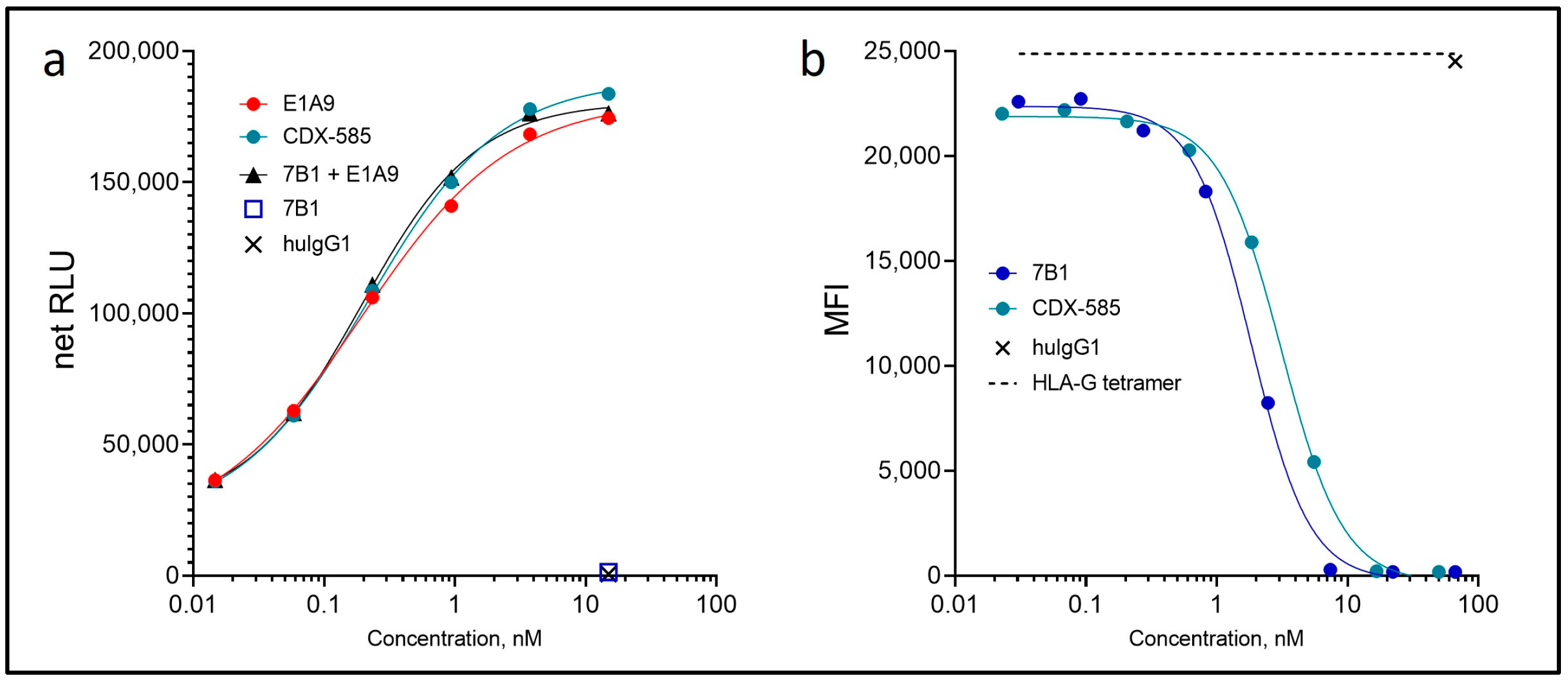
3.3. CDX-585 Potently Inhibits PD-1 Signaling and HLA-G Ligand Binding

Inhibition of PD-1/PD-L1 signaling by CDX-585 was determined using a cell-based reporter assay in which PD-1 effector cells and PD-L1 APCs were co-cultured in the presence of titrated antibodies. CDX-585 and E1A9 inhibited PD-1 reporter activation with IC50 values of 0.21 nM and 0.19 nM, respectively (Figure 3a). Additionally, CDX-585 and 7B1 effectively blocked fluorescently-labeled HLA-G tetramer from binding to HEK-293 cells overexpressing ILT4, with IC50 values of 3.12 nM and 1.83 nM, respectively (Figure 3b).

3.4. CDX-585 Drives M1 Macrophage Polarization

The ILT4 blockade has been reported to induce M1 macrophage polarization and promote a pro-inflammatory microenvironment [32]. To test whether CDX-585 can induce an inflammatory macrophage phenotype, we differentiated human donor monocytes into mature macrophages in the presence of CDX-585 or parental mAbs. Following differentiation, monocyte-derived macrophages were activated with LPS overnight to induce a pro-inflammatory phenotype, and cell-conditioned media were collected the following day to assess pro- and anti-inflammatory cytokine production by ELISA. CDX-585 enhanced TNF-α secretion (Figure 4a) and led to a marked inhibition of IL-10 production to a similar extent as the parental mAb 7B1 (Figure 4b). Further, CDX-585 treatment resulted in decreased PD-L1 (CD274) cell surface expression (Figure 4c), as assessed by flow cytometry. These data indicate that the blockade of ILT4 with CDX-585 during cellular differentiation drives macrophages towards a M1 pro-inflammatory phenotype.

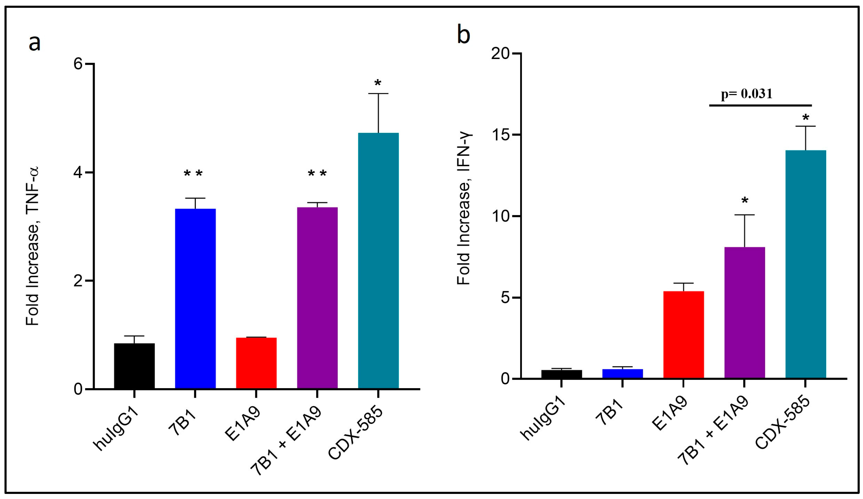
3.5. CDX-585 Enhances Both Myeloid and T Cell Activation

We next looked at the ability of CDX-585 to shift the inflammatory phenotype of already differentiated macrophages and DCs. Monocyte-derived macrophages or DCs were incubated overnight with titrated CDX-585 or parental mAbs in the presence of LPS. CDX-585 induced a significant increase in TNF-α production in both macrophages (Figure 4d) and DCs (Figure 4e) compared to isotype controls, further suggesting that CDX-585 treatment promotes a pro-inflammatory phenotype in myeloid cells. Additionally, human PBMCs pre-incubated overnight with a suboptimal dose of the TCR-stimulatory mAb OKT3, resulting in enhanced INF-γ secretion with the subsequent addition of CDX-585, indicating its ability to enhance T cell activation in vitro (Figure 4f). These data indicate that a dual blockade of ILT4 and PD-1 drives inflammatory responses in both myeloid and T cell populations.

3.6. CDX-585 Enhances T Cell Activation in Mixed Lymphocyte Reactions

We further assessed the ability of CDX-585 to directly influence the T cell compartment using a mixed lymphocyte reaction (MLR) assay. CD4+ T cells were co-cultured with LPS-activated allogeneic DCs for four days (Figure 5). Addition of the ILT4 antagonist mAb 7B1 did not significantly increase T cell activation, as measured by INF-γ and IL-2 secretion, whereas the anti-PD-1 mAb E1A9 enhanced T cell responses by preventing PD-1 mediated suppression of T cell activation. CDX-585 significantly induced more IFN-γ and IL-2 production from T cells than a single-agent blockade with 7B1 or E1A9 mAbs alone or the combination of the 7B1 and E1A9 mAbs. Interestingly, relieving both myeloid and T cell checkpoints via CDX-585 led to the synergistic activation of CD4+ T cells in vitro in most donor pairs we tested, although, in some donor pairs, the MLR was largely driven by the PD-1 blockade, and CDX-585 showed equal activity to parental mAbs (data not shown).

3.7. CDX-585 Potentiates CD40-Mediated Immune Cell Activation

Ligation of CD40-expressing DCs with its ligand CD40L (CD154) on CD4+ helper T cells leads to DC-induced priming and activation of CD8+ effector T cells [39,40]. CD40-activated macrophages can be tumoricidal [41]. Therefore, CD40, expressing APCs, can play an important role in effective immune responses. We sought to determine whether CDX-585 could enhance CD40-mediated immune cell activation in a manner similar to Toll-like receptor 4 (TLR4) agonism with LPS, which is typically used to demonstrate the pro-inflammatory effects of the ILT4 blockade, but it is less relevant in a clinical setting. To promote CD40 signaling, we used the previously described CD40 agonist mAb CDX-1140 [42]. In monocyte-derived DCs primed with CDX-1140, CDX-585 significantly increased TNF-α production (Figure 6a), and they had the ability to activate allogeneic co-cultured CD4+ T cells, as shown by the enhanced IFN-γ production (Figure 6b). Monocyte-derived macrophages also demonstrated enhanced TNF-α secretion by CDX-585 when primed with CDX-1140. These data indicate that APCs primed with CDX-1140 have enhanced pro-inflammatory effects when combined with CDX-585 treatment, similar to what is observed with LPS.

3.8. CDX-585 Enhances Antitumor Activity in a SKMEL-5 Mouse Model

To investigate whether CDX-585 can drive anti-tumor activity in vivo, we tested its activity in a SK-MEL-5 melanoma tumor model using CD34+ cord blood-engrafted humanized NCG mice (CD34-NCG). Starting on the day following tumor implantation, mice were administered human IgG1 isotype control, 7B1, E1A9, 7B1 and E1A9, or CDX-585, once a week, for five weeks. Beginning approximately three weeks after implantation, co-inhibition of ILT4 and PD-1 receptors via CDX-585 led to a statistically enhanced decrease in tumor growth of SK-MEL-5 tumors compared to the single agent or combination arms (Figure 7). By day 35, CDX-585 treatment resulted in a 56% decrease in tumor volume compared to isotype control treated mice, while 7B1 (25%), E1A9 (11%), or their combination (31%) treatments failed to reach statistically significant tumor volume reductions.

3.9. Non-GLP Pilot Study in Non-Human Primates

A pilot study was performed with a single 10 mg/kg slow bolus i.v. injection of CDX-585 in 3 non-naïve cynomolgus monkeys (one male, two females) to generate preliminary safety and PK data. CDX-585 was well tolerated in the study. There were no CDX-585-related clinical observations, including body weights and body temperature. Clinical chemistry and hematological parameters remained unchanged or were considered non-adverse. PK analysis was performed on samples up to day 20 (Figure 8a). Blood samples were assayed for circulating levels of monocyte chemoattractant protein (MCP-1), macrophage inflammatory protein (MIP)-1β, macrophage-derived chemokine (MDC), along with six other chemokines (eotaxin, thymus, and activation-regulated chemokine (TARC), interferon γ-induced protein (IP)-10, MIP-1α, IL-8, and MCP-4). During sample analysis, it was discovered that one female animal had pre-existing anti-drug antibodies and exhibited rapid drug clearance; this animal was removed from the analysis. Data from the remaining two individual animals demonstrated a clear transient increase in MCP-1 and MIP-1β and sustained increase in MDC over 8 h post dosing, indicative of myeloid cell activation (Figure 8b). The PK and safety data from the pilot non-human primate study were assessed prior to initiation of a full toxicology study. A repeat dose non-human primate GLP-compliant toxicology study in cynomolgus monkeys supported a good safety and PK profile, with a non-observed adverse effect level (NOAEL) at the highest dose tested at 60 mg/kg/dose.

4. Discussion

While T cell checkpoint therapies have demonstrated remarkable activity in several solid tumors, immunosuppression from tumor-infiltrating myeloid cells has been recognized as an important mechanism of resistance. Dual targeting of both myeloid and T cell checkpoints has been explored as a promising approach for overcoming these roadblocks. ILT4 is a well described ITIM-containing receptor that acts as a myeloid cell checkpoint and has been shown preclinically to restrict myeloid cell activation, particularly in macrophages, MDSCs, and DCs. Furthermore, anti-ILT4 mAbs have demonstrated early clinical efficacy, particularly in combination with PD-1 antagonists without adding toxicity [35]. Based on the strong rationale for dual ILT4/PD-1 inhibition, we designed a bsAb, designated CDX-585, to simultaneously block both axes with a single molecule.
CDX-585 was developed as a tetravalent IgG1–scFv construct that exhibited good pharmacological and biophysical properties, and in vitro activity comparable to its parental mAbs. CDX-585 retained high-affinity binding and blockade of both ILT4 and PD-1 and could simultaneously engage both receptors, as demonstrated by bifunctional ELISAs and cell bindings assays. CDX-585 effectively inhibited ligand–receptor interaction for both ILT4 (HLA-G and HLA-A2) and PD-1 (PD-L1/L2). CDX-585 was designed to abrogate FcγR binding to prevent effector functionality and reduce the likelihood of potentially harmful effector-induced antibody-dependent cellular cytotoxicity or phagocytosis against PD-1 expressing T cells, while enhancing FcRn binding to promote PK, both of which were confirmed by bio-layer interferometry.
Efficient blockade of the innate immune cell checkpoint ILT4 has previously been shown to promote M1 macrophage polarization and a pro-inflammatory microenvironment [32,33], which can suppress tumor growth [43,44,45]. We set out to understand the effect CDX-585 has on macrophage polarization. Using human donor monocytes, we demonstrated the differentiation of monocytes into macrophages in the presence of CDX-585, resulting in a more robust M1 phenotype. When pre-stimulated with LPS, CDX-585 differentiated macrophages increased TNF-α secretion, decreased IL-10 secretion, and decreased PD-L1 (CD274) cell surface expression. Classical M1 polarization agonists, such as the TLR4 ligand LPS, resulted in the upregulation of both pro- and anti-inflammatory cytokines [46]. However, CDX-585 exerts a dual effect on local macrophages, leading to the potent upregulation of pro-inflammatory molecules and marked inhibition of immunosuppressive molecules. Importantly, we further demonstrated that ILT4 blockade in the context of a more biologically relevant mechanism of myeloid cell activation, through CD40 agonism, can further enhance the pro-inflammatory phenotype of these cells and lead to improved antitumor activity. Additionally, these data suggest that the additive effect exerted by CDX-585 may be applied in combinations with other pro-inflammatory stimuli in the clinic.
ILT4 partners with a wide range of classical MHC class I ligands, including HLA-A, -B, -C, as well as non-classical ligands, such as HLA-F, -G, angiopoietin-like protein 2, MAG, and Nogo66 [47]. Unlike many ligands, MHC class I is constitutively expressed on the cell surface of all nucleated cells, and ILT4 has been shown to compete with CD8 for HLA-A binding, resulting in the perpetual immunosuppression of the myeloid cell population [48]. With regards to this, we further showed that CDX-585 can modulate the inflammatory signature of differentiated innate immune cells that have been exposed to constitutively expressed ligands in culture. Monocyte-derived macrophages or DCs co-cultured with LPS and CDX-585 resulted in a marked increase in TNF-α secretion compared to cells treated with parental mAbs alone or in combination. These results indicate that CDX-585 can shift the inflammatory signature of previously differentiated myeloid cells to a more M1-like state. Further, IFN-γ has been shown to play a direct role in promoting a pro-inflammatory phenotype by antagonizing IL-10 [49,50] and TGF-β [51], inhibiting Treg differentiation [52,53], and promoting apoptosis of human cancer cells [54]. PBMCs sub-optimally primed with OKT3 and cocultured with CDX-585 resulted in a significant increase in IFN-γ secretion. Collectively, these data indicate that the dual blockade of ILT4 and PD-1 checkpoints via CDX-585 promotes a robust pro-inflammatory phenotype for both the myeloid and T cell compartments.
Previous reports have shown that treatment with PD-(L)1 inhibitors led to enhanced T cell activation in MLR assays [55,56,57,58] and that this can be potentiated by the ILT4 blockade [32]. Indeed, in our MLR assays, combined ILT4 and PD-1 mAbs led to significantly greater T cell activation than the individual ILT4 and PD-1 mAbs. Surprisingly, we observed a significant increase in MLR activity with CDX-585 relative to the combination of ILT4 and PD-1 mAbs, suggesting that co-engagement of the receptors with CDX-585 resulted in greater T cell activation. The enhanced activity of CDX-585 relative to parental mAbs may be attributed to more effective bridging of APCs and T cells, contributing to a more productive immune synapse formation. In support of this theory, a separate bsAb targeting PD-L1xILT4, which could primarily target APCs, did not demonstrate this enhanced effect over the combined mAb approach in our MLR assays (data not shown). Further, this enhanced activity was reduced at high CDX-585 concentrations (100 nM). Additionally, we discovered donor pairs influenced CDX-585 synergistic activity in these MLR assays. The mechanisms underlying this discrepancy are still unclear and may include differences in target expression or MHC haplotype. These data suggest that target expression could be a consideration for choosing patient populations in the clinic and, even so, there might be a narrow treatment window to achieve enhanced activity of CDX-585 over combination treatments. While CDX-585 does not need to co-engage both targets to be effective, this co-engagement mechanism might improve upon the clinical efficacy of mAb monotherapy or combination treatments, where these types of interactions are not expected to occur.
While relief of TAM/MDSC immunosuppression can lead to improved antitumor activity, other innate mechanisms can lead to tumor escape. For example, DCs can downregulate the expression of MHC Class I molecules, masking them from CD8+-infiltrating T cells [59] or through the upregulation of PD-L1 to inhibit T cell effector function [60]. An important function of CD40 with its ligand CD40L (CD154) is to license DCs to prime CD8+ effector T cells. We have previously shown that CD40 activation with the anti-CD40 agonist mAb, CDX-1140, promotes DC licensing, B cell proliferation, and CD95 (FAS) expression, leading to increased antitumor activity [42]. Combining the activities of DC licensing with both myeloid and T cell checkpoint inhibition would further polarize the pro-inflammatory microenvironment, potentially leading to improved antitumor activity. We tested this hypothesis by combining CDX-585 with cells primed by the CD40 agonist, CDX-1140, and demonstrated increased immune activation and antitumor activity by combining DC licensing with macrophage re-polarization and T cell checkpoint inhibition. Additionally, it is plausible that the ILT4 blockade, through CDX-585, frees up HLA-A to bind to CD8+ T cells, improving T cell mediated effector functions [61]. These results may provide the basis for simultaneous targeting of DCs, TAM/MDSCs, and T cells in a clinical setting.
To study in vivo activity of CDX-585, we utilized humanized CD34-NCG mice. These humanized mice were implanted with human melanoma SK-MEL-5 tumor cells, which express ILT4 and PD-(L)1 [62,63]. CDX-585 provided a superior response over the combination of ILT4 and PD-1 mAbs in this SK-MEL-5 melanoma model, consistent with the effect observed in in vitro co-culture assays. These data indicate treatment with CDX-585 promotes tumor volume reduction through direct inhibition of both ILT4 and PD-1.
Incorporation of an intact human IgG1 Fc domain, including YTE point mutations, promoted mAb-like PK through enhanced FcRn binding. In cynomolgus monkeys, we discovered transient increases in pro-inflammatory serum cytokine levels, consistent with the expected mechanism of CDX-585. Additionally, no obvious toxicity was observed. These data support direct activation of innate immune pathways in vivo by CDX-585. One of the animals developed strong antidrug antibodies (ADA) that resulted in rapid drug clearance that hampered longer term evaluation. A repeat dose non-human primate GLP-compliant toxicology study in cynomolgus monkeys supported a good safety and PK profile, with a NOAEL at the highest dose tested at 60 mg/kg/dose.
Along with current clinical data, collectively, these data support CDX-585 as a promising approach to enhance the clinical activity of the PD-1 blockade. Currently, a clinical study has been opened, evaluating CDX-585 in treating patients with solid tumors (NCT05788484). Engineering ILT4 and PD-1 blockade into a single molecule will provide us additional flexibility to effectively target more than two modalities at once. Therefore, in our clinical development strategy, CDX-585 is poised to be used in combination treatments with other therapeutic modalities, such as CD40 agonists, chemotherapy, or radiation, which may provide additional benefit to patients.

Supplementary Materials

The following supporting information can be downloaded at: https://www.mdpi.com/article/10.3390/immuno3030018/s1, Figure S1: Binding of 7B1 to primary immune cells. Figure S2: Binding affinity of CDX-585 to human and cynomolgus PD-1 and ILT4.

Author Contributions

Conceptualization, T.K., J.G., H.C.M., D.A. and L.V.; methodology, T.K., J.G., H.C.M., D.A., L.V., M.B.M. and L.J.T.; validation, T.K., J.G., H.C.M., D.A., L.V., M.B.M. and L.J.T.; formal analysis, T.K., J.G., H.C.M., D.A, L.J.T., M.B.M., L.V., T.O., J.L., A.W., J.W. (Jeff Weidlick), J.W. (Jenifer Widger), L.M.-C., A.C., C.P., J.B. and A.R.B.; investigation M.B.M., L.V., T.O., J.L., A.W., J.W. (Jeff Weidlick), J.W. (Jenifer Widger), L.M.-C., A.C., C.P., J.B. and A.R.B.; writing original draft, M.B.M., D.A. and T.K.; writing review and editing, M.B.M., D.A., T.K., L.V., J.G., H.C.M., L.J.T., A.W., S.X., Z.P., M.C., H.M.D. and M.M. All authors have read and agreed to the published version of the manuscript.

Funding

This work was funded by Celldex Therapeutics, Inc.

Institutional Review Board Statement

The animal study protocol was approved by the Institutional Animal Care and Use Committee (IACUC) of Celldex Therapeutics or Envol Biomedical (study code 08-2020, approved, August 2020; study Code CE2101, approved May 2021).

Informed Consent Statement

Not applicable.

Data Availability Statement

The data presented in this study are available upon request from the corresponding author.

Conflicts of Interest

The authors declare no conflict of interest. M.B.M, L.V., T.O., J.L., L.M., A.W., J.W., J.W., L.M.-C., A.C., C.P., J.B., A.R.B., R.A.H., J.G., L.J.T., D.A., H.C.M. and T.K. are full time employees of Celldex Therapeutics. S.X., Z.P., M.C., H.M.D. and M.M. are full-time employees of Biosion Inc.

References

  1. Solinas, C.; Gombos, A.; Latifyan, S.; Piccart-Gebhart, M.; Kok, M.; Buisseret, L. Targeting Immune Checkpoints in Breast Cancer: An Update of Early Results. ESMO Open 2017, 2, e000255. [Google Scholar] [CrossRef] [Green Version]
  2. Rini, B.I.; Battle, D.; Figlin, R.A.; George, D.J.; Hammers, H.; Hutson, T.; Jonasch, E.; Joseph, R.W.; McDermott, D.F.; Motzer, R.J.; et al. The Society for Immunotherapy of Cancer Consensus Statement on Immunotherapy for the Treatment of Advanced Renal Cell Carcinoma (RCC). J. Immunother. Cancer 2019, 7, 354. [Google Scholar] [CrossRef] [Green Version]
  3. Haslam, A.; Prasad, V. Estimation of the Percentage of US Patients With Cancer Who Are Eligible for and Respond to Checkpoint Inhibitor Immunotherapy Drugs. JAMA Netw. Open 2019, 2, e192535. [Google Scholar] [CrossRef] [Green Version]
  4. Sharma, P.; Allison, J.P. The Future of Immune Checkpoint Therapy. Science 2015, 348, 56–61. [Google Scholar] [CrossRef]
  5. Carlino, M.S.; Larkin, J.; Long, G.V. Immune Checkpoint Inhibitors in Melanoma. Lancet 2021, 398, 1002–1014. [Google Scholar] [CrossRef]
  6. Lopez-Beltran, A.; Cimadamore, A.; Blanca, A.; Massari, F.; Vau, N.; Scarpelli, M.; Cheng, L.; Montironi, R. Immune Checkpoint Inhibitors for the Treatment of Bladder Cancer. Cancers 2021, 13, 131. [Google Scholar] [CrossRef]
  7. Onoi, K.; Chihara, Y.; Uchino, J.; Shimamoto, T.; Morimoto, Y.; Iwasaku, M.; Kaneko, Y.; Yamada, T.; Takayama, K. Immune Checkpoint Inhibitors for Lung Cancer Treatment: A Review. J. Clin. Med. 2020, 9, 1362. [Google Scholar] [CrossRef]
  8. Jenkins, R.W.; Barbie, D.A.; Flaherty, K.T. Mechanisms of Resistance to Immune Checkpoint Inhibitors. Br. J. Cancer 2018, 118, 9–16. [Google Scholar] [CrossRef] [Green Version]
  9. Bonavida, B.; Chouaib, S. Resistance to Anticancer Immunity in Cancer Patients: Potential Strategies to Reverse Resistance. Ann. Oncol. Off. J. Eur. Soc. Med. Oncol. 2017, 28, 457–467. [Google Scholar] [CrossRef]
  10. Kim, S.K.; Cho, S.W. The Evasion Mechanisms of Cancer Immunity and Drug Intervention in the Tumor Microenvironment. Front. Pharmacol. 2022, 13, 868695. [Google Scholar] [CrossRef]
  11. Fleming, V.; Hu, X.; Weber, R.; Nagibin, V.; Groth, C.; Altevogt, P.; Utikal, J.; Umansky, V. Targeting Myeloid-Derived Suppressor Cells to Bypass Tumor-Induced Immunosuppression. Front. Immunol. 2018, 9, 398. [Google Scholar] [CrossRef] [Green Version]
  12. Ai, L.; Mu, S.; Wang, Y.; Wang, H.; Cai, L.; Li, W.; Hu, Y. Prognostic Role of Myeloid-Derived Suppressor Cells in Cancers: A Systematic Review and Meta-Analysis. BMC Cancer 2018, 18, 1220. [Google Scholar] [CrossRef] [Green Version]
  13. Wang, J.; Yang, J. Identification of CD4+CD25+CD127− Regulatory T Cells and CD14+HLA−DR−/Low Myeloid-Derived Suppressor Cells and Their Roles in the Prognosis of Breast Cancer. Biomed. Rep. 2016, 5, 208–212. [Google Scholar] [CrossRef] [Green Version]
  14. Yuan, L.; Yuan, P.; Du, J.; Chen, G.; Wan, X.; Li, Z.; Xu, B. Relationship of preoperative and postoperative myeloid-derived suppressor cells percentage with the prognosis in rectal cancer patients. Zhonghua Wei Chang Wai Ke Za Zhi Chin. J. Gastrointest. Surg. 2015, 18, 1139–1143. [Google Scholar]
  15. Adah, D.; Hussain, M.; Qin, L.; Qin, L.; Zhang, J.; Chen, X. Implications of MDSCs-Targeting in Lung Cancer Chemo-Immunotherapeutics. Pharmacol. Res. 2016, 110, 25–34. [Google Scholar] [CrossRef]
  16. Monu, N.R.; Frey, A.B. Myeloid-Derived Suppressor Cells and Anti-Tumor T Cells: A Complex Relationship. Immunol. Investig. 2012, 41, 595–613. [Google Scholar] [CrossRef] [Green Version]
  17. Komohara, Y.; Jinushi, M.; Takeya, M. Clinical Significance of Macrophage Heterogeneity in Human Malignant Tumors. Cancer Sci. 2014, 105, 1–8. [Google Scholar] [CrossRef] [Green Version]
  18. Ruffell, B.; Coussens, L.M. Macrophages and Therapeutic Resistance in Cancer. Cancer Cell 2015, 27, 462–472. [Google Scholar] [CrossRef] [Green Version]
  19. Wagtmann, N.; Rojo, S.; Eichler, E.; Mohrenweiser, H.; Long, E.O. A New Human Gene Complex Encoding the Killer Cell Inhibitory Receptors and Related Monocyte/Macrophage Receptors. Curr. Biol. 1997, 7, 615–618. [Google Scholar] [CrossRef] [Green Version]
  20. Colonna, M.; Navarro, F.; Bellón, T.; Llano, M.; García, P.; Samaridis, J.; Angman, L.; Cella, M.; López-Botet, M. A Common Inhibitory Receptor for Major Histocompatibility Complex Class I Molecules on Human Lymphoid and Myelomonocytic Cells. J. Exp. Med. 1997, 186, 1809–1818. [Google Scholar] [CrossRef] [Green Version]
  21. Allan, D.S.; Colonna, M.; Lanier, L.L.; Churakova, T.D.; Abrams, J.S.; Ellis, S.A.; McMichael, A.J.; Braud, V.M. Tetrameric Complexes of Human Histocompatibility Leukocyte Antigen (HLA)-G Bind to Peripheral Blood Myelomonocytic Cells. J. Exp. Med. 1999, 189, 1149–1156. [Google Scholar] [CrossRef] [Green Version]
  22. Lewis Marffy, A.L.; McCarthy, A.J. Leukocyte Immunoglobulin-Like Receptors (LILRs) on Human Neutrophils: Modulators of Infection and Immunity. Front. Immunol. 2020, 11, 857. [Google Scholar] [CrossRef]
  23. Tedla, N.; Gibson, K.; McNeil, H.P.; Cosman, D.; Borges, L.; Arm, J.P. The Co-Expression of Activating and Inhibitory Leukocyte Immunoglobulin-like Receptors in Rheumatoid Synovium. Am. J. Pathol. 2002, 160, 425–431. [Google Scholar] [CrossRef] [Green Version]
  24. Colonna, M.; Samaridis, J.; Cella, M.; Angman, L.; Allen, R.L.; O’Callaghan, C.A.; Dunbar, R.; Ogg, G.S.; Cerundolo, V.; Rolink, A. Human Myelomonocytic Cells Express an Inhibitory Receptor for Classical and Nonclassical MHC Class I Molecules. J. Immunol. 1998, 160, 3096–3100. [Google Scholar] [CrossRef]
  25. Sloane, D.E.; Tedla, N.; Awoniyi, M.; MacGlashan, D.W.; Borges, L.; Austen, K.F.; Arm, J.P. Leukocyte Immunoglobulin-like Receptors: Novel Innate Receptors for Human Basophil Activation and Inhibition. Blood 2004, 104, 2832–2839. [Google Scholar] [CrossRef] [Green Version]
  26. Baudhuin, J.; Migraine, J.; Faivre, V.; Loumagne, L.; Lukaszewicz, A.-C.; Payen, D.; Favier, B. Exocytosis Acts as a Modulator of the ILT4-Mediated Inhibition of Neutrophil Functions. Proc. Natl. Acad. Sci. USA 2013, 110, 17957–17962. [Google Scholar] [CrossRef]
  27. Köstlin, N.; Ostermeir, A.-L.; Spring, B.; Schwarz, J.; Marmé, A.; Walter, C.B.; Poets, C.F.; Gille, C. HLA-G Promotes Myeloid-Derived Suppressor Cell Accumulation and Suppressive Activity during Human Pregnancy through Engagement of the Receptor ILT4. Eur. J. Immunol. 2017, 47, 374–384. [Google Scholar] [CrossRef]
  28. Zhang, P.; Guo, X.; Li, J.; Yu, S.; Wang, L.; Jiang, G.; Yang, D.; Wei, Z.; Zhang, N.; Liu, J.; et al. Immunoglobulin-like Transcript 4 Promotes Tumor Progression and Metastasis and up-Regulates VEGF-C Expression via ERK Signaling Pathway in Non-Small Cell Lung Cancer. Oncotarget 2015, 6, 13550–13563. [Google Scholar] [CrossRef] [Green Version]
  29. Liu, J.; Wang, L.; Gao, W.; Li, L.; Cui, X.; Yang, H.; Lin, W.; Dang, Q.; Zhang, N.; Sun, Y. Inhibitory Receptor Immunoglobulin-like Transcript 4 Was Highly Expressed in Primary Ductal and Lobular Breast Cancer and Significantly Correlated with IL-10. Diagn. Pathol. 2014, 9, 85. [Google Scholar] [CrossRef] [Green Version]
  30. Fanger, N.A.; Cosman, D.; Peterson, L.; Braddy, S.C.; Maliszewski, C.R.; Borges, L. The MHC Class I Binding Proteins LIR-1 and LIR-2 Inhibit Fc Receptor-Mediated Signaling in Monocytes. Eur. J. Immunol. 1998, 28, 3423–3434. [Google Scholar] [CrossRef]
  31. Liang, S.; Ristich, V.; Arase, H.; Dausset, J.; Carosella, E.D.; Horuzsko, A. Modulation of Dendritic Cell Differentiation by HLA-G and ILT4 Requires the IL-6—STAT3 Signaling Pathway. Proc. Natl. Acad. Sci. USA 2008, 105, 8357–8362. [Google Scholar] [CrossRef]
  32. Chen, H.-M.; van der Touw, W.; Wang, Y.S.; Kang, K.; Mai, S.; Zhang, J.; Alsina-Beauchamp, D.; Duty, J.A.; Mungamuri, S.K.; Zhang, B.; et al. Blocking Immunoinhibitory Receptor LILRB2 Reprograms Tumor-Associated Myeloid Cells and Promotes Antitumor Immunity. J. Clin. Investig. 2018, 128, 5647–5662. [Google Scholar] [CrossRef] [PubMed] [Green Version]
  33. Gao, A.; Liu, X.; Lin, W.; Wang, J.; Wang, S.; Si, F.; Huang, L.; Zhao, Y.; Sun, Y.; Peng, G. Tumor-Derived ILT4 Induces T Cell Senescence and Suppresses Tumor Immunity. J. Immunother. Cancer 2021, 9, e001536. [Google Scholar] [CrossRef] [PubMed]
  34. Ma, G.; Pan, P.-Y.; Eisenstein, S.; Divino, C.M.; Lowell, C.A.; Takai, T.; Chen, S.-H. Paired Immunoglobin-like Receptor-B Regulates the Suppressive Function and Fate of Myeloid-Derived Suppressor Cells. Immunity 2011, 34, 385–395. [Google Scholar] [CrossRef] [PubMed] [Green Version]
  35. Siu, L.L.; Wang, D.; Hilton, J.; Geva, R.; Rasco, D.; Perets, R.; Abraham, A.K.; Wilson, D.C.; Markensohn, J.F.; Lunceford, J.; et al. First-in-Class Anti-Immunoglobulin-like Transcript 4 Myeloid-Specific Antibody MK-4830 Abrogates a PD-1 Resistance Mechanism in Patients with Advanced Solid Tumors. Clin. Cancer Res. Off. J. Am. Assoc. Cancer Res. 2022, 28, 57–70. [Google Scholar] [CrossRef]
  36. Alvarado, D.; Maurer, M.; Gedrich, R.; Seibel, S.B.; Murphy, M.B.; Crew, L.; Goldstein, J.; Crocker, A.; Vitale, L.A.; Morani, P.A.; et al. Anti-KIT Monoclonal Antibody CDX-0159 Induces Profound and Durable Mast Cell Suppression in a Healthy Volunteer Study. Allergy 2022, 77, 2393–2403. [Google Scholar] [CrossRef]
  37. Vidarsson, G.; Dekkers, G.; Rispens, T. IgG Subclasses and Allotypes: From Structure to Effector Functions. Front. Immunol. 2014, 5, 520. [Google Scholar] [CrossRef] [Green Version]
  38. Dall’Acqua, W.F.; Kiener, P.A.; Wu, H. Properties of Human IgG1s Engineered for Enhanced Binding to the Neonatal Fc Receptor (FcRn). J. Biol. Chem. 2006, 281, 23514–23524. [Google Scholar] [CrossRef] [Green Version]
  39. Lederman, S.; Yellin, M.J.; Krichevsky, A.; Belko, J.; Lee, J.J.; Chess, L. Identification of a Novel Surface Protein on Activated CD4+ T Cells That Induces Contact-Dependent B Cell Differentiation (Help). J. Exp. Med. 1992, 175, 1091–1101. [Google Scholar] [CrossRef]
  40. Noelle, R.J.; Roy, M.; Shepherd, D.M.; Stamenkovic, I.; Ledbetter, J.A.; Aruffo, A. A 39-KDa Protein on Activated Helper T Cells Binds CD40 and Transduces the Signal for Cognate Activation of B Cells. Proc. Natl. Acad. Sci. USA 1992, 89, 6550–6554. [Google Scholar] [CrossRef]
  41. Beatty, G.L.; Chiorean, E.G.; Fishman, M.P.; Saboury, B.; Teitelbaum, U.R.; Sun, W.; Huhn, R.D.; Song, W.; Li, D.; Sharp, L.L.; et al. CD40 Agonists Alter Tumor Stroma and Show Efficacy against Pancreatic Carcinoma in Mice and Humans. Science 2011, 331, 1612–1616. [Google Scholar] [CrossRef] [PubMed] [Green Version]
  42. Vitale, L.A.; Thomas, L.J.; He, L.-Z.; O’Neill, T.; Widger, J.; Crocker, A.; Sundarapandiyan, K.; Storey, J.R.; Forsberg, E.M.; Weidlick, J.; et al. Development of CDX-1140, an Agonist CD40 Antibody for Cancer Immunotherapy. Cancer Immunol. Immunother. 2019, 68, 233–245. [Google Scholar] [CrossRef] [PubMed]
  43. Mosser, D.M.; Edwards, J.P. Exploring the Full Spectrum of Macrophage Activation. Nat. Rev. Immunol. 2008, 8, 958–969. [Google Scholar] [CrossRef]
  44. Romieu-Mourez, R.; Solis, M.; Nardin, A.; Goubau, D.; Baron-Bodo, V.; Lin, R.; Massie, B.; Salcedo, M.; Hiscott, J. Distinct Roles for IFN Regulatory Factor (IRF)-3 and IRF-7 in the Activation of Antitumor Properties of Human Macrophages. Cancer Res. 2006, 66, 10576–10585. [Google Scholar] [CrossRef] [Green Version]
  45. Pollard, J.W. Macrophages Define the Invasive Microenvironment in Breast Cancer. J. Leukoc. Biol. 2008, 84, 623–630. [Google Scholar] [CrossRef] [Green Version]
  46. Hu, X.; Chakravarty, S.D.; Ivashkiv, L.B. Regulation of IFN and TLR Signaling During Macrophage Activation by Opposing Feedforward and Feedback Inhibition Mechanisms. Immunol. Rev. 2008, 226, 41–56. [Google Scholar] [CrossRef] [Green Version]
  47. Burshtyn, D.N.; Morcos, C. The Expanding Spectrum of Ligands for Leukocyte Ig-like Receptors. J. Immunol. 2016, 196, 947–955. [Google Scholar] [CrossRef] [Green Version]
  48. Shiroishi, M.; Tsumoto, K.; Amano, K.; Shirakihara, Y.; Colonna, M.; Braud, V.M.; Allan, D.S.J.; Makadzange, A.; Rowland-Jones, S.; Willcox, B.; et al. Human Inhibitory Receptors Ig-like Transcript 2 (ILT2) and ILT4 Compete with CD8 for MHC Class I Binding and Bind Preferentially to HLA-G. Proc. Natl. Acad. Sci. USA 2003, 100, 8856–8861. [Google Scholar] [CrossRef]
  49. Reprogramming of IL-10 Activity and Signaling by IFN-γ|The Journal of Immunology. Available online: https://www.jimmunol.org/content/171/10/5034 (accessed on 5 May 2022).
  50. Hu, X.; Paik, P.K.; Chen, J.; Yarilina, A.; Kockeritz, L.; Lu, T.T.; Woodgett, J.R.; Ivashkiv, L.B. IFN-γ Suppresses IL-10 Production and Synergizes with TLR2 by Regulating GSK3 and CREB/AP-1 Proteins. Immunity 2006, 24, 563–574. [Google Scholar] [CrossRef] [PubMed] [Green Version]
  51. Park, I.-K.; Letterio, J.J.; Gorham, J.D. TGF-Β1 Inhibition of IFN-γ-Induced Signaling and Th1 Gene Expression in CD4+ T Cells Is Smad3 Independent but MAP Kinase Dependent. Mol. Immunol. 2007, 44, 3283–3290. [Google Scholar] [CrossRef] [Green Version]
  52. Cutting Edge: The Th1 Response Inhibits the Generation of Peripheral Regulatory T Cells|The Journal of Immunology. Available online: https://www.jimmunol.org/content/184/1/30 (accessed on 5 May 2022).
  53. B Cells Expressing IFN-γ Suppress Treg-cell Differentiation and Promote Autoimmune Experimental Arthritis—Olalekan—2015—European Journal of Immunology—Wiley Online Library. Available online: https://onlinelibrary.wiley.com/doi/10.1002/eji.201445036 (accessed on 5 May 2022).
  54. Detjen, K.M.; Farwig, K.; Welzel, M.; Wiedenmann, B.; Rosewicz, S. Interferon γ Inhibits Growth of Human Pancreatic Carcinoma Cells via Caspase-1 Dependent Induction of Apoptosis. Gut 2001, 49, 251–262. [Google Scholar] [CrossRef] [Green Version]
  55. Li, Y.; Carpenito, C.; Wang, G.; Surguladze, D.; Forest, A.; Malabunga, M.; Murphy, M.; Zhang, Y.; Sonyi, A.; Chin, D.; et al. Discovery and Preclinical Characterization of the Antagonist Anti-PD-L1 Monoclonal Antibody LY3300054. J. Immunother. Cancer 2018, 6, 31. [Google Scholar] [CrossRef] [Green Version]
  56. Stewart, R.; Morrow, M.; Hammond, S.A.; Mulgrew, K.; Marcus, D.; Poon, E.; Watkins, A.; Mullins, S.; Chodorge, M.; Andrews, J.; et al. Identification and Characterization of MEDI4736, an Antagonistic Anti–PD-L1 Monoclonal Antibody. Cancer Immunol. Res. 2015, 3, 1052–1062. [Google Scholar] [CrossRef] [Green Version]
  57. Wang, C.; Thudium, K.B.; Han, M.; Wang, X.-T.; Huang, H.; Feingersh, D.; Garcia, C.; Wu, Y.; Kuhne, M.; Srinivasan, M.; et al. In Vitro Characterization of the Anti-PD-1 Antibody Nivolumab, BMS-936558, and In Vivo Toxicology in Non-Human Primates. Cancer Immunol. Res. 2014, 2, 846–856. [Google Scholar] [CrossRef] [PubMed]
  58. Vitale, L.A.; He, L.-Z.; Thomas, L.J.; Wasiuk, A.; O’Neill, T.; Widger, J.; Crocker, A.; Mills-Chen, L.; Forsberg, E.; Weidlick, J.; et al. Development of CDX-527: A Bispecific Antibody Combining PD-1 Blockade and CD27 Costimulation for Cancer Immunotherapy. Cancer Immunol. Immunother. 2020, 69, 2125–2137. [Google Scholar] [CrossRef] [PubMed]
  59. Rabinovich, G.A.; Gabrilovich, D.; Sotomayor, E.M. Immunosuppressive Strategies That Are Mediated by Tumor Cells. Annu. Rev. Immunol. 2007, 25, 267–296. [Google Scholar] [CrossRef] [PubMed] [Green Version]
  60. Kroemer, G.; Galluzzi, L. Combinatorial Immunotherapy with Checkpoint Blockers Solves the Problem of Metastatic Melanoma-An Exclamation Sign with a Question Mark. Oncoimmunology 2015, 4, e1058037. [Google Scholar] [CrossRef] [PubMed] [Green Version]
  61. Thaiss, C.A.; Semmling, V.; Franken, L.; Wagner, H.; Kurts, C. Chemokines: A New Dendritic Cell Signal for T Cell Activation. Front. Immunol. 2011, 2, 31. [Google Scholar] [CrossRef] [Green Version]
  62. Johnson, D.B.; Estrada, M.V.; Salgado, R.; Sanchez, V.; Doxie, D.B.; Opalenik, S.R.; Vilgelm, A.E.; Feld, E.; Johnson, A.S.; Greenplate, A.R.; et al. Melanoma-Specific MHC-II Expression Represents a Tumour-Autonomous Phenotype and Predicts Response to Anti-PD-1/PD-L1 Therapy. Nat. Commun. 2016, 7, 10582. [Google Scholar] [CrossRef] [Green Version]
  63. Sanlorenzo, M.; Vujic, I.; Floris, A.; Novelli, M.; Gammaitoni, L.; Giraudo, L.; Macagno, M.; Leuci, V.; Rotolo, R.; Donini, C.; et al. BRAF and MEK Inhibitors Increase PD-1-Positive Melanoma Cells Leading to a Potential Lymphocyte-Independent Synergism with Anti–PD-1 Antibody. Clin. Cancer Res. 2018, 24, 3377–3385. [Google Scholar] [CrossRef] [Green Version]
Figure 1. Characterization of anti-ILT4 antibody 7B1 and anti-PD-1 antibody E1A9. (a) Immunoglobulin-like transcript (ILT) family members with the highest homology to ILT4 were transiently expressed on the surface of HEK-293 cells. The 7B1 mAb or human IgG1 control was allowed to bind to cells and detected by flow cytometry with a phycoerythrin (PE)-labeled goat anti-human IgG Fc-specific polyclonal reagent. (b) HEK-293 cells expressing human ILT4 were pre-incubated with antibodies followed by fluorescently labeled HLA-G tetramer. HLA-G binding was assessed by flow cytometry. (c) HEK-293 cells expressing human ILT4 were pre-incubated with antibodies followed by fluorescently labeled HLA-A2 tetramer. HLA-A2 binding was assessed by flow cytometry. (d) Monocyte-derived macrophages (differentiated with M-CSF 100 ng/mL for 6 days) were incubated overnight with antibodies and lipopolysaccharide (LPS) 10 ng/mL. Supernatant was harvested and analyzed for TNF-α production by ELISA. (e) ELISA plates were coated with recombinant human PD-1, as well as blocked and incubated with mAbs. Bound mAb was detected with a goat anti-human IgG Fc-specific polyclonal antibody reagent. (f) The HEK-293 cell line expressing PD-1 was incubated with mAbs and bound mAb was detected by flow cytometry with a PE-labeled goat anti-human IgG Fc-specific polyclonal reagent. (g) PD-1 effector cells and PD-L1 APCs were co-cultured in the presence of mAbs. Activation of the NFAT pathway via PD-1/PD-L1 blockade was detected as increased luminescence using the Bio-Glo™ reagent (Promega kit J1250). (h). ELISA plates were coated with recombinant human PD-1, blocked, and incubated with E1A9, followed by PD-L2. Bound PD-L2 was detected with an anti-mouse Fc horse radish peroxidase (HRP) reagent. This is a figure. Schemes follow the same formatting.
Immuno 03 00018 g001
Figure 2. Analytical characterization of CDX-585. (a) Schematic of CDX-585. (b) SDS-PAGE of CDX-585 and E1A9 mAb run under reducing conditions. (c) HPLC profile of CDX-585 and E1A9 mAb on a TSK3000 size exclusion chromatography (SEC) column. (d) Bifunctional binding of CDX-585 to both antigens. HEK-293-ILT4 cells were incubated with CDX-585 or controls, followed by soluble PD-1-mouse Fc fusion. The bound PD-1 was detected with PE-labeled anti-mouse IgG.
Immuno 03 00018 g002
Figure 3. CDX-585 inhibits PD-1 signaling and HLA-G binding. (a) PD-1 effector cells and PD-L1 APCs were co-cultured in the presence of antibodies. Activation of the NFAT pathway via PD-1/PD-L1 blockade was detected by increasing luminescence using Bio-Glo™ reagent (Promega kit J1250). (b) Titrated antibodies preincubated with ILT4-expressing 293 cells, block fluorescently labeled HLA-G tetramer binding.
Immuno 03 00018 g003
Figure 4. CDX-585 drives M1 macrophage polarization and proinflammatory cytokine release. Human monocytes were incubated for six days with M-CSF in the presence of antibodies (6.7 nM). After differentiation, cells were activated with LPS overnight. Supernatant was harvested and analyzed for TNF-α (a) and IL-10 (b) production by ELISA. The cells were stained for PD-L1 (CD274) expression and analysis by flow cytometry (c). Macrophages (cultured with M-CSF) (d) or dendritic cells (cultured with GM-CSF/IL-4) (e) were incubated overnight with antibodies and LPS. Supernatant was harvested and analyzed for TNF-α production by ELISA. (f) Human peripheral blood mononuclear cells (PBMC) were incubated overnight with a sub-optimal concentration of anti-CD3 antibody (OKT3) before the addition of antibodies (33 nM) and then incubated for three days. Supernatant was harvested and analyzed for IFN-γ production by ELISA. Statistical significance vs. human IgG1 control was measured by Student’s paired t-test, * = p < 0.05, ** = p < 0.01, *** = p < 0.001.
Immuno 03 00018 g004
Figure 5. CDX-585 enhances T cell activation in mixed lymphocyte reactions. Purified CD4+ T cells and dendritic cells were prepared from independent donor PBMCs (n = 8). Dendritic cells were activated overnight with LPS, and then they were co-cultured with allogeneic CD4+ T cells in the presence of antibodies (5 nM) for four days. Supernatant was harvested and analyzed for IFN-γ and IL-2 production by ELISA. Statistical significance vs. human IgG1 control was measured by Student’s paired t-test, ** = p < 0.01.
Immuno 03 00018 g005
Figure 6. CDX-585 potentiates CD40-mediated immune cell activation. (a) Monocyte-derived dendritic cells (cultured with GM-CSF/IL4) were incubated overnight with antibodies (6.7 nM) and CDX-1140 (5 µg/mL). Supernatant was harvested and analyzed for TNF-a production by ELISA. (b) Purified CD4+ T cells and monocyte-derived dendritic cells were prepared from independent donors. Dendritic cells were activated overnight with CDX-1140 (0.5 µg/mL), and then they were co-cultured with allogeneic CD4+ T cells in the presence of antibodies (5 nM) for four days. Supernatant was harvested and analyzed for IFN-γ production by ELISA. Statistical significance vs. human IgG1 control was measured by Student’s paired t-test, * = p < 0.05, ** = p < 0.005.
Immuno 03 00018 g006
Figure 7. Anti-tumor activity of CDX-585 in a humanized mouse melanoma model. Thirty HuCD34-NCG mice (Charles River Laboratories) were divided into five groups of six mice each. Mice were implanted with 2 × 10⁷ SKMEL-5 cells subcutaneously. Starting on the day following implantation, mice were treated as follows: Group 1: Human IgG1 isotype control (25 mg/kg), Group 2: CDX-585 (25 mg/kg), Group 3: 7B1 (18.75 mg/kg), and Group 4: E1A9 (18.75 mg/kg), and Group 5: 7B1 (18.75 mg/kg) and E1A9 (18.75 mg/kg). Mice were dosed once a week for five weeks. Tumor volumes were measured three times per week. Statistical significance vs. human IgG1 control was measured by Student’s t-test, * = p < 0.05, *** = p < 0.005.
Immuno 03 00018 g007
Figure 8. Pharmacokinetics of CDX-585 in cynomolgus macaques. Three non-naïve cynomolgus monkeys (one male, two females) were administered a single 10 mg/kg slow bolus intravenous injection of CDX-585. (a) Serum concentrations of CDX-585 were measured by ELISA. (b) Blood levels of MCP-1, MIP-1β, and MDC following a single intravenous dose of CDX-585 using a Mesoscale Discovery method are shown. Each line represents the data from a single animal (Animal #1 being male and Animal #2 being female).
Immuno 03 00018 g008
Disclaimer/Publisher’s Note: The statements, opinions and data contained in all publications are solely those of the individual author(s) and contributor(s) and not of MDPI and/or the editor(s). MDPI and/or the editor(s) disclaim responsibility for any injury to people or property resulting from any ideas, methods, instructions or products referred to in the content.

