Review

Long COVID (PASC) Is Maintained by a Self-Sustaining Pro-Inflammatory TLR4/RAGE-Loop of S100A8/A9 > TLR4/RAGE Signalling, Inducing Chronic Expression of IL-1b, IL-6 and TNFa: Anti-Inflammatory Ezrin Peptides as Potential Therapy

by
Rupert Donald Holms
Newal R&D Ltd., London NW1 7SX, UK
Immuno 2022, 2(3), 512-533; https://doi.org/10.3390/immuno2030033
Submission received: 1 August 2022 / Revised: 31 August 2022 / Accepted: 5 September 2022 / Published: 8 September 2022
(This article belongs to the Section Infectious Immunology and Vaccines)

Abstract

:
Long COVID, also referred to as Post-Acute Sequelae of COVID (PASC), is probably triggered during SARS-CoV-2 infection and acute COVID-19 by SARS-CoV-2 Spike-protein binding and hyper-activating the cell-membrane expressed Receptor for Advance Glycation End-products (mRAGE) and Toll-Like Receptor 4 (TLR4). SARS-CoV-2 infects lung monocytes by Spike binding to mRAGE (not ACE2). During acute COVID-19, high levels of IL-6 hyper-stimulate S100A8/A9 expression and secretion. Although no viral protein nor mRNA can be detected in half of long COVID (PASC) patients, there is a significant elevation of serum levels of IL-1b, IL-6, TNFa, and S100A8/A9. It appears that a pathological pro-inflammatory feedback loop (the TLR4/RAGE-loop) is established during acute COVID-19, which is maintained by S100A8/A9 > RAGE/TLR4 chronic inflammatory signalling, even after SARS-CoV-2 has been cleared from the body. During long COVID/PASC, Ca2+-binding protein S100A8/A9 chronically stimulates TLR4/RAGE-signalling to induce chronic expression of IL-1b, IL-6 and TNFa. Secreted IL-6 binds to its IL-6R receptor on the surface of other cells and signals via STAT3 and C/EBPb for more S100A8/A9 expression. Secreted IL-1b binds to its receptor IL-1R on other cells, and signals via NFkB for more mRAGE and TLR4 expression. New S100A8/A9 can bind and activate cell-surface mRAGE and TLR4 to stimulate expression of more IL-1b, IL-6 and TNFa. This process establishes a pathogenic pro-inflammatory TLR4/RAGE-loop: IL-1b + IL-6 > IL-1R + IL-6R > TLR4/mRAGE + S100A8/A9 > IL-1b + IL-6, which generates multi-organ inflammation that persists in the blood vessels, the brain, the liver, the heart, the kidneys, the gut and the musculo-skeletal system, and is responsible for all the complex pathologies associated with long COVID/PASC. Chronic expression of IL-1, IL-6 and TNFa is critical for the maintenance of the TLR4/RAGE-loop and persistence of long COVID/PASC. Ezrin peptides are inhibitors of IL-1, IL-6, IL-8 and TNFa expression, so are now being investigated as potential therapy for long COVID/PASC. There is preliminary anecdotal evidence of symptomatic relief (not confirmed yet by formal clinical trials) from a few long COVID/PASC patient volunteers, after treatment with ezrin peptide therapy.

1. Introduction

This review focuses on the possibility of an underlying chronic self-stimulated inflammatory mechanism that causes long COVID and Post-Acute Sequelae of COVID-19 (PASC). Observations in long COVID/PASC patients show significant chronic elevations of serum concentrations of the pro-inflammatory cytokines: Interleukin-1 beta (IL-1b); Interleukin-6 (IL-6); Tumour Necrosis Factor alpha (TNFa); together with Ca2+-binding protein S100A8/A9 and High Mobility Group Box-1 protein (HMGB1). In contrast, viral mRNA and protein is undetectable in half of long COVID/PASC patients. The hypothesis is that during acute COVID-19, SARS-CoV-2 Spike protein induces an inflammation signalling circuit that persists in long COVID/PASC even after the virus has been cleared. In summary, the TLR4/RAGE-loop is a signalling and expression circuit: IL-1b + IL-6 > IL-1R + IL-6R > TLR4/mRAGE + S100A8/A9 > IL-b + IL-6, which is self-sustaining.

2. Long COVID and Post-Acute Sequelae of COVID-19 (PASC)

2.1. Prevalence of Long COVID/PASC

Even after SARS-CoV-2 mRNA and proteins cannot be detected in half of long COVID/PASC patients, COVID-19 sequelae related to multi-organ chronic inflammation persist for months. Generally, persistence of symptoms between 4 and 12 weeks after SARS-CoV-2 infection is known as long COVID. However, no SARS-CoV-2 mRNA can be detected in half of symptomatic long COVID patients at a 55-day mean time-point after confirmed infection [1].
Persistence of symptoms beyond 90 days is common and is generally referred to as Post-Acute Sequelae of COVID-19 (PASC). At least seventy different organ-specific disorders have been reported to be associated with long COVID/PASC symptoms, but all seem to originate from a common systemic inflammatory process initiated by SARS-CoV-2 infection.
In the UK as of 4 June 2022, the prevalence of ongoing long COVID following SARS-CoV-2 infection was 2 million people (3% of the whole UK population). The number of long COVID cases increased by ten per cent over the prior three months since an earlier report on 5 March 2022. Of the UK population suffering long COVID, 405,000 (21%) had been infected with SARS-CoV-2 less than 12 weeks previously, 1.4 million people (74%) more than 12 weeks previously, 807,000 (41%) more than one year previously and 403,000 (21%) more than two years previously [2].
All SARS-CoV-2 variants caused long COVID: 570,000 (29%) first had COVID-19 before the Alpha variant became dominant in November 2020; 237,000 (12%) during the Alpha variant wave, 394,000 (20%) during the Delta variant wave between May and December 2021, and 642,000 (33%) during the subsequent Omicron variant period.
There is no significant difference between the prevalence of long COVID/PASC symptoms between hospitalized and non-hospitalized acute COVID-19 patients. Many patients with only mild acute COVID-19 go on to develop long COVID symptoms. The prevalence of long COVID symptoms in the UK is higher in women compared with men, while the age-group estimated to be most greatly affected by long COVID symptoms is 35–69.
The UK Government estimated that a total of 22.2 million people had been infected with SARS-CoV-2 since the start of the pandemic, suggesting that long COVID/PASC developed in 13.5% of all SARS-Cov-2 infected people [3]. Studies from different countries suggest that the UK estimate understates the scale of the long COVID/PASC problem.

2.2. The Frequency of Long COVID/PASC Due to Infection and Re-Infection

A study of the outcomes of SARS-CoV-2 infection versus re-infection was performed using data collected by The Department of Veterans Affairs, USA, to determine the six-month burdens of all-cause mortality, hospitalization, and a set of pre-specified incident outcomes. The study also detected the incidence and prevalence of long COVID/PASC. The cohort comprised of first infection (n = 257,427), re-infection (two or more infections, n = 38,926), and a non-infected control group (n = 5,396,855) [4].
The first infection symptom burden per thousand persons was compared to re-infection symptom burden per thousand persons, six months after SARS-CoV-2 infection and ranked by burden frequency (Table 1).
Ranking the data by burden per thousand persons six months after estimated date of infection revealed the frequency of long COVID/PASC in this cohort: of the first infected, 36% had long COVID/PASC at 6 months (at least 1 sequela) and 55% of the re-infected group had long COVID/PASC at 6 months (at least 1 sequela). People with a re-infection also had an increased frequency of hospitalization and death compared to those with first infection.
In people with a first SARS-CoV-2 infection, re-infection contributed additional risks of all-cause mortality, hospitalization, and adverse health outcomes of long COVID/PASC. The descending order of frequency of symptoms and organ pathologies for long COVID/PASC was revealed to be: mental health problems, neurologic, musculo-skeletal, gastro-intestinal, cardio-vascular, diabetes, pulmonary, fatigue, coagulation-haematology and kidneys.
These risks of long COVID/PASC were significant in those individuals who were unvaccinated, who had had 1 vaccine shot, or had had 2 or more vaccine shots, prior to a second SARS-CoV-2 infection. Evidence is accumulating that the re-infection risk is significantly higher with the SARS-CoV-2 B.1.1.529 Omicron and its descendants. This highly infectious variant appeared suddenly in November 2021 in Botswana but there is still no evidence of the expected prior natural ancestor variants. Generally, re-infection with SARS-CoV-2 increases the risk of developing long COVID/PASC.

3. SARS-CoV-2 Infection, Acute COVID-19 and Long COVID

3.1. Markers of Immune Perturbation after Acute COVID-19

SARS-CoV-2 infection causes reduced activity of the interferon system, pathological hyper-activation of inflammatory mechanisms, malfunction of early macrophage mobilisation, alterations in the induction of adaptive immune response due to stimulation of effector T-cells with pro-inflammatory properties, followed by ineffective elimination of SARS-CoV-2 [5].
Recovered acute COVID-19 patients have elevated levels of pro-inflammatory cytokines: IL-1b, IL-6, TNFa, INFg, macrophage inflammatory protein-1 (MIP-1) and vascular endothelial growth factor, even six months after infection, compared to uninfected control subjects. long COVID/PASC patients maintain excessively high plasma levels of pro-inflammatory IL-1b, IL-6, and TNFa that are also associated with higher levels of antiplasmin (2AP) leading to hyper-coagulability and formation of fibrinolysis-resistant blood micro-clots. Dysregulation of the immune system in long COVID/PASC patients is also characterized by pathological changes in CD4+ and CD8+ lymphocyte subpopulations, dysregulation of the CD14+/CD16+ monocyte subset, reduced HLA-DR expression and deficits of B-lymphocytes.

3.2. Complexity of Symptoms in long COVID/PASC Patients

A range of symptoms can remain long after SARS-CoV-2 infection and acute COVID-19. However, the relative intensity of symptoms vary between patients, but can be categorized by the relative effects on different organ systems. For example, in the lungs and airways, long COVID patients can suffer chronic sore throat and cough, dyspnoea (breathing difficulty), chest pain, and evidence of chronic inflammation. Inflammation within and around the airways may induce concentric fibrosis around the bronchioles, resulting in airway narrowing or obliteration. This is termed constrictive (or obliterative) bronchiolitis, the development of which may result in persistent dyspnea after resolution of the acute infection [6]. In the heart, long COVID patients suffer chest pains, palpitations and myocardial inflammation. In the blood vessels, there is evidence of vessel damage, coagulopathy, microangiopathy, and chronic inflammation [7].
In relation to the brain, long COVID/PASC patients report cognitive impairment, concentration problems, sleep disturbances, depression, anxiety, symptoms similar to post-traumatic stress disorder, and evidence of ongoing inflammation. In the gut, long COVID symptoms include nausea, dysbiosis and diarrhoea. In the spleen: depressed T-cell and B-cell counts and evidence of chronic inflammation. In the liver: elevated aspartate-aminotransferase and alanine aminotransferase, and evidence of chronic inflammation. In the kidneys: renal impairment, damage and chronic inflammation. In the pancreas: injury, pancreatitis and evidence of chronic inflammation. In the musculo-skeletal system: fatigue, muscle pain, joint pain, mitochondrial dysfunction and evidence of chronic inflammation (Figure 1).
The origin of this inflammatory process can be traced to the alveoli of the lungs where sustained production of pro-inflammatory cytokines and reactive oxygen species (ROS) is originally released into the surrounding tissue and bloodstream in response to SARS-CoV-2 infection. Endothelial damage caused by the inflammatory response triggers the activation of fibroblasts, which deposit collagen and fibronectin resulting in fibrosis. Endothelial injury, complement activation, platelet activation, and platelet-leukocyte interactions also enhance the release of pro-inflammatory cytokines and disrupt normal coagulant pathways. The hypoxia and hypercoagulable state increases the risk of thrombosis, which affects all organs.

3.3. Long COVID/PASC Compared and Contrasted with ME/CFS

The molecular mechanism that causes sustained symptoms in Myalgic Encephalomyelitis/Chronic Fatigue Syndrome (ME/CFS) seems to be related to the self-stimulated persistent immune cell perturbation in long COVID/PASC [8].
ME/CFS is a disease triggered by an acute viral infection but maintained by a systemic inflammatory cytokine response, chronic immune cell activation and dysregulation. There is no conclusive evidence of any long-term chronic viral infection that maintains ME/CFS, while evidence is accumulating of a chronic self-sustained immune dysfunction.
Similar to long COVID/PASC, early in disease development of ME/CFS, there is a characteristic pattern of pro-inflammatory cytokines in the plasma, in which protein concentrations of IL-1beta, IL-6, IL-8, TNFalpha and INFgamma are elevated, together with other inflammatory cytokines. However, in ME/CFS patients, in contrast to long COVID, the most upregulated gene expression is for the inflammatory cytokine IL-8, (5.6× more mRNA than healthy controls) [9].
ME/CFS patients also express 2.4× more mRNA from NFKBIA than healthy controls, which results in the production of the negative regulator NF-kB-Inhibitor-Alpha (also known as IkBa protein), a suppressor of transcription factor NFkB. In addition, ME/CFS patients also express 3.6× more mRNA from TNFAIP3, which results in the production of Tumour-Necrosis-Factor-Alpha-Induced-Protein-3 (also known as A20 protein), which inhibits further TNFa expression, in response to TNFa-induced NF-kB activation. Pro-inflammatory cascades that activate transcription factor NF-kB induce IL-1, IL-6 IL-8 and TNFa, plus a multitude of other cytokine and chemokine receptors required for immune recognition, antigen presentation, and cell adhesion.
In addition, in ME/CFS, there is also evidence of active suppression (compared to healthy controls) of expression of the cytokine-like pro-inflammatory calcium binding proteins S100A8 and S100A9, which trigger inflammatory RAGE and TLR4 signalling. In contrast, S100A8 and S100A9 expression is elevated in long COVID/PASC, and the significance of this shall be discussed later.
Similarly to long COVID/PASC patients, ME/CFS patients suffer brain-centred symptoms of neuroinflammation, including loss of homeostatic control, “brain fog” affecting cognitive ability, impaired circadian clock function, lack of refreshing sleep, and poor response to small stresses. In ME/CFS, it appears that pro-inflammatory cytokines chronically activate microglia in the brain, which further promotes inflammation and neurological dysfunction.
Neuroinflammation activates microglia to release Reactive Oxygen Species (ROS) that have a destructive effect on mitochondria. Neuroinflammation is closely associated with mitochondrial dysfunction, compromised energy production and further oxidative stress. Immortalized lymphocytes from ME/CFS patients have dysregulated mitochondrial function and inefficient production of ATP by Complex-V of the mitochondrial electron transport chain. The interdependence of oxidative stress, neuroinflammation and mitochondrial dysfunction are causative of many of the common symptoms seen in ME/CFS and long COVID/PASC.
In addition, both CD4+ T-cells and CD8+ T-cells from ME/CFS patients have reduced glycolysis at rest and CD8+ T-cells also have reduced glycolysis following activation. However, resting glycolysis in long COVID is higher than healthy controls, in contrast to ME/CFS.

4. Chronic Expression of Pro-Inflammatory Cytokines in Long COVID/PASC

4.1. IL-1b, IL-6 and TNFa Expression in Long COVID/PASC

In a study of 121 Post-Acute Sequelae of COVID (PASC) patients, markers of immune activation and inflammation correlated with the persistence of a long COVID state. Inflammatory cytokines were sampled “Early” between 38 and 64 days after infection, and again “Later” between 116 and 136 days, and compared with a Control Group who had recovered completely from acute COVID-19 [10].
Comparison between Early and Later sampling of the PASC group showed IL-6 increased over time by up to 50 pg/mL in some PASC patients, and was on average 44% higher than the control group in Later sampling, while IL-6 declined in the Control Group. TNFa was also significantly higher in the PASC group than the Control Group, up to 8 pg/mL higher in some patients, but TNFa decreased over time in both the PASC group and Control Group. There was no significant difference in the serum levels of anti-inflammatory cytokine IL-10 between the PASC group and the Control Group, and in both the Early and Later sampling, there were only low levels of IL-10, approximately 0.8 pg/mL.
IL-6 and TNFa are both proinflammatory cytokines that contribute to leukocyte recruitment, activation, and differentiation, as well as B-cell maturation and the expansion of T-helper cell subsets. The data supports the important roles for chronic expression of IL-6 and TNFa in driving long COVID/PASC. However, IL-1b was not measured in this study.
In DigiHero, a digital epidemiology study in Germany that collected information on consequences of acute COVID-19, 8077 individuals responded to questionnaires and a sub-group was followed-up in a long COVID/PASC-focused survey. Generally, the clinical spectrum of PASC symptoms included fatigue and exercise intolerance, brain fog, shortness of breath, joint pain, fever, sleep and anxiety disorders, as well as gastrointestinal symptoms and palpitations. It was found that after only mild acute COVID-19, post-acute sequelae could persist from 8 months up to 24 months in 60% of patients and the severity ranged from mild to debilitating [11].
Using a 21 cytokine panel assay that included IL-1b, IL-4, IL-6, IL-8, IL-13, IL-17a, TNFa, LTA and INFa2, cytokine plasma levels were measured in n = 641 Long COVID /PASC patients, and compared with plasma samples from never-infected control participants and previously SARS-CoV-2 infected/acute COVID-19 recovered participants without PASC. Despite the 8-month interval between acute SARS-CoV-2 infection and blood sampling, participants with prior acute COVID-19 showed patterns of systemic cytokine dysregulation. However, it was found that only chronic elevated expression of IL-1b, IL-6 and TNFa was significantly associated with at least a 10-month persistence of chronic PASC symptoms.
In PASC patients, plasma levels of IL-1b, IL-6 and TNFa remained high and stable, whereas in non-PASC patients they decreased in the post-infection/post-acute COVID-19 phase. The plasma concentrations of these three cytokines also positively correlated with each other in PASC patients, suggesting the expression of IL-1b, IL-6 and TNFa was being driven by a common pro-inflammatory mechanism.
In acute COVID-19, highly elevated levels of plasma IL-6 and high levels of IL-1b and TNFa are associated with over-activated CD14+CD16+ monocytes and macrophages and CD38+HLA-DR+ myeloid (bone-marrow derived) cells in lung tissue and bronchoalveolar fluid (BALF), but not with the activation of peripheral blood monocytes. Single-cell analysis of lung tissue and BALF, compared to the peripheral blood, revealed that the lung monocytes and macrophages are uniquely hyper-activated and dysregulated during acute COVID-19. In addition, BALF-derived macrophages from patients with acute COVID-19 show especially high response scores for IL-1b and TNFa when measuring the respective cytokine receptors IL1R1, IL1R2, TNFRSF1A and TNFRSF1B.
There was no evidence that elevated autoantibodies are associated with PASC, even in patients with rheumatoid arthritis co-morbidity. In addition, vaccination post-SARS-CoV-2 infection had no effect on the progress of PASC symptoms, despite some reports of the detection of viral particles months after infection.
It appears that long COVID/PASC is triggered by SARS-CoV-2 infection but maintained by other mechanisms in the absence of viral proteins. The combination of the blood plasma data, lung single-cell (BALF) data and vaccination data suggested that the chronic induction of IL-1b, IL-6, and TNFa was due to a self-sustaining immunological process (a positive feedback loop), which was independent of viral antigens. The DigiHero cohort data suggests that the phase of uncontrolled hyper-inflammation during acute COVID-19 (particularly in the lungs) had induced a pro-inflammatory state of self-stimulated and persistent immune cell perturbation, which correlated with long COVID/PASC multi-organ symptoms.

4.2. IL-6 > IL-6R Driven S100A8/A9 Expression in Acute COVID-19

A small clinical study of 12 acute COVID-19 patients compared to 10 healthy controls was performed to assess the efficacy of monoclonal anti-body Tocilizumab, a competitive inhibitor of IL-6 binding to IL-6R.
COVID-19 patients (n = 10) were recruited and compared to age and sex-matched healthy control subjects (n = 13). At various time points Peripheral Blood Mononuclear Cell (PBMC) samples were collected and isolated. Monocytes from patients with progressive acute COVID-19 were expressing high levels of S100A8 and S100A9 proteins, but low levels of HLA-DR. As expected, patients with severe acute COVID-19 had lymphopenia and increased blood levels of inflammatory biomarkers such as C-Reactive Protein (CRP), IL-1b, IL-6, IL-8, and TNFa [12].
S100A8, S100A9 and hetero-dimer S100A8/A9 all trigger mRAGE and TLR4 signalling which results in high levels of NFkB mediated IL-6 expression. In addition, secreted IL-6 binds IL-6R and stimulates S100A8 and S100A9 expression in monocytes via activation of STAT3 and C/EBPb. These stimulated monocytes release S100A8/A9 that binds and activates mRAGE and TLR4 to induce much more IL-6. This pro-inflammatory positive-feedback loop between IL-6 expression and S100A8/9 expression is a rapid amplifier of innate pro-inflammatory responses. As expected, anti-IL-6R tocilizumab treatment inhibited IL-6 signalling in the acute COVID-19 patients. However, in addition, tocilizumab treatment also inhibited S100A8 and S100A9 expression and reduced levels of S100A8/A9 in the serum.

5. Other Pro-Inflammatory Factors in Acute COVID-19 (Metabolic Reprogramming, AGEs and HMGB1)

5.1. Virally Induced Metabolic Re-Programming

Recent studies have revealed that SARS-CoV-2 infection distorts and re-programs human metabolic pathways. One hypothesis is that Long COVID/PASC may be due to the chronic pro-inflammatory signalling arising from metabolic re-programming by SARS-CoV-2 proteins, driving high serum concentrations of AGEs and chronic AGE > RAGE signalling (see next section). Virally induced metabolic re-programming inhibits pyruvate and ATP production, while increasing the rate of glycolysis and stimulating synthesis of AGEs, which then amplifies AGE > RAGE pro-inflammatory signalling [13].
Hours after SARS-CoV-2 infection of cells, there is inhibition of the production of pyruvate, the end product of glycolysis. This causes a significant amplification of the rate of glycolysis and an accumulation of up-stream metabolic intermediates. The reduced supply of pyruvate to the mitochondria, which is normally catabolized by the Tricarboxylic Acid (TCA) cycle to provide fuel for oxidative phosphorylation, reduces the production of ATP, the basic energy molecule of living systems.
Experimental transfection of plasmids expressing SARS-CoV-2 proteins into lung epithelial BEAS-2B cells demonstrated that SARS-CoV-2 proteins disrupt the terminal step of glycolysis. Normally phosphoenolpyruvate (PEP) is converted into pyruvate, catalysed by isoforms of Pyruvate Kinase Muscle (PKM). PKM has two distinct isoforms: PKM1 and PKM2. PKM1 is continuously expressed and constitutively active, while the enzyme activity of PKM2 is controlled allosterically. PKM2 is only enzymatically active in its tetrameric form, but inactive as a monomer or in dimers.
In lung epithelial BEAS-2B cells, SARS-CoV-2 proteins stimulate increases in the expression of the PKM2 isoform and also stimulate increases in the expression of Polypyrimidine Tract Binding Protein (1PTBP1), which interferes with tyrosine kinases. Virally-induced phosphorylation of PKM2 at Tyrosine-105 inhibits the formation of the active PKM2-tetramer, blocking enzymatic activity. The result is that the conversion of PEP into pyruvate stops and inactive PKM2 dimers accumulate. In this virally perturbed environment, metabolic regulatory feed-back loops cause the rate of glycolysis to increase and increases the concentrations of early intermediates.
Measurement of the mitochondrial Oxygen Consumption Rate (OCR) and the Extracellular Acidification Rate (ECAR) in lung epithelial BEAS-2B cells shows that transfection of SARS-CoV-2 Spike protein also decreases the reaction rate of the TCA cycle and inhibits mitochondrial oxidative phosphorylation.
SARS-CoV-2 induced metabolic re-programming and the elevated concentrations of early intermediates of glycolysis stimulate the non-enzymic chemical synthesis of Advanced Glycation End-products (AGEs). SARS-CoV-2 proteins induce increases in the concentration of AGEs such as N-Carboxymethyl-lysine (CML) and N-carboxy-ethyl-lysine (CEL) in the serum, during the early phase of SARS-CoV-2 infection.
High serum concentrations of CML, CEL and other AGEs trigger mRAGE signalling, causing expression of the pro-inflammatory cytokines IL-1b, IL-6 and TNFa. In addition, the elevated concentrations of AGEs and increased mRAGE-signalling results in higher levels of mRAGE-expression, creating a self-sustaining positive-feedback loop that further amplifies inflammation.

5.2. The Role of AGEs in Acute COVID-19

Advanced Glycation End-products (AGEs) are a heterogeneous group of compounds produced by glycation of amino acids, lipids, and DNA molecules by non-enzymic chemical reactions with glucose and fructose glycolysis intermediates in the presence of inflammation-induced Reactive Oxygen Species (ROS). Serum concentrations of AGEs in SARS-CoV-2 infected patients are significantly higher in those who develop severe acute COVID-19, compared to asymptomatic SARS-CoV-2 infected patients [14].
Some AGEs are produced by threonine and lipid peroxidation in reactions that result in the generation of highly reactive alpha dicarbonyl groups. Methylglyoxal (MGO), a by-product in glycolysis during the conversion of dihydroxyacetone phosphate to glyceraldehyde-3, can chemically modify amino acids such as lysine and arginine. AGES, such as N-Carboxymethyl-lysine (CML) and N-carboxy-ethyl-lysine (CEL), are produced in hyperglycaemic disorders such as diabetes. In cardiovascular disease, chronic expression of pro-inflammatory cytokines is implicated in increased ROS production that drives higher serum-AGE concentrations, which favour platelet aggregation, atherosclerotic plaque formation, the entry of inflammatory cells into atherosclerotic plaque lesions and a higher risk of inflammatory thrombocytosis, atherosclerosis and hypertension.
Increased plasma concentrations of AGEs also correlate with age-associated increases in systemic inflammation, sometimes referred to as ‘‘inflammaging’’. AGEs accumulate gradually, and in adults aged 65 and older, serum concentrations of AGEs correlate with an increased risk of mortality due to cardiovascular disease.

5.3. AGE > RAGE-Signalling in Acute COVID-19

Generally, AGEs in the extra-cellular medium trigger pro-inflammatory cytokine expression, particularly in monocytes and macrophages, by binding to cell-membrane Receptor for Advanced Glycation End-products (mRAGE) [15]. Clinical studies on acute COVID-19 show that higher cell-surface expression and signalling activity of mRAGE correlates with acute COVID-19 severity. In addition, the comorbidities that are risk factors for severe acute COVID-19 are also associated with hyper-activated RAGE signalling. The COVID-19 co-morbidities include: ageing, diabetes, obesity, atherosclerosis, cancer, and Chronic Obstructive Pulmonary Disease (COPD). RAGE is associated with a broad range of inflammatory, degenerative and hyperproliferative diseases, including sepsis, rheumatoid arthritis, diabetic nephropathy, atherosclerosis, cancer, and neurological disorders [16].
Membrane-expressed RAGE (mRAGE) belongs to the immunoglobulin superfamily. It comprises of five domains: an extra-cellular ligand binding Ig-V domain, on top of a Ig-C1 domain, sitting on a Ig-C2 domain that is connected to its Transmembrane domain and Cytoplasmic tail, which binds sub-membrane signalling proteins. Cell-surface mRAGE is expressed in high density on the linings of the airways. In the lungs, RAGE is found on the luminal membrane of type I pneumocytes, as well as monocytes, macrophages and endothelial cells. RAGE signalling also plays a role in the differentiation of monocytes and macrophages in the lungs. mRAGE is also expressed on the surface of many types of cell including epithelial cells, fibroblasts, endothelial cells, monocytes, macrophages, neutrophils, dendritic cells, vascular cells, neuronal cells, cardiomyocytes, adipocytes, podocytes and many others.
mRAGE binds Pathogen-Associated Molecular Patterns (PAMPs) in macromolecules of invading micro-organisms. mRAGE-signalling plays a key role in rapid triggering of innate immune responses to new infections and molecular patterns that indicate host tissue damage. RAGE signalling is also involved in the induction of phagocytotic activity in neutrophils, in controlling the adhesion and transmigration of granulocytes, and in the maturation of dendritic cells. mRAGE is also triggered by “alarmins”, such as High Mobility Group Box-1 protein (HMGB1) and S100 proteins, and other Damage Associated Molecular Pattern molecules (DAMPs) such as β2-Integrin, Macrophage-1-antigen (Mac-1) and CD11b.
RAGE is highly expressed during embryonic development, but generally at low levels in most tissues of healthy adults. The significant exception is that mRAGE remains abundant on type 1 Alveolar Epithelial Cells (AECs), monocytes and macrophages in adult lung tissue. However, RAGE is re-expressed and over-expressed in age-related chronic inflammatory diseases, such as atherosclerosis, cardiovascular disease, liver disease, type 2 diabetes, osteoarthritis, nephropathy, neuropathy, brain diseases and in cancer. RAGE is encoded by the AGER gene that has multiple single-nucleotide polymorphisms. AGER genetic polymorphisms correlate with the differential severity of pathological and age-related inflammatory conditions. AGE > RAGE signalling sustains “inflammaging”, the low-grade chronic inflammation that develops during aging.
RAGE also exists in serum as soluble RAGE (sRAGE), which is the proteolytic product of Matrix-Metallo-Proteinase (MMP) cleavage of mRAGE. Soluble sRAGE in the intra-cellular medium acts as a competitor and regulator of mRAGE signalling by binding and inactivating RAGE ligands, such as AGEs. High levels of RAGE-signalling in severe acute COVID-19 correlates with the detection of high levels of sRAGE in serum [17].

5.4. SARS-CoV-2 Spike-Protein Binds mRAGE to Infect Lung Monocytes

SARS-CoV-2 infection induces a severe immune dysregulation in monocytes and macrophages, which determines the severity of acute COVID-19 and the probability of long COVID/PASC. However, circulating primary monocytes and macrophages express neither the ACE2 Receptor nor TMPRSS2, the main cell membrane proteins assumed essential for cell invasion by SARS-CoV-2.
It has been recently established in vitro, in Peripheral Blood Mononuclear Cells (PBMCs), that SARS-CoV-2 infects monocytes as a result of specific binding of the viral Spike protein S1-RBD domain to the Receptor for Advanced Glycation End-products (RAGE). This interaction between SARS-CoV-2 Spike and RAGE was verified in co-immunoprecipitation and anti-body blocking experiments. Spike-binding RAGE induces cell signalling via p38MAPK, activation of transcription factor NFkB and up-regulation of expression of the inflammatory cytokines: IL-1, IL-6 IL-8 and TNFa [18].

5.5. Virus Induction of HMGB1 > RAGE Signalling in Acute COVID-19

In healthy individuals, High Mobility Group Box-1 protein (HMGB1) is found in the nucleus where it binds to DNA. However, HMGB1 also acts as a Damage Associated Molecular Pattern molecule (DAMP) in the serum, where it forms complexes with extra-cellular DNA and RNA arising from infection and cell damage. HMGB1-RNA/DNA complexes bind to mRAGE, and Toll-Like Receptors such as TLR2 and TLR4, to initiate pro-inflammatory responses [19].
In respiratory epithelial cells, monocytes and macrophages, HMGB1 triggering of mRAGE and TLRs leads to p38MAPK phosphorylation and activation of transcription factor NFkB. This causes expression of pro-inflammatory cytokines IL-1, IL-6, IL-8 and TNFa, together with chemokines and adhesion molecules, such as ICAM-1. In addition, the activation of mRAGE receptors triggers ROS-dependent activation of MEK > ERK > IKKb signalling, which also amplifies NF-κB signalling pathways and pro-inflammatory cytokine expression.
HMGB1 plays a significant role in the disease progression of acute COVID-19. In acute COVID-19 patients, high HMGB1 serum levels from SARS-CoV-2 lysed cells and dying non-immune cells cause hyper-expression of inflammatory cytokines, Acute Lung Injury (ALI), Acute Respiratory Distress Syndrome (ARDS) and mortality. Elevations of serum concentrations of pro-inflammatory cytokines trigger the secretion of even more HMGB1 from monocytes and macrophages. High serum levels of HMGB1 are associated with “late” amplification of inflammation and ALI.
HMGB1-driven pro-inflammatory processes observed in SARS-CoV-2 infection and acute COVID-19 resemble pro-inflammatory processes induced by other viral infections. For example, Respiratory Syncytial Virus (RSV) causes lower respiratory tract infection and acute bronchiolitis in children, which correlates with high concentrations of HMGB1 in the nasopharyngeal samples. Dengue virus induces systemic inflammation and endothelial injury that is caused by the release of HMGB1 from monocytes.
Obesity and kidney disease also increase serum HMGB1 concentrations and are co-morbidities that increase the risk of severe acute COVID-19. In the obese, dying adipocytes release HMGB1 and trigger mRAGE, causing the secretion of inflammatory cytokines and production of reactive-oxygen species (ROS). In kidney disease, HMGB1 stimulates RAGE-mediated production of ROS and sustained nephropathy. Generally, high serum HMGB1 concentrations in the lungs trigger endothelial inflammation, the formation of Neutrophil Extracellular Traps (NETs), the release of Platelet Derived Growth Factor (PDGF) and Transforming Growth Factor (TGF). Activation of platelets leads to coagulopathy, micro-clots and thrombosis. In acute COVID-19, high serum concentrations of HMGB1 predispose patients to high risk of acute ischemic stroke.

5.6. S100A8/A9 > RAGE-Signalling in Acute COVID-19 and Long COVID

The expression of genes for calcium-binding proteins S100A8 and S100A9 are significantly upregulated in severe forms of acute COVID-19 and Acute Respiratory Distress Syndrome (ARDS). In severely ill acute COVID-19 patients, serum levels of S100A8/A9 hetero-dimer protein (also known as calprotectin) are highly elevated. S100A8/A9 acts like a pro-inflammatory cytokine and is associated with the development of severe acute COVID-19 and increased mortality. High serum concentrations of S100A8/A9 trigger both TLR and RAGE signalling, driving hyper-inflammation in severe acute COVID-19.
In acute COVID-19, mature neutrophils containing large amounts of S100A8/A9 migrate into the lungs, where the S100A8/A9 is released. Retro-analysis of clinical studies of acute COVID-19 patients revealed that S100A8/A9 heterodimer levels predicted cases associated with poor clinical outcomes. In acute COVID-19 patients, S100A8 expression was markedly higher compared to healthy subjects, and serum concentrations of S100A8/A9 correlated with the severity of acute COVID-19 [20,21,22,23,24].
Elevated serum levels of S100A8/A9 at hospital admission of SARS-CoV-2-infected patients that correlate with inferior clinical outcomes with acute COVID-19 are between 5 micro-gram (10−6g) /mL and 10 micro-gram (10−6g) /mL. In contrast, the pro-inflammatory cytokines IL-1b and IL-6 can activate cells at serum concentrations of 10 pico-grams (10−12g) per ml. S100A8/A9 is approximately a million times less potent than IL-1b and IL-6 as a pro-inflammatory effector, but its serum concentrations are huge. In vitro experiments show that 15 μg/mL S100A8/A9 can induce IL-8 release from bronchial epithelial cells, 50 μg/mL S100A8/A9 induces freshly isolated monocytes to express significant levels of TNFa, and 100 μg/mL S100A8/A9 directly activates endothelial cells to induce pro-inflammatory chemokines and adhesion molecules [25].
Long-term longitudinal whole-blood RNA sequencing of SARS-CoV-2 infected patients who had suffered acute COVID-19 has revealed specific perturbations of the peripheral immune system, even six months after their confirmed date of SARS-CoV-2 infection. Cell analysis revealed expression of S100A8/A9 and HMGB1 were still strongly up-regulated six months after SARS-CoV-2 infection. Long COVID/PASC also correlates with over-expression of S100A8/A9 heterodimers in CD4+ T-cells. So far, no clinical study on the serum concentrations of S100A8/A9 in long COVID/PASC patients has been published [26,27].
However, a clinical study of Systemic Lupus Erythematosus (SLE) patients, indicates that a concentration of S100A8/A9 as low as 1.5 μg/mL correlates with brain inflammation, “brain fog” and fatigue. Generally, physiological concentrations of S100A8/A in healthy individuals are below 1 μg/mL [28].

6. A Brief Review of S100A8/A9 Research

6.1. S100A8/A9 Ca2+-Binding Protein

During the investigations of the properties of various S100A proteins, they accumulated a number of different names. These include S100A4 (Calvasculin or metastasin), S100A6 (Calcyclin), S100A8 (Calgranulin A), S100A9 (Calgranulin B), the hetero-dimer S100A8/A9 (Calprotectin), and S100A12 (Calgranulin C or EN-RAGE).
S100A8 and S100A9 are related small charged proteins that contain an “EF-hand Ca2+-binding” motif, which on engagement of a divalent metal cation (usually Ca2+, but also Zn2+ or Cu2+) undergo a conformational change that allows dimerization to form the hetero-dimer S100A8/A9 (Calprotectin). S100A8/A9 protein occurs in the nucleus, the cytoplasm, and is abundant in the extra-cellular medium, where it regulates a wide range of cellular processes, including Ca2+ homeostasis, energy metabolism, proliferation, differentiation, migration and apoptosis.
S100A8/A9 is a Danger-Associated-Molecular-Pattern (DAMP) recognized by TLR4 and mRAGE. Serum S100A8/A9 triggers both mRAGE and TLR4 to upregulate innate immune inflammatory responses. S100A8/A9 is involved in the innate defence of the lungs, where mRAGE and TLR4 are expressed in high density in the bronchial epithelium [29,30,31,32,33].

6.2. Constitutive and Induced Expression of S100A8/A9

S100A8 and S100A9 proteins are constitutively expressed together to form S100A8/A9 in the cytoplasm. S100A8/A9 occurs in many tissues: in mucosal epithelial cells lining the airways, neutrophils, granulocytes, monocytes and macrophages, dendritic cells, fibroblasts and keratinocytes. Constitutive expression of S100A8/A9 protein in human monocytes is suppressed by the combination of IL-4 and IL-10.
In healthy individuals, the serum concentration of S100A8/A9 is less than one micro-gram per millilitre. However, during acute and chronic inflammation, serum concentrations of S100A8/A9 may increase 2x to 50x. The pro-inflammatory cytokine IL-6 induces expression of S100A8 and S100A9 genes in human macrophages, dendritic cells and microvascular endothelial cells, leading to the higher serum levels of S100A8/A9. In mice, S100A8/A9 expression can be induced by LPS > TLR4 signalling, leading to IL-6 expression [34]. In addition, high levels of serum IL-6 induce high levels of INFg and IL-10 expression leading to oxidative stress, which also induces S100A8/A9 expression. SARS-CoV-2 infection also induces S100A8/A9 in monocytes, macrophages and endothelial cells of the lungs. High serum levels of serum S100A8/A9 stimulates S100A8/A9 > RAGE and S100A8/A9 > TLR4 signalling, in a positive feedback loop which amplifies the production of more S100A8/A9 [35].

6.3. The Functions of S100A8/A9 > RAGE and TLR4 Signalling

S100A8/A9 is expressed in neutrophils and monocytes in response to acute pro-inflammatory signalling. S100A8/A9 can be present at up to 50% of total cytosolic protein in neutrophils, compared to only 1% in monocytes. S100A8/A9 is released into the extra-cellular medium by Neutrophil Extracellular Traps (NETs), forming inflammatory foci that upregulate local RAGE and TLR4 mediated pro-inflammatory cytokine expression.
S100A8/A9 binds and activates RAGE and TLR4 receptors expressed on monocytes and macrophages, neutrophils, lymphocytes, mast cells, epithelial cells, fibroblasts, articular chondrocytes, smooth muscle cells, endothelial cells (particularly in the vasculature) myoblasts and cardiomyocytes neurons, microglia, astrocytes and Schwann cells.
Activation of local RAGE and TLR4 induces pro-inflammatory transcription factor NF-kB, and expression of pro-inflammatory cytokines IL-1b, IL-6, IL-8 and TNFa. S100A8/A9-induced RAGE and TLR4 signalling also stimulates the production of Reactive Oxygen Species (ROS) and Nitrous Oxide (NO). The increase of ROS production in mitochondria can cause mitochondrial damage, lysosomal activation and cell death. S100A8/A9 also scavenges intracellular ROS and stabilizes NO in neutrophils, providing protection from oxidative damage.
S100A8/A9 signalling also induces migration, proliferation and cell differentiation. S100A8/A9 regulates CD11b expression, a chemotactic factor for neutrophils that facilitates transmigration, adhesion and accumulation. In phagocytes, S100A8/A9 enhances the expression of beta-2 integrin and CD11b mediated cell adhesion. S100A8/A9 also induces chemokines such as CCL2, VEGF andCXCL11.

6.4. Intra-Cellular Signalling Induced by S100A8/A9

Immunoprecipitation and dissociation experiments confirm that S100A8/A9 specifically binds to both mRAGE and TLR4. S100A8/A9 binding the extra-cellular V-domain of mRAGE causes conformational changes, oligomerization and the phosphorylation of Ser391 on the cytoplasmic domain of RAGE by sub-membrane PKC.
Phosphorylated mRAGE associates in the sub-membrane with a TIRAP+MyD88 complex. Downstream signalling results in phosphorylation and activation of p38MAPK signalling cascades, induction of transcription factor NFkB and expression of IL-1b, IL-6 and TNFa. Ligation of TLR4 can also activate the TIRAP+MyD88 complex to stimulate a second signalling pathway via IRAKs and TRAF6, activation of TAK1 and TABs, phosphorylation of IκB, nuclear translocation of NF-κB and expression of the pro-inflammatory cytokines. TAK1 also activates MAP kinase cascades, leading to activation of transcription factor AP-1.
The pro-inflammatory cytokine expression also stimulates the production of reactive nitric oxide (NO), reactive oxygen species (ROS), and also more mRAGE expression. S100A8/A9 also inhibits the activity of MMPs, which cleave and regulate mRAGE, resulting in sustained mRAGE signalling. S100A8/A9 also acts synergistically with AGEs to amplify mRAGE signalling. S100A8/A9 can even hyper-activate TLR4 to over-express pro-inflammatory cytokines, the cause of endotoxin-induced shock [36,37,38].

6.5. S100A8/A9 over-Expression in Chronic Inflammatory Diseases

S100A8/A9 is over-expressed in chronic inflammatory diseases; in healthy individuals, S100A8/A9 serum concentrations are typically below 1 micro-gram per mL, but serum concentrations can increase up to 100x following inflammation. Elevated serum levels of S100A8/A9 (2 to 50 micro-grams per mL) are associated with acute COVID-19. Very high concentrations of S100A8/A9 are secreted by activated immobilized immune cells at sites of local damage or infection. Increased plasma S100A8/A9 levels are associated with rheumatoid arthritis, cardiovascular disease, myocardial tissue inflammation, atherogenesis, fibrotic scar formation, plaque, myocardial infarction, heart failure and neuro-inflammation in the brain [39,40].
Chronic high levels of serum S100A8/A9 correlates with age-related inflammation and aging, and involves a wide range of tissues, including the central nervous system. In metabolic inflammatory diseases, such as gout, diabetes and obesity, S100A8/A9 is secreted and distributed in a disease-specific manner, and elevated levels of S100A8/A9 have been detected in both the serum and at inflammatory sites. Elevated serum levels of S100A8/A9 are also associated with Rheumatoid Arthritis (RA), Ulcerative Colitis (UC), Inflammatory Bowel Disease (IBD) and Systemic Lupus Erythematosus (SLE). S100A8/A9 also causes vascular inflammation and damages endothelial integrity. S100A8/A9 activation of RAGE and TLR4 is directly involved in microglial-induced neuro-inflammatory diseases.

6.6. S100A8/A9 Inflammation in Rheumatoid Arthritis

Serum S100A8/A9 increases in the early phase of Rheumatoid Arthritis. Cell stress or inflammation induces the release of S100A8/A9 proteins into the extra-cellular medium. As the concentration of S100A8/A9 in the serum and synovial fluid increases, S100A8/A9 binds to mRAGE on activated tissue macrophages, inducing p38MAPK and transcription factor NF-kB, to amplify pro-inflammatory cytokine production [30,41].
In Rheumatoid Arthritis, IL-6 stimulates chondrocytes express large amounts of S100A8 and S100A9, which is secreted as S100A8/A9 and stimulates the synovial membrane to generate pro-inflammatory cytokines: IL-1b, IL-6, IL-8 and TNFa. Higher S100A8/A9 concentrations in peripheral blood of patients with Rheumatoid Arthritis is associated with the severity of this inflammatory arthritis. The NF-kB transcription activity is induced by S100A8/A9 in a dose-dependent manner.
This dose-dependent S100A8/A9-mediated cytokine production can be partly suppressed by p38MAPK mitogen-activated protein kinase inhibitors, and almost completely suppressed by Nuclear Factor kappa B (NFkB) inhibitors. In contrast, S100A8/A9 does not induce IL-10 expression in Rheumatoid Arthritis. Extracellular S100A8/A9 also enhances CD11b expression of macrophages, which can bind intercellular adhesion molecule-1 (ICAM-1) on other cells. CD11b expression correlates with trans-endothelial migration of macrophages [41].

6.7. S100A8/A9 Inflammation in Ulcerative Colitis

S100A8/A9 over-expression is associated with Ulcerative Colitis (UC) and Inflammatory Bowel Disease (IBD). Serum concentrations of S100A8/A9 and IL-6 are central to the generation of the chronic colonic inflammation. S100A8/A9 induces infiltration of granulocytes that cause the disintegration of the colonic epithelia. Colonic epithelial cells and leukocytes express only low levels of TLR4 under normal conditions but in Ulcerative Colitis patients the cell surface density of TLR4 increases and IL-6 production is enhanced [42].
Dextran Sulphate Solution (DSS) poisoning in mice triggers an acute pro-inflammatory response in the gut and is used as a disease model for Ulcerative Colitis. IL-6 is abundantly expressed in the DSS-inflamed colon, and triggers IL- 6Ra expression on Colonic Epithelial Cells (CECs), on the basal surface near microvasculature.
IL-6 > IL-6Ra triggering leads to the activation of Janus-Kinase-2 (JAK2) and the downstream effectors of Signal-Transducer-and-Activator-of-Transcription-3 (STAT3), Nuclear Factor kappa B (NFkB) activation and expression of S100A8/A9. STAT3 activation induces phospho-STAT3 that can bind directly to the S100A9 gene promoter in Colonic Epithelial Cells (CECs). The expression level of S100A9 in intestinal epithelial cells (IECs) is a marker for Ulcerative Colitis. Elevated S100A8/A9 causes the recruitment of immune cells into the colonic epithelial area and sustains inflammation.

6.8. S100A8/A9 Inflammation in Cardiovascular Disease

High serum levels of S100A8/A9 is associated with inflammatory cardiovascular disease and stroke. S100A8/A9 is expressed at high levels in human atherosclerotic lesions and the blood levels are high in the patients with coronary artery diseases. Increased plasma S100A8/A9 levels are associated with atherogenesis, plaque vulnerability, myocardial infarction, cardiovascular death and heart failure. In a mouse model of angiotensin-induced cardiac damage, it was shown that S100A8/A9 activated mRAGE on granulocytes and upregulated pro-inflammatory gene expression, which led to release of cytokines and chemokines that promoted myocardial tissue inflammation and fibrotic scar formation. High concentration of S100A8/A9 may influence cardiomyocyte contractility and cause arrythmia [43].

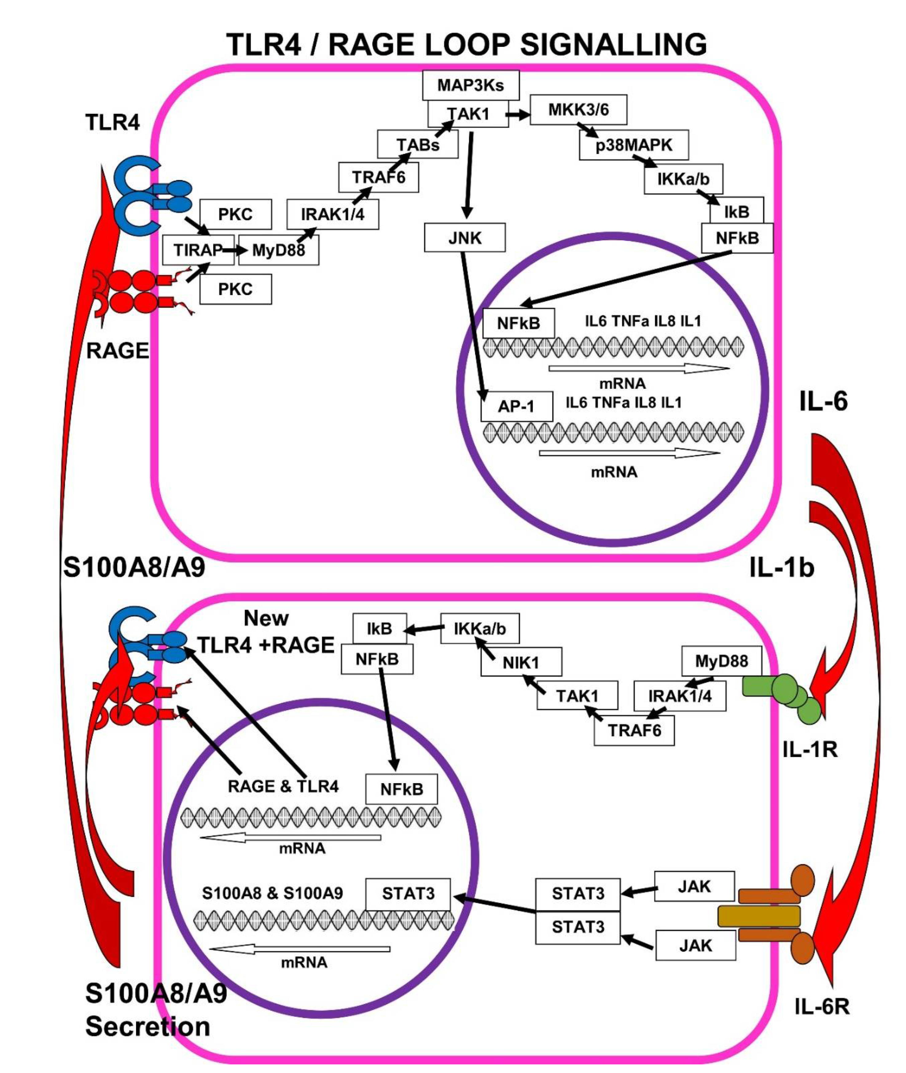
7. The Self-Sustaining TLR4/RAGE-Loop

IL-1b+IL-6 > IL-1R+IL-6R > TLR4/mRAGE+S100A8/A9 > IL-1b+IL-6 (Figure 2).

7.1. IL-1b > IL-1R Signalling Enhances mRAGE and TLR4 Expression

In Rheumatoid Arthritis (RA), serum concentrations of pro-inflammatory IL-1b and IL-6 are high and mRAGE is over-expressed in synovial tissues. RAGE-expression is driven by IL-1b binding to its cell surface receptor IL-1R, triggering the MyD88 > IRAK-TRAF6 > TAB1 + TAK1 > IKKb > NFkB and AP-1 signalling cascade. In patient-derived fibroblast-like synoviocytes, an in vitro RA model, IL-1b increased the levels of RAGE mRNA expression, which was amplified further by the addition of IL-17. In contrast, TNFa had no effect on RAGE expression in patient derived fibroblast-like synoviocytes [44].
TLR4 gene expression is also induced by IL-1b > IL-1R signalling and activation of transcription factors NFkB and AP-1. Increased expression of TLR4 is detected in inflammatory atherosclerotic and cardiovascular disease, where part of the amplification of TLR4 gene expression is due to oxidative stress, inducing post-transcriptional stabilization of TLR4 mRNA in vascular cells [45]. TLR4 signalling also stimulates IL-1b > IL-1R, by upregulating IL-1R expression in alveolar macrophages, which promotes lung inflammation [46].

7.2. IL-6 > IL-6R Signalling Enhances S100A8/A9 Expression

IL-6 binding to cell surface IL-6R receptor on human monocytes, macrophages and fibroblasts induces JAK/STAT3 activation, followed by binding of both STAT3 and transcription factor CAAT-Enhancing-Binding-Protein-alpha (C/EBPa) to adjacent gene-promoter regions of the S100A8 gene (and the S100A9 gene), leading to activation of S100A8 and S100A9 expression and S100A8/A9 secretion into the inter-cellular medium [47].
The 2178-bp to 234-bp region in the S100A8 gene-promoter contains important cis-regulatory binding sites for STAT3, C/EBP, AP-1, Ets, and NF-1, that contribute to its transcriptional activation. Transcription factors STAT3 and AP-1 binding to the S100A8 and S100A9 promoters commences mRNA transcription. STAT3 controls the induction of pro-inflammatory responses and the increase of S100A8/A9 secretion. In colonic epithelial cells and Caco-2 Cells, treatment with STAT3 inhibitors inhibits IL-6 mediated S100A9 expression. SOCS3 is an inhibitor of STAT3 activity and a negative controller of inflammatory signalling. If SOCS3 is inhibited, IL-6 strongly induces S100A8/A9 secretion. However, transcription factor C/EBPd is important for the constitutive expression of S100A8 and S100A9 during development and differentiation of monocytes. IL-6 > IL-6R induction of S100A8 and S100A9 genes is a “late” inflammation event that needs new protein synthesis [42,48].

7.3. S100A8/A9 Pushes the TLR4/RAGE-Loop to Signal for More Cytokines

Excessive expression of S100A8/A9 magnifies the inflammatory response and stimulates neutrophils and macrophages to release more cytokines, inducing the TLR4/RAGE-loop amplification of inflammation. The TLR4/RAGE-loop amplifies multiple cellular processes such as inflammation, differentiation, proliferation, migration and apoptosis. In inflammatory Psoriasis, TNFa induces S100A8 expression in keratinocytes, leading to inflammatory processes independent of viral or microbial triggering, a process sometimes referred to as “sterile inflammation” [49].
Elevated concentration of serum S100A8/A9 triggers both TLR4 and RAGE, inducing intracellular signalling pathways, which lead mainly to activation of transcription factor NFkB and the expression and secretion of IL-1b, IL-6 and TNFa. In a mouse model, recombinant S100A8/A9 binds TLR4 and signals via TIRAP+MyD88 to induce more pro-inflammatory cytokines and also more S100A8/A9 expression. In tissue macrophages from obese patients, fat-derived S100A8/A9 stimulates the TLR4>TIRAP+MyD88 cascade to enhance the expression of IL-1b mRNA [39].
These signalling cascades amplify expression of S100A8/A9, which in turn is a pro-inflammatory ligand for mRAGE and TLR4, which signal for more pro-inflammatory cytokines, thus forming the TLR4/RAGE-loop. The potential for rapid auto-amplification of inflammation by this system means it is highly regulated in healthy individuals, but effective TLR4/RAGE regulation breaks down in acute COVID-19-19 and in many chronic inflammatory diseases, including long COVID [23,50].

7.4. SARS-CoV-2 in hACE2 Mice and Inhibition of S100A8/A9 > RAGE-Signalling

TLR4/RAGE-loop signalling was demonstrated in hACE2-mice (mice expressing human ACE2) infected with human SARS-CoV-2. In SARS-CoV-2 infected hACE2-mice, the inhibition of RAGE-signalling, limited disease pathogenesis and promoted survival [51].
There were two distinct phases of disease in this mouse-SARS-CoV-2-infection model. hACE2-mice were intranasally infected with a 1 × 10⁴ TCID50 (50% tissue culture infective dose) of SARS-CoV-2. During the first day following infection of hACE2 mice, SARS-CoV-2 virus replicated to high titres within the lungs, triggering acute inflammation.
The second disease phase commenced with significant cell death occurring on day 2 after infection. Viral-induced cell death in non-myeloid cell populations led to progressive tissue damage, which activated RAGE signalling cascades and hyper-inflammation. On day 2 after infection, there was mRAGE hyper-activation, elevation of MMP-9 protease cleavage and production of inhibitory sRAGE. There was a rise in serum concentrations of S100A8/A9 and HMGB1, which induced chemotactic cellular recruitment and more inflammation (S100A9 was used as a marker for S100A8/A9).
These events were followed by a rapid decrease in blood-oxygen saturation, metabolic re-programming of glycolysis, interstitial lung pneumonia, perivascular inflammation and endothelial hyperplasia. Up to 80% of the hACE2 mice became so moribund between days 4 and 6 after infection, they required humane euthanasia as early as 4.5 days after infection.
An mRAGE-inhibitor called FPS-ZM1, which specifically blocks the binding of RAGE-ligands, such as AGEs, S100A8/A9 or HMGB1, to the extra-cellular V domain of RAGE (KI: 25 ± 5 nM), limited the virus-induced inflammation and its associated perivascular pathology. FPS-ZM1 also improved survival and extended the median time to death of SARS-CoV-2 infected hACE2-mice.
FPS-ZM1 treatment reduced levels of MMP-9 on day 2 after infection, evidence of that FPS-ZM1 treatment was inhibiting mRAGE activation. FPS-ZM1 treatment also reduced the level of pro-inflammatory cytokines and chemokines in the lungs of the mice on day 4 after infection.
In addition, during viral infection there was a time-dependent decrease in the serum concentrations of S100A8/A9. Part of the mechanism of the FPS-ZM1 therapeutic benefit, appeared to be related to inhibition of RAGE-loop dependent S100A8/A9 expression, which resulted from the inhibition of RAGE signalling.
In contrast, FPS-ZM1 had no effect on SARS-CoV-2 viral load. Although FPS-ZM1 treatment reduced baseline levels of the RAGE-agonist HMGB1 in the lungs, it did not alter the trajectory of increasing HMGB1 levels as the viral infection progressed.
These results supported the hypothesis that S100A8/A9 triggering of mRAGE and pro-inflammatory TLR4/RAGE-loop signalling, has a key role in the inflammatory disease pathogenesis of acute COVID-19.

7.5. SARS-CoV-2 in hACE2 Mice and Inhibition of S100A8/A9 >TLR4-Signalling

The central role of TLR4 in TLR4/RAGE-loop signalling was also established in hACE2-mice (mice expressing human ACE2) infected with human SARS-CoV-2. In SARS-CoV-2-infected hACE2 mice, the inhibition of TLR4-signalling limited disease pathogenesis and promoted survival [50].
High serum levels of S100A8/A9 mediates activation of aberrant neutrophils in acute COVID-19. A TLR4-MD2 receptor protein complex is formed by 25-kD glycoprotein Myeloid Differentiation protein-2 (MD2) binding to the extracellular domain of TLR4. The binding of serum S100A8/A9 to the TLR4-MD2 complex, induces hyper-inflammation, recruitment of myeloid cells, increased vascular permeability, and activation of coagulation cascades [52].
In infected hACE2-mice, S100A8 and S100A9 expression, leading to S100A8/A9 secretion, was significantly upregulated by coronaviruses such as human SARS-CoV-2 and Mouse Hepatitis Virus (MHV). In contrast, the encephalomyocarditis virus (EMCV) and herpes simplex virus 1 (HSV-1) neither induced S100A8 hyper-expression nor neutrophil chemotaxis genes in hACE2-mice.
Serum S100A8/A9 not only triggers mRAGE signalling, it also triggers Toll-like receptor 4 (TLR4) signalling. S100A8/A9 acting on both mRAGE and TLR4 triggers multiple pro-inflammatory pathways, including the induction of neutrophil chemotaxis genes. In SARS-CoV-2- and MHV-infected mice, some neutrophils expressed a characteristic pattern of three cell surface proteins: CD45, CD11b and Ly6G-variable, in which Ly6G expression was inhibited relative to control mice. SARS-CoV-2 Spike-protein hyper-activation of both mRAGE and TLR4 had led to an abnormal group of neutrophils three to five days after infection, which invaded the lungs and secreted large amounts of S100A8/A9.
Paquinimod is a specific inhibitor of S100A9 binding to TLR4. Intranasal treatment of mice with Paquinimod led to almost 100% survival from a normally lethal infection titre of mouse hepatitis virus (MHV) and a substantial reduction of viral load in SARS-CoV-2-infected mice. Histopathological and immunohistochemical staining of the lungs showed that Paquinimod treatment had suppressed pulmonary damage, the invasion of neutrophils, and coronavirus infection. Most neutrophils in Paquinimod-treated mice returned to normal CD45, CD11b, and Ly6G-high levels.
Resatorvid is a selective TLR4-inhibitor, which also downregulates expression of TLR4. Resatorvid treatment of SARS-CoV-2 infected hACE2-mice led to a significant reduction of viral replication in the lungs of the infected mice, a reduction in aberrant neutrophils, and also inhibited S100A8 expression. These results show that inhibition of the TLR4/RAGE-loop suppresses IL-1b, IL-6, TNFa and S100A8/A9 mediated hyper-inflammation and coronavirus replication.
In addition, supporting evidence that chronic TLR4 signalling is associated with long COVID is an open clinical study in 38 long COVID patients (median time from diagnosis of COVID-19 until enrolment was 333 days) using low dose Naltrexone as a TLR4 inhibitor, (1 mg once daily for month one per patient, 2 mg once daily per patient for month two per patient), resulted in some symptomatic improvement. Adverse events in two patients (diarrhoea and fatigue in both) resulted in discontinuation [53].

8. Ezrin Peptides as Potential Therapies for Long COVID/PASC

Ezrin Peptides HEP-1 and RepG3 Inhibit Pro-Inflammatory Cytokine Expression

Overexpression of IL-1, IL-6 and TNFa is significantly associated with severe acute COVID-19 and long COVID. In contrast, oral administration of ezrin peptide solution (1mg/mL) of HEP-1 or RepG3 in humans, inhibits IL-1, IL-6, IL-8 and TNFa pro-inflammatory cytokine expression. I/P injection of ezrin peptides HEP-1 and RepG3 inhibit IL-1b and IL-6 expression in the Mouse DSS colitis model and alleviate DSS-induced experimental colitis. DSS poisoning of mice causes colon tissue to hyper-express pro-inflammatory cytokines: IL-6 mRNA increased by 29x, IL-1b mRNA increased by 12x and NOS2 mRNA increased by 6x in the inflamed colons of DSS colitis mice, relative to colon tissue of healthy control mice.
Oral ezrin peptide solution therapy in mice significantly inhibited the elevated expression of pro-inflammatory cytokines IL-1b, IL-6, and NOS2, which had been significantly elevated by DSS poisoning. HEP-1 ezrin peptide treatment significantly decreased the elevated gene expression of pro-inflammatory cytokines: IL-1b down 2.8x, IL-6 down 3.4x and TNFa down 3.2x (RepG3 has twice the activity of HEP-1 in suppressing pro-inflammatory cytokine expression) [54].
Ezrin peptides HEP-1 and RepG3 inhibit the expression of IL-1b, IL-6, IL-8 and TNFa in human peripheral blood mononuclear cells stimulated with PHA or LPS. Human ezrin peptides also enhance HLA-DR expression and adaptive immune B-cell and T-cell responses [55]. Severe acute COVID-19 and long COVID are also associated with depressed HLA-DR and CD44 expression. In vitro and clinical trials of ezrin peptide HEP-1 in HIV patients in Russia (10 mg per day of HEP-1 by oral administration of 1 mg/mL HEP-1 solution for 30 days, increased the expression of HLA-DR and CD44 expression during and after the treatment period. HEP-1 was registered for human use in Russia in 2001, and HEP-1 and RepG3 have been used as effective treatment for acute COVID-19 in volunteers, but so far there have been no large scale clinical trials [56,57].
There is preliminary anecdotal evidence of symptomatic relief (not confirmed yet by formal clinical trials) during treatment with ezrin peptide therapy in a few long COVID/PASC patient volunteers. For example, Volunteer L, a chronic Myalgic Encephalomyelitis/Chronic Fatigue Syndrome (ME/CFS) patient, whose symptoms worsened after acute COVID-19, has used spray-inhaled RepG3 0.5 mL per day of 1 mg/mL RepG3 for ten days, and self-reported symptom improvement in cognition and mood, resolution of dry cough and sore throat, improvement of inflammatory bowel symptoms and alleviation of general fatigue. There was some symptom relapse following a ten day break in therapy, but the patient felt healthier again on retreatment, noting particularly her improved gut function and mood. There are other anecdotal reports of successful therapy with HEP-1 and one male long COVID patient self-reported he felt cured. RepG3 treatment of long COVID must be evaluated in new clinical studies.

9. Conclusions

After SARS-CoV-2 infection, viral Spike protein specifically binds to membrane Receptor for Advanced Glycation End Products (mRAGE) and Toll Like Receptor 4 (TLR4), leading to intense pro-inflammatory cytokine stimulation and acute COVID-19.
TLR4 and mRAGE activate expression of the pro-inflammatory cytokines IL-1b, IL-6 and TNFa. The pro-inflammatory cytokines IL-1b, IL-6 and TNFa are then released by virally infected cells. IL-1b binds its receptor IL-1R on the surface of other cells and signals via NFkB for more mRAGE and TLR4 expression. IL-6 binds to its IL-6R receptor on other cells, and signals via STAT3 and C/EBPb for more S100A8/A9 expression. Elevated serum concentrations of S100A8/A9 can bind and activate cell-surface mRAGE and TLR4. The TLR4/RAGE-loop is amplified further by elevated AGEs and HMGB1 in the serum. As serum concentrations of S100A8/A9 increase and bind to the new mRAGE and TLR4, downstream signalling induces more IL-1b, IL-6 and TNFa, forming a signalling circuit that can amplify inflammatory responses in the absence of viral proteins.
The pathological positive feedback that sustains the TLR4/RAGE-loop, is probably the fundamental immunological dysregulation responsible for long COVID/PASC. TLR4-RAGE-loop inflammation generates a multi-organ inflammation which is particularly intense in the lungs, where damaged epithelia, endothelia and dysregulated monocytes act together to hyper-express pro-inflammatory cytokines. These pro-inflammatory cytokines induce further pro-inflammatory responses affecting almost all organs, including upregulation of TLR4/RAGE signalling in the blood vessels, the liver, the heart and the brain. TLR4/RAGE-loop inflammation is likely to be the basic cause of the wide range of downstream symptoms reported in long COVID/PASC.
Generally, inhibition of pro-inflammatory IL-1, IL-6 and TNFa cytokine expression and S100A8/A9 expression stops TLR4/RAGE-loop inflammation. The anti-inflammatory properties of ezrin peptides suggest RepG3 as potential therapy for Long COVID/PASC and some preliminary results are already promising.

Funding

This research received no external funding.

Institutional Review Board Statement

Not applicable.

Informed Consent Statement

Not applicable.

Data Availability Statement

Not applicable.

Conflicts of Interest

Rupert D Holms is inventor of ezrin peptide technology and issued patents in various territories are owned by NewalR&D Ltd, London, a company owned by Rupert D Holms.

Abbreviations

Alphabetically grouped.
ABCD
AC Adenylyl Cyclase; ACE2 Angiotensin-Converting Enzyme 2 receptor; ATF1 cAMP-Transcription Factor 1; AGEs Advanced Glycation End-products. ALI Acute Lung Injury; AP-1 transcription-factor Activator-Protein-1; ARF6 ADP-Ribosylation-Factor-6; ATF Activating Transcription Factor; BALF Broncho Alveolar Lavage Fluid; C-Reactive Protein IL-6-induced plasma protein-pentamer; CBP CREB-Binding Protein; c-Fos part of the “Fos” family of transcription factors; c-Jun part of the “Jun” family of transcription factors; Cdc42 a small GTPase of the Rho family, CML-HSA N-(CarboxyMethyl)Lysine-modified Human Serum Albumin; COX-2 Cyclo-Oxygenase-2; CREB cAMP Response Element-Binding transcription factor; CREM cAMP-Responsive-Element-Modulator; CFTR (Cl-) Cystic Fibrosis Transmembrane Conductance Regulator; CXCL2 macrophage inflammatory protein 2-alpha; DAMPs Damage-Associated-Molecular-Patterns.
EFGHIJKL
eIF4E Eukaryotic Translation Initiation Factor 4E; ERK Extra-cellular-signal-Regulated protein-Kinase 1 or 2; GSK-3b Glycogen Synthase Kinase-3b; GTPase Guanosine Tri-Phosphatase; HEP-1 Human Ezrin Peptide 1; HMGB1 High Mobility Group Box-1 proteins, also known as Amphoterin; IκB Inhibitor of nuclear factor kappa B; IKK I Kappa B Kinase; IL-1 pro-inflammatory Interleukin-1; IL-6 pro-inflammatory Interleukin-6; IL-8 pro-inflammatory Interleukin-8 (also known as CXCL8); IL-10 anti-inflammatory Interleukin-10; IRAK4 Interleukin-1 Receptor-Associated Kinase 4; JAKs Janus Kinases; JDP Jun Dimerization Protein; JNKs c-Jun-N-terminal Kinases; KPNA2 Karyopherin A2.
MNOPQ
MAP3Ks Mitogen Activated Protein (MAP) kinase kinase kinases; MAP2Ks Mitogen-activated protein kinase kinases, e.g., MKK3 or MKK6; p38MAPK mitogen-activated protein kinase p38; MCP-1 monocyte chemoattractant protein; M-CSF Macrophage-Colony-Stimulating-Factor MEK MAPK/ERK Kinase 1 or 2; MKK3/6 Mitogen Activated Kinase Kinase 3 or 6; MMP-9 Matrix-Metallo-Proteinase-9; MNK1 MAP kinase-interacting serine/threonine-protein kinase 1; m-RAGE cell-membrane-expressed Receptor-for-Advanced-Glycation-End-products (V+C1+C2+cyto-tail); MyD88 Myeloid Differentiation factor 88; NFκB Nuclear-transcription-Factor kappa-B; NHERF1 Na+ H+ Exchanger Regulatory Factor 1; NHE Na+ H+ Exchanger; nsp 6/12/13 non-structural protein; ORF6 Open Reading Frame 6 PAMPs Pathogen-Associated-Molecular-Patterns, PBMC Peripheral Blood Mononuclear Cells; PI3K Phosphatidyl-Inositol-3-kinase; PIP2 Phosphatidyl-Inositol 4,5-bisPhosphate; PI PIP3 3,4,5-trisphosphate; PIP5K phosphatidyl-Inositol 4-phosphate 5-kinase; PKC Protein-Kinase-C; PI3K Phosphatidyl-Inositol-3-Kinase; PKA cAMP-dependent-Protein-Kinase-A; PKB/Akt Protein Kinase B also known as Akt; PDK1 Phosphoinositide-Dependent Kinase 1; PI Phosphatidyl-Inositol PRR Pattern-Recognition-Receptor; p300 E1A-binding-protein-p300.
RSTUVWXYZ
Rac1 Ras-related C3 botulinum-toxin substrate 1; Raf Rapidly accelerated fibrosarcoma kinase1; Ras p21 small GTPase; RepG3 Regulatory-Ezrin-Peptide-Glycine-3; ROS Reactive Oxygen Species; RSK p90 Ribosomal S6 Kinase; S100 Small calcium-binding-protein-100 (Calgranulin); SAPK Stress-Activated-Protein-Kinase; SOS Son of Sevenless; SP-1 Specificity Protein 1 (a zinc-finger transcription factor); s-RAGE soluble Receptor for Advance Glycation End product (exterior V+C1+C2 domains only); STATs Signalling-Transducer and Activator of Transcription; TIRAP Toll-Interleukin-1-Receptor-Adaptor-Protein; THP-1 cells leukaemia peripheral blood monocyte-like cell line; TLRs Toll-Like Receptors; TNFα Tumour Necrosis Factor alpha; TMPRSS2 Trans-Membrane-Serine-Protease-2; VIP Vasoactive Intestinal Peptide.

References

  1. Tejerina, F.; Catalan, P.; Rodriguez-Grande, C.; Adan, J.; Rodriguez-Gonzalez, C.; Muñoz, P.; Aldamiz, T.; Diez, C.; Perez, L.; Fanciulli, C.; et al. Post-COVID-19 syndrome. SARS-CoV-2 RNA detection in plasma, stool, and urine in patients with persistent symptoms after COVID-19. BMC Infect. Dis. 2022, 22, 211. [Google Scholar] [CrossRef]
  2. Available online: https://www.ons.gov.uk/peoplepopulationandcommunity/healthandsocialcare/conditionsanddiseases/bulletins/prevalenceofongoingsymptomsfollowingcoronaviruscovid19infectionintheuk/7july2022 (accessed on 18 July 2022).
  3. Available online: https://coronavirus.data.gov.uk/details/cases (accessed on 18 July 2022).
  4. Al-Aly, Z.; Bowe, B.; Xie, Y. Outcomes of SARS-CoV-2 Reinfection. Res. Sq. 2022. [Google Scholar] [CrossRef]
  5. Fernández-Lázaro, D.; Sánchez-Serrano, N.; Mielgo-Ayuso, J.; García-Hernández, J.L.; González-Bernal, J.J.; Seco-Calvo, J. Long COVID a New Derivative in the Chaos of SARS-CoV-2 Infection: The Emergent Pandemic? J. Clin. Med. 2021, 10, 5799. [Google Scholar] [CrossRef]
  6. Elicker, B. What Are the Long-term Pulmonary Sequelae of COVID-19 Infection? Radiology 2022, 304, 193–194. [Google Scholar] [CrossRef]
  7. Crook, H.; Raza, S.; Nowell, J.; Young, M.; Edison, P. Long covid—Mechanisms, risk factors, and Management. BMJ 2021, 374, n1648. [Google Scholar] [CrossRef] [PubMed]
  8. Tate, W.; Walker, M.; Sweetman, E.; Helliwell, A.; Peppercorn, K.; Edgar, C.; Blair, A.; Chatterjee, A. Molecular Mechanisms of Neuroinflammation in ME/CFS and Long COVID to Sustain Disease and Promote Relapses. Front. Neurol. 2022, 13, 877772. [Google Scholar] [CrossRef] [PubMed]
  9. Hornig, M.; Montoya, J.G.; Klimas, N.G.; Levine, S.; Felsenstein, D.; Bateman, L.; Peterson, D.L.; Gottschalk, C.G.; Schultz, A.F.; Che, X.; et al. Distinct plasma immune signatures in ME/CFS are present early in the course of illness. Sci. Adv. 2015, 1, e1400121. [Google Scholar] [CrossRef] [PubMed]
  10. Peluso, M.J.; Lu, S.; Tang, A.F.; Durstenfeld, M.S.; Ho, H.E.; Goldberg, S.A.; Forman, C.A.; Munter, S.E.; Hoh, R.; Tai, V.; et al. Markers of Immune Activation and Inflammation in Individuals With Postacute Sequelae of Severe Acute Respiratory Syndrome Coronavirus 2 Infection. J. Infect. Dis. 2021, 224, 1839–1848. [Google Scholar] [CrossRef]
  11. Schultheiβ, C.; Willscher, E.; Paschold, L.; Gottschick, C.; Klee, B.; Henkes, S.S.; Bosurgi, L.; Dutzmann, J.; Sedding, D.; Frese, T.; et al. The IL-1b, IL-6, and TNF cytokine triad is associated with post-acute sequelae of COVID-19. Cell Rep. Med. 2022, 3, 100663. [Google Scholar] [CrossRef]
  12. Unterman, A.; Sumida, T.S.; Nouri, N.; Yan, X.; Zhao, A.Y.; Gasque, V.; Schupp, J.C.; Asashima, H.; Liu, Y.; Cosme, C.; et al. Single-cell multi-omics reveals dyssynchrony of the innate and adaptive immune system in progressive COVID-19. Nat. Commun. 2022, 13, 440. [Google Scholar] [CrossRef]
  13. Allen, C.N.S.; Santerre, M.; Arjona, S.P.; Ghaleb, L.J.; Herzi, M.; Llewellyn, M.D.; Shcherbik, N.; Sawaya, B.E. SARS-CoV-2 Causes Lung Inflammation through Metabolic Reprogramming and RAGE. Viruses 2022, 14, 983. [Google Scholar] [CrossRef] [PubMed]
  14. Sellegounder, D.; Zafari, P.; Rajabinejad, M.; Taghadosi, M.; Kapahi, P. Advanced glycation end products (AGEs) and its receptor, RAGE, modulate age-dependent COVID-19 morbidity and mortality. Int. Immunopharmacol. 2021, 98, 107806. [Google Scholar] [CrossRef] [PubMed]
  15. Senatus, L.M.; Schmidt, A.M. The AGE-RAGE Axis: Implications form Age-Associated Arterial Diseases. Front. Genet. 2017, 8, 187. [Google Scholar] [CrossRef]
  16. Chiappalupi, S.; Salvadori, L.; Donato, R.; Riuzzi, F.; Sorci, G. Hyperactivated RAGE in Comorbidities as a Risk Factor for Severe COVID-19—The Role of RAGE-RAS Crosstalk. Biomolecules 2021, 11, 876. [Google Scholar] [CrossRef] [PubMed]
  17. Saputra, G.; Yudhawati, R.; Fitriah, M. Association of soluble receptor for advanced glycation end-products (sRAGE) serum on COVID-19 severity: A cross-sectional study. Ann. Med. Surg. 2022, 74, 103303. [Google Scholar] [CrossRef]
  18. Angioni, R.; Bonfanti, M.; Caporale, N.; Sánchez-Rodríguez, R.; Munari, F.; Savino, A.; Buratto, D.; Pagani, I.; Bertoldi, N.; Zanon, C.; et al. RAGE engagement by SARS-CoV-2 enables monocyte infection and underlies COVID-19 severity. bioRxiv 2022, preprint. [Google Scholar] [CrossRef]
  19. Al-kuraishy, H.; Al-Gareeb, A.; Alkazmi, L.; Habotta, O.; Batiha, G. High-mobility group box 1 (HMGB1) in COVID-19: Extrapolation of dangerous liaisons. Inflammopharmacology 2022, 30, 811–820. [Google Scholar] [CrossRef]
  20. Deguchi, A.; Yamamoto, T.; Shibata, N.; Maru, Y. S100A8 may govern hyper-inflammation in severe COVID-19. FASEB J. 2021, 35, e21798. [Google Scholar] [CrossRef]
  21. Chapuis, N.; Ibrahimi, N.; Belmondo, T.; Goulvestre, C.; Berger, A.E.; Mariaggi, A.A.; Andrieu, M.; Chenevier-Gobeaux, C.; Bayle, A.; Campos, L.; et al. Dynamics of circulating calprotectin accurately predict the outcome of moderate COVID-19 patients. eBioMedicine 2022, 80, 104077. [Google Scholar] [CrossRef]
  22. Mao, Q.; Wang, C.; Wen, W.; Zhou, M.; Tang, J.; Chen, C.; Cheng, Y.; Wu, Q.; Zhang, X.; Feng, Z.; et al. A meta-analysis of the association between calprotectin and the severity of COVID-19. J. Infect. 2022, 84, e31–e33. [Google Scholar] [CrossRef]
  23. Mellett, L.; Khader, S.A. S100A8/A9 in COVID-19 pathogenesis: Impact on clinical outcomes. Cytokine Growth Factor Rev. 2022, 63, 90–97. [Google Scholar] [CrossRef] [PubMed]
  24. Silvin, A.; Chapuis, N.; Dunsmore, G.; Goubet, A.G.; Dubuisson, A.; Derosa, L.; Almire, C.; Hénon, C.; Kosmider, O.; Droin, N.; et al. Elevated calprotectin and abnormal myeloid cell subsets discriminate severe from mild COVID-19. Cell 2020, 182, 1401–1418.e18. [Google Scholar] [CrossRef] [PubMed]
  25. Chen, L.; Long, X.; Xu, Q.; Tan, J.; Wang, G.; Cao, Y.; Wei, J.; Luo, H.; Zhu, H.; Huang, L.; et al. Elevated serum levels of S100A8/A9 and HMGB1 at hospital admission are correlated with inferior clinical outcomes in COVID-19 patients. Cell. Mol. Immunol. 2020, 17, 992–994. [Google Scholar] [CrossRef]
  26. Ryan, F.J.; Hope, C.M.; Masavuli, M.G.; Lynn, M.A.; Mekonnen, Z.A.; Yeow, A.E.L.; Garcia-Valtanen, P.; Al-Delfi, Z.; Gummow, J.; Ferguson, C.; et al. Long-term perturbation of the peripheral immune system months after SARS-CoV-2 infection. BMC Med. 2022, 20, 26. [Google Scholar] [CrossRef] [PubMed]
  27. Cheon, I.S.; Li, C.; Son, Y.M.; Goplen, N.P.; Wu, Y.; Cassmann, T.; Wang, Z.; Wei, X.; Tang, J.; Li, Y.; et al. Immune signatures underlying post-acute COVID-19 lung sequelae. Sci. Immunol. 2021, 6, eabk1741. [Google Scholar] [CrossRef] [PubMed]
  28. Zervides, K.A.; Jern, A.; Nystedt, J.; Gullstrand, B.; Nilsson, P.C.; Sundgren, P.C.; Bengtsson, A.A.; Jönsen, A. Serum S100A8/A9 concentrations are associated with neuropsychiatric involvement in systemic lupus erythematosus: A cross-sectional study. BMC Rheumatol. 2022, 6, 38. [Google Scholar] [CrossRef]
  29. Gonzalez, L.; Karin Garrie, K.; Mark, D.; Turner, M. Role of S100 proteins in health and disease. BBA—Mol. Cell Res. 2020, 1867, 118677. [Google Scholar] [CrossRef] [PubMed]
  30. Xia, C.; Braunstein, Z.; Toomey, A.C.; Zhong, J.; Rao, X. S100 Proteins As an Important Regulator of Macrophage Inflammation. Front. Immunol. 2018, 8, 1908. [Google Scholar] [CrossRef]
  31. Donato, R.; R cannon, B.; Sorci, G.; Riuzzi, F.; Hsu, K.; Weber, D.J.; Geczy, C.L. Functions of S100 Proteins. Curr. Mol. Med. 2013, 13, 24–57. [Google Scholar] [CrossRef]
  32. Hsu, K.; Champaiboon, C.; Guenther, B.D.; Sorenson, B.S.; Khammanivong, A.; Ross, K.F.; Geczy, C.L.; Herzberg, M.C. Anti-Infective Protective Properties of S100 Calgranulins. Antiinflamm Antiallergy Agents Med. Chem. 2009, 8, 290–305. [Google Scholar] [CrossRef] [Green Version]
  33. Henke, M.; Renner, A.; Rubin, B.; Gyves, J.; Eva Lorenz, E.J.; Koo, J. Up-Regulation of S100A8 And S100A9 Protein in Bronchial Epithelial Cells by Lipopolysaccharide. Exp. Lung. Res. 2006, 32, 331–347. [Google Scholar] [CrossRef] [PubMed]
  34. Yen, T.; Harrison, C.A.; Devery, J.M.; Leong, S.; Iismaa, S.E.; Yoshimura, T.; Geczy, C.L. Induction of the S100 chemotactic protein, CP-10, in murine microvascular endothelial cells by proinflammatory stimuli. Blood 1997, 90, 4812. [Google Scholar] [CrossRef] [PubMed]
  35. Xu, K.; Yen, T.; Carolyn, L. Geczy IL-10 Up-Regulates Macrophage Expression of the S100 Protein S100A8. J. Immunol. 2001, 166, 6358–6366. [Google Scholar] [CrossRef]
  36. Hofmann, M.A.; Drury, S.; Fu, C.; Qu, W.; Taguchi, A.; Lu, Y.; Avila, C.; Kambham, N.; Bierhaus, A.; Nawroth, P.; et al. RAGE Mediates a Novel Proinflammatory Axis: A Central Cell Surface Receptor for S100/Calgranulin Polypeptides. Cell 1999, 97, 889–901. [Google Scholar] [CrossRef]
  37. Leclerc, E.; Fritz, G.; Vetter, S.; Heizmann, C. Binding of S100 proteins to RAGE: An update. Biochim. Biophys. Acta 2009, 1793, 993–1007. [Google Scholar] [CrossRef]
  38. Sakaguchi, M.; Murata, H.; Yamamoto, K.I.; Ono, T.; Sakaguchi, Y.; Motoyama, A.; Hibino, T.; Kataoka, K.; Huh, N.H. TIRAP, an Adaptor Protein for TLR2/4, Transduces a Signal from RAGE Phosphorylated upon Ligand Binding. PLoS ONE 2011, 6, e23132. [Google Scholar] [CrossRef] [PubMed]
  39. Wang, S.; Song, R.; Wang, Z.; Jing, Z.; Wang, S.; Ma, J. S100A8/A9 in Inflammation. Front. Immunol. 2018, 9, 1298. [Google Scholar] [CrossRef]
  40. Ma, L.; Sun, P.; Zhang, J.C.; Zhang, Q.; Yao, S.L. Proinflammatory effects of S100A8/A9 via TLR4 and RAGE signaling pathways in BV-2 microglial cells. Int. J. Mol. Med. 2017, 40, 31–38. [Google Scholar] [CrossRef]
  41. Sunahori, K.; Yamamura, M.; Yamana, J.; Takasugi, K.; Kawashima, M.; Yamamoto, H.; Chazin, W.J.; Nakatani, Y.; Yui, S.; Makino, H. The S100A8/A9 heterodimer amplifies proinflammatory cytokine production by macrophages via activation of nuclear factor kappa B and p38 mitogen-activated protein kinase in rheumatoid arthritis. Arthritis Res. Ther. 2006, 8, R69. [Google Scholar] [CrossRef]
  42. Lee, M.J.; Lee, J.K.; Choi, J.W.; Lee, C.S.; Sim, J.H.; Cho, C.H.; Lee, K.H.; Cho, I.H.; Chung, M.H.; Kim, H.R.; et al. Interleukin-6 Induces S100A9 Expression in Colonic Epithelial Cells through STAT3 Activation in Experimental Ulcerative Colitis. PLoS ONE 2012, 7, e38801. [Google Scholar] [CrossRef] [Green Version]
  43. Cotoi, O.; Dunér, P.; Ko, N.; Hedblad, B.; Nilsson, J.; Björkbacka, H.; Schiopu, A. Plasma S100A8/A9 Correlates With Blood Neutrophil Counts, Traditional Risk Factors, and Cardiovascular Disease in Middle-Aged Healthy Individuals. Arterioscler. Thromb. Vasc. Biol. 2014, 34, 202. [Google Scholar] [CrossRef] [PubMed]
  44. Heo, Y.J.; Oh, H.J.; Jung, Y.O.; Cho, M.L.; Lee, S.Y.; Yu, J.G.; Park, M.K.; Kim, H.R.; Lee, S.H.; Park, S.H.; et al. The expression of the receptor for advanced glycation end-products (RAGE) in RA-FLS is induced by IL-17 via Act-1. Arthritis Res. Ther. 2011, 13, R113. Available online: http://arthritis-research.com/content/13/4/R113 (accessed on 22 July 2022). [CrossRef] [PubMed]
  45. Yan, Z.-Q. Regulation of TLR4 Expression Is a Tale About Tail. Arterioscler. Thromb. Vasc. Biol. 2006, 26, 2582–2584. [Google Scholar] [CrossRef] [PubMed]
  46. He, X.; Qian, Y.; Li, Z.; Fan, E.K.; Li, Y.; Wu, L.; Billiar, T.R.; Wilson, M.A.; Shi, X.; Fan, J. TLR4-Upregulated IL-1β and IL-1RI Promote Alveolar Macrophag Pyroptosis and Lung Inflammation through an Autocrine Mechanism. Sci. Rep. 2016, 6, 31663. [Google Scholar] [CrossRef]
  47. Jauch-Speer, S.L.; Herrera-Rivero, M.; Ludwig, N.; De Carvalho, B.C.V.; Martens, L.; Wolf, J.; Chasan, A.I.; Witten, A.; Markus, B.; Schieffer, B.; et al. C/EBPδ-induced epigenetic changes control the dynamic gene transcription of S100a8 and S100a9. ELife 2022, 11, e75594. [Google Scholar] [CrossRef]
  48. Hsu, K.; Chung, Y.M.; Endoh, Y.; Geczy, C.L. TLR9 Ligands Induce S100A8 in Macrophages via a STAT3-Dependent Pathway which Requires IL-10 and PGE2. PLoS ONE 2014, 9, e103629. [Google Scholar] [CrossRef]
  49. Vogl, T.; Stratis, A.; Wixler, V.; Völler, T.; Thurainayagam, S.; Jorch, S.K.; Zenker, S.; Dreiling, A.; Chakraborty, D.; Fröhling, M.; et al. Autoinhibitory regulation of S100A8/S100A9 alarmin activity locally restricts sterile inflammation. J. Clin. Investig. 2018, 128, 1852–1866. [Google Scholar] [CrossRef]
  50. Guo, Q.; Zhao, Y.; Li, J.; Liu, J.; Yang, X.; Guo, X.; Kuang, M.; Xia, H.; Zhang, Z.; Cao, L.; et al. Induction of alarmin S100A8/A9 mediates activation of aberrant neutrophils in the pathogenesis of COVID-19. Cell Host Microbe 2021, 29, 222–235. [Google Scholar] [CrossRef]
  51. Jessop, F.; Schwarz, B.; Scott, D.; Lydia MRoberts, L.; Bohrnsen, E.; Hoidal, J.; Bosio, M. Impairing RAGE signaling promotes survival and limits disease pathogenesis following SARS-CoV-2 infection in mice. JCI Insight. 2022, 7, e155896. [Google Scholar] [CrossRef]
  52. Sohn, K.M.; Lee, S.G.; Kim, H.J.; Cheon, S.; Jeong, H.; Lee, J.; Kim, I.S.; Silwal, P.; Kim, Y.J.; Paik, S.; et al. COVID-19 patients upregulate toll-like receptor 4-mediated inflammatory signalling that mimics bacterial sepsis. bioRxiv 2020, preprint. [Google Scholar] [CrossRef]
  53. O’Kelly, B.; Vidal, L.; McHugh, T.; Woo, J.; Avramovic, G.; Lambert, J. Safety and efficacy of low dose naltrexone in a long covid cohort; an interventional pre-post study. Brain Behav. Immun.—Health 2022, 24, 100485. [Google Scholar] [CrossRef] [PubMed]
  54. Chulkina, M.; Pichugin, A.; Ataullakhanov, R. Pharmaceutical grade synthetic peptide Thr-Glu-Lys-Lys-Arg-Arg-Glu-Thr-Val-Glu-A,rg-Glu-Lys-Glu ameliorates DSS-induced murine colitis by reducing the number and pro-inflammatory activity of colon tissue-infiltrating Ly6G+ granulocytes and Ly6C+ monocytes. Peptides 2020, 132, 170364. [Google Scholar] [CrossRef] [PubMed]
  55. Holms, R.D.; Ataullakhanov, R.I. Ezrin Peptide Therapy from HIV to COVID: Inhibition of Inflammation and Amplification of Adaptive Anti-Viral Immunity. Int. J. Mol. Sci. 2021, 22, 11688. [Google Scholar] [CrossRef] [PubMed]
  56. Holms, R.D. The COVID-19 Cell Signalling Problem: Spike, RAGE, PKC, p38, NFkB & IL-6 Hyper-Expression and the Human Ezrin Peptide, VIP, PKA-CREB Solution. Immuno 2022, 2, 260–282. [Google Scholar] [CrossRef]
  57. Holms, R.D.; Ataullakhanov, R.I. Ezrin Peptide Therapy: A Potential Treatment for COVID. J. Bioprocess. Biotech. 2021, 12, 1–4. [Google Scholar]
Figure 1. The TLR4/RAGE loop causes chronic secretion of pro-inflammatory cytokines IL-1b and IL-6, which act on IL-1R and IL-6R, to induce more RAGE and TLR4 and secretion of pro-inflammatory S100A8/A9. Chronic systemic inflammation driven by TLR4/RAGE Loop signalling gives rise to chronic symptoms and pathologies in all organ systems. Brain inflammation causes cognitive impairment, sleep disturbances, depression and anxiety. Lung and airway inflammation causes cough, sore throat, breathlessness, low oxygen saturation and chest pain. Inflammation in blood vessels and heart causes myocardial inflammation, coagulopathy and micro-angiopathy. Liver inflammation causes ALT and AST elevation. Inflammation in the gut causes nausea, dysbiosis, diarrhoea and pain. Inflammation also causes renal impairment and pancreatitis. Inflammation in the musculo-skeletal system causes fatigue, and aches and pains in muscles and joints.
Immuno 02 00033 g001
Figure 2. High serum concentrations of S100A8/A9 trigger TLR4 and RAGE to activate the TIRAP+Md88 to NFkB and AP-1 signalling cascade, which results in hyper-expression of pro-inflammatory IL-1b and IL-6, which is secreted by the activated cells. High levels of serum IL-1b trigger IL-1R and the MyD88 to NFkB signalling pathway that upregulates the expression of new TLR4 and RAGE. High levels of serum IL-6 trigger IL-6R and the JAK/STAT signalling pathway which induces the expression of more pro-inflammatory S100A8 and S100A9 genes. After translation of the mRNA to polypeptides, S100A8/A9 hetero-dimer is secreted and the high concentrations in the serum trigger more TLR4 and RAGE, completing a pro-inflammation circuit, which does not require viral protein to persist.
Immuno 02 00033 g002
Table 1. The frequency of long COVID/PASC due to infection and re-infection.
At least 1 sequela:1st infect, 357re-infect: 553per 1000 persons at 6 months
Mental problem:1st infect, 146re-infect: 227per 1000 persons at 6 months
Neurologic:1st infect, 100re-infect: 136per 1000 persons at 6 months
Hospitalization:1st infect, 52re-infect: 148per 1000 persons at 6 months
Musculo-skeletal:1st infect, 46re-infect: 59per 1000 persons at 6 months
Gastro-intestinal:1st infect, 42re-infect: 69per 1000 persons at 6 months
Cardio-vascular:1st infect, 38re-infect: 88per 1000 persons at 6 months
Diabetic:1st infect, 36re-infect: 58per 1000 persons at 6 months
Pulmonary:1st infect, 35re-infect: 86per 1000 persons at 6 months
Fatigue:1st infect, 25re-infect: 58per 1000 persons at 6 months
Coagulation-blood:1st infect, 22re-infect: 48per 1000 persons at 6 months
Kidneys:1st infect, 21re-infect: 35per 1000 persons at 6 months
All-cause mortality:1st infect, 21re-infect: 45per 1000 persons at 6 months
Publisher’s Note: MDPI stays neutral with regard to jurisdictional claims in published maps and institutional affiliations.

