Review

Recent Progress on the Roles of Regulatory T Cells in IgG4-Related Disease

Department of Gastroenterology and Hepatology, Kochi Medical School, Kochi University, Kochi 783-8505, Japan
Immuno 2022, 2(2), 430-442; https://doi.org/10.3390/immuno2020026
Submission received: 15 April 2022 / Revised: 22 May 2022 / Accepted: 25 May 2022 / Published: 31 May 2022
(This article belongs to the Special Issue Is IgG4-Related Disease a Systemic Autoimmune Disease?)

Abstract

:
IgG4-related disease (RD) is a proposed concept of systemic inflammatory condition from Japanese researchers. Patients with IgG4-RD manifest several immunological and histological characterizations in the organs involved, including elevated levels of serum IgG4 and lympho-plasmacytic infiltration, storiform fibrosis, IgG4-positive plasma cells infiltration, and obstructive phlebitis. Nevertheless, the pathogenesis of IgG4-RD still remains unclear. It has been made clear that several immune cells with regulatory function play a vital part in several diseases. In particular, abnormalities in the function and proportion of regulatory T cells (Tregs) are implicated in several diseases, and their part in IgG4-RD has been investigated. This review offers an overview of the research in IgG4-RD related to Tregs. Herein, the basic information of Tregs, knowledge gained from animal models involving Tregs, and the role of IgG4-RD has been provided. We also included the immunological mechanisms of IgG4-RD based on the data accumulated so far in our hypothesis.

1. Introduction

Recently, Immunoglobulin G4-related disease (IgG4-RD) has been recognized as a systemic inflammatory disease which is histologically characterized by lymphoplasmacytic infiltration, IgG4-positive plasma cell infiltration, storiform fibrosis, and obliterative phlebitis. These histological findings are based on lymphoplasmacytic sclerosing pancreatitis, (LPSP) and which were published in a 1991 study by Kawaguchi et al. [1]. IgG4-RD can affect systemic involved organs resulting in swelling and hyperplasia, such as that of the pachymeninx, hypophysis, lacrimal gland, eye, salivary gland, thyroid gland, lung, pancreas, bile duct, retroperitonium, kidney, prostate, aorta, and other arteries [2].
In 2001, Hamano et al. published the first report relating to IgG4-RD as “sclerosing pancreatitis”, which is currently known as type I autoimmune pancreatitis (AIP) [3]. AIP was proposed by Yoshida et al. before Hamano’s report in 1995. They presented a case with hyperglobulinemia, increased levels of serum IgG levels, autoantibody-positive, diffusely enlarged pancreas, diffuse narrowing of the main pancreatic duct, histopathologic changes with sever fibrosis, infiltration of lymphocytes, and the effectiveness of steroidal therapy [4]. On the basis of the cases available, AIP is known to often develop extrapancreatic lesions (other organs involvement) and is thus regarded as a systemic disorder instead of a pancreatitis. Subsequently, in 2006, Kamisawa et al. proposed the term “IgG4-related sclerosing disease” [5]. Similarly, the term “IgG4-related plasmacytic disease” was coined by Yamamoto et al. from the point of view of Mikulicz’s disease in 2006 [6]. Masaki et al. named “IgG4-related multiorgan lymphoproliferative syndrome” from the perspective of a lymphoproliferative disturbance in 2009 [7]. Several terms have been proposed to present this new clinical entity. Based on these similar concepts, in 2012 these terms were unified under the umbrella term “IgG4-RD” [8].
In 2011, International Consensus Diagnostic Criteria for Autoimmune Pancreatitis divided AIP into two types: type 1 and type 2 [9]. Type 1 AIP is related to IgG4 and is identified as a part of IgG4-RD. Presently, only the term “type 1 AIP” remains in IgG4-RD. For instance, the names of Riedel thyroiditis and Mikulicz’s disease were changed to IgG4-related thyroiditis and IgG4-related dacryo-sialoadenitis, respectively. Type 2 AIP histologically exhibits the infiltration of neutrophils within the lumen and epithelium of the interlobular pancreatic ducts and granulocytic epithelial lesion (GEL), and it is completely different from type 1 AIP. These pathological characteristics were proposed as idiopathic duct centric chronic pancreatitis (IDCP) or AIP with GEL by Notohara et al. in 2003 [10] or Zamboni et al. in 2004 [11], respectively.
Interleukin (IL)-10 is well known and is one of the major anti-inflammatory and immunosuppressive cytokines that was first discovered as a cytokine secreted from T helper Th2 cells, and it inhibits inflammatory changes by activated Th1 cells [12]. Th2 immune response plays an important role in the pathophysiology of IgG4-RD [13]. Several IL-10-producing regulatory cells have been recognized, including T cells [14], B cells [15], dendritic cells [16,17], and macrophages [18]. The pathophysiology of several immune-mediated diseases is considered to be related to regulatory T cells (Tregs) [19]. Herein, we highlight the role of Tregs in IgG4-RD.

2. History of Regulatory T cells

Neonatal thymectomy is known to cause organ-specific autoimmune diseases in certain mouse strains. In 1995, Sakaguchi et al. termed Tregs that were characterized as CD4+CD25+ (IL-2 receptor α-chain) as T cells. They reported that neonatal thymectomy induces the elimination and disappearance of Tregs from the peripheral blood to facilitate the supply of Tregs from the thymus, and initiates autoimmune reaction similar to those in human counterparts such as type 1 diabetes, thyroiditis, and autoimmune gastritis from peripheral tolerance. Moreover, the development of this autoimmune reaction was inhibited by the reconstitution of CD4+CD25+ T cells in neonatal thymectomized mice [20].
In 2001, Tregs were identified as CD4+CD25high T cells from peripheral blood by several independent researchers in humans [21,22,23,24]. The master key gene of Tregs was discovered as the transcription factor forkhead box P3 (Foxp3) in 2003 [25,26,27]. Foxp3 is a part of the winged helix/forkhead family of transcription factors and plays an essential role in the conservation of immune tolerance against self-antigens and, therefore, in suppressing autoimmune diseases [19]. Activated effector Tregs (eTregs) inhibit the function, activation, and proliferation of immunocompetent cells against microbial, environmental and self-antigens, including T cells, B cells, several antigen-presenting cells, natural killer cells, and NKT cells [28]. Inactivating mutations of Foxp3 result in the early onset of fatal autoimmune diseases, which are known as immunodysregulation, polyendocrinopathy, enteropathy, and X-linked (IPEX) syndrome in humans [19,29].
Various suppressive mechanisms have been reported via Tregs, including cell/cell contact-dependent mechanisms through cytokines (e.g., IL-2, IL-10, IL-35, and transforming growth factor-beta (TGF-β)), cell surface molecules (glucocorticoid-induced TNFR-related protein (GITR), cytotoxic T lymphocyte antigen 4 (CTLA-4), T-cell immunoreceptor with immunoglobulin and ITIM domains (TIGIT), CD25, CD39, and CD73), and intracellular or secreted molecules (cyclic AMP, granzymes, and indoleamine 2,3-dioxygenase (IDO)), that result in the release of suppressive cytokines or the generation of suppressive metabolic products [19,30,31,32,33,34]. This ability of immunoregulation enables Foxp3+ Tregs to control the immunological reaction in autoimmune diseases, allergic diseases, tumor immunity, microbial infection, immunometabolic diseases, and tissue repair effectively, as well as to maintain the immune tolerance of allograft and pregnancy [19].
What is the number of Tregs in actual diseases? Several investigators have previously counted the number of Tregs in various diseases. Decreased numbers of CD4+CD25high Tregs in peripheral blood have been presented in several diseases, specifically in the patients with systemic lupus erythematosus (SLE) [35], psoriatic arthritis [36], juvenile idiopathic arthritis [37,38], autoimmune liver disease [39], hepatitis C virus-associated mixed cryoglobulinemia [40], and Kawasaki disease [41]. No significant difference has been reported in patients with type I diabetes [42,43], or multiple sclerosis compared with healthy controls [44,45]. In Sjögren’s syndrome, the numbers of peripheral Tregs increased [46]. Opposing results have been published for the number of circulating Tregs in patients with myasthenia gravis [47,48], rheumatoid arthritis (RA) [49,50,51,52], and inflammatory bowel disease [53,54], with either similar or decreased levels observed.
Therefore, the number of Tregs in peripheral blood varies according to the disease and reports, and hence it remains controversial. However, several investigators agree that there is an increased number of FOXP3-positive Tregs in the inflamed tissue in not only immune-mediated but also nonimmune-mediated inflammatory organs [55,56]. The common findings of the recruitment of Tregs in the inflamed tissues might suggest that the Tregs function is insufficient due to either cell-intrinsic or -extrinsic factors.

3. Regulatory T Cells in Animal Models of IgG4-Related Disease

Some animal models are available for understanding the role of Tregs in IgG4-RD. As mentioned in the previous section, neonatal thymectomy induces organ-specific autoimmune response. The depletion of Tregs due to neonatal thymectomy initiates autoimmunity from peripheral tolerance [18]. Several autoantibodies have been detected in type 1 AIP. Asada et al. reported that the positive ratio of anti-lactoferrin (LF) antibody was 73.1%, and the positive ratio of anti-carbonic anhydrase (CA)-II was 53.8% in patients with type 1 AIP [57]. To assess the role of Tregs in IgG4-RD development, the authors immunized neonatally thymectomized mice with LF or CA-II. These immunized mice developed inflammatory changes in the pancreas, salivary gland, and around the bile duct. Hence, neonatally thymectomized mice immunized with LF or CA-II sere as animal model of human IgG4-RD, including type 1 AIP [58].
Another model is the WBN/Kob rat, which spontaneously develops pancreatitis, sclerosinging cholangitis, sialadenitis, thyroiditis, and tubulointerstitial nephritis similar to IgG4-RD because of congenitally decreased peripheral Tregs. A large amount of CD8+ T cells were infiltrated. Although the target antigens are unknown, CD8+ T cells might be effector cells. The deposition of IgG2b is observed in the inflamed pancreas and lachrymal glands [59]. Although IgG4 is not found in rodents, rat IgG2b appears to be similar to human IgG4 [60]. Therefore, these mouse and rat models are helpful for investigating the role of Tregs on the initiation and progression of IgG4-RD.
From the viewpoint of the function of Tregs, another mice model has been established based on the deletion of the mammalian sterile 20–like kinase-1 (Mst1) gene. Mst1 is a serine/threonine kinase which is recognized to be crucial in the regulation of cell trafficking of immune cells. Mst1 was also determined to be involved in the selection of thymocyte and was critical for the maintenance of Tregs. Mst1-knocked out mice spontaneously developed inflammatory changes in multiple organs, which increased as the mice grew older and autoantibodies were also detected. Mst1−/− mice showed lymphoplasmacytic infiltration in the pancreas, liver, kidney, and lung, similar to human IgG4-RD. In more detail, Tregs require cell–cell contact to demonstrate their immunosuppressing ability [61]. Integrin (LFA-1) is a key molecule for cell–cell contact via Rap1 and RapL, which is regulated by Mst-1. Therefore, Mst1-deficient Tregs cannot contact antigen-presenting cells and cannot suppress immune reaction [61]. Mst1 kinase is critical in maintaining Treg function through the regulation of the cell–cell contact-depending regulatory mechanism. Moreover, Mst1-knocke out Tregs failed to inhibit antigen-specific inhibition. These data indicated that this gene controls the function of Tregs [62].
In target organs of IgG4-RD, the infiltration of Tregs is widely recognized, as described subsequently. The data of MRL/Mp mice stimulated with poly I:C supports this result. Poly I:C-treated MRL/Mp mice developed inflammatory change and fibrosis in the pancreas associated with autoantibodies comparable to human type 1 AIP [63]. Moreover, the immunological features of six different models stimulated with poly I:C in three different strains of mice (MRL/Mp, C57BL/6, and BALB/c) and in three different models of pancreatitis (cerulein-administrated, ligation of pancreatic duct-treated, and both cerulein-administrated and ligation of pancreatic duct-treated) by conducting a study to compare the immunohistochemical staining of FOXP3+ Tregs in the pancreas. These three groups provide as mouse models for acute pancreatitis (pancreatic duct ligation-treated), chronic pancreatitis (cerulein-administrated), and severe pancreatitis (cerulein-administrated and pancreatic duct ligation-treated). At the early stage before developing pancreatitis, abundant FOXP3+ Tregs infiltrated the poly I:C-administrated MRL/Mp mice unlike other models [64]. These findings indicate that Tregs might be affected in the initiation of type 1 AIP.

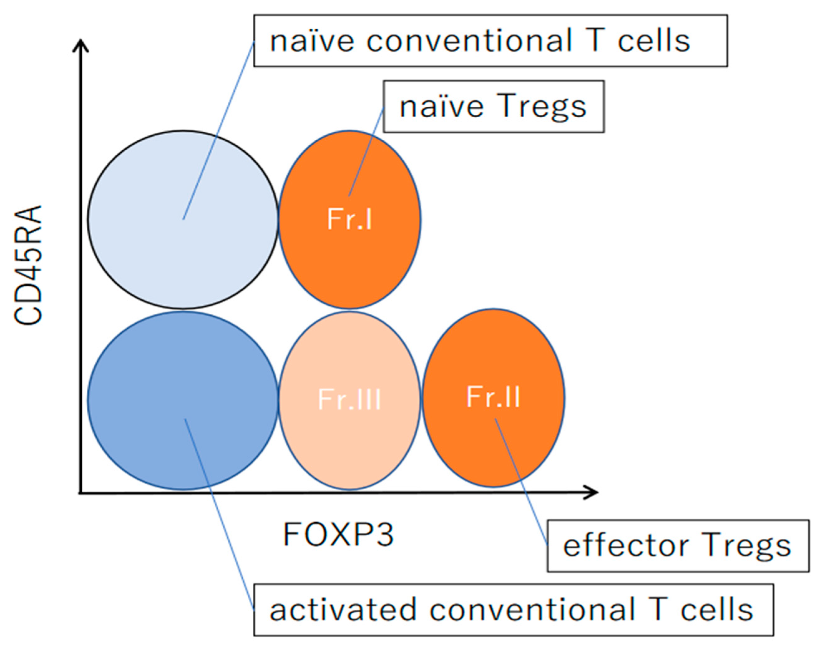
4. Differentiation of Regulatory T Cells in Humans

The lineages from naive CD4+ T-cells toward conventional T cells and Tregs is able to be identified in accordance with surface markers. In the thymus, entire T cell lineages develop and start as naïve CD45RA+ T cells. Following activation in the periphery, naive T cells differentiate into both conventional and regulatory T cells [19]. Conventional T cells differentiate into memory T cells that are able to be reactivated. CD45RA+ (naive) Tregs are reported in humans from two independent research groups in 2006 [65,66].
Circulating CD4+ FOXP3+ T cells are able to be separated into three fractions through the surface makers CD4, CD25, and CD45RA, viz., fraction I (Fr. I) CD45RA+FOXP3low/CD25low resting or naive Tregs; Fr. II CD45RAFOXP3high/CD25high eTregs; and Fr. III CD45RAFOXP3low/CD25low cells (Figure 1) [67]. Fr. I is naïve Tregs and revealed several naïve T-cell markers as CD45RA, CD62L, and CCR7. Fr. II is eTregs. Human CD45RA+ naïve Tregs (Fr. I) differentiate into eTregs (Fr. II) with the downregulation of CD45RA and the upregulation of FOXP3 and CD25 by TCR stimulation. Fr. III is activated conventional T cells and not Tregs despite weakly expressed FOXP3 [67]. Not only Tregs but also non-Tregs express FOXP3+. However, it has been reported that some types of regulatory cells may be included in Fr III. CD45RACXCR5+CD25lowFOXP3low T follicular regulatory (Tfr) cells are the commonly known regulatory cells present in Fr. III [68]. Recent studies have demonstrated that thymus-derived Tregs compose the major part of Tregs (tTregs) in the periphery, whereas some conventional T cells in peripheral sites acquire steady FoxP3 expression and differentiate into peripherally derived Tregs (pTregs) [69]. Naïve conventional T cells can also differentiate in vitro to express FoxP3 in the existence of TGF-β and IL-2, constituting in vitro induced Tregs (iTregs) [70].

5. Regulatory T Cells in IgG4-Related Disease

5.1. Circulating Regulatory T Cells in Patients with IgG4-Related Disease

Miyoshi et al. investigated the Treg phenotype related to circulating nTreg (CD4+CD25+CD45RA+) and eTregs (CD4+CD25high) in patients with type 1 AIP. They also investigated patients with alcoholic or idiopathic pancreatitis and healthy controls by comparison. In patients with type 1 AIP, the ratio of circulating nTregs was significantly lower, whereas that of circulating eTregs was significantly higher. They cannot show the difference between patients with type 1 AIP undergoing steroid treatment and those not undergoing it. Serum levels of IgG4 positively correlated with the proportion of eTregs in the untreated patients with type 1 AIP. [71].
Two functionally different subsets of eTregs in the periphery, inducible costimulatory molecules (ICOS)+ or ICOS eTregs, generate IL-10 or TGF-β, which is the suppressive cytokine, respectively [73]. Kusuda et al. reported the ratio of peripheral ICOS+ Tregs and IL-10+ Tregs in patients with type 1 AIP. They found that the proportion of ICOS+ Tregs was significantly higher in peripheral blood in patients with type 1 AIP than in the control groups (other pancreatic diseases (alcoholic pancreatitis or idiopathic) and healthy controls). Circulating IL-10+ Tregs also significantly expanded in patients with type 1 AIP compared with the control groups. Furthermore, in patients with type 1 AIP, the proportion of IL-10+ Tregs correlated positively with the serum IgG4 levels. These data indicated that a decrease in the number of nTregs plays an initial role in the development of type 1 AIP. The increase of the number of eTregs might be caused by a natural immunological reaction toward inflammatory changes. These expanded numbers of eTregs include tTregs and pTregs. Unfortunately, increased numbers of eTregs cannot control the disease condition. In addition, an increase in the proportion of ICOS+ Tregs might encourage IgG4 production through IL-10. In contrast, ICOS Tregs might be affected in fibrosis through TGF-β [74].
Through which mechanism the number of eTregs increases in patients with type 1 AIP? Newly discovered cytokine as IL-35 produced by Tregs have a potent of a powerful immunosuppression and anti-inflammation. IL-35 belongs to the IL-12 cytokine family, which also includes IL-12, IL-23, and IL-27. IL-35 can encourage the expansion of IL-35-producing Tregs and induce the development of regulatory B cells (Bregs) [75,76]. IL-35 has two subunits termed IL-12A (p35) and Epstein–Barr virus-induced 3 (EBi3) [76]. Ito et al. demonstrated that plasma levels of IL-35 were significantly increased in the patients with type 1 AIP in comparison with patients with alcoholic pancreatitis and healthy subjects. Both IL-12p35 and EBI3 double-positive cells were also infiltrated in the pancreas of patients with type 1 AIP. Some of these double-positive cells also expressed FOXP3 [77]. Elevated levels of plasma IL-35 and expression of IL-35 subunits in the tissue in patients with type 1 AIP could suppress inflammatory change through an increased number of eTregs. The proportion of circulating CD19+CD24highCD38high regulatory B cells was increased in patients with type 1 AIP [78]. IL-35 might also be associated with the development of CD19+CD24highCD38high Bregs (Figure 2).
Recent research has clarified that rituximab was efficacious for IgG4-RD, as was glucocorticoid [79]. CD19+CD27+CD20CD38hi plasmablasts that are mostly IgG4-positive take a vital part in the disease activity of IgG4-RD [79]. The depletion of CD20-positive precursor cells led to a rapid decrease in the proportion of CD19+CD27+CD38hi plasmablasts when treated with rituximab, because CD19+CD27+CD38hi plasmablasts are negative for CD20 [80]. There is significant focus on B cells in the pathophysiology of IgG4-RD. As mentioned earlier, Tfr cells participate in the regulation of GC formation and the class-switch recombination of B cells [81]. The proportion of circulating Tfr cells that produced IL-10, was reported to be markedly increased in patients with IgG4-RD compared with that in healthy elderly subjects. Furthermore, in patients with IgG4-RD, the proportion of circulating Tfr cells significantly correlated with several clinical findings, including serum IgG4 levels and the number of involved organs [72].

5.2. Regulatory T Cells in the Involved Organ in IgG4-Related Disease

Zen et al. clarified that Th2 immune balance Foxp3+ Tregs and regulatory cytokines (IL-10 and TGF-β) have a vital role in type 1 AIP and IgG4-related sclerosing cholangitis (SC). Using immunohistochemical analysis, they showed that Foxp3+ cells were more frequently found in patients with type 1 AIP and IgG4-SC than other disease controls [13]. They also analyzed the gene expression patterns of various cytokines in the patients with control diseases. Their findings showed that in patients with type 1 AIP and IgG4-SC and those with other organ involvements (IgG4-related dacrioadenitis and sialadenitis (DS)), the levels of comparative gene expression of IL-4, IL-10, and TGF-β were all significantly greater in those in patients with IgG4-RD than those in primary sclerosing cholangitis (PSC) and primary biliary cirrhosis (PBC) [13]. Another report also stated that in the pancreas of patients with type 1 AIP, the proportion of Foxp3-positive cells that infiltrated mononuclear cells (Foxp3/Mono) was significantly increased compared with alcoholic chronic pancreatitis (CP); in addition, in patients with type 1 AIP, the proportion of IgG4/Mono correlated positively with Foxp3/Mono [74]. Koyabu et al. analyzed immunohistochemical findings in the livers of patients with IgG4-SC and PSC. They found that the proportion of IgG4/G1-positive cells was significantly higher in patients with IgG4-SC than in patients with PSC. In patients with IgG4-SC, the ratio of Foxp3/Mono was significantly higher than that in patients with PSC. In addition, the number of Foxp3-positive cells in patients with IgG4-SC correlated positively with that of IgG4-positive cells, whereas no such correlation was shown in the other groups [82]. These results indicate that the practice of circulating eTregs parallels that of infiltrated Tregs.
Tanaka et al. provided data about Th1/Th2 cytokines and Tregs in IgG4-related sialadenitis (Mikulicz’s disease) using mRNA expression analysis and immunohistochemistry [83]. In the labial salivary glands (LSGs), various subsets of lymphocyte infiltrated in that of patients with Sjögren’s syndrome and, however, IgG4-positive plasma cells and Tregs selectively infiltrated in IgG4-related sialadenitis. The expression levels of mRNA of Th2 cytokines were significantly increased in in the LSGs of patients with IgG4-related sialadenitis than in controls, whereas the expression levels of both Th1 and Th2 cytokines in patients with Sjögren’s syndrome were significantly increased compared to control groups. Moreover, the gene expression of IL-4 or IL-10 in the LSGs correlated positively with the proportion of IgG4/IgG-positive cells.
Two research groups have reported about IgG4-related kidney disease (RKD). Mizushima et al. examined the histological characteristics of infiltrated cells including Tregs in patients with IgG4-related tubulointerstitial nephritis (TIN) before and after steroid therapy [84]. They clarified that before steroid treatment, histological findings showed dense lymphoplasmacytic infiltration, infiltration of IgG4+ cells and Foxp3+ cells, and interstitial fibrosis. However, after steroid therapy, these histological characteristics disappeared in the renal interstitium. In particular, even in the initial stage of steroid treatment, the numbers of Foxp3+ and IgG4+ cells decreased significantly, whereas a certain degree of CD4+ or CD8+ T cells infiltration persisted into the persistent inflamed site and continued in the later stage [85]. Another study focused on Tregs in IgG4-RKD from the view point of the fibrosis and IgG4 production. It was observed that infiltration of lymphocyte, plasmacyte, and eosinophil and fibrosis in the renal interstitium were more severe in patients with IgG4-RKD than in patients with idiopathic TIN (ITIN) and kidney lesions with Sjögren’s syndrome. The proportions of Foxp3+/CD3+ cells, IgG4+/IgG+ plasma cells, and TGF-β1+ cells to the whole number of infiltrated cells were increased in patients with IgG4-RKD compared with that in ITIN and those with Sjögren’s syndrome. The proportion of Foxp3+/CD3+ cells corelated positively with that of IgG4+/IgG+ in patients with IgG4-RKD. Moreover, in patients with IgG4-RKD, a positive correlation was found between the ratio of Foxp3+/CD3+ cells and that of TGF-β1+ cells to the whole number of infiltrating cells. In addition, in patients with IgG4-RKD, Foxp3+ cells and TGF-β1+ cells were colocalized in the interstitium. In patients with IgG4-RKD the proportion of TGF-β1+ cells to the whole infiltrated cells also correlated significantly with the intensity of fibrotic change. The distribution of type III and type IV collagen in the interstitium was also denser in patients with IgG4-RKD compared with those with Sjögren’s syndrome. Regarding the Th1/Th2 immune environment, the proportion of CXCR3+/CD3+ cells which is Th1 was increased in patients with Sjögren’s syndrome compared to those with IgG4-RKD and ITIN. Conversely, there is no significant difference in the proportion of CCR4+/CD3+ (Th2) cells among three diseases groups [86].
Altogether, these studies on the role of Tregs in not only type 1 AIP but also IgG4-RD have clearly demonstrated that the disease is associated with substantial Treg infiltration in the involved organs [13,74,83,84,85,86]. Tregs might be involved in the initiation of IgG4 production via IL-10. Furthermore, Tregs might be participating in fibrosis via TGF-β [74].

6. Functional Disorders of Regulatory T Cells in IgG4-Related Disease

From the data of the previous research results which is described in the abovementioned section, the following type of question is pertinent. Why can inflammatory reaction not be effectively controlled in patients with IgG4-RD despite the augmentation of Tregs in the involved organs? There are two possible explanations: the severity of inflammation is extremely severe for the Tregs to overcome and/or that the function of Tregs is insufficiently reacted in these inflammations. As mentioned earlier, the phenotype of Mst-1−/− mice is similar to that of IgG4-RD. Tregs from Mst1−/− mice lose their regulatory function. Hence, Mst1−/− mice are not able to prevent the immunological reaction, because Tregs from Mst1−/− mice lose their regulatory function. Hence, Mst1−/− mice cannot prevent an autoimmune reaction [61,62]. Fukuhara et al. reported that the gene expression of MST1 was downregulated in the Tregs of patients with type 1 AIP significantly in comparison to that in healthy controls. Furthermore, they also identified that the number of CpG methylation sites in the 5ʹ region of MST1 was increased in patients with type 1 AIP. This increased frequency of methylated CpG sites correlated positively with the number of involved extrapancreatic lesions. These data indicate that the decrease in the expression of the MST1 gene in Tregs in patients with type 1 AIP as a result of hypermethylation of the promoter involved in the pathophysiology dose influence systemic manifestation in IgG4-RD [87].

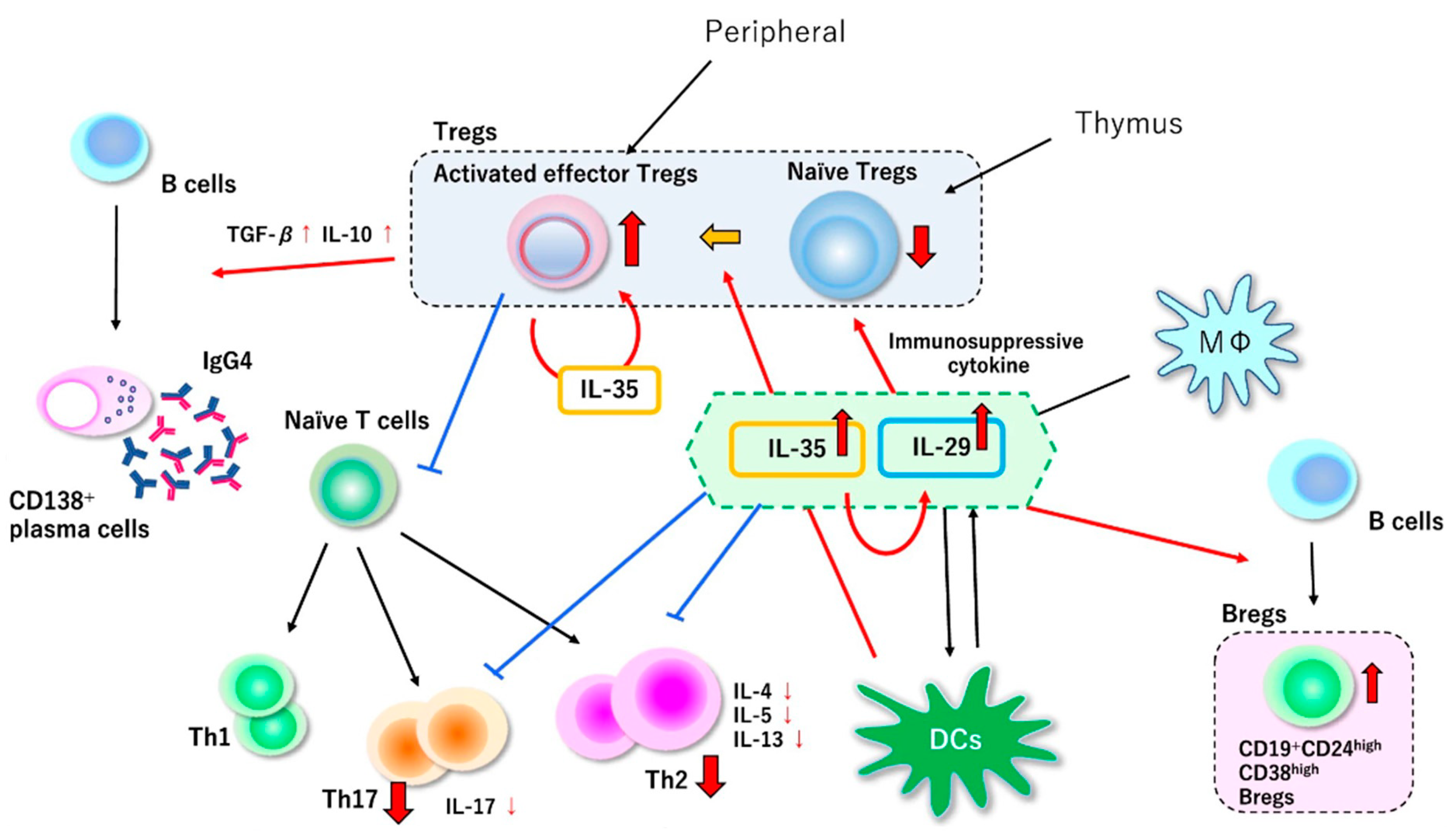
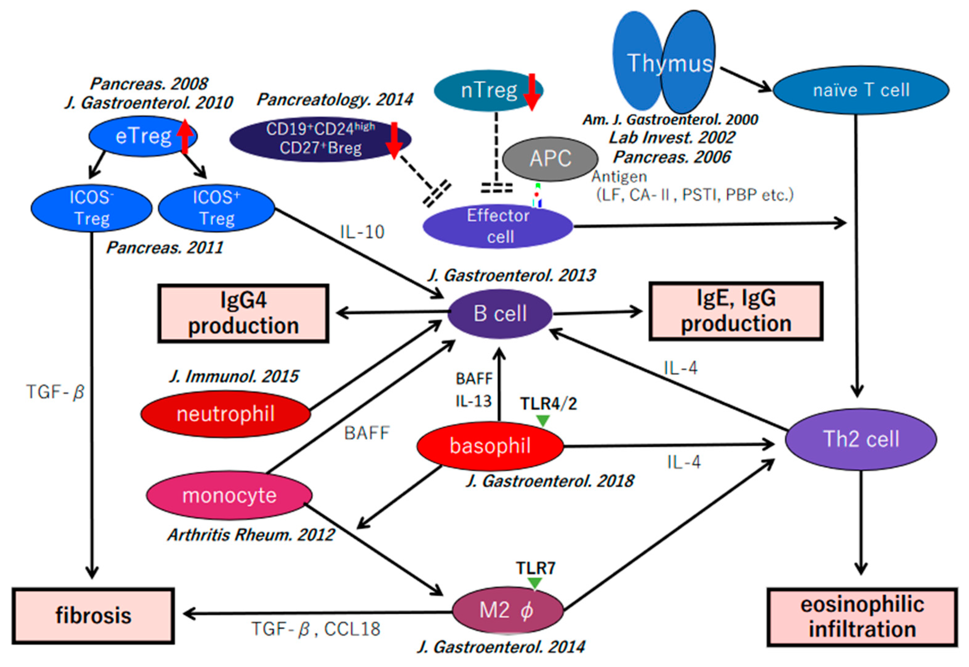
7. Our Hypothesis of the Pathophysiology of IgG4-Related Disease

On the basis of the data accumulated so far and summarized herein, we will provide the following hypothesis for the pathophysiological mechanisms of IgG4-RD. The decrease of the numbers of naive Tregs and CD19+CD24highCD27+ Bregs play an essential role in the development of IgG4-RD. Furthermore, the number of eTregs and that of CD19+CD24+CD38high Bregs expand because of a natural reaction against inflammation through IL-35. Basophils in the involved organs recruit inflamed monocytes and differentiate to M2 macrophages from inflamed monocytes. The Th2 immune environment derived from basophils and M2 macrophages promote disease progression. The increased numbers of eTregs are divided into two subtypes: ICOS-positive and ICOS-negative Tregs. In adaptive immunity, IL-10 produced by ICOS+ Tregs induce the differentiation from B cells to IgG4-producing plasma cells. In an innate immune reaction, monocytes and basophils are also regulating IgG4-production through BAFF via TLR and nucleotide-binding oligomerization domain (NOD)-like receptor signaling. Neutrophils also influence IgG4 production through neutrophil extracellular traps (NETs). ICOS-negative Tregs and M2 macrophages, which perform a suppressive function against inflammation, may regulate fibrosis via TGF-β. Both adaptive and innate immunity may influence the production of IgG4 and fibrosis (Figure 3) [72].

8. Conclusions

Tregs may be critically important in the pathogenesis of IgG4-RD. However, the precise mechanism underlying the immune reaction mediated by Tregs is still unclear. It will be extremely important to clarify the role of Tregs in IgG4-RD.

Funding

This study was partially supported by (1) JSPS KAKENHI Grant Numbers 24591020, 15K09052, 18K07921, 21K07943 and (2) a Grant-in-Aid for “Research for Intractable Diseases” Program from the Ministry of Health, Labor and Welfare of Japan.

Institutional Review Board Statement

Not applicable.

Informed Consent Statement

Not applicable.

Data Availability Statement

The data presented in this review are available on request from the corresponding author.

Acknowledgments

Figure 2 was reprinted from J. Gastroenterol., Ito T et al. Interleukin-35 promotes the differentiation of regulatory T cells and suppresses Th2 response in IgG4-related type 1 autoimmune pancreatitis. J. Gastroenterol. 2020, 55, 789–799, with permission from SPRINGER NATURE.

Conflicts of Interest

The author declare no conflict of interest.

References

  1. Kawaguchi, K.; Koike, M.; Tsuruta, K.; Okamoto, A.; Tabata, I.; Fujita, N. Lymphoplasmacytic sclerosing pancreatitis with cholangitis: A variant of primary sclerosing cholangitis extensively involving pancreas. Hum. Pathol. 1991, 22, 387–395. [Google Scholar] [CrossRef]
  2. Umehara, H.; Okazaki, K.; Kawa, S.; Takahashi, H.; Goto, H.; Matsui, S.; Ishizaka, N.; Akamizu, T.; Sato, Y.; Kawano, M.; et al. The 2020 revised comprehensive diagnostic (RCD) criteria for IgG4-RD. Mod. Rheumatol. 2021, 31, 529–533. [Google Scholar] [CrossRef] [PubMed]
  3. Hamano, H.; Kawa, S.; Horiuchi, A.; Unno, H.; Furuya, N.; Akamatsu, T.; Fukushima, M.; Nikaido, T.; Nakayama, K.; Usuda, N.; et al. High serum IgG4 concentrations in patients with sclerosing pancreatitis. N. Engl. J. Med. 2001, 344, 732–738. [Google Scholar] [CrossRef]
  4. Yoshida, K.; Toki, F.; Takeuchi, T.; Watanabe, S.; Shiratori, K.; Hayashi, N. Chronic pancreatitis caused by an autoimmune abnormality. Dig. Dis. Sci. 1995, 40, 1561–1568. [Google Scholar] [CrossRef] [PubMed]
  5. Kamisawa, T.; Nakajima, H.; Egawa, N.; Funata, N.; Tsuruta, K.; Okamoto, A. IgG4-related sclerosing disease incorporating sclerosing pancreatitis, cholangitis, sialadenitis and retroperitoneal fibrosis with lymphadenopathy. Pancreatology 2006, 6, 132–137. [Google Scholar] [CrossRef]
  6. Yamamoto, M.; Takahashi, H.; Ohara, M.; Suzuki, C.; Naishiro, Y.; Yamamoto, H.; Shinomura, Y.; Imai, K. A new conceptualization for Mikulicz’s disease as an IgG4-related plasmacytic disease. Mod. Rheumatol. 2006, 16, 335–340. [Google Scholar] [CrossRef]
  7. Masaki, Y.; Dong, L.; Kurose, N.; Kitagawa, K.; Morikawa, Y.; Yamamoto, M.; Takahashi, H.; Shinomura, Y.; Imai, K.; Saeki, T.; et al. Proposal for a new clinical entity, IgG4-positive multiorgan lymphoproliferative syndrome: Analysis of 64 cases of IgG4-related disorders. Ann. Rheum. Dis. 2009, 68, 1310–1315. [Google Scholar] [CrossRef]
  8. Umehara, H.; Okazaki, K.; Masaki, Y.; Kawano, M.; Yamamoto, M.; Saeki, T.; Matsui, S.; Sumida, T.; Mimori, T.; Tanaka, Y.; et al. A novel clinical entity, IgG4-related disease (IgG4RD): General concept and details. Mod. Rheumatol. 2012, 22, 1–14. [Google Scholar] [CrossRef]
  9. Shimosegawa, T.; Chari, S.T.; Frulloni, L.; Kamisawa, T.; Kawa, S.; Mino-Kenudson, M.; Kim, M.H.; Klöppel, G.; Lerch, M.M.; Löhr, M.; et al. International consensus diagnostic criteria for autoimmune pancreatitis: Guidelines of the International Association of Pancreatology. Pancreas 2011, 40, 352–358. [Google Scholar] [CrossRef]
  10. Notohara, K.; Burgart, L.J.; Yadav, D.; Chari, S.; Smyrk, T.C. Idiopathic chronic pancreatitis with periductal lymphoplasmacytic infiltration: Clinicopathologic features of 35 cases. Am. J. Surg. Pathol. 2003, 27, 1119–1127. [Google Scholar] [CrossRef]
  11. Zamboni, G.; Lüttges, J.; Capelli, P.; Frulloni, L.; Cavallini, G.; Pederzoli, P.; Leins, A.; Longnecker, D.; Klöppel, G. Histopathological features of diagnostic and clinical relevance in autoimmune pancreatitis: A study on 53 resection specimens and 9 biopsy specimens. Virchows Arch. 2004, 445, 552–563. [Google Scholar] [CrossRef] [PubMed]
  12. Moore, K.W.; Vieira, P.; Fiorentino, D.F.; Trounstine, M.L.; Khan, T.A.; Mosmann, T.R. Homology of cytokine synthesis inhibitory factor (IL-10) to the Epstein–Barr virus gene BCRFI. Science 1990, 248, 1230–1234. [Google Scholar] [CrossRef] [PubMed]
  13. Zen, Y.; Fujii, T.; Harada, K.; Kawano, M.; Yamada, K.; Takahira, M.; Nakanuma, Y. Th2 and regulatory immune reactions are increased in immunoglobin G4-related sclerosing pancreatitis and cholangitis. Hepatology 2007, 45, 1538–1546. [Google Scholar] [CrossRef] [PubMed]
  14. Groux, H.; O’Garra, A.; Bigler, M.; Rouleau, M.; Antonenko, S.; de Vries, J.E.; Roncarolo, M.G. A CD4+ T-cell subset inhibits antigen-specific T-cell responses and prevents colitis. Nature 1997, 389, 737–742. [Google Scholar] [CrossRef]
  15. Mizoguchi, A.; Mizoguchi, A.; Mizoguchi, E.; Takedatsu, H.; Blumberg, R.S.; Bhan, A.K. Chronic intestinal inflammatory condition generates IL-10-producing regulatory B cell subset characterized by CD1d upregulation. Immunity 2002, 16, 219–230. [Google Scholar] [CrossRef] [Green Version]
  16. Fujita, S.; Seino, K.; Sato, K.; Sato, Y.; Eizumi, K.; Yamashita, N.; Taniguchi, M.; Sato, K. Regulatory dendritic cells act as regulators of acute lethal systemic inflammatory response. Blood 2006, 107, 3656–3664. [Google Scholar] [CrossRef] [Green Version]
  17. Hoshino, S.; Kurishima, A.; Inaba, M.; Ando, Y.; Fukui, T.; Uchida, K.; Nishio, A.; Iwai, H.; Yokoi, T.; Ito, T.; et al. Amelioration of 2,4,6-trinitrobenzene sulfonic acid-induced colitis in mice by immunoregulatory dendritic cells. J. Gastroenterol. 2011, 46, 1368–1381. [Google Scholar] [CrossRef]
  18. Lechner, A.; Bohnacker, S.; Esser-von Bieren, J. Macrophage regulation & function in helminth infection. Semin Immunol. 2021, 53, 101526. [Google Scholar]
  19. Sakaguchi, S.; Mikami, N.; Wing, J.B.; Tanaka, A.; Ichiyama, K.; Ohkura, N. Regulatory T cells and Human Disease. Annu. Rev. Immunol. 2020, 38, 541–566. [Google Scholar] [CrossRef] [Green Version]
  20. Sakaguchi, S.; Sakaguchi, N.; Asano, M.; Itoh, M.; Toda, M. Immunologic self-tolerance maintained by activated T cells expressing IL-2 receptor alpha-chains (CD25). Breakdown of a single mechanism of self-tolerance causes various autoimmune diseases. J. Immunol. 1995, 155, 1151–1164. [Google Scholar]
  21. Baecher-Allan, C.; Brown, J.A.; Freeman, G.J.; Hafler, D.A. CD4+CD25 high regulatory cells in human peripheral blood. J. Immunol. 2001, 167, 1245–1253. [Google Scholar] [CrossRef] [PubMed] [Green Version]
  22. Dieckmann, D.; Plottner, H.; Berchtold, S.; Berger, T.; Schuler, G. Ex vivo isolation and characterization of CD4(+) CD25(+) T cells with regulatory properties from human blood. J. Exp. Med. 2001, 193, 1303–1310. [Google Scholar] [CrossRef] [PubMed]
  23. Jonuleit, H.; Schmitt, E.; Stassen, M.; Tuettenberg, A.; Knop, J.; Enk, A.H. Identification and functional characterization of human CD4(+) CD25(+) T cells with regulatory properties isolated from peripheral blood. J. Exp. Med. 2001, 193, 1285–1294. [Google Scholar] [CrossRef] [PubMed] [Green Version]
  24. Levings, M.K.; Sangregorio, R.; Roncarolo, M.G. Human CD25(+) CD4(+) T regulatory cells suppress naive and memory T cell proliferation and can be expanded in vitro without loss of function. J. Exp. Med. 2001, 193, 1295–1302. [Google Scholar] [CrossRef] [PubMed]
  25. Hori, S.; Nomura, T.; Sakaguchi, S. Control of regulatory T cell development by the transcription factor Foxp3. Science 2003, 299, 1057–1061. [Google Scholar] [CrossRef] [Green Version]
  26. Fontenot, J.D.; Gavin, M.A.; Rudensky, A.Y. Foxp3 programs the development and function of CD4+CD25+ regulatory T cells. Nat. Immunol. 2003, 4, 330–336. [Google Scholar] [CrossRef]
  27. Khattri, R.; Cox, T.; Yasayko, S.A.; Ramsdell, F. An essential role for Scurfin in CD4+CD25+ T regulatory cells. Nat. Immunol. 2003, 4, 337–342. [Google Scholar] [CrossRef]
  28. Morikawa, H.; Sakaguchi, S. Genetic and epigenetic basis of Treg cell development and function: From a FoxP3-centered view to an epigenome-defined view of natural Treg cells. Immunol. Rev. 2014, 259, 192–205. [Google Scholar] [CrossRef]
  29. Katoh, H.; Zheng, P.; Liu, Y. FOXP3: Genetic and epigenetic implications for autoimmunity. J. Autoimmun. 2013, 41, 72–78. [Google Scholar] [CrossRef] [Green Version]
  30. Schmidt, A.; Oberle, N.; Krammer, P.H. Molecular mechanisms of Treg-mediated T cell suppression. Front. Immunol. 2012, 3, 51. [Google Scholar] [CrossRef] [Green Version]
  31. Sakaguchi, S.; Wing, K.; Onishi, Y.; Prieto-Martin, P.; Yamaguchi, T. Regulatory T cells: How do they suppress immune responses? Int. Immunol. 2009, 21, 1105–1111. [Google Scholar] [CrossRef] [PubMed]
  32. Roncarolo, M.G.; Gregori, S.; Battaglia, M.; Bacchetta, R.; Fleischhauer, K.; Levings, M.K. Interleukin-10-secreting type 1 regulatory T cells in rodents and humans. Immunol. Rev. 2006, 212, 28–50. [Google Scholar] [CrossRef] [PubMed]
  33. Sakaguchi, S.; Yamaguchi, T.; Nomura, T.; Ono, M. Regulatory T cells and immune tolerance. Cell 2008, 133, 775–787. [Google Scholar] [CrossRef] [PubMed] [Green Version]
  34. Sakaguchi, S.; Miyara, M.; Costantino, C.M.; Hafler, D.A. FOXP3+ regulatory T cells in the human immune system. Nat. Rev. Immunol. 2010, 10, 490–500. [Google Scholar] [CrossRef] [PubMed]
  35. Crispin, J.C.; Martínez, A.; Alcocer-Varela, J. Quantification of regulatory T cells in patients with systemic lupus erythematosus. J. Autoimmun. 2003, 21, 273–276. [Google Scholar] [CrossRef]
  36. Cao, D.; van Vollenhoven, R.; Klareskog, L.; Trollmo, C.; Malmström, V. CD25bright CD4+ regulatory T cells are enriched in inflamed joints of patients with chronic rheumatic disease. Arthritis Res. Ther. 2004, 6, 335–346. [Google Scholar] [CrossRef] [Green Version]
  37. De Kleer, I.M.; Wedderburn, L.R.; Taams, L.S.; Patel, A.; Varsani, H.; Klein, M.; de Jager, W.; Pugayung, G.; Giannoni, F.; Rijkers, G.; et al. CD4CD25bright regulatory T cells actively regulate inflammation in the joints of patients with the remitting from of juvenile idiopathic arthritis. J. Immunol. 2004, 172, 6435–6443. [Google Scholar] [CrossRef] [Green Version]
  38. Cao, D.; Malmström, V.; Baecher-Allan, C.; Hafler, D.; Klareskog, L.; Trollmo, C. Isolation and functional characterization of regulatory CD25bright CD4+ T cells from the target organ of patients with rheumatoid arthritis. Eur. J. Immunol. 2003, 33, 215–223. [Google Scholar] [CrossRef]
  39. Longhi, M.S.; Ma, Y.; Bogdanos, D.P.; Cheeseman, P.; Mieli-Vergani, G.; Vergani, D. Impairment of CD4+CD25+ regulatory T cells in autoimmune liver disease. J. Hepatol. 2004, 41, 31–37. [Google Scholar] [CrossRef]
  40. Boyer, O.; Saadoun, D.; Abriol, J.; Dodille, M.; Piette, J.C.; Cacoub, P.; Klatzmann, D. CD4+CD25+ regulatory T-cell deficiency in patients with hepatitis C-mixed cryoglobulinemia vasculitis. Blood 2004, 103, 3428–3430. [Google Scholar] [CrossRef]
  41. Furuno, K.; Yuge, T.; Kusuhara, K.; Takada, H.; Nishio, H.; Khajoee, V.; Ohno, T.; Hara, T. CD25+CD4+ regulatory T cells in patients with Kawasaki disease. J. Pediatr. 2004, 145, 385–390. [Google Scholar] [CrossRef] [PubMed]
  42. Lindley, S.; Dayan, C.M.; Bishop, A.; Roep, B.O.; Peakman, M.; Tree, T.I. Defective suppressor function in CD4(+) CD25(+) T-cells from patients with type I diabetes. Diabetes 2005, 193, 1303–1310. [Google Scholar] [CrossRef] [PubMed] [Green Version]
  43. Putnam, A.L.; Vendrame, F.; Dotta, F.; Gottlieb, P.A. CD4+CD25high regulatory T cells in human autoimmune diabetes. J. Autoimmun. 2005, 24, 55–62. [Google Scholar] [CrossRef] [PubMed]
  44. Putheti, P.; Pettersson, A.; Soderstrom, M.; Link, H.; Huang, Y.M. Circulating CD4+CD25+ T regulatory cells are not altered in multiple sclerosis and unaffected by disease-modulating drugs. J. Clin. Immunol. 2004, 24, 155–161. [Google Scholar] [CrossRef]
  45. Viglietta, V.; Baecher-Allan, C.; Weiner, H.L.; Hafler, D.A. Loss of functional suppression by CD4+CD25+ regulatory T patients with multiple sclerosis. J. Exp. Med. 2004, 199, 971–979. [Google Scholar] [CrossRef] [Green Version]
  46. Gottenberg, J.E.; Lavie, F.; Abbed, K.; Gasnault, J.; Le Nevot, E.; Delfraissy, J.F.; Taoufik, Y.; Mariette, X. CD4 CD25high regulatory T cells are not impaired in patients with primary Sjögren’s syndrome. J. Autoimmun. 2005, 24, 235–242. [Google Scholar] [CrossRef]
  47. Balandina, A.; Lecart, S.; Dartevelle, P.; Saoudi, A.; Berrih-Aknin, S. Functional defect of regulatory CD4+CD25+ T cells in the thymus of patients with autoimmune Myasthenia Gravis. Blood 2005, 105, 734–741. [Google Scholar] [CrossRef] [Green Version]
  48. Fattorossi, A.; Battaglia, A.; Buzzonetti, A.; Ciaraffa, F.; Scambia, G.; Evoli, A. Circulating and thymic CD4+CD25+ T regulatory cells in myasthenia gravis: Effect of immunosuppressive treatment. Immunology 2005, 116, 134–141. [Google Scholar] [CrossRef]
  49. Van Amelsfort, J.M.; Jacobs, K.M.; Bijlsma, J.W.; Bijlsma, J.W.; Lafeber, F.P.; Taams, L.S. CD4+CD25+ regulatory T cells in rheumatoid arthritis: Differences in the presence, phenotype, and function between peripheral blood and synovial fluid. Arthritis Rheum. 2004, 50, 2775–2785. [Google Scholar] [CrossRef]
  50. Ehrenstein, M.R.; Evans, J.G.; Singh, A.; Moore, S.; Warnes, G.; Isenberg, D.A.; Mauri, C. Compromised function of regulatory T cells in rheumatoid arthritis and reversal by anti-TNF alpha therapy. J. Exp. Med. 2004, 200, 277–285. [Google Scholar] [CrossRef]
  51. Möttönen, M.; Heikkinen, J.; Mustonen, I.; Isomäki, P.; Luukkainen, R.; Lassila, O. CD4+ CD25+ T cells with the phenotypic and functional characteristics of regulatory T cells are enriched in the synovial fluid of patients with rheumatoid arthritis. Clin. Exp. Immunol. 2004, 140, 360–367. [Google Scholar] [CrossRef] [PubMed]
  52. Vigna-Pérez, M.; Abud-Mendoza, C.; Portillo-Salazar, H.; Alvarado-Sánchez, B.; Cuevas-Orta, E.; Moreno-Valdés, R.; Baranda, L.; Paredes-Saharopulos, O.; González-Amaro, R. Immune effects of therapy with adalimumab in patients with rheumatoid arthritis. Clin. Exp. Immunol. 2005, 141, 372–380. [Google Scholar] [CrossRef] [PubMed]
  53. Makita, S.; Kanai, T.; Oshima, S.; Uraushihara, K.; Totsuka, T.; Sawada, T.; Nakamura, T.; Koganei, K.; Fukushima, T.; Watanabe, M. CD4+CD25bright T cells in human intestinal lamina propria as regulatory cells. J. Immunol. 2004, 173, 3119–3130. [Google Scholar] [CrossRef] [PubMed] [Green Version]
  54. Maul, J.; Loddenkemper, C.; Mundt, P.; Berg, E.; Giese, T.; Stallmach, A.; Zeitz, M.; Duchmann, R. Peripheral and intestinal regulatory CD4+ CD25(high) T cells in inflammatory bowel disease. Gastroenterology 2005, 128, 1868–1878. [Google Scholar] [CrossRef]
  55. Nakajima, T.; Ueki-Maruyama, K.; Oda, T.; Ohsawa, Y.; Ito, H.; Seymour, G.J.; Yamazaki, K. Regulatory T-cells infiltrate periodontal disease tissues. J. Dent. Res. 2005, 84, 639–643. [Google Scholar] [CrossRef] [Green Version]
  56. Okui, T.; Ito, H.; Honda, T.; Amanuma, R.; Yoshie, H.; Yamazaki, K. Characterization of CD4+ FOXP3+ T-cell clones established from chronic inflammatory lesions. Oral Microbiol. Immunol. 2008, 23, 49–54. [Google Scholar] [CrossRef]
  57. Asada, M.; Nishio, A.; Uchida, K.; Kido, M.; Ueno, S.; Uza, N.; Kiriya, K.; Inoue, S.; Kitamura, H.; Ohashi, S.; et al. Identification of a novel autoantibody against pancreatic secretory trypsin inhibitor in patients with autoimmune pancreatitis. Pancreas 2006, 33, 20–26. [Google Scholar] [CrossRef] [Green Version]
  58. Uchida, K.; Okazaki, K.; Nishi, T.; Uose, S.; Nakase, H.; Ohana, M.; Matsushima, Y.; Omori, K.; Chiba, T. Experimental immune-mediated pancreatitis in neonatally thymectomized mice immunized with carbonic anhydrase II and lactoferrin. Lab. Investig. 2002, 82, 411–424. [Google Scholar] [CrossRef] [Green Version]
  59. Sakaguchi, Y.; Inaba, M.; Tsuda, M.; Quan, G.K.; Omae, M.; Ando, Y.; Uchida, K.; Okazaki, K.; Ikehara, S. The Wistar Bonn Kobori rat, a unique animal model for autoimmune pancreatitis with extrapancreatic exocrinopathy. Clin. Exp. Immunol. 2008, 152, 1–12. [Google Scholar] [CrossRef]
  60. Bowman, M.; Holt, G. Selective enhancement of systemic Th1 immunity in immunologically immature rats with an orally administered bacterial extract. Infect Immun. 2001, 69, 3719–3727. [Google Scholar] [CrossRef] [Green Version]
  61. Ueda, Y.; Katagiri, K.; Tomiyama, T.; Yasuda, K.; Habiro, K.; Katakai, T.; Ikehara, S.; Matsumoto, M.; Kinashi, T. Mst1 regulates integrin-dependent thymocyte trafficking and antigen recognition in the thymus. Nat. Commun. 2012, 3, 1098. [Google Scholar] [CrossRef] [PubMed]
  62. Tomiyama, T.; Ueda, Y.; Katakai, T.; Kondo, N.; Okazaki, K.; Kinashi, T. Antigen-specific suppression and immunological synapse formation by regulatory T cells require the Mst1 kinase. PLoS ONE 2013, 8, e73874. [Google Scholar] [CrossRef] [PubMed]
  63. Yamashina, M.; Nishio, A.; Nakayama, S.; Okazaki, T.; Uchida, K.; Fukui, T.; Okazaki, K. Comparative study on experimental autoimmune pancreatitis and its extrapancreatic involvement in mice. Pancreas 2012, 41, 1255–1262. [Google Scholar] [CrossRef] [PubMed]
  64. Koyabu, M.; Uchida, K.; Sakaguchi, Y.; Fukata, N.; Kusuda, T.; Miyoshi, H.; Yoshida, K.; Sumimoto, K.; Mitsuyama, T.; Fukui, T.; et al. Possible involvement of Foxp3(+) regulatory T cells in the development of immune-mediated pancreatitis in MRL/Mp mice treated with polyinosinic:polycytidylic acid. Int. J. Rheumatol. 2013, 2013, 367325. [Google Scholar] [CrossRef]
  65. Seddiki, N.; Santner-Nanan, B.; Tangye, S.G.; Alexander, S.I.; Solomon, M.; Lee, S.; Nanan, R.; Fazekas de Saint Groth, B. Persistence of naive CD45RA+ regulatory T cells in adult life. Blood 2006, 107, 2830–2838. [Google Scholar] [CrossRef] [Green Version]
  66. Fritzsching, B.; Oberle, N.; Pauly, E.; Geffers, R.; Buer, J.; Poschl, J.; Krammer, P.; Linderkamp, O.; Suri-Payer, E. Naive regulatory T cells: A novel subpopulation defined by resistance toward CD95L-mediated cell death. Blood 2006, 108, 3371–3378. [Google Scholar] [CrossRef]
  67. Miyara, M.; Yoshioka, Y.; Kitoh, A.; Shima, T.; Wing, K.; Niwa, A.; Parizot, C.; Taflin, C.; Heike, T.; Valeyre, D.; et al. Functional delineation and differentiation dynamics of human CD4+ T cells expressing the FoxP3 transcription factor. Immunity 2009, 30, 899–911. [Google Scholar] [CrossRef] [Green Version]
  68. Wing, J.B.; Kitagawa, Y.; Locci, M.; Hume, H.; Tay, C.; Morita, T.; Kidani, Y.; Matsuda, K.; Inoue, T.; Kurosaki, T.; et al. A distinct subpopulation of CD25− T- follicular regulatory cells localizes in the germinal centers. Proc. Natl. Acad. Sci. USA 2017, 114, E6400–E6409. [Google Scholar] [CrossRef] [Green Version]
  69. Abbas, A.K.; Benoist, C.; Bluestone, J.A.; Campbell, D.J.; Ghosh, S.; Hori, S.; Jiang, S.; Kuchroo, V.K.; Mathis, D.; Roncarolo, M.G.; et al. Regulatory T cells: Recommendations to simplify the nomenclature. Nat. Immunol. 2013, 14, 307–308. [Google Scholar] [CrossRef]
  70. Chen, W.; Jin, W.; Hardegen, N.; Lei, K.J.; Li, L.; Marinos, N.; McGrady, G.; Wahl, S.M. Conversion of peripheral CD4+CD25− naive T cells to CD4+CD25+ regulatory T cells by TGF-β induction of transcription factor Foxp3. J. Exp. Med. 2003, 198, 1875–1886. [Google Scholar] [CrossRef]
  71. Miyoshi, H.; Uchida, K.; Taniguchi, T.; Yazumi, S.; Matsushita, M.; Takaoka, M.; Okazaki, K. Circulating naïve and CD4+CD25high regulatory T cells in patients with autoimmune pancreatitis. Pancreas 2008, 36, 133–140. [Google Scholar] [CrossRef] [PubMed]
  72. Ito, F.; Kamekura, R.; Yamamoto, M.; Takano, K.; Takaki, H.; Yabe, H.; Ikegami, I.; Shigehara, K.; Himi, T.; Takahashi, H.; et al. IL-10 + T follicular regulatory cells are associated with the pathogenesis of IgG4-related disease. Immunol Lett. 2019, 207, 56–563. [Google Scholar] [CrossRef] [PubMed]
  73. Ito, T.; Hanabuchi, S.; Wang, Y.H.; Park, W.R.; Arima, K.; Bover, L.; Qin, F.X.; Gilliet, M.; Liu, Y.J. Two functional subsets of FOXP3+ regulatory T cells in human thymus and periphery. Immunity 2008, 28, 870–880. [Google Scholar] [CrossRef] [Green Version]
  74. Kusuda, T.; Uchida, K.; Miyoshi, H.; Koyabu, M.; Satoi, S.; Takaoka, M.; Shikata, N.; Uemura, Y.; Okazaki, K. Involvement of inducible costimulator- and interleukin 10-positive regulatory T cells in the development of IgG4-related autoimmune pancreatitis. Pancreas 2011, 40, 1120–1130. [Google Scholar] [CrossRef] [PubMed]
  75. Collison, L.W.; Workman, C.J.; Kuo, T.T.; Boyd, K.; Wang, Y.; Vignali, K.M.; Cross, R.; Sehy, D.; Blumberg, R.S.; Vignali, D.A. The inhibitory cytokine IL-35 contributes to regulatory T-cell function. Nature 2007, 450, 566–569. [Google Scholar] [CrossRef] [PubMed]
  76. Collison, L.W.; Delgoffe, G.M.; Guy, C.S.; Vignali, K.M.; Chaturvedi, V.; Fairweather, D.; Satoskar, A.R.; Garcia, K.C.; Hunter, C.A.; Drake, C.G.; et al. The composition and signaling of the IL-35 receptor are unconventional. Nat. Immunol. 2012, 13, 290–299. [Google Scholar] [CrossRef] [PubMed] [Green Version]
  77. Ito, T.; Tanaka, T.; Nakamaru, K.; Tomiyama, T.; Yamaguchi, T.; Ando, Y.; Ikeura, T.; Fukui, T.; Uchida, K.; Nishio, A.; et al. Interleukin-35 promotes the differentiation of regulatory T cells and suppresses Th2 response in IgG4-related type 1 autoimmune pancreatitis. J. Gastroenterol. 2020, 55, 789–799. [Google Scholar] [CrossRef]
  78. Sumimoto, K.; Uchida, K.; Kusuda, T.; Mitsuyama, T.; Sakaguchi, Y.; Fukui, T.; Matsushita, M.; Takaoka, M.; Nishio, A.; Okazaki, K. The role of CD19+ CD24high CD38high and CD19+ CD24high CD27+ regulatory B cells in patients with type 1 autoimmune pancreatitis. Pancreatology 2014, 14, 193–200. [Google Scholar] [CrossRef] [Green Version]
  79. Khosroshahi, A.; Carruthers, M.N.; Deshpande, V.; Unizony, S.; Bloch, D.B.; Stone, J.H. Rituximab for the treatment of IgG4-related disease: Lessons from 10 consecutive patients. Medicine 2012, 91, 57–66. [Google Scholar] [CrossRef]
  80. Mattoo, H.; Mahajan, V.S.; Della-Torre, E.; Sekigami, Y.; Carruthers, M.; Wallace, Z.S.; Deshpande, V.; Stone, J.H.; Pillai, S. De novo oligoclonal expansions of circulating plasmablasts in active and relapsing IgG4-related disease. J. Allergy Clin. Immunol. 2014, 134, 679–687. [Google Scholar] [CrossRef] [Green Version]
  81. Sage, P.T.; Sharpe, A.H. T follicular regulatory cells in the regulation of B cell responses. Trends Immunol. 2015, 36, 410–418. [Google Scholar] [CrossRef] [PubMed] [Green Version]
  82. Koyabu, M.; Uchida, K.; Miyoshi, H.; Sakaguchi, Y.; Fukui, T.; Ikeda, H.; Takaoka, M.; Hirohara, J.; Nishio, A.; Uemura, Y.; et al. Analysis of regulatory T cells and IgG4-positive plasma cells among patients of IgG4-related sclerosing cholangitis and autoimmune liver diseases. J. Gastroenterol. 2010, 45, 732–741. [Google Scholar] [CrossRef] [PubMed]
  83. Tanaka, A.; Moriyama, M.; Nakashima, H.; Miyake, K.; Hayashida, J.N.; Maehara, T.; Shinozaki, S.; Kubo, Y.; Nakamura, S. Th2 and regulatory immune reactions contribute to IgG4 production and the initiation of Mikulicz disease. Arthritis Rheum. 2012, 64, 254–263. [Google Scholar] [CrossRef] [PubMed]
  84. Mizushima, I.; Yamada, K.; Fujii, H.; Inoue, D.; Umehara, H.; Yamagishi, M.; Yamaguchi, Y.; Nagata, M.; Matsumura, M.; Kawano, M. Clinical and histological changes associated with corticosteroid therapy in IgG4-related tubulointerstitial nephritis. Mod. Rheumatol. 2012, 22, 859–870. [Google Scholar] [CrossRef]
  85. Kawamura, E.; Hisano, S.; Nakashima, H.; Takeshita, M.; Saito, T. Immunohistological analysis for immunological response and mechanism of interstitial fibrosis in IgG4-related kidney disease. Mod. Rheumatol. 2015, 25, 571–578. [Google Scholar] [CrossRef]
  86. Fukuhara, T.; Tomiyama, T.; Yasuda, K.; Ueda, Y.; Ozaki, Y.; Son, Y.; Nomura, S.; Uchida, K.; Okazaki, K.; Kinashi, T. Hypermethylation of MST1 in IgG4-related autoimmune pancreatitis and rheumatoid arthritis. Biochem. Biophys. Res. Commun. 2015, 463, 968–974. [Google Scholar] [CrossRef]
  87. Uchida, K.; Okazaki, K. Clinical and pathophysiological aspects of type 1 autoimmune pancreatitis. J. Gastroenterol. 2018, 53, 475–483. [Google Scholar] [CrossRef] [Green Version]
Figure 1. Subpopulations of FOXP3+ T cells in humans using CD25, CD45RA, and FOXP3. CD4+ CD25+ FOXP3+ cells analyzed using CD45RA. FOXP3 CD45RA+ cells are naïve conventional T cells. FOXP3 CD45RA cells are activated conventional T cells. FOXP3+ cells are divided into three groups. Fr. I is naïve Tregs. Fr. II is eTregs. Human CD45RA+ naive Tregs (nTregs) (Fr. I) differentiate into effector Tregs (eTregs) (Fr. II) with the downregulation of CD45RA and the upregulation of FOXP3 and CD25 by TCR stimulation [19,67]. In IgG4-RD, circulating nTregs were decreased and e Tregs were increased [71]. Fr. III is primarily activated in conventional T cells and not Tregs, which exhibit weak positive FOXP3 expression. However, CD45RACXCR5+CD25lowFOXP3low circulating T follicular regulatory (Tfr) cells are Fr. III [19,67]. Tfr cells were increased and correlated with clinical manifestations in IgG4-RD [72].
Immuno 02 00026 g001
Figure 2. Proposed pathophysiological mechanisms of type 1 AIP focusing on IL-35. The decrease of the proportion of naïve regulatory T cells (nTregs) might participate in the initiation of type 1 AIP. The proportion of effector regulatory T cells (eTregs) increased through a natural immune reaction. Plasma levels of IL-35 were significantly increased in the patients with type 1 AIP. Serum IL-35 and IL-29 elevated with various regulatory cells. IL-35 reacted on eTregs and advanced IL-35 production. In type 1 AIP, CD19+CD24highCD38high regulatory B cells (Bregs) were increased [78]. IL-35 also increased CD19+CD24highCD38high Bregs. This figure was reprinted from [77], with kind permission from Springer Nature.
Immuno 02 00026 g002
Figure 3. Proposed hypothesis for the pathophysiology of IgG4-RD. A reduction in the numbers of naïve regulatory T cells (Tregs) and CD19+CD24highCD27+ regulatory B cells (Bregs) might be responsible for the initiation of IgG4-RD, including type 1 AIP. The number of effector Tregs (eTregs) and CD19+CD24+CD38high Bregs increased reactively. Basophils recruit inflamed monocytes into target organs and also differentiate to M2 macrophages. ICOS-positive Tregs might control IgG4 production via IL-10, monocytes and basophil also control IgG4 production via BAFF through the pathway using NOD-like receptor and TLR signaling. M2 macrophages and ICOS-negative Tregs might regulate fibrosis. M2 Φ and basophil might also influence the environment in IgG4-RD. Neutrophils also contribute to IgG4 production through NETs. The figure was reprinted from [72].
Immuno 02 00026 g003
Publisher’s Note: MDPI stays neutral with regard to jurisdictional claims in published maps and institutional affiliations.

