Article

Impact of hMLH1 −93G>A (rs1800734) and hMSH2 1032G>A (rs4987188) Polymorphisms on Colorectal Cancer Susceptibility

1
Laboratory of Human Genetics, Genetic Resources Institute, Ministry of Science and Education, Baku AZ1106, Azerbaijan
2
Department of Natural Sciences, Western Caspian University, Ahmad Rajabli st., 17A, Baku AZE1072, Azerbaijan
3
Department of Oncology, Oncological Clinic, Azerbaijan Medical University, Baku AZ1022, Azerbaijan
4
Department of Medical Biology and Genetics, Azerbaijan Medical University, Baku AZ1022, Azerbaijan
5
Department of Biochemistry, Istanbul University, Istanbul 34452, Türkiye
6
Department of Surgery, Scientific Surgery Center Named after Academician M.A. Topchubashov, Baku AZ1122, Azerbaijan
7
Department of Cytology, Embryology, and Histology, Azerbaijan Medical University, Baku AZ1078, Azerbaijan
8
Department of Surgery, Azerbaijan Medical University, Baku AZ1022, Azerbaijan
*
Authors to whom correspondence should be addressed.
J. Mol. Pathol. 2025, 6(3), 15; https://doi.org/10.3390/jmp6030015
Submission received: 2 May 2025 / Revised: 26 June 2025 / Accepted: 4 July 2025 / Published: 8 July 2025

Abstract

Background: This study is the first to investigate the association between colorectal cancer (CRC) risk and the hMLH1 −93G>A and hMSH2 1032G>A polymorphisms of mismatch repair (MMR) genes in the Azerbaijani population. Methods: Peripheral blood samples containing EDTA were collected from the study subjects (134 patients and 137 controls), and genomic DNA was extracted using the non-enzymatic salting-out method. Genotypes were determined by polymerase chain reaction–restriction fragment length polymorphism (PCR-RFLP), and the results were visualized through agarose gel electrophoresis. Results: Overall, no statistically significant correlation was observed between CRC risk and the hMLH1 −93G>A polymorphism in the heterozygous GA (OR = 0.760; 95% CI = 0.374–1.542; p = 0.446), the mutant AA (OR = 1.474; 95% CI = 0.738–2.945; p = 0.270), or the A allele (OR = 1.400; 95% CI = 0.984–1.995; p = 0.062). However, in contrast to the dominant model, a statistically significant association was found between the recessive model and an increased CRC risk, with an odds ratio of 1.788 (95% CI = 1.102–2.900; p = 0.018). The hMLH1 −93G>A polymorphism was identified at a significantly higher frequency across the TNM stages, with the distribution showing statistical significance (p < 0.05). Additionally, no statistically significant association was observed between the hMSH2 1032G>A polymorphism and CRC risk. Conclusions: Although no overall association was observed for hMLH1 −93G>A, our findings suggest a potential link with increased colorectal cancer risk under the recessive model in the Azerbaijani population. Further studies are warranted to confirm this model-specific association and investigate the underlying biological mechanisms.

1. Introduction

Colorectal cancer (CRC) is one of the most prevalent malignancies worldwide, ranking as the third most commonly diagnosed cancer and the second leading cause of cancer-related deaths. Both genetic predispositions and environmental factors contribute to its development [1,2]. DNA repair is an essential pathway in CRC pathogenesis, which maintains genomic stability by managing replication errors. Critical alterations in the DNA mismatch repair (MMR) system, including mutations and polymorphisms in key genes such as hMLH1 and hMSH2, have been strongly implicated in CRC susceptibility [3,4,5].
The hMLH1 and hMSH2 genes encode proteins that form a part of the MMR complex, which corrects base mismatches and insertion–deletion loops generated during DNA replication. The dysfunction of these genes leads to microsatellite instability (MSI), a hallmark of hereditary nonpolyposis colorectal cancer (HNPCC) and a significant factor in sporadic CRC cases [6,7,8].
Several single-nucleotide polymorphisms (SNPs) in these genes have been reported to affect gene expression and protein function, potentially modifying CRC risk. One of the most extensively studied polymorphisms in the hMLH1 gene is −93G>A (rs1800734), located in the promoter region. It has been linked to altered transcriptional activity and increased promoter methylation, contributing to reduced gene expression and colorectal tumorigenesis. Studies have shown that individuals carrying the A allele have a higher risk of developing CRC, particularly in the presence of other genetic or environmental factors [9]. Similarly, the hMSH2 1032G>A (rs4987188) polymorphism in exon 13 has been investigated for its potential role in cancer predisposition. Although its direct effect on protein function remains unclear, certain studies suggest that it may be linked to CRC susceptibility [10,11,12]. Recent genome-wide association studies (GWAS) have identified multiple SNPs associated with CRC risk, including polymorphisms in DNA repair genes. However, findings have been inconsistent across different ethnic groups, necessitating further investigation. Given the crucial role of DNA repair mechanisms in maintaining genomic stability, identifying genetic variations that influence CRC susceptibility is essential for improving early detection and personalized treatment strategies [13,14].
Despite their functional relevance, these variants have not been studied in the Azerbaijani population. Considering that genetic associations can vary significantly across populations, examining these SNPs in a regionally understudied group offers meaningful insights and addresses an important gap in current colorectal cancer research.
This study aims to assess the association between the hMLH1 −93G>A and hMSH2 1032G>A polymorphisms and CRC risk by analyzing their distribution in patients and healthy controls. By investigating these polymorphisms, we aim to deepen our understanding of genetic risk factors for colorectal cancer and explore their potential as useful biomarkers for early diagnosis and treatment planning.

2. Materials and Methods

2.1. Subjects

A total of 134 patients diagnosed with CRC were enrolled in the study of Azerbaijan Medical University and the M.A. Topchubashov Scientific Surgery Center. The study included sporadic CRC cases, excluding cases of inflammatory bowel disease and hereditary CRC syndromes. The histopathological data of the tumors, including tumor grade and stage, were specified in the pathology report.
One hundred thirty-seven healthy individuals were recruited from the general outpatient population with no personal or family history of cancer. They were frequency-matched to cases by age and sex. All control participants had previously undergone colonoscopy as a part of routine health check-ups, confirming the absence of colorectal abnormalities. Additionally, they were free of chronic disorders and tested negative for HIV, hepatitis B, and hepatitis C. Upon obtaining informed consent, additional data—including age, smoking status, and alcohol consumption—were collected from both patients and control subjects. The study protocol was approved by the Ethics Committee of the Institute of Genetic Resources, and written informed consent was obtained from each patient.

2.2. Genotyping

Blood samples were collected from patients and controls, followed by DNA extraction, which was performed at the Human Genetics Laboratory of the Institute of Genetic Resources using the non-enzymatic salting-out method [15]. The extracted DNA samples were then stored at −20 °C until required for the subsequent stage of the experiment. The quantitative and qualitative parameters of the DNA were measured using a NanoDrop™ 2000/2000 c spectrophotometer (Thermo Fisher Scientific, Waltham, MA, USA). PCR-RFLP (restriction fragment length polymorphism) methods were used to determine the hMLH1 −93G>A (rs1800734) and hMSH2 1032G>A (rs4987188) gene polymorphisms. The PCR reaction mixture (20 µL) was prepared according to the instructions. The PCR reaction mixture consists of 2 µL genomic DNA, 2 µL 10Xbuffer [10 mM Tris-HCl pH 8.0, 50 mM KCl], 2 µL MgCl2, 0.2 µL (20 mM) dNTP mixture, 0.5 µL (100 µM) primer, and 0.2 µL (5 U/µL) Taq polymerase enzyme. The PCR reaction consisted of initial denaturation (5 min at 95 °C), 35 cycles (30 sec at 95 °C, 1 min at 57 °C, 2 min at 72 °C), and final extension (5 min at 72 °C).
The primer sequences for determining the −93G>A polymorphism of the hMLH1 gene are listed below: 5′-CCGAGCTCCTAAAAACGAAC-3′; reverse strand: 5′-CTGGCCGCTGGATAACTTC-3′. In the PCR reaction, the amplified 387 bp region of the gene was restricted with the PvuII (Thermo Scientific, USA) enzyme at 37 °C for 5 h. Samples were then electrophoresed on a 2% agarose gel. For the hMSH2 1032G>A (rs4987188) polymorphism, the following primers were used, i.e., sense 5′-GTTTTCACTAATGAGCTTGC-3′ and antisense 5′-AGTGGTATAATCATGTGGGT-3′. In the PCR experiment, the amplified 252 bp fragment of the gene was subjected to restriction using the HinfI enzyme (Thermo Scientific, Waltham, MA, USA) at 37 °C for 5 h. The wild-type GG genotype produced undigested fragments of 252 bp, whereas the heterozygous GA genotype yielded fragments of 252, 182, and 70 bp. The homozygous AA genotype produced fragments of 182 and 70 bp.
A randomly selected 10% study group was retested with PCR-RFLP, and the results were found to be 100% consistent.

2.3. Statistical Analysis

The comparisons of genotype and allele distributions between colorectal cancer patients and control subjects were conducted using the chi-square (χ2) test or Fisher’s exact test when appropriate. For contingency tables larger than 2 × 2, Fisher’s exact test was performed using the Social Science Statistics platform (http://www.socscistatistics.com/tests/chisquare2/Default2.aspx, accessed on 2 April 2025). To assess the relationship between genetic variants and CRC risk, odds ratios (ORs) with 95% confidence intervals (CIs) were calculated through binary logistic regression. Genetic association was evaluated under both dominant and recessive models. All tests were two-tailed, with statistical significance defined as p < 0. Data analysis was carried out using SPSS software, version 22 (IBM Corp., Chicago, IL, USA). A post hoc power analysis was performed using G*Power version 3.1.9.7 (https://www.psychologie.hhu.de/arbeitsgruppen/allgemeine-psychologie-und-arbeitspsychologie/gpower, accessed on 15 May 2025). The genotype distribution in the control group was in accordance with the Hardy–Weinberg equilibrium (χ2 = 0.37, p > 0.05).

3. Results

Among the patients, 76 (56.7%) were male, and 58 (43.3%) were female, while the control group comprised 64 (43.3%) males and 73 (56.7%) females (Table 1). The patients’ ages ranged from 25 to 85 years, whereas the control group ranged from 32 to 82 years. The mean ages of the patient and control groups were 60 ± 10.2 and 60 ± 11.3 years, respectively. Tumor grade was observed to be G1 in 11 patients (8.2%), G2 in 90 patients (67.1%), and G3 in 33 patients (24.7%).
The homozygous AA genotype produced undigested fragments of 387 bp, while the homozygous GG genotype yielded two digested fragments of sizes 207 bp and 180 bp. The AG heterozygous condition revealed three bands sized 387 bp, 207 bp, and 180 bp (Figure 1).
The total number of patients diagnosed with each tumor stage was as follows: T1 (2.1%), T2 (9.3%), T3 (81.5%), and T4 (7.1%). The patient survey revealed that 34.3% of patients were smokers, while 60% were non-smokers. In the control group, these percentages were 33.5% and 59.8%, respectively. It should be noted that smoking data were unavailable for some individuals. Among the patients, 36 (32.1%) consumed alcohol, 90 (62.2%) did not, and, for 8 (5.7%), no information was available. In the control group, these values were recorded as 59 (36%), 93 (56.6%), and 5 (3.3%), respectively. A comparison of gender, age, smoking, and alcohol consumption between the study groups revealed no statistically significant relationship (p > 0.05).
The genotype and allele frequencies of the hMLH1 gene −93G>A polymorphism were calculated and statistically analyzed for both groups (Table 2). The frequency of the GG genotype in the patient group was 15.7%, heterozygous GA was 32.1%, and mutant AA was 52.2%. In contrast, in the control group, the frequencies of GG, GA, and mutant AA genotypes were 16.8%, 45.2%, and 38%, respectively. No significant association was found between disease risk and the heterozygous GA (OR = 0.760; 95% CI = 0.374–1.542; p = 0.446) or homozygous mutant AA (OR = 1.474; 95% CI = 0.738–2.945; p = 0.270) genotypes. In the dominant model (GG vs. GA + AA), no statistically significant association with disease risk was observed (OR = 0.921; 95% CI = 0.569–2.070; p = 0.803). However, in the recessive model (GG + GA vs. AA), a statistically significant association with disease risk was identified (OR = 1.788; 95% CI = 1.102–2.900; p = 0.018). Additionally, the G allele was found in 53.1% of patients and 56.1% of the control group, while the mutant A allele was observed in 46.9% of patients and 43.9% of the control group. Statistical analyses revealed no significant association between the mutant A allele and CRC risk (p = 0.062).
Post hoc power was estimated using G*Power based on the significant result under the recessive model. The achieved power was approximately 70%, supporting the validity of the association despite a limited sample size (Cohen’s w = 0.15; n = 271).
The genotype frequencies of the hMLH1 gene based on gender were compared between both study groups (Table 3). The heterozygous GA genotype was found in 32.9% of male patients, while it was more common in healthy males (43.8%). The homozygous mutant AA genotype was more frequently observed in male patients (48.7%) compared to healthy males (40.6%). In female patients, the heterozygous GA genotype (31%) was less common than in controls (46.6%), while the mutant AA genotype was more prevalent in female patients (56.9%) compared to healthy females (35.6%). However, despite these observations, when comparing the two groups based on gender, no statistically significant correlation was found between genotype and disease risk (p > 0.05).
Table 4 demonstrates that the study groups were compared according to their genotypes, with the average age taken into account. In healthy individuals younger than 60 years, the heterozygous GA genotype (38.2%) was more common than in patients, while the mutant AA genotype was more prevalent in patients (54.2%) than in healthy individuals. In contrast, in individuals older than 60 years, the heterozygous GA genotype was more prevalent in the control group (47.4%), while the mutant AA genotype was more frequent in patients (51.1%). However, no statistically significant correlation was observed between these parameters (p > 0.05).
Additionally, we calculated the distribution of genotypes in patients based on smoking and alcohol consumption (Table 5). The heterozygous GA genotype was found more frequently in non-smoking patients (32.9%) compared to smokers (29.5%). In contrast, the mutant AA genotype was more prevalent in smokers (54.5%) than in non-smokers (52.5%). No statistically significant association was observed between genotypes and CRC risk in both smoking and non-smoking patients. Similarly, in patients who do not consume alcohol, the heterozygous GA genotype (33.3%) was more common, whereas in alcohol-consuming patients, the mutant AA genotype (61.1%) was observed at a higher frequency. No statistically significant difference was found between genotypes and disease risk with regard to alcohol consumption (p > 0.05).
In our study, we also analyzed how the hMLH1 gene −93G>A polymorphism varies with the stage and grade of the tumor (Table 6). The heterozygous GA genotype was primarily observed in grade G3 (54.5%), while the mutant AA genotype was more commonly found in grade G2 (20%). No statistically significant difference was found between the tumor grade and hMLH1 gene −93G>A genotypes. Regarding the distribution of genotypes across cancer stages, the heterozygous GA genotype (46.8%) was most frequently observed in stage T3, while the homozygous AA genotype was more prevalent in stage T1 (33.3%). A positive statistical correlation was found between the tumor stages and genotype distribution (p < 0.05).
An evaluation of the genotype and allele frequencies of the hMSH2 gene 1032G>A polymorphism was conducted, and the corresponding risk values were determined (Table 7). It was observed that this polymorphism is uncommon among patients, with the heterozygous GA genotype present in 17.2% of patients and 9.5% of the control group. The homozygous mutant AA genotype was present in only one patient, and in the control group, only two individuals had the AA genotype. Although no statistically significant differences were observed between the genotype and allele frequencies and cancer risk (p > 0.05), the interpretation of this result remains constrained by the very low number of AA genotype carriers, which may have limited the sensitivity to detect a true association.

4. Discussion

Deficiencies in DNA repair mechanisms are a hallmark of many cancers. While mutations in DNA repair genes are commonly found in malignant tumors, only a few studies have highlighted the critical role that these genes play in determining disease prognosis and shaping responses to chemotherapy and radiotherapy [16]. The contribution of MMR gene polymorphisms to CRC risk has been widely acknowledged, but the picture remains incomplete, namely, in genetically understudied populations. This study aimed to evaluate two well-characterized polymorphisms, hMLH1 −93G>A and hMSH2 1032G>A, in a cohort of Azerbaijani patients and controls, contributing to a broader understanding of CRC genetics.
While the inherited mutations and promoter methylation of hMLH1 and hMSH2 are well-established contributors to Lynch syndrome (hereditary nonpolyposis colorectal cancer, HNPCC), this study focused on patients with sporadic CRC. Although SNPs in these genes do not directly cause cancer, they are recognized as potential risk modifiers in certain individuals, particularly in the absence of a strong family history [17].
In this study, we found that the genotypic and allelic frequencies of hMLH1 −93G>A (rs1800734) and hMSH2 1032G>A (rs4987188) do not significantly contribute to the risk of CRC. Despite this, the hMLH1 −93G>A polymorphism was more frequently observed in patients, whereas the hMSH2 1032G>A polymorphism occurred at a lower frequency. Notably, a statistical link was identified between the hMLH1 −93G>A polymorphism and disease risk when analyzed under a recessive model.
An analysis conducted in China, which included 12 studies with 4128 cancer cases and 4678 control subjects, showed that the hMLH1 −93G>A polymorphism is not associated with an increased risk of cancer development [12]. Wang and colleagues obtained similar results in their study on CRC patients from Northeastern China. Researchers found no link between the hMSH2 gene polymorphisms and cancer risk [18]. Consistent with our findings, two different studies conducted in a Chinese population found no significant association between the −93G>A polymorphism and susceptibility to CRC [19,20]. Moreover, a large meta-analysis conducted in 2015 investigated the SNPs of hMLH1 (rs1800734, rs1799977, and rs63750448), and no association was found between rs1800734 and CRC risk in both overall and subgroup analyses [21].
Nevertheless, several studies have reported that this polymorphism increases the risk of CRC. Recent studies in Chinese and Turkish populations showed that the hMLH1 −93G>A polymorphism is associated with CRC risk under both dominant and recessive models, leading to the suggestion that variants in the hMLH1 gene could potentially act as predictors of the disease [18,22]. For instance, Kaya et al. reported an OR = 1.91 (95% CI: 1.24–2.95, p = 0.003) under the dominant model and an OR = 1.84 (95% CI: 1.10–3.06, p = 0.019) under the recessive model in a Turkish cohort. Similarly, Wang et al. found an OR = 0.66 (95% CI: 0.45–0.99, p = 0.04) under the dominant model and an OR = 1.90 (95% CI: 1.14–3.17, p = 0.01) under the recessive model in a Chinese population. In our study, a statistically significant association was observed only under the recessive model. This selective significance, while constrained by sample size, partially aligns with findings from Nizam et al., who reported a stronger association under the heterozygous model (AG vs. GG: OR = 1.9, 95% CI: 1.1–3.4) in Malaysian patients, highlighting possible differences in genetic impact across populations and inheritance patterns [23].
This observation is further supported by a meta-analysis of 38 case–control studies, which reported no significant overall association between the MLH1 −93G>A polymorphism and CRC. However, in subgroup analysis, a statistically significant association was identified under the recessive model specifically in Asians (OR = 1.17, 95% CI = 1.03–1.34), while no such association was observed in Caucasian populations. This pattern aligns with our findings and suggests that genetic background may influence CRC risk, further pointing to the possible importance of this variant in certain populations [10].
In a cohort of 1518 CRC patients, homozygosity for the MLH1 −93A variant was significantly associated with a threefold increased risk of CRC (OR = 2.8, 95% CI = 1.7–4.8), accompanied by a loss of MLH1 protein expression, as assessed by the immunohistochemical analysis [24]. Similarly, a comprehensive meta-analysis suggested that the MLH1 −93G>A polymorphism may play a role in increasing an individual’s susceptibility to CRC and could serve as a potential risk factor for microsatellite instability-positive CRC [11]. Supporting this, the first study conducted in the Lower Northeastern Region of Thailand reported that the hMLH1 −93G>A polymorphism is associated with an increased risk of CRC [25].
In the study conducted by Mik and his colleagues, a correlation was identified between the −93G>A hMLH1 polymorphism and an increased risk of CRC. However, no association was found between the MSH2 Gly322Asp genotypes and the risk of the disease [17]. Similarly, Liu et al. classified Gly322Asp as a benign polymorphism without clear segregation in hereditary cancer families [26]. These findings are in concordance with our results; this variant was less frequent among CRC patients and showed no significant association with disease risk. Interestingly, the MSH2 Gly322Asp polymorphism was proposed as a potential marker for patients at risk of disease recurrence [17].
The given polymorphism has been investigated in relation to various cancers, although available studies remain limited, and findings are inconsistent. While few studies have specifically evaluated this variant in endometrial cancer, Romanowicz and the authors demonstrated that the Gly322Asp significantly increased the risk of disease, particularly among carriers of the Asp/Asp genotype. Interestingly, Smolarz et al. also reported that this polymorphism may have a protective effect against triple-negative breast cancer, highlighting that the impact of Gly322Asp might vary depending on the cancer type and genetic background. Taken together, while our findings support the lack of a strong association with CRC risk, the available evidence suggests that the clinical significance of this variant may differ across tumor types, warranting further investigation [27,28].
Furthermore, our research stratified the research groups according to age, gender, smoking and alcohol consumption, and tumor grade and stage. Among these variables, a statistically significant association was found between the hMLH1 −93G>A polymorphism and CRC stages. In contrast to these findings, a study has reported an association between the −93G>A polymorphism and smoking [29]. In another study, the association of increased age at diagnosis and tumor characteristics with MSI tumors linked to the MLH1 −93 G>A polymorphism suggests that this polymorphism may influence colorectal carcinogenesis at a later stage, promoting the progression of CIMP+ tumors via the MSI pathway [30]. Whiffin et al. provided further evidence supporting the role of the MLH1 −93A polymorphism as a low-penetrance susceptibility variant for CRC. In the same study, the rs1800734 polymorphism was found to be associated with age and sex in MSI-H CRC cases, which is more frequent in older individuals and females. No association was found with tumor site, stage, grade, or family history of CRC [31]. In contrast, Zhang and colleagues reported comparable findings, showing a positive association between the tumor stage and hMLH1 −93 G>A polymorphism [20].
The given investigation possesses several limitations. The relatively small sample size limits the ability to detect weak associations. Furthermore, the study did not incorporate molecular analyses such as methylation profiling or protein expression, which could have provided deeper insights into genotype–phenotype correlations. Lastly, although adjustments were made for demographic and major clinical variables, the inclusion of more comprehensive environmental data would enhance the understanding of gene–environment interactions in this population.

5. Conclusions

Our study investigated two common polymorphisms, hMLH1 −93G>A and hMSH2 1032G>A, in patients with sporadic CRC from the Azerbaijani population. Although the overall distribution of genotypes and alleles of the hMLH1 −93G>A polymorphism did not differ significantly between patients and controls, a statistically significant association was observed under the recessive model suggesting a potential mode-specific contribution to CRC susceptibility. These findings align with prior evidence indicating a possible link between the −93G>A polymorphism and colorectal cancer risk under certain inheritance models.
In contrast, the hMSH2 1032G>A polymorphism did not show a statistically significant association with colorectal cancer risk in this cohort. However, given the rarity of this variant and the limited number of homozygous carriers, the result should be interpreted with caution.
By investigating these polymorphisms in an underrepresented population, our study contributes to the growing understanding of genetic risk variation in colorectal cancer. Nonetheless, the results should be interpreted cautiously, and future research incorporating larger sample sizes, functional validation, and environmental data is warranted to better define the role of these variants in disease pathogenesis and risk prediction.

Author Contributions

Conceptualization, B.B. and N.K.; methodology, B.B.; software, Z.S.; validation, N.K., R.K. and R.S.; formal analysis, O.I.; investigation, B.B.; resources, N.M.; data curation, H.V. and H.A.; writing—original draft preparation, B.B.; writing—review and editing, N.K.; visualization, O.I.; supervision, N.B. All authors have read and agreed to the published version of the manuscript.

Funding

This study was funded by the Science Development Foundation under the President of the Republic of Azerbaijan within the framework of Grant No. AEF-MCG-2023-1(43)-13/08/3-M-08.

Institutional Review Board Statement

The study was carried out in full compliance with the principles outlined in the Declaration of Helsinki and received ethical approval from the Institutional Ethics Committee of the Genetic Resources Institute under the Ministry of Science and Education (protocol no. 68-12/258; approval date: 20 March 2023).

Informed Consent Statement

All experimental procedures in this study were carried out at the M.A. Topchubashov Scientific Surgery Center, the Educational-Surgical Clinic of Azerbaijan Medical University, and the Genetic Resources Institute. Prior to participation, all individuals received detailed information regarding the study protocol and voluntarily provided their written informed consent. Furthermore, written consent for publication was obtained from all participants.

Data Availability Statement

The entirety of the data employed in this research is comprehensively presented in the tables included in this article.

Acknowledgments

We would like to thank the Science Development Foundation under the President of the Republic of Azerbaijan for supporting this research.

Conflicts of Interest

The authors declare no conflicts of interest.

Abbreviations

The following abbreviations are used in this manuscript:
CRCColorectal cancer
SNPSingle-nucleotide polymorphism
MMRMismatch repair
GWASGenome-wide association studies
PCRPolymerase chain reaction
RFLPRestriction fragment length polymorphism
OROdds ratios
CIConfidence interval
HNPCCHereditary nonpolyposis colorectal cancer

References

  1. Matsuda, T.; Fujimoto, A.; Igarashi, Y. Colorectal Cancer: Epidemiology, Risk Factors, and Public Health Strategies. Digestion 2025, 106, 91–99. [Google Scholar] [CrossRef]
  2. Roshandel, G.; Ghasemi-Kebria, F.; Malekzadeh, R. Colorectal Cancer: Epidemiology, Risk Factors, and Prevention. Cancers 2024, 16, 1530. [Google Scholar] [CrossRef] [PubMed]
  3. Chung, D.C.; Rustgi, A.K. The Hereditary Nonpolyposis Colorectal Cancer Syndrome: Genetics and Clinical Implications. Ann. Intern. Med. 2003, 138, 560–570. [Google Scholar] [CrossRef]
  4. De’ Angelis, G.L.; Bottarelli, L.; Azzoni, C.; De’ Angelis, N.; Leandro, G.; Di Mario, F.; Gaiani, F.; Negri, F. Microsatellite instability in colorectal cancer. Acta Biomed. Atenei Parmensis 2018, 89, 97–101. [Google Scholar] [CrossRef]
  5. dos Santos, I.B.; da Costa, A.C.A.; Gellen, L.P.A.; Sales, L.L.S.; Monte, N.; de Moraes, F.C.A.; Santo, M.O.M.; Rodrigues, J.C.G.; de Assumpção, P.P.; Guerreiro, J.F.; et al. Identification of genomic variants associated with colorectal cancer heredity in indigenous populations of the Amazon. Sci. Rep. 2025, 15, 14616. [Google Scholar] [CrossRef] [PubMed]
  6. Söreide, K.; Janssen, E.A.M.; Söiland, H.; Körner, H.; Baak, J.P.A. Microsatellite instability in colorectal cancer. Br. J. Surg. 2006, 93, 395–406. [Google Scholar] [CrossRef]
  7. Boland, C.R.; Goel, A. Microsatellite Instability in Colorectal Cancer. Gastroenterology 2010, 138, 2073–2087.e3. [Google Scholar] [CrossRef]
  8. Vilar, E.; Gruber, S.B. Microsatellite instability in colorectal cancer—The stable evidence. Nat. Rev. Clin. Oncol. 2010, 7, 153–162. [Google Scholar] [CrossRef] [PubMed]
  9. Pan, X.-M.; Yang, W.-Z.; Xu, G.-H.; Bai, P.; Qin, H.-J.; Zhang, L.-S.; Zhai, X.-D.; Tang, M.; Deng, W.; Zhang, L.; et al. The association between MLH1 -93 G>A polymorphism of DNA mismatch repair and cancer susceptibility: A meta-analysis. Mutagenesis 2011, 26, 667–673. [Google Scholar] [CrossRef]
  10. Zare, M.; Jafari-Nedooshan, J.; Jafari, M.; Neamatzadeh, H.; Abolbaghaei, S.M.; Foroughi, E.; Nasiri, R.; Zare-Shehneh, M. Relevance of hMLH1 -93G>A, 655A>G and 1151T>A polymorphisms with colorectal cancer susceptibility: A meta-analysis based on 38 case-control studies. Rev. Assoc. Méd. Bras. 2018, 64, 942–951. [Google Scholar] [CrossRef]
  11. Wang, T.; Liu, Y.; Sima, L.; Shi, L.; Wang, Z.; Ni, C.; Zhang, Z.; Wang, M. Association between MLH1 -93G>A Polymorphism and Risk of Colorectal Cancer. PLoS ONE 2012, 7, e50449. [Google Scholar] [CrossRef]
  12. He, Y.; Xu, X.; Chen, H.; Wang, J.; Xiong, W.; Xu, Y.; Liu, J. The hMLH1 promoter polymorphisms and cancer susceptibility in Asian populations: A meta-analysis. Gene 2013, 523, 199–204. [Google Scholar] [CrossRef] [PubMed]
  13. Law, P.J.; Timofeeva, M.; Fernandez-Rozadilla, C.; Broderick, P.; Studd, J.; Fernandez-Tajes, J.; Farrington, S.; Svinti, V.; Palles, C.; Orlando, G.; et al. Association analyses identify 31 new risk loci for colorectal cancer susceptibility. Nat. Commun. 2019, 10, 2154. [Google Scholar] [CrossRef]
  14. Pardini, B.; Corrado, A.; Paolicchi, E.; Cugliari, G.; Berndt, S.I.; Bezieau, S.; Bien, S.A.; Brenner, H.; Caan, B.J.; Campbell, P.T.; et al. DNA repair and cancer in colon and rectum: Novel players in genetic susceptibility. Int. J. Cancer 2020, 146, 363–372. [Google Scholar] [CrossRef] [PubMed]
  15. Koshy, L.; Anju, A.L.; Harikrishnan, S.; Kutty, V.R.; Jissa, V.T.; Kurikesu, I.; Jayachandran, P.; Nair, A.J.; Gangaprasad, A.; Nair, G.M.; et al. Evaluating genomic DNA extraction methods from human whole blood using endpoint and real-time PCR assays. Mol. Biol. Rep. 2017, 44, 97–108. [Google Scholar] [CrossRef] [PubMed]
  16. Torgovnick, A.; Schumacher, B. DNA repair mechanisms in cancer development and therapy. Front. Genet. 2015, 6, 157. [Google Scholar] [CrossRef]
  17. Mik, M.; Dziki, L.; Malinowska, K.; Trzcinski, R.; Majsterek, I.; Dziki, A. Polymorphism of MSH2 Gly322Asp and MLH1-93G>A in non-familial colon cancer—A case-controlled study. Arch. Med. Sci. 2017, 6, 1295–1302. [Google Scholar] [CrossRef]
  18. Wang, Y.; Li, G.; Hu, F.; Bi, H.; Wu, Z.; Zhao, X.; Li, Y.; Li, S.; Li, D.; Cui, B.; et al. The prognostic significance of polymorphisms in hMLH1/hMSH2 for colorectal cancer. Med. Oncol. 2014, 31, 975. [Google Scholar] [CrossRef]
  19. Li, G.; Hu, F.; Yuan, F.; Fan, J.; Yu, Z.; Wu, Z.; Zhao, X.; Li, Y.; Li, S.; Rong, J.; et al. Intronic and promoter polymorphisms of hMLH1/hMSH2 and colorectal cancer risk in Heilongjiang Province of China. J. Cancer Res. Clin. Oncol. 2015, 141, 1393–1404. [Google Scholar] [CrossRef]
  20. Zhang, L.-L.; Tang, X.-J.; Wang, X.-Y.; Zhu, Y.-W.; Peng, X.-B.; Gong, L. A promoter polymorphism in the hMLH1 gene (-93G/A) associated with sporadic colorectal cancer. Oncol. Lett. 2016, 12, 4035–4040. [Google Scholar] [CrossRef]
  21. Chen, H.; Shen, Z.; Hu, Y.; Xiao, Q.; Bei, D.; Shen, X.; Ding, K. Association between MutL homolog 1 polymorphisms and the risk of colorectal cancer: A meta-analysis. J. Cancer Res. Clin. Oncol. 2015, 141, 2147–2158. [Google Scholar] [CrossRef] [PubMed]
  22. Oztas, E.; Cilingir, U.; Akyuz, A.; Yanar, H.T.; Ozhan, G. MLH1 -93G>A and I219V polymorphisms are susceptible to increased risk of sporadic colorectal cancer in a Turkish population. Istanb. J. Pharm. 2017, 47, 63–67. [Google Scholar] [CrossRef]
  23. Nizam, Z.M.; Abdul Aziz, A.A.; Kaur, G.; Abu Hassan, M.R.; Mohd Sidek, A.S.; Lee, Y.Y.; Mazuwin, M.; Ankathil, R. Contribution of the MLH1 -93G>A Promoter Polymorphism in Modulating Susceptibility Risk in Malaysian Colorectal Cancer Patients. Asian Pac. J. Cancer Prev. 2013, 14, 619–624. [Google Scholar] [CrossRef]
  24. Allan, J.M.; Shorto, J.; Adlard, J.; Bury, J.; Coggins, R.; George, R.; Katory, M.; Quirke, P.; Richman, S.; Scott, D.; et al. MLH1 -93G>A promoter polymorphism and risk of mismatch repair deficient colorectal cancer. Int. J. Cancer 2008, 123, 2456–2459. [Google Scholar] [CrossRef] [PubMed]
  25. Pongsavee, M.; Wisuwan, K.; Pongsavee, K. MLH1 rs1800734 Pathogenic Variant among Patients with Colorectal Cancer in the Lower Northeastern Region of Thailand. Asian Pac. J. Cancer Prev. 2023, 24, 2911–2916. [Google Scholar] [CrossRef]
  26. Liu, T.; Stathopoulos, P.; Lindblom, P.; Rubio, C.; Wasteson Arve, B.; Iselius, L.; Holmberg, E.; Grönberg, H.; Lindblom, A. MSH2 codon 322 Gly to Asp seems not to confer an increased risk for colorectal cancer susceptibility. Eur. J. Cancer 1998, 34, 1981. [Google Scholar] [CrossRef]
  27. Romanowicz, H.; Bieńkiewicz, J.; Szaflik, T.; Malinowski, J.; Smolarz, B. Association between Gly322Asp polymorphism of hMSH2 (1032G>A, rs4987188) and endometrial cancer. Int. J. Clin. Exp. Pathol. 2017, 10, 2199–2204. [Google Scholar]
  28. Smolarz, B.; Makowska, M.; Samulak, D.; Michalska, M.M.; Romanowicz, H. Gly322Asp and Asn127Ser single nucleotide polymorphisms (SNPs) of hMSH2 mismatch repair gene and the risk of triple-negative breast cancer in Polish women. Fam. Cancer 2015, 14, 81–88. [Google Scholar] [CrossRef] [PubMed]
  29. Campbell, P.T.; Curtin, K.; Ulrich, C.M.; Samowitz, W.S.; Bigler, J.; Velicer, C.M.; Caan, B.; Potter, J.D.; Slattery, M.L. Mismatch repair polymorphisms and risk of colon cancer, tumour microsatellite instability and interactions with lifestyle factors. Gut 2009, 58, 661–667. [Google Scholar] [CrossRef]
  30. Samowitz, W.S.; Curtin, K.; Wolff, R.K.; Albertsen, H.; Sweeney, C.; Caan, B.J.; Ulrich, C.M.; Potter, J.D.; Slattery, M.L. The MLH1 -93 G>A promoter polymorphism and genetic and epigenetic alterations in colon cancer. Genes Chromosomes Cancer 2008, 47, 835–844. [Google Scholar] [CrossRef]
  31. Whiffin, N.; Broderick, P.; Lubbe, S.J.; Pittman, A.M.; Penegar, S.; Chandler, I.; Houlston, R.S. MLH1-93G>A is a risk factor for MSI colorectal cancer. Carcinogenesis 2011, 32, 1157–1161. [Google Scholar] [CrossRef] [PubMed]
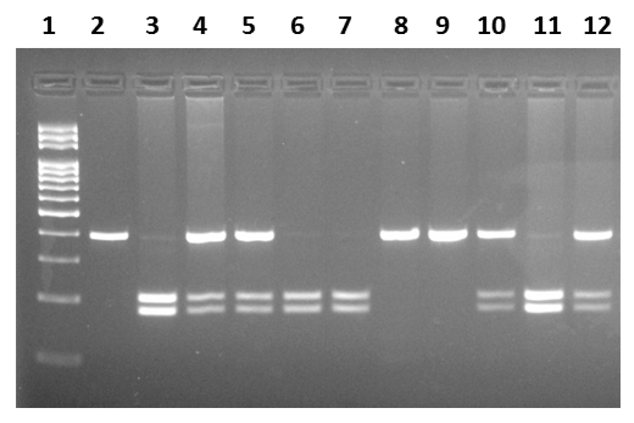
Figure 1. hMLH1 −93G>A polymorphism determined by PCR-RFLP method. Ladder (100 nc): 1. Wild-type GG: 2, 8, 9. Heterozygous GA: 4, 5, 10, 12. Homozygous mutant AA: 3, 6, 7, 11.
Jmp 06 00015 g001
Table 1. The clinical and demographic characteristics of the study groups.
Patients
n = 134 (%)
Controls
n = 137 (%)
p Value
Gender
Male
Female

76 (56.7)
58 (43.3)

64 (43.3)
73 (56.7)

0.256
Age
Average
Mean

25–85
60 ± 10.2

32–82
60 ± 11.3
Grade
G1
G2
G3

11 (8.2)
90 (67.1)
33 (24.7)
Stage
T I
T II
T III
T IV

3 (2.1)
12 (9.3)
109 (81.5)
10 (7.1)
Smoking
Smokers
Non-smokers
Unknown

44 (34.3)
82 (60)
8 (5.7)

55 (33.5)
98 (59.8)
11 (6.7)

0.942
Alcohol consumption
User
Non-user
Unknown

36 (32.1)
90 (62.2)
8 (5.7)

59 (36)
93 (56.6)
12 (7.4)

0.410
Table 2. The genotype and allele frequencies of the −93G>A polymorphism in the hMLH1 gene among patient and control groups.
GenotypesPatients
n = 134 (%)
Controls
n = 137 (%)
OR (95%CI)p Value
GG
GA
AA
Dominant
GG
GA + AA
Recessive
GG + GA
AA
21 (15.7)
43 (32.1)
70 (52.2)

21 (15.7)
113 (84.3)

64 (47.8)
70 (52.2)
23 (16.8)
62 (45.2)
52 (38)

23 (16.8)
114 (83.2)

85 (62)
52 (38)
1
0.760 (0.374–1.542)
1.474 (0.738–2.945)

1
0.921 (0.569–2.070)

1
1.788 (1.102–2.900)
-
0.446
0.270

-
0.803

-
0.018
Allele
G
A

85 (53.1)
183 (46.9)

108 (56.1)
166 (43.9)

1
1.400 (0.984–1.995)

-
0.062
Table 3. The distribution of −93G>A polymorphism of the hMLH1 gene in terms of gender factor.
MalesPatients
n = 76 (%)
Controls
n = 64 (%)
OR (95%CI)p Value
Genotypes
GG
GA
AA

14 (18.4)
25 (32.9)
37 (48.7)

10 (15.6)
28 (43.8)
26 (40.6)

1
0.638 (0.241–1.690)
1.016 (0.392–2.639)

-
0.364
0.973
Femalen = 58 (%)n = 73 (%)
Genotypes
GG
GA
AA

7 (12.1)
18 (31)
33 (56.9)

13 (17.8)
34 (46.6)
26 (35.6)

1
0.983 (0.333–2.901)
2.357 (0.823–6.755)

-
0.976
0.110
Table 4. The distribution of genotype differences among groups according to the average age.
Age
≤60
Patients
n = 48 (%)
Controls
n = 96 (%)
OR (95%CI)p Value
GG
GA
AA
6 (12.5)
16 (33.3)
26 (54.2)
17 (39.2)
44 (38.2)
35 (22.6)
1
1.030 (0.346–3.072)
2.105 (0.729–6.075)
-
0.957
0.169
>60n = 86 (%)n = 41 (%)
GG
GA
AA
15 (17.5)
27 (31.4)
44 (51.1)
6 (26.3)
18 (47.4)
17 (26.3)
1
0.600 (0.196–1.837)
1.035 (0.345–3.110)
-
0.370
0.951
Table 5. The prevalence of the −93G>A hMLH1 polymorphism among patients who smoke and drink alcohol.
GenotypesSmokers
n = 44 (%)
Non-Smokers
n = 82 (%)
OR (95%CI)p Value
GG
GA
AA
7 (16)
13 (29.5)
24 (54.5)
12 (14.6)
27 (32.9)
43 (52.5)
1
0.825 (0.263–2.589)
0.957 (0.332–2.755)
-
0.742
0.935
Alcohol consumer
n = 36 (%)
Non-drinkers
n = 90 (%)
GG
GA
AA
4 (11.1)
10 (27.8)
22 (61.1)
15 (16.7)
30 (33.3)
45 (50)
1
1.250 (0.336–4.655)
1.833 (0.544–6.179)
-
0.739
0.328
Table 6. The HMLH1 gene −93G>A polymorphism and tumor grade/stage.
GG, (%)GA, (%)AA, (%)p Value
Tumor grade
G 1
G 2
G 3

6 (54.5)
38 (42.2)
11 (33.3)

4 (36.4)
34 (37.8)
18 (54.5)

1 (9.1)
18 (20)
4 (12.2)

0.277
Tumor stage
T I
T II
T III
T IV

1 (33.3)
6 (50)
45 (41.3)
4 (40)

1 (33.3)
4 (33.3)
51 (46.8)
4 (40)

1 (33.3)
2 (16.7)
13 (11.9)
2 (20)

0.033
Table 7. The genotype and allele frequencies of the hMSH2 gene 1032G>A polymorphism in the patient and control groups.
GenotypesPatients
n = 134 (%)
Controls
n = 137 (%)
OR (95%CI)p Value
GG
GA
AA
Dominant
GG
GA + AA
Recessive
GG + GA
AA
110 (82)
23 (17.2)
1 (0.8)

110 (82.1)
24 (17.9)

133 (99.2)
1 (0.8)
122 (89.1)
13 (9.5)
2 (1.4)

122 (89.1)
15 (10,9)

135 (98.6)
2 (1.4)
1
1.962 (0.948–4.061)
0.555 (0.050–6.201)

1
1.775 (0.886–3.555)

1
0.508 (0.045–5.664)
-
0.066
0.627

-
0.103

-
0.508
Allele
G
A

243 (90.7)
25 (9.3)

257 (93.8)
17 (6.2)

1
1.555 (0.820–2.951)

-
0.174
Disclaimer/Publisher’s Note: The statements, opinions and data contained in all publications are solely those of the individual author(s) and contributor(s) and not of MDPI and/or the editor(s). MDPI and/or the editor(s) disclaim responsibility for any injury to people or property resulting from any ideas, methods, instructions or products referred to in the content.