Article

Methylene Blue Increases Active Mitochondria and Cellular Survival Through Modulation of miR16–UPR Signaling Axis

by
Carlos Garcia-Padilla
1,2,†,
David García-Serrano
1,† and
Diego Franco
1,2,*
1
Cardiovascular Research Group, Department of Experimental Biology, University of Jaen, 23071 Jaen, Spain
2
Medina Foundation, Technology Park of Health Sciences, 18016 Granada, Spain
*
Author to whom correspondence should be addressed.
These authors contributed equally to this work.
J. Mol. Pathol. 2025, 6(3), 16; https://doi.org/10.3390/jmp6030016
Submission received: 9 May 2025 / Revised: 2 July 2025 / Accepted: 17 July 2025 / Published: 23 July 2025

Abstract

Background: Methylene blue (MB), a versatile redox agent, is emerging as a promising therapeutic in diseases associated with mitochondrial dysfunction. Its ability to optimize the electron transport chain increases ATP synthesis (30–40%) and reduces oxidative stress, protecting cellular components such as mitochondrial DNA. The protective role of this compound has been described in several neurodegenerative disease such as Alzheimer’s and Parkinson’s diseases. However, its role in cardiovascular disease has been poorly explored. Methods: In this study, we explored the impact of MB on murine (HL1) and human (AC16) cardiomyocyte redox signaling and cellular survival using RT-Qpcr analysis and immunochemistry assays. Results: Our results revealed that MB increased functional mitochondria, reversed H2O2-induced oxidative damage, and modulated antioxidant gene expression. Furthermore, it regulated the microRNA16–UPR signaling axis, reducing CHOP expression and promoting cell survival. Conclusions: These findings underscore its potential in cardioprotective therapy; however, its putative use as a drug requires in vivo validation in preclinical animal models.

1. Introduction

Methylene blue (MB), a chemical compound with a long history of medical applications, has emerged as a versatile therapeutic agent in addressing diseases linked to mitochondrial dysfunction [1,2]). Its ability to modulate bioenergetic and redox processes positions it as a promising tool for maintaining cellular homeostasis, particularly in several disorders caused and/or affected by alterations in this critical organelle [3,4,5]. The mitochondria, as the primary generators of ATP and regulators of redox metabolism, depend on the integrity of their electron transport chain (ETC) [6]. Curiously, Methylene blue acts as a redox catalyst, alternating between oxidized (electron acceptor) and reduced (electron donor) forms, allowing electron flow to be restored when the ETC is compromised [7]. This property gives it two key functions: (1) Optimizing energy production: by bridging damaged mitochondrial complexes, it increases ATP synthesis by up to 30–40%, even under hypoxic conditions; (2) Reducing oxidative stress: by decreasing electron leakage, it minimizes the formation of reactive oxygen species (ROS), protecting cellular components such as mitochondrial DNA [8,9,10,11,12]. Furthermore, several reports have highlighted that Methylene blue increases the mitochondrial biogenesis modulating the activation of key factor PGC-1α, a main transcription factor involved in the biogenesis of this organelle [13,14]. In addition, Methylene blue promotes the selective elimination of damaged mitochondria by activating pathways such as PINK1/Parkin, preventing the accumulation of dysfunctional organelles that generate ROS and reducing the oxidative environment in the cell [15,16]. Moreover, not only does it protect against the ROS generated by dysfunctional organelles, but it also has the ability to reduce the endogenous production of ROS at the intracellular level. For example, Methylene blue blocks nitric oxide synthase (NOS), reducing the production of nitric oxide (NO), a molecule that, in excess, inhibits the respiratory chain and generates peroxynitrite, a well-known reactive oxygen species that primarily affects cellular proteostasis [17,18].
This protective role of methylene blue has promoted its use as a drug against various diseases, including Alzheimer’s and Parkinson’s. In Alzheimer’s disease, methylene blue reverses the inhibition of cytochrome oxidase, increasing cerebral oxygen consumption by 35% and reducing the accumulation of β-amyloid and tau protein by improving mitochondrial autophagy [19]. On the other hand, in Parkinson’s disease, it protects dopaminergic neurons by normalizing mitochondrial membrane potential and reducing apoptosis [20,21,22]). However, the role of Methylene blue has been poorly explored in cardiovascular diseases. Recently, Fernandes Junior et al. [23] demonstrated a bivalent role of this compound during reperfusion after a heart attack. The authors showed that administration of this compound before the onset of a heart attack is cytotoxic to the heart, while its administration after reperfusion protects the heart from the deleterious effects associated with this process [23]. On the other hand, Methylene blue applied in porcine and rat models of cardiac arrest showed promising results, improving overall survival of cardiomyocytes and reducing oxidative stress in the brain [24,25].
Given the importance of mitochondrial dysfunction in cardiovascular disease and cardiac regeneration, understanding the role of MB in cardiomyocyte mitochondrial homeostasis is essential to understand the role of this compound as a potential drug against the damage associated with heart attack [26]. In this study, we aimed to explore the impact of Methylene blue in mitochondrial function and its modulation of UPR signaling on two different lines of cardiomyoctes: HL1, a mouse atrial cardiomyocyte cell line, and AC16, a human ventricular cardiomyoblast cell line. Our results demonstrated that in both cell lines, MB increased the number of active mitochondria and modulated the expression of several genes involved in ROS protection. In addition, MB reversed the effect of the oxidative environment generated by H2O2 administration. Furthermore, our results demonstrated that MB modulated the microRNA16–UPR signaling axis in both cell lines, increasing cell survival by triggering UPR signaling and reducing CHOP pro-apoptotic protein expression.

2. Materials and Methods

2.1. Cell Culture Treatments

HL1 cell line was cultured in Claycomb medium supplemented with 10% Fetal Bovine serum, 100 U/mL penicillin/streptomycin, 200 nM of L-glutamine, and Norepinephrine 0.1 mM, as previously reported. AC16 Human cardiomyocyte cell line was cultured in DMEM F12, supplemented with FBS 12.5%, 100 U/mL penicillin/ streptomycin, and 200 nM of L-glutamine. Both lines were cultured in 100 cm2 culture disks, previously treated with gelatin-fibronectin (2% and 1%, respectively) at 37 °C in a humidified atmosphere of 5% CO2. Sub-cultured cells (40.000 cells/well) were treated with Methylene blue (Sigma, Aldrich, Munich, Germany), and Hydrogen peroxide (Sigma, Aldrich, Munich, Germany) at 10 μM and 25 μM, respectively, for 24 h. Four experimental conditions were analyzed in both cell lines: (1) Control, (2) Methylene Blue (10 μM), (3) Hydrogen Peroxide (25 μM), and (4) Methylene Blue + Hydrogen Peroxide (10 μM and 25 μM, respectively).

2.2. Cell Viability Assay

Cell viability of HL1 and AC16 exposed to different experimental conditions—(1) Control, (2) Methylene Blue, (3) Hydrogen Peroxide, and (4) Methylene Blue + Hydrogen Peroxide—was evaluated using an Apoptosis/Necrosis Detection Kit (Blue, Red, Green) ab176750 (Abcam Limited, Cambridge, UK) following the manufacturer’s instructions.

2.3. RNA Isolation

RNA from treated cells was isolated using Reliaprep RNA Miniprep System (Promega Corp., Madison, WI, USA) following the manufacturer’s protocol. In all cases, at least three different pools of each condition were collected from the different culture wells. RNA isolated was stored at −80 °C until use.

2.4. Primary Cardiomyocyte Isolation

Primary cardiomyocytes were isolated from neonatal mice CD1 using the protocol described by Bongiovanni et al., 2024 [27]. Ten hearts were used for each condition and 60,000 primary cardiomyocytes were seed per well and experimental condition.

2.5. cDNA Synthesis and qPCR Analysis

RNA retrotranscription was performed using a PrimeScript RT Reagent Kit (Takara Bio Co., Ltd., Shiga, Japan). To perform real-time qPCR, we used the corresponding cDNA, primers, and Go Taq Syber Green (Promega Corp., Madison). Reactions were performed at 10 μL total volume, using 96-well or 384-well plates and optical sealing tape in a CFX96TM or CFX384TM thermocycler (Bio-Rad, Hercules, CA, USA), respectively. Amplification conditions were a denaturalization step of 95 °C for 10 min, followed by 45 cycles of 95 °C for 5 s, 60 °C for 10 s, and 75 °C for 7 s, with a final step of 95 °C for 10 s. To determine the relative expression of the different genes, Gadph was used as normalization control. Each qPCR reaction was performed in triplicate and repeated three times to obtain representative means. Primers were designed using primer3 software 3.0 and validated using the last version of Blast software from ncbi databse. No amplification was observed in the PCR control reactions with only water as template and RT reactions. The Livak and Schmittgen [28] method for relative qPCR analysis was used and normalized to control (wild-type) values. Primer sequence is described in Supplementary Table S1.

2.6. Mitotracker Labeling

The number of active mitochondria was measured using Mitotracker Green FM labelling (Thermofisher #M7514, Waltham, MA, USA), following the manufacturer’s guidelines. Mitotracker staining was analyzed using a Leica TCS SP5 II confocal scanning laser microscope (Leica, Wetzlar, Germany). Images were subsequently quantified using ImageJ software 4.0.

2.7. Immunochemestry Analysis

For immunohistochemical analyses, cell cultures were rinsed twice in PBS 1× and fixed in 4% PFA for 10 min at room temperature. Subsequently, cell cultures were rinsed twice in PBS 1× and incubated with permeabilizing solution (Ammonium chloride 50 mM and Triton 0.2% in PBS 1×) for 10 min at room temperature. Afterward, cell cultures were incubated twice with blocking solution (gelatin 2% in PBS 1×) for 15 min at room temperature to block nonspecific binding sites. As primary antibodies, polyclonal rabbit Bip (Cell signaling #3177S) and monoclonal mouse CHOP (Cell signaling #2895S) were used, diluted (1:200) in PBS, and applied to each cell culture overnight at 4 °C. Subsequently, the cell cultures were rinsed three times (for 1 h each) in PBS to remove excess primary antibodies and incubated overnight at 4 °C with Alexa-Fluor 488 anti-rabbit and Alexa-Fluor 546 anti-mouse (1:100; Invitrogen carlsbad ca U.S.A) as secondary antibodies, respectively. After incubation with the secondary antibodies, the cell cultures were rinsed and incubated with DAPI (1:1000; Sigma, Aldrich, Munich, Germany) for 10 min at room temperature and rinsed three times in PBS for 5 min each. Finally, the cell cultures were analyzed using a Leica TCS SP5 II confocal scanning laser microscope (Leica, Wetzlar, Germany). Negative controls, lacking primary antibody incubation, resulted in all cases having no detectable signal. Images were subsequently quantified using ImageJ software.

2.8. Statistical Analyses

Statistical analysis was performed using ANOVA test. Significance levels of p values are stated in each figure legend. p < 0.05 was considered statistically significant.

3. Results

3.1. Methylene Blue Increases the Number of Active Mitochondria and Modulates the Expression of Stress Oxidative Genes in HL1 and AC16 Cell Lines

Firstly, we explored the possible role of MB and Hydrogen peroxide (H2O2) in the activation of mitochondria in mouse atrial cardiomyocytes (HL1) and human cardiomyoblasts (AC16). Mitotracker (MTDR) labelling showed that both cell lines treated with MB displayed an increased number of active mitochondria with respect to the control, whereas H2O2 treatment resulted in a lower number of active mitochondria (Figure 1A–G). Furthermore, co-treatment with MB and H2O2 in HL1 and AC16 displayed a similar active mitochondria number with respect to the control, showing that MB reversed the negative effect of H2O2 on mitochondrial activity (Figure 1D–J) and suggesting a possible protective function of MB against the oxidative environment derived from H2O2 treatment. Given the harmful role of H2O2 in the generation of oxidative stress, we analyzed the expression of several genes involved in protection against this process: –SOD1 and SOD2, enzymes that catalyze the dismutation of superoxide into hydrogen peroxide, a less reactive molecule; oxygen PRDX2, 3, and 5, pivotal enzymes that act as scavenging of peroxides; GSR, an enzyme that regenerates reduced glutathione (GSH) from oxidized glutathione (GSSG), thus allowing the neutralization of reactive oxygen species; and GPRX1, that primarily functions to reduce hydrogen peroxide to water, thus limiting its harmful effects and modulating ROS-mediated signaling pathways (Figure 1K–Q). qPCR analysis demonstrated that MB increased the expression of GSR but not the other genes analyzed in HL1 (Figure 1Q). On the other hand, MB treatment in AC16 increased the expression of PRDX2, PRDX3, PRDX5, and GSR, while repressing SOD1, SOD, and GPRX1 expression (Figure 1K–Q). H2O2 treatment reduced the expression of SOD2, PRDX3, PRDX3, and GSR, whereas it increased SOD1 expression in HL1, while in AC16 cells, H2O2 treatment repressed expression of PRDX3, PRDX5, GPRX1, and GRS but increased expression of SOD2 (Figure 1K–Q). Interestingly, co-treatment of MB and H2O2 translated into a similar expression of GSR compared with control conditions in both cell lines, suggesting that MB can reverse the repressive role of H2O2 on GSR gene and that this genetic compensation may be conserved across species. In line with this reversion, co-treatment in HL1 displayed a reduction in SOD1 expression compared to the H2O2 condition, while AC16 exhibited a similar expression pattern to PRDX3, PRDX5, GSR, and SOD2 between MB + H2O2 and MB treatments and contrary to what was observed in cells treated with H2O2, supporting the protective role of MB against H2O2 (Figure 1K–Q). Finally, to determine whether the observable differences in the modulation of genes involved in defense against oxidative stress between HL1 and AC16 cells were a consequence of the cell type studied or due to differences between species, we isolated mouse neonatal cardiomyocytes and checked the regulation of these genes in the different experimental treatments by qPCR. Our results demonstrated primary cardiomyocytes exhibited the same response as HL1 cells to the different treatments, with the exception of peroxiredoxin 5, suggesting that the observed differences may have been a consequence of species differences and not due to the HL1 cell line (Supplementary Figure S1).

3.2. Methylene Blue Modulates miR16–UPR Signalling Axis, Increasing the Cellular Supervivence of Cardiomyocytes and Cardiomyoblast

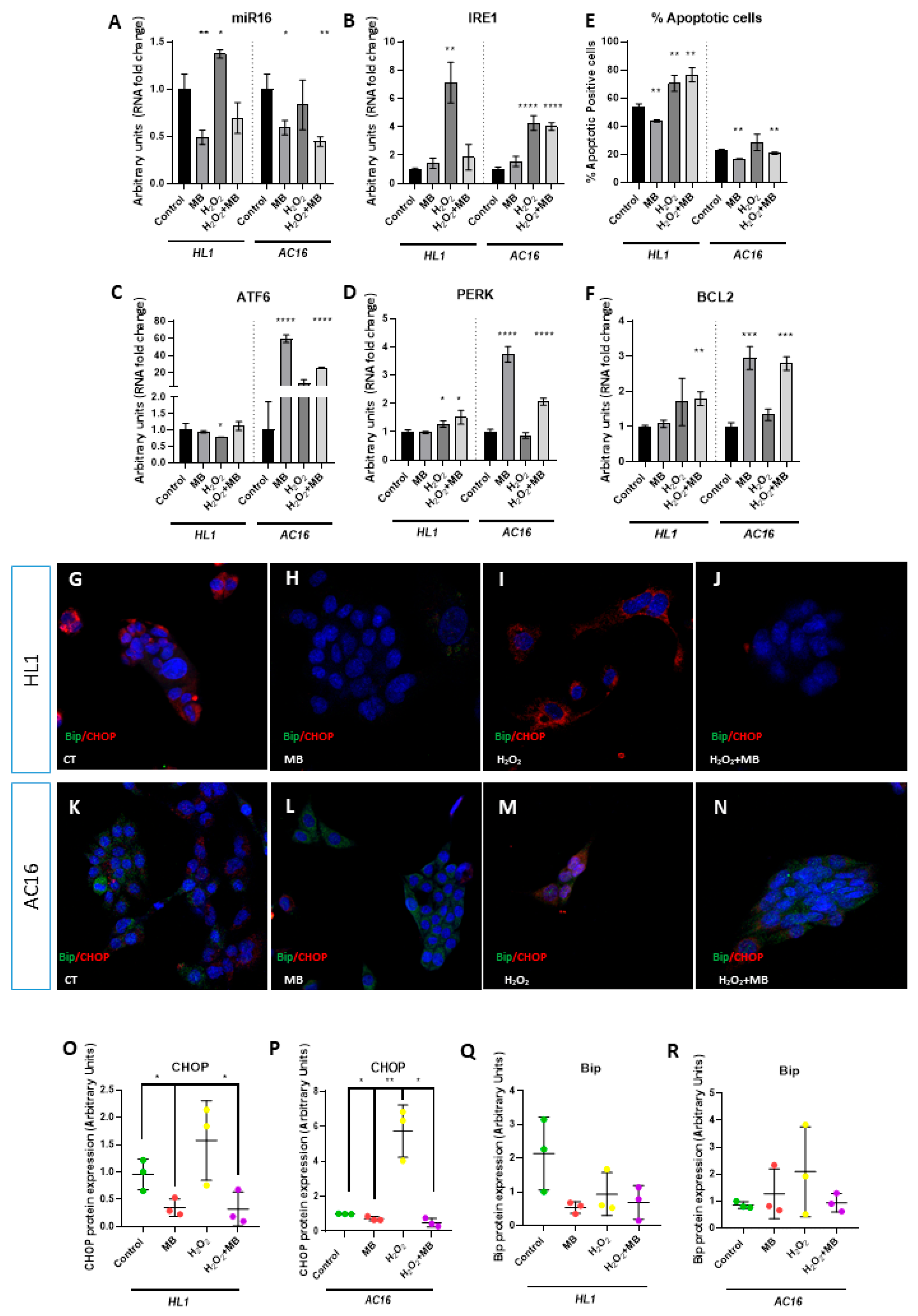
Recently, Toro et al. (2022) demonstrated that miR-16a exerts a deleterious effect on mitochondrial function and cellular survival by repressing UPR signaling by targeting ATF6 3′UTR [29]. Given the link between mitochondria homeostasis and activation of endoplasmic reticulum stress (ERS), we wondered if miR16–UPR signaling axis may be modulated by MB in the HL1 cell line. To check our hypothesis, we performed qPCR analysis of miR-16 expression and anchoring genes involved in UPR signaling. Our results demonstrated that MB reduced expression of miR-16 in both cell lines, whereas H2O2 treatment increased the expression of this microRNA in HL1 but not in AC16 (Figure 2A). In line with the possible protective role of MB, co-treatment with H2O2 and MB resulted in the reversion of miR-16 upregulation observed in H2O2 treatment in HL1 (Figure 2A). MB treatment in AC16, but not in HL1, increased ATF6 and PERK expression. Unlike in AC16, MB treatment did not modulate the expression of the genes dependent on the UPR signaling pathway in HL1. On the other hand, H2O2 treatment reduced the expression of ATF6, a direct target of miR-16, while increasing PERK and IRE1 expression in HL1. Curiously, H2O2 treatment in AC16 resulted in an IRE1 upregulation similar to that observed in HL1. MB + H2O2 co-treatment increased ATF6, PERK, and IRE1 expression in AC16, it whereas increased the expression of PERK in HL1 (Figure 2B–D). Since the UPR pathway may act through prosurvival or proapoptotic signaling, we analyzed the impact of several treatments on cellular viability by flow cytometry (Supplementary Figure S2). Our data demonstrated that MB treatment resulted in a lower number of apoptotic cells compared with the control condition in both cell lines. Furthermore, H2O2 and MB + H2O2 treatments displayed increased apoptotic cells in HL1. In AC16 cells, H2O2 treatment displayed a trend of increased apoptotic cells, without reaching statistical significance, whereas MB + H2O2 treatment translated into lower apoptotic cells (Figure 2E). Curiously, BCL2 expression was upregulated in MB + H2O2 but not in H2O2 and/or MB treatments in HL1, while BCL2 expression was upregulated in MB and MB + H2O2 but not in H2O2 treatment for AC16 (Figure 2F). In line with these results, CHOP protein expression, an inducer of UPR-dependent apoptosis, was downregulated in MB and MB + H2O2 but not in the H2O2 treatment in both cell lines, whereas changes in the Bip protein expression were not detected (Figure 2G–R). These data suggest that MB increased cellular viability, activating UPR-dependent prosurvival signals by repressing miR16 expression in HL1 and repressing the expression of the pro-apoptotic protein CHOP in both cell lines.

4. Discussion

The protective role of MB in mitochondrial homeostasis has been described in various neurodegenerative diseases [1,2]. However, the role of this compound as a protector in cardiac diseases and its impact on mitochondrial function in cardiomyocytes have been poorly addressed. Given the importance of proper mitochondrial activity for correct cardiac function and homeostasis, it is essential to analyze the role of MB as a possible drug in cardiomyocytes [30,31]. Our results demonstrated that, in both cell lines AC16 and HL1, MB reversed H2O2-induced mitochondrial dysfunction, normalizing the number of active mitochondria. This effect may be explained by the observed ability of MB to restore electron flow by acting as an alternative electron acceptor; however, a TMRM analysis should be performed to confirm this possibility [32,33]). Furthermore, by decreasing electron leakage, it limits superoxide formation, protecting critical components such as mtDNA, avoiding mitophagy, and leading to a higher number of active mitochondria [34].
The impact of MB not only affected the number of active mitochondria, MB also modulated the expression of genes involved in oxidative stress response, suggesting an important protective role in this process. In the AC16 cell line, MB exerted a selective induction of peroxiredoxins PRDX3 and PRDX5 but not PRDX2, whereas this regulation was not observed in the HL1 cell line, demonstrating a species-specific difference in redox regulation of these genes. Unlike the PRDX3/5 differences observed between AC16 and HL1, MB restored GSR levels in both cell lines. GSR is essential for recycling oxidized glutathione (GSSG), maintaining the cellular antioxidant pool [35]. This GSR modulation may explain the protection against apoptosis observed, along with the triggering of the UPR pathway. On the other hand, downregulation of SOD2 through MB treatment and the restoration of normal levels after co-treatment with MB and H2O2 was not observed in HL1, suggesting a different response to the ROS generated inside of mitochondria in the two cell lines. Furthermore, MB repressed the expression of cytosolic enzymes, SOD1 and GPRX1, in both cell lines, suggesting that MB promotes the activation of enzymes that exert their function inside the mitochondria but not in the cellular cytoplasm [36]. These data demonstrated that MB activated the protective genes involved in the removal of ROS from mitochondria. Furthermore, the major activation of antioxidant enzymes observed in AC16 cells compared to HL1 cells suggests a more effective role of this compound in human cardiomyoblasts compared to mouse cardiomyocytes. This effectiveness could have been mediated by the difference in cellular differentiation in the two cell lines—cardiomyoblasts versus adult cardiomyocytes—or by the presence of additional molecular mechanisms activated in response to ROS increases in each cell type or species [37,38,39]. On the other hand, the differences we observed between HL1 and AC16 cells may have been a consequence of these cells’ greater or lesser resistance to oxidative stress. Various studies have shown that HL1 cells exhibit greater resistance to oxidative stress compared to AC16 cells. This is partly due to the fact that HL-1 cells exhibit high catalase activity (absent in AC16) and more stable mitochondrial superoxide dismutase (SOD2). Similarly, AC16 cells are more dependent on the glutathione system and have a lower GSH regenerative capacity compared to HL1 cells. This differential resistance may affect the protective role of MB against oxidative stress, being more necessary and evident in those cells that present a worse defense against oxidative stress, such as AC16 compared to HL1 [40,41,42,43].
The physiological connection between mitochondria and the endoplasmic reticulum is essential for proper cellular homeostasis [44,45,46,47]. Alterations in mitochondria can lead to the development of a cellular oxidative environment. This increase in cytoplasmic oxidation translates into a decrease in proteostasis and the subsequent activation of the UPR pathway [48,49]. In addition, the role of microRNAs in the modulation of several biological processes, such as development, differentiation, and homeostasis, has been widely described [50,51]. Recently, Toro et al. demonstrated that miR-16a was upregulated in patients with dilated cardiomyopathy. The upregulation of this microRNA reduced the ER’s ability to establish and trigger proper activation of the UPR pathway, the main protective pathway against proteostasis failure. In order to analyze if MB may modulate the UPR signaling, our data suggest that MB reduced expression of miR16 both AC16 and HL1, subsequently reducing apoptotic cells and increasing cardiomyocyte survival, possibly through activating several UPR signaling branches. Curiously, MB addition reversed the upregulation of CHOP- proapoctotic protein [52,53] in the H2O2 treatment, pinpointing the importance of this compound as a protective agent against apoptosis triggering by oxidative stress. Importantly, CHOP triggering was dependent on IRE1 activation [54], which was upregulated upon treatment with H2O2 in both cell lines. However, co-treatment with MB did not reduce IRE1 expression in either cell line, suggesting that the increase in cell survival and the consequent reduction in CHOP may have been the result of the activation of other compensatory pathways, such as ATF6 in the case of AC16, or the inhibition of pro-apoptotic genes, such as microRNA 16a in both cell lines.
Taking all our data together, our data suggest a protective role of MB against oxidative stress by modulating GSR expression gene, in mouse and human cardiomyocytes, and the modulation of SOD2 and PRDX3 and 5 in human cardiomyocytes. Furthermore, we suggest that MB treatment reduces CHOP pro-apoptotic protein expression, reversing the enhanced expression of this protein after an increase in oxidative stress. Our study provides the first data demonstrating the protective role of this compound in cardiomyocyte mitochondria. Furthermore, we suggest that the deleterious effects of H2O2 treatment are attenuated by MB administration, revealing the potential role of this compound as a possible drug, as an activator of mitochondrial defenses against oxidative stress. However, it is important to highlight the limitations of our study, since it was carried out in vitro and not in vivo. Further in vivo research is required to understand whether MB can be used as a therapeutic drug to improve mitochondrial response during heart failure. In addition, Fernandes et al. (2024) demonstrated that the timing of MB administration and the recurrence of this administration are essential for its protective or detrimental function during heart failure, suggesting that the effect of MB in vivo may be more complex than that observed in vitro [23].

Supplementary Materials

The following supporting information can be downloaded at https://www.mdpi.com/article/10.3390/jmp6030016/s1, Supplementary Figure S1: Neonatal cardiomyocyte analysis of ROS genes related; Supplementary Figure S2: Flow cytometry plots; Supplementary Table S1: Primer’s sequences.

Author Contributions

Conceptualization, D.F. and D.G.-S.; methodology, D.G.-S. and C.G.-P.; software, D.G.-S.; validation, D.F. and C.G.-P.; formal analysis, C.G.-P.; investigation, D.G.-S. and C.G.-P.; resources, D.F.; data curation, C.G.-P. and D.G.-S.; writing—original draft preparation, C.G.-P.; writing—review and editing, D.F.; supervision, D.F.; project administration, D.F.; funding acquisition, D.F. All authors have read and agreed to the published version of the manuscript.

Funding

This work was supported by grants of the Ministerio de Innovación y Ciencia of the Spanish Government to DF (PID2022-138163OB-C32) and of the Consejería de Universidad, Investigación e Innovación of the Junta de Andalucia Regional Council to DF (ProyExcel_00409).

Institutional Review Board Statement

This study was performed in line with the principles of the Declaration of Helsinki. Approval was granted by the Ethics Committee of the University of Jaén (code 14/03/2022/038).

Informed Consent Statement

Not applicable.

Data Availability Statement

The datasets generated during and/or analyzed during the current study are available from the corresponding author on reasonable request.

Conflicts of Interest

The authors declare that they have no competing interests.

References

  1. Schirmer, R.H.; Adler, H.; Pickhardt, M.; Mandelkow, E. “Lest we forget you—methylene blue...”. Neurobiol. Aging 2011, 32, 2325.e7–2325.e16. [Google Scholar] [CrossRef] [PubMed]
  2. Oz, M.; Lorke, D.E.; Hasan, M.; Petroianu, G.A. Cellular and molecular actions of Methylene Blue in the nervous system. Med. Res. Rev. 2011, 31, 93–117. [Google Scholar] [CrossRef] [PubMed] [PubMed Central]
  3. Akbar, M.; Essa, M.M.; Daradkeh, G.; Abdelmegeed, M.A.; Choi, Y.; Mahmood, L.; Song, B.J. Mitochondrial dysfunction and cell death in neurodegenerative diseases through nitroxidative stress. Brain Res. 2016, 1637, 34–55. [Google Scholar] [CrossRef] [PubMed] [PubMed Central]
  4. Rambani, V.; Hromnikova, D.; Gasperikova, D.; Skopkova, M. Mitochondria and mitochondrial disorders: An overview update. Endocr. Regul. 2022, 56, 232–248. [Google Scholar] [CrossRef] [PubMed]
  5. Sun, Y.; Jin, L.; Qin, Y.; Ouyang, Z.; Zhong, J.; Zeng, Y. Harnessing Mitochondrial Stress for Health and Disease: Opportunities and Challenges. Biology 2024, 13, 394. [Google Scholar] [CrossRef] [PubMed] [PubMed Central]
  6. Nolfi-Donegan, D.; Braganza, A.; Shiva, S. Mitochondrial electron transport chain: Oxidative phosphorylation, oxidant production, and methods of measurement. Redox Biol. 2020, 37, 101674. [Google Scholar] [CrossRef] [PubMed] [PubMed Central]
  7. Sváb, G.; Kokas, M.; Sipos, I.; Ambrus, A.; Tretter, L. Methylene Blue Bridges the Inhibition and Produces Unusual Respiratory Changes in Complex III-Inhibited Mitochondria. Studies on Rats, Mice and Guinea Pigs. Antioxidants 2021, 10, 305. [Google Scholar] [CrossRef] [PubMed] [PubMed Central]
  8. Atamna, H.; Nguyen, A.; Schultz, C.; Boyle, K.; Newberry, J.; Kato, H.; Ames, B.N. Methylene blue delays cellular senescence and enhances key mitochondrial biochemical pathways. FASEB J. 2008, 22, 703–712. [Google Scholar] [CrossRef] [PubMed]
  9. Atamna, H.; Kumar, R. Protective role of methylene blue in Alzheimer’s disease via mitochondria and cytochrome c oxidase. J. Alzheimers Dis. 2010, 20 (Suppl. S2), S439–S452. [Google Scholar] [CrossRef] [PubMed]
  10. Roy Choudhury, G.; Winters, A.; Rich, R.M.; Ryou, M.G.; Gryczynski, Z.; Yuan, F.; Yang, S.H.; Liu, R. Methylene blue protects astrocytes against glucose oxygen deprivation by improving cellular respiration. PLoS ONE 2015, 10, e0123096. [Google Scholar] [CrossRef] [PubMed] [PubMed Central]
  11. Haouzi, P.; Gueguinou, M.; Sonobe, T.; Judenherc-Haouzi, A.; Tubbs, N.; Trebak, M.; Cheung, J.; Bouillaud, F. Revisiting the physiological effects of methylene blue as a treatment of cyanide intoxication. Clin. Toxicol. 2018, 56, 828–840. [Google Scholar] [CrossRef] [PubMed] [PubMed Central]
  12. Duicu, O.M.; Privistirescu, A.; Wolf, A.; Petruş, A.; Dănilă, M.D.; Raţiu, C.D.; Muntean, D.M.; Sturza, A. Methylene blue improves mitochondrial respiration and decreases oxidative stress in a substrate-dependent manner in diabetic rat hearts. Can. J. Physiol. Pharmacol. 2017, 95, 1376–1382. [Google Scholar] [CrossRef] [PubMed]
  13. Xiong, Z.M.; Choi, J.Y.; Wang, K.; Zhang, H.; Tariq, Z.; Wu, D.; Ko, E.; LaDana, C.; Sesaki, H.; Cao, K. Methylene blue alleviates nuclear and mitochondrial abnormalities in progeria. Aging Cell 2016, 15, 279–290. [Google Scholar] [CrossRef] [PubMed] [PubMed Central]
  14. Gureev, A.P.; Syromyatnikov, M.Y.; Gorbacheva, T.M.; Starkov, A.A.; Popov, V.N. Methylene blue improves sensorimotor phenotype and decreases anxiety in parallel with activating brain mitochondria biogenesis in mid-age mice. Neurosci. Res. 2016, 113, 19–27. [Google Scholar] [CrossRef] [PubMed]
  15. Poudel, S.B.; Frikha-Benayed, D.; Ruff, R.R.; Yildirim, G.; Dixit, M.; Korstanje, R.; Robinson, L.; Miller, R.A.; Harrison, D.E.; Strong, J.R.; et al. Targeting mitochondrial dysfunction using methylene blue or mitoquinone to improve skeletal aging. Aging 2024, 16, 4948–4964. [Google Scholar] [CrossRef] [PubMed] [PubMed Central]
  16. Krutskikh, E.P.; Potanina, D.V.; Samoylova, N.A.; Gryaznova, M.V.; Sadovnikova, I.S.; Gureev, A.P.; Popov, V.N. Brain Protection by Methylene Blue and Its Derivative, Azur B, via Activation of the Nrf2/ARE Pathway in Cisplatin-Induced Cognitive Impairment. Pharmaceuticals 2022, 15, 815. [Google Scholar] [CrossRef] [PubMed] [PubMed Central]
  17. Mayer, B.; Brunner, F.; Schmidt, K. Inhibition of nitric oxide synthesis by methylene blue. Biochem. Pharmacol. 1993, 45, 367–374. [Google Scholar] [CrossRef] [PubMed]
  18. Saha, B.K.; Burns, S.L. The Story of Nitric Oxide, Sepsis and Methylene Blue: A Comprehensive Pathophysiologic Review. Am. J. Med. Sci. 2020, 360, 329–337. [Google Scholar] [CrossRef] [PubMed]
  19. Hashmi, M.U.; Ahmed, R.; Mahmoud, S.; Ahmed, K.; Bushra, N.M.; Ahmed, A.; Elwadie, B.; Madni, A.; Saad, A.B.; Abdelrahman, N. Exploring Methylene Blue and Its Derivatives in Alzheimer’s Treatment: A Comprehensive Review of Randomized Control Trials. Cureus 2023, 15, e46732. [Google Scholar] [CrossRef] [PubMed] [PubMed Central]
  20. Xie, L.; Li, W.; Winters, A.; Yuan, F.; Jin, K.; Yang, S. Methylene blue induces macroautophagy through 5′ adenosine monophosphate-activated protein kinase pathway to protect neurons from serum deprivation. Front. Cell Neurosci. 2013, 7, 56. [Google Scholar] [CrossRef] [PubMed] [PubMed Central]
  21. Smith, E.S.; Clark, M.E.; Hardy, G.A.; Kraan, D.J.; Biondo, E.; Gonzalez-Lima, F.; Cormack, L.K.; Monfils, M.; Lee, H.J. Daily consumption of methylene blue reduces attentional deficits and dopamine reduction in a 6-OHDA model of Parkinson’s disease. Neuroscience 2017, 359, 8–16. [Google Scholar] [CrossRef] [PubMed]
  22. Bariotto-Dos-Santos, K.; Padovan-Neto, F.E.; Bortolanza, M.; Dos-Santos-Pereira, M.; Raisman-Vozari, R.; Tumas, V.; Del Bel, E. Repurposing an established drug: An emerging role for methylene blue in L-DOPA-induced dyskinesia. Eur. J. Neurosci. 2019, 49, 869–882. [Google Scholar] [CrossRef] [PubMed]
  23. Fernandes Junior, H.J.; de Araújo, E.A.; Machado Junior, J.A.; Lutz Motta, F.M.; Guarize, G.F.; Cheng, L.C.; Tantray, J.; Medeiros, J.V.R.; Nicolau, L.A.D.; Barbosa, A.H.P.; et al. Cardiotoxic and Cardioprotective Effects of Methylene Blue in the Animal Model of Cardiac Ischemia and Reperfusion. Biomedicines 2024, 12, 2575. [Google Scholar] [CrossRef] [PubMed] [PubMed Central]
  24. Miclescu, A.; Basu, S.; Wiklund, L. Cardio-cerebral and metabolic effects of methylene blue in hypertonic sodium lactate during experimental cardiopulmonary resuscitation. Resuscitation 2007, 75, 88–97. [Google Scholar] [CrossRef] [PubMed]
  25. Xavier, M.S.; Vane, M.F.; Vieira, R.F.; Oliveira, C.C.; Maia, D.R.R.; de Castro, L.U.C.; Carmona, M.J.C.; Costa Auler JOJr Otsuki, D.A. Methylene blue as an adjuvant during cardiopulmonary resuscitation: An experimental study in rats. Braz. J. Anesthesiol. 2024, 74, 744470. [Google Scholar] [CrossRef] [PubMed] [PubMed Central]
  26. Liu, M.; Lv, J.; Pan, Z.; Wang, D.; Zhao, L.; Guo, X. Mitochondrial dysfunction in heart failure and its therapeutic implications. Front. Cardiovasc. Med. 2022, 9, 945142. [Google Scholar] [CrossRef] [PubMed] [PubMed Central]
  27. Bongiovanni, C.; Miano, C.; Sacchi, F.; Da Pra, S.; Del Bono, I.; Boriati, S.; D’Uva, G. Protocol for isolating and culturing neonatal murine cardiomyocytes. STAR Protoc. 2024, 5, 103461. [Google Scholar] [CrossRef] [PubMed] [PubMed Central]
  28. Livak, K.J.; Schmittgen, T.D. Analysis of relative gene expression data using real-time quantitative PCR and the 2(-Delta Delta C(T)) Method. Methods 2001, 25, 402–408. [Google Scholar] [CrossRef] [PubMed]
  29. Toro, R.; Pérez-Serra, A.; Mangas, A.; Campuzano, O.; Sarquella-Brugada, G.; Quezada-Feijoo, M.; Ramos, M.; Alcalá, M.; Carrera, E.; García-Padilla, C.; et al. miR-16-5p Suppression Protects Human Cardiomyocytes against Endoplasmic Reticulum and Oxidative Stress-Induced Injury. Int. J. Mol. Sci. 2022, 23, 1036. [Google Scholar] [CrossRef] [PubMed] [PubMed Central]
  30. Siasos, G.; Tsigkou, V.; Kosmopoulos, M.; Theodosiadis, D.; Simantiris, S.; Tagkou, N.M.; Tsimpiktsioglou, A.; Stampouloglou, P.K.; Oikonomou, E.; Mourouzis, K.; et al. Mitochondria and cardiovascular diseases-from pathophysiology to treatment. Ann. Transl. Med. 2018, 6, 256. [Google Scholar] [CrossRef] [PubMed] [PubMed Central]
  31. Poznyak, A.V.; Ivanova, E.A.; Sobenin, I.A.; Yet, S.F.; Orekhov, A.N. The Role of Mitochondria in Cardiovascular Diseases. Biology 2020, 9, 137. [Google Scholar] [CrossRef] [PubMed] [PubMed Central]
  32. Wen, Y.; Li, W.; Poteet, E.C.; Xie, L.; Tan, C.; Yan, L.J.; Ju, X.; Liu, R.; Qian, H.; Marvin, M.A.; et al. Alternative mitochondrial electron transfer as a novel strategy for neuroprotection. J. Biol. Chem. 2011, 286, 16504–16515. [Google Scholar] [CrossRef] [PubMed] [PubMed Central]
  33. Rojas, J.C.; Bruchey, A.K.; Gonzalez-Lima, F. Neurometabolic mechanisms for memory enhancement and neuroprotection of methylene blue. Prog. Neurobiol. 2012, 96, 32–45. [Google Scholar] [CrossRef] [PubMed] [PubMed Central]
  34. Shen, J.; Xin, W.; Li, Q.; Gao, Y.; Yuan, L.; Zhang, J. Methylene Blue Reduces Neuronal Apoptosis and Improves Blood-Brain Barrier Integrity After Traumatic Brain Injury. Front. Neurol. 2019, 10, 1133. [Google Scholar] [CrossRef] [PubMed] [PubMed Central]
  35. Handy, D.E.; Loscalzo, J. Responses to reductive stress in the cardiovascular system. Free Radic. Biol. Med. 2017, 109, 114–124. [Google Scholar] [CrossRef] [PubMed] [PubMed Central]
  36. D’Oria, R.; Schipani, R.; Leonardini, A.; Natalicchio, A.; Perrini, S.; Cignarelli, A.; Laviola, L.; Giorgino, F. The Role of Oxidative Stress in Cardiac Disease: From Physiological Response to Injury Factor. Oxid. Med. Cell Longev. 2020, 2020, 5732956. [Google Scholar] [CrossRef] [PubMed] [PubMed Central]
  37. Villalpando-Rodriguez, G.E.; Gibson, S.B. Reactive Oxygen Species (ROS) Regulates Different Types of Cell Death by Acting as a Rheostat. Oxid. Med. Cell Longev. 2021, 2021, 9912436. [Google Scholar] [CrossRef] [PubMed] [PubMed Central]
  38. Kuznetsov, A.V.; Javadov, S.; Sickinger, S.; Frotschnig, S.; Grimm, M. H9c2 and HL-1 cells demonstrate distinct features of energy metabolism, mitochondrial function and sensitivity to hypoxia-reoxygenation. Biochim. Biophys. Acta 2015, 1853, 276–284. [Google Scholar] [CrossRef] [PubMed] [PubMed Central]
  39. Eimre, M.; Paju, K.; Pelloux, S.; Beraud, N.; Roosimaa, M.; Kadaja, L.; Gruno, M.; Peet, N.; Orlova, E.; Remmelkoor, R.; et al. Distinct organization of energy metabolism in HL-1 cardiac cell line and cardiomyocytes. Biochim. Biophys. Acta. 2008, 1777, 514–524. [Google Scholar] [CrossRef] [PubMed]
  40. ENCODE Project Consortium; Moore, J.E.; Purcaro, M.J.; Pratt, H.E.; Epstein, C.B.; Shoresh, N.; Adrian, J.; Kawli, T.; Davis, C.A.; Dobin, A.; et al. Expanded encyclopaedias of DNA elements in the human and mouse genomes. Nature 2020, 583, 699–710, Erratum in Nature 2022, 605, E3.. [Google Scholar] [CrossRef] [PubMed] [PubMed Central]
  41. Anmann, T.; Guzun, R.; Beraud, N.; Pelloux, S.; Kuznetsov, A.V.; Kogerman, L.; Kaambre, T.; Sikk, P.; Paju, K.; Peet, N.; et al. Different kinetics of the regulation of respiration in permeabilized cardiomyocytes and in HL-1 cardiac cells. Importance of cell structure/organization for respiration regulation. Biochim. Biophys. Acta 2006, 1757, 1597–1606. [Google Scholar] [CrossRef] [PubMed]
  42. Alan, L.; Opletalova, B.; Hayat, H.; Markovic, A.; Hlavackova, M.; Vrbacky, M.; Mracek, T.; Alanova, P. Mitochondrial metabolism and hypoxic signaling in differentiated human cardiomyocyte AC16 cell line. Am. J. Physiol.-Cell Physiol. 2025, 328, C1571–C1585. [Google Scholar] [CrossRef] [PubMed]
  43. Zhou, B.; Wang, X.; Li, F.; Wang, Y.; Yang, L.; Zhen, X.; Tan, W. Mitochondrial activity and oxidative stress functions are influenced by the activation of AhR-induced CYP1A1 overexpression in cardiomyocytes. Mol. Med. Rep. 2017, 16, 174–180. [Google Scholar] [CrossRef] [PubMed] [PubMed Central]
  44. Huang, Y.; Ji, J.; Zhao, Q.; Song, J. Editorial: Regulation of endoplasmic reticulum and mitochondria in cellular homeostasis. Front. Cell Dev. Biol. 2022, 10, 1004376. [Google Scholar] [CrossRef] [PubMed] [PubMed Central]
  45. Caballano-Infantes, E.; Terron-Bautista, J.; Beltrán-Povea, A.; Cahuana, G.M.; Soria, B.; Nabil, H.; Bedoya, F.J.; Tejedo, J.R. Regulation of mitochondrial function and endoplasmic reticulum stress by nitric oxide in pluripotent stem cells. World J. Stem Cells 2017, 9, 26–36. [Google Scholar] [CrossRef] [PubMed] [PubMed Central]
  46. Zhang, P.; Konja, D.; Zhang, Y.; Wang, Y. Communications between Mitochondria and Endoplasmic Reticulum in the Regulation of Metabolic Homeostasis. Cells 2021, 10, 2195. [Google Scholar] [CrossRef] [PubMed] [PubMed Central]
  47. Martinez-Amaro, F.J.; Garcia-Padilla, C.; Franco, D.; Daimi, H. LncRNAs and CircRNAs in Endoplasmic Reticulum Stress: A Promising Target for Cardiovascular Disease? Int. J. Mol. Sci. 2023, 24, 9888. [Google Scholar] [CrossRef] [PubMed] [PubMed Central]
  48. Burkewitz, K.; Feng, G.; Dutta, S.; Kelley, C.A.; Steinbaugh, M.; Cram, E.J.; Mair, W.B. Atf-6 Regulates Lifespan through ER-Mitochondrial Calcium Homeostasis. Cell Rep. 2020, 32, 108125. [Google Scholar] [CrossRef] [PubMed] [PubMed Central]
  49. Wang, S.; Hu, B.; Ding, Z.; Dang, Y.; Wu, J.; Li, D.; Liu, X.; Xiao, B.; Zhang, W.; Ren, R.; et al. ATF6 safeguards organelle homeostasis and cellular aging in human mesenchymal stem cells. Cell Discov. 2018, 4, 2. [Google Scholar] [CrossRef] [PubMed] [PubMed Central]
  50. Garcia-Padilla, C.; Dueñas, A.; Franco, D.; Garcia-Lopez, V.; Aranega, A.; Garcia-Martinez, V.; Lopez-Sanchez, C. Dynamic MicroRNA Expression Profiles During Embryonic Development Provide Novel Insights into Cardiac Sinus Venosus/Inflow Tract Differentiation. Front. Cell Dev. Biol. 2022, 9, 767954. [Google Scholar] [CrossRef] [PubMed] [PubMed Central]
  51. Garcia-Padilla, C.; Garcia-Lopez, V.; Aranega, A.; Franco, D.; Garcia-Martinez, V.; Lopez-Sanchez, C. Inhibition of RhoA and Cdc42 by miR-133a Modulates Retinoic Acid Signalling during Early Development of Posterior Cardiac Tube Segment. Int. J. Mol. Sci. 2022, 23, 4179. [Google Scholar] [CrossRef] [PubMed] [PubMed Central]
  52. Li, Y.; Guo, Y.; Tang, J.; Jiang, J.; Chen, Z. New insights into the roles of CHOP-induced apoptosis in ER stress. Acta Biochim. Biophys. Sin. 2014, 46, 629–640, Erratum in Acta Biochim. Biophys. Sin. 2015, 47, 146–147. [Google Scholar] [CrossRef] [PubMed]
  53. Hu, H.; Tian, M.; Ding, C.; Yu, S. The C/EBP Homologous Protein (CHOP) Transcription Factor Functions in Endoplasmic Reticulum Stress-Induced Apoptosis and Microbial Infection. Front. Immunol. 2019, 9, 3083. [Google Scholar] [CrossRef] [PubMed] [PubMed Central]
  54. Huang, R.; Hui, Z.; Wei, S.; Li, D.; Li, W.; Daping, W.; Alahdal, M. IRE1 signaling regulates chondrocyte apoptosis and death fate in the osteoarthritis. J. Cell Physiol. 2022, 237, 118–127. [Google Scholar] [CrossRef] [PubMed] [PubMed Central]
Figure 1. Analysis of number of active mitochondria and modulation of several genes related to oxidative stress by MB (methylene blue), H2O2 (hydrogen peroxide), and H2O2 + MB (methylene blue and hydrogen peroxide) treatments. Mitrotracker (MTDR) labelling—green signal—of HL1 and AC16 cell lines (AJ). Note that the number of active mitochondria increased with treatment of MB, whereas it decreased with H2O2. RT-qPCR analysis of oxidative stress genes (KQ). Note that GSR was similarly modulated in both cell lines by the different treatments. Three biological samples were used in each analysis. Statistical analysis: ANOVA (95% confidence interval); * p-value < 0.05; ** p-value < 0.01; *** p-value < 0.001; **** p-value < 0.0001.
Jmp 06 00016 g001
Figure 2. Analysis of miR16–UPR signaling axis and cellular survival with MB (methylene blue), H2O2 (hydrogen peroxide), and H2O2 + MB (methylene blue and hydrogen peroxide) treatments. RT-qPCR analysis of miR16 and UPR signaling genes (AD). Note that MB treatment decreased the expression of miR16 in both cell lines, whereas H2O2 treatment resulted in an upregulation of this microRNA in HL1 but not in AC16. Subsequently, expression of ATF6 was downregulated in the H2O2 treatment, while PERK and IRE1 expression was upregulated in this condition in HL1, whereas expression of ATF6 and PERK was upregulated in MB and H2O2 + MB treatments in AC16. qPCR analysis expression of BCL2 (F). Observe that BCL2 was increased in MB and H2O2 + MB treatments in AC16 but not in HL1. Flox cytometry of apoptotic cell labelling (E). Note that MB treatment reduced the apoptotic cellular number in both cell lines, whereas H2O2 and H2O2 + MB treatments increased the number of apoptosis cells in HL1, and H2O2 + MB treatment reduced the apoptotic cellular number in AC16. Immunochemistry of CHOP—red signal—and Bip proteins—green signal—(GR). Note that MB and H2O2 + MB reduced the pro-apoptotic protein dependently on UPR signaling, but CHOP did not modulate Bip protein expression in both cell lines. Three biological samples were used in each analysis. Statistical analysis: ANOVA (95% confidence interval); * p-value < 0.05; ** p-value < 0.01; *** p-value < 0.001; **** p-value < 0.0001.
Jmp 06 00016 g002
Disclaimer/Publisher’s Note: The statements, opinions and data contained in all publications are solely those of the individual author(s) and contributor(s) and not of MDPI and/or the editor(s). MDPI and/or the editor(s) disclaim responsibility for any injury to people or property resulting from any ideas, methods, instructions or products referred to in the content.