Review

Different Subtypes of Osteosarcoma: Histopathological Patterns and Clinical Behaviour

1
School of Biomedical Sciences, The University of Western Australia, Perth, WA 6009, Australia
2
Medical School, The University of Western Australia, Perth, WA 6009, Australia
*
Author to whom correspondence should be addressed.
J. Mol. Pathol. 2023, 4(2), 99-108; https://doi.org/10.3390/jmp4020011
Submission received: 8 March 2023 / Revised: 29 April 2023 / Accepted: 4 May 2023 / Published: 16 May 2023

Abstract

:
Osteosarcoma (OS) is a primary malignant bone tumour that usually occurs in children and adolescents. OS is a highly aggressive tumour type with a propensity for local invasion and systemic early metastasis to the lungs or other bones. According to the World Health Organization, there are different subtypes of OS, including conventional OS (osteoblastic, chondroblastic, fibroblastic), telangiectatic OS, low-grade OS, small-cell OS, parosteal OS, periosteal OS, and high-grade surface OS. In this mini review, we will discuss the background of OS and histopathological patterns and clinical behaviour of the disease. Understanding the subtypes of OS and their pathogenesis is crucial for developing more precise and effective therapies for OS patients.

1. Introduction to Sarcoma
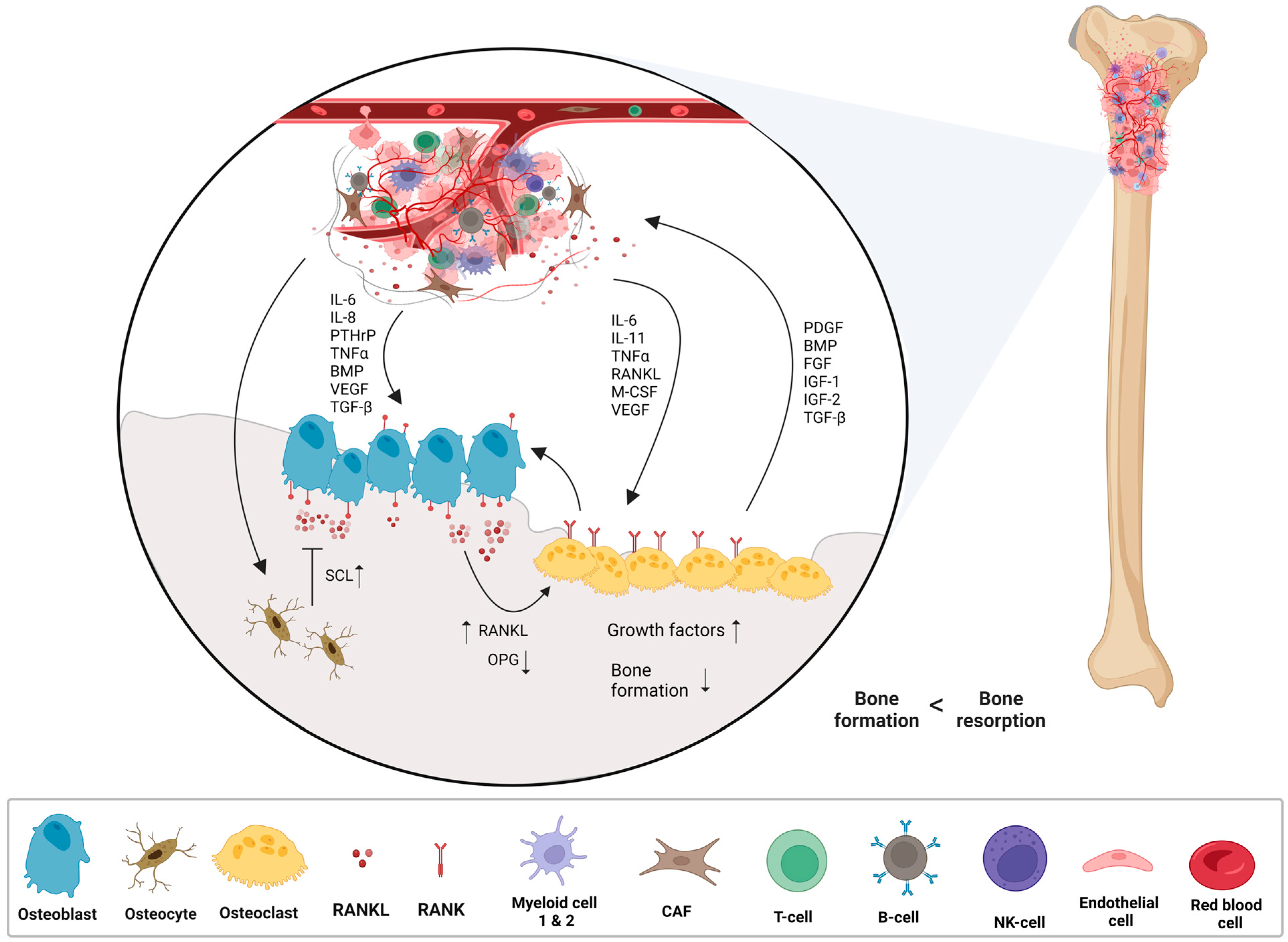
The term “sarcoma” refers to a tumour of connective tissue which derives from the Greek language—sarkos (flesh) and sarkōma (fleshy excrescence) [1]. Sarcomas are rare malignant solid tumours that mesenchymal cells derived cancer of the bones (osteosarcoma (OS), chondrosarcoma, and Ewing sarcoma) and soft tissues (muscle, fat, blood vessels, peripheral nerves and fibrous connective tissue), which represents approximately 1% of all adults and 15% of paediatric malignancies [2,3,4]. The World Health Organisation (WHO) defined and divided sarcoma tumours into more than 100 subtypes, depending on their distinct morphology and genetic alternations [5]. Consequently, the diagnosis of sarcoma can be challenging for pathologists.
Sarcomas may present aslocal tumours and are treated with excisional surgery and radiation. However, recent findings highlighted that 50% of sarcoma cases metastasise within the first years, depending on the tumour’s site, grade and subtype [6]. In addition, the 5-year relative survival rate for sarcoma patients with distant metastasis is less than 20% [7]. Several subtypes of sarcoma have specific systemic therapies to reduce tumour sizes such as tyrosine kinase inhibitor and imatinib for gastrointestinal sarcomas (gastrointestinal stromal tumour) [8] and alone or a cocktail of cytotoxic chemotherapy drugs (methotrexate, ifosfamide, doxorubicin, and cisplatin) for rhabdomyosarcoma, Ewing sarcoma, and OS [9,10].

2. Osteosarcoma

OS, also known as osteogenic sarcoma, is the most common primary malignant solid tumour of a bone among children and adolescents. There is a second, minor peak in older people due to Paget’s disease [11]. It is characterised by the formation of immature bone or osteoid tissue by tumour cells and malignant osteoblasts [12]. OS is a locally aggressive tumour and frequently produces early systemic metastases to the lungs, leading to a 5-year overall survival rate of 70%. OS usually occurs near the metaphysis of the long bones such as a distal femur, proximal tibia and proximal humerus, and less common sites include the skull, jaw, and pelvis [12,13].
OS can be classified into various subtypes based on tumour location, histology, and genetic characteristics. According to the WHO, the histological classification of OS can be divided into two categories, central (medullary) and surface (peripheral) tumours, with several subtypes within each group [14]. The central (medullary) OS subtypes are conventional, telangiectatic, low-grade, and small-cell, whereas the surface (peripheral) OS subtypes are parosteal, periosteal, and high-grade surface (Figure 1).

2.1. Central (Medullary) Osteosarcoma

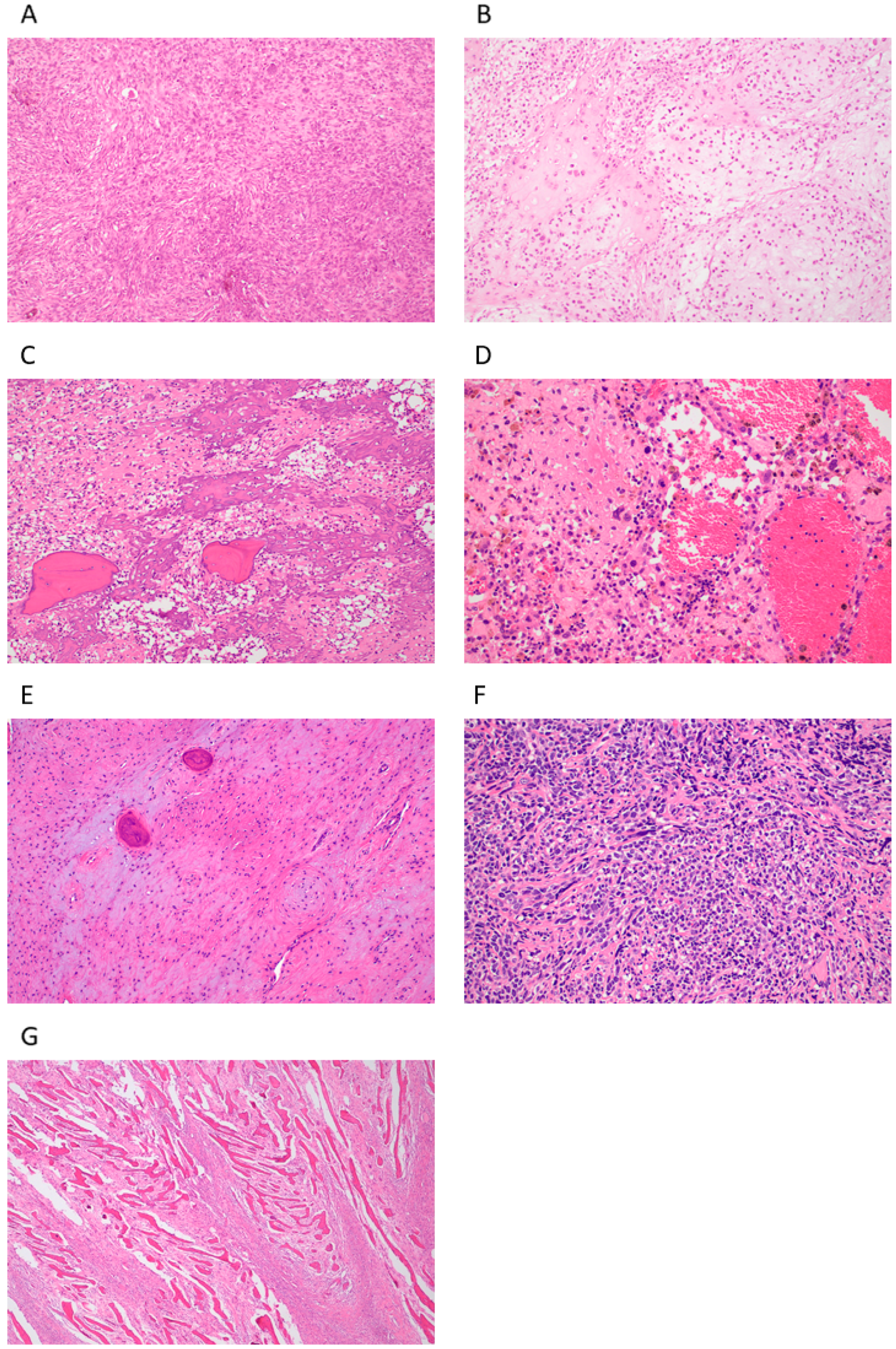
2.1.1. Conventional Central Osteosarcoma (COS)

COS can be divided into three groups including osteoblastic, chondroblastic, and fibroblastic. It is the most common pathological subtype of OS, which represents 80% of all the cases. It is characterised by a high-grade malignant bone-forming mesenchymal neoplasm producing osteoid, high necrosis, and atypical mitosis [15,16], although, for diagnosis, evidence of bone or osteoid production by the tumour cells within the tumour is required. The radiographic appearance of COS is usually an osteolytic lesion with cortical destruction, and the tumour extends into surrounding soft tissues [14]. In some cases, Codman’s triangle is observed on an X-ray. The presence of Codman’s triangle is often indicative of an aggressive bone lesion or tumour that is eroding the bone and pushing against the periosteum, resulting in bone lifting with a characteristic triangular shape [17].

2.1.2. Telangiectatic Osteosarcoma (TOS)

TOS accounts for <4% of all OS cases. It has radiographic and histological features similar to an aneurysmal bone cyst (ABC) or giant cell tumour [14]. The main characteristics of the tumour are an aggressive asymmetric expansion, ossification, and osteolytic lesion with permeative destruction [18]. Histologically, TOS usually has multiple cystic cavities containing blood, with septa composed of an osteoclast-like multinucleate giant and anaplastic cancer cells [18].

2.1.3. Low-Grade Osteosarcoma (LGOS, Intraosseous, Well-Differentiated)

LGOS is a rare intramedullary bone-producing tumour that accounts for 1–2% of all OS [19]. It is characterized by a low-grade malignant appearance, with slow growth and a relatively good prognosis compared to other types of OS [20]. LGOS generally affects older individuals in the third or fourth decade of life [14]. Radiologically, the appearance of LGOS is often confused with that of fibrous dysplasia, which often results in a delayed diagnosis or misdiagnosis [19]. The histological features of LGOS have included irregularly shaped bony spicules surrounded by hypocellular spindle cell stroma [21].

2.1.4. Small-Cell Osteosarcoma (SCOS)

SCOS is a rare subtype of OS which consists of sheets of round cells that produce an osteoid matrix. The tumour diagnosis may be confusing with Ewing sarcoma if the osteoid matrix is not included in the biopsy [22]. SCOS and Ewing sarcoma share extreme similarities, such as in both cases, the cells are round hypo-chromatic nuclei with little nuclear polymorphism [14]. Generally, radiological images show lytic lesions and sclerosis [14]. SCOS is a highly aggressive malignancy that primarily affects young adults. The prognosis for the disease is generally poor, with a reported 5-year survival rate of approximately 30% [23]. The high rate of metastasis and poor responses to chemotherapy contribute to the poor prognosis [23,24].

2.2. Surface (Peripheral) Osteosarcoma

2.2.1. Parosteal Osteosarcoma (POS)

POS is a slow-growing bone tumour arising from the cortical bone surface that originates from the periosteum, and commonly occurs in young women [25,26]. Radiologically, POS demonstrates densely ossified and lobulated mass and, histologically, exhibits atypical spindle cells between regularly arranged osseous trabeculae. The tumour diagnosis can be mistaken for fibrous dysplasia [25]. The aetiology of POS is not completely understood, but it has been associated with genetic abnormalities, such as the amplification of MDM2 and CDK4 oncogenes [27,28]. The overall prognosis for POS is generally favourable, with a reported 5-year survival rate of approximately 90% [26]. However, the risk of local recurrence and distant metastasis can persist, particularly in cases with inadequate surgical margins or high-grade histology.

2.2.2. Periosteal Osteosarcoma (PeOS)

PeOS is a rare primary malignant bone tumour arising from the inner layer of the periosteum [29]. The tumour contains osteoid-producing primitive MSCs between the cartilage lobules [29]. PeOS is characterized by a slow-growing, low-grade tumour that is typically less aggressive with a lower propensity to metastasize compared to other subtypes of OS [30]. The prognosis for PeOS is generally good, with a 91% overall survival rate at 5 years with surgical treatment alone [30].

2.2.3. High-Grade Surface Osteosarcoma (HGSO)

HGSO is an extremely rare subtype of OS, representing only <1% of all the tumours. It usually develops on the surface of the bone from the outer cortex [31]. It is characterized by a high-grade tumour with aggressive growth and a tendency to metastasize early [31,32]. The prognosis for HGSO is guarded, with a higher risk of local recurrence and metastasis compared to other OS subtypes [31].

3. Epidemiology and Aetiology

OS is still considered a rare cancer, with an annual incidence rate of 4.7 per million people in children and adolescents between the ages of 0 to 19 years and causes 8.9% of cancer-related deaths in children [33]. Some studies suggested that OS affects males slightly more frequently than females, with a male-to-female ratio of approximately 1.43:1 [34].
In the United States, research utilizing population data from the Surveillance, Epidemiology, and End Results (SEER) program has revealed that the incidence of OS varies by race and age group [35,36]. Among children and adolescents, the highest incidence is observed in people of Asian/Pacific Islands [36]. The highest rate of incidence is seen in people of African descent between the ages 25 and 59 years, while in those over 60 years of age, Caucasian people have the greatest incidence of the disease [37]. In addition, Italy, Latin America, Sudan, and Uganda have reported higher rates of childhood OS compared to other populations worldwide, whereas lower rates have been documented in Western Australia in contrast to the United States [34,35,36,37,38,39]. Moreover, higher rates of OS in the elderly population have been observed in the United Kingdom and Australia [34].
The quality of life of OS patients has improved over the last few decades, although its aetiology remains unknown. However, several risk factors related to the progression of the disease, including Paget’s disease, hereditary retinoblastoma, Li–Fraumeni syndrome, Rothmund–Thomson syndrome, RAPADILINO, Diamond–Blackfan anaemia, Bloom syndrome, Werner syndrome, ionizing radiation, exposure to beryllium, FBJ virus, osteochondromatosis, enchondromatosis, fibrous dysplasia, orthopaedic prosthetics, and alkylating agents [33,40,41]. An investigation of pre-therapeutic biopsy specimens has revealed the amplifications of chromosomes 6p21, 8q24, and 12q14 and the loss of chromosomes 9, 10, 13, and 17. Consequently, it can be said that chromosomal abnormalities correlate with OS [33]. In addition, recent studies have shown that insulin-like growth factors are usually highly expressed in OS tissues compared to healthy bone samples [33].
OS development has been associated with osteolysis, which is the destruction of periprosthetic bony tissue. Further, there is a positive correlation between OS aggressiveness and osteolysis levels. The receptor activator of nuclear factor kappa-Β (RANK, expressed on the cell surface of osteoclast precursors) and RANK ligand (RANKL, produced by osteoblasts and osteocytes) are responsible for regulating osteolysis [42]. Therefore, in the OS tumour microenvironment, osteoclast activity results in a vicious cycle between OS cell proliferation and bone degradation, leading to the release of pro-tumour factors such as insulin-like growth factor 1 (IGF1), insulin-like growth factor 2 (IGF2), transforming growth factor-β (TGF-β), fibroblast growth factor (FGF), bone morphogenetic protein (BMP), or platelet-derived growth factor (PDGF) from the bone matrix [42]. Further, the tumour produces interleukin (IL) -6 and -11, transforming growth factor- α (TGF-α), RANKL, macrophage colony-stimulating factor (M-CSF), vascular endothelial growth factor (VEGF), BMP, and parathyroid hormone-related protein (PTHrP) with aims of increasing bone resorption and promoting OS metastasis [42,43,44,45] (Figure 2).
The next-generation single-cell RNA sequencing highlighted that the OS tumour microenvironment is highly heterogeneous [46]. The tumour is composed of bone cells such as osteoclasts and osteoblasts as well as fibroblasts, endothelial cells, immune cells including myeloid cells 1 and 2, natural killer cells, T cells, B cells, monocytes, and plasmacytes [46]. Complexity and high heterogeneity increase the difficulty of diagnosis and treatment of OS. Moreover, a tumour with significant heterogeneity often conveys resistance to chemotherapy drugs, giving rise to relapse and metastasis to the lungs or other bones [47].

4. Symptoms, Diagnosis, and Treatment

OS symptoms usually include local pain, swelling, tenderness, and redness (inflammation) near the affected bone [48]. Patients may also experience weight loss, fever, fatigue, or malaise [49].
Diagnosis and progression of the disease are always achieved by a combination of medical imaging such as X-ray, magnetic resonance imaging (MRI), positron emission tomography (PET), and computed tomography (CT) scans. Further, the diagnosis is confirmed by the histological examination of a microscopic examination of suspected tissue that has been excised by biopsy [50].
In cancer, therapeutic approaches are always based on the stage of the tumour, the age of the patient, the patient’s general condition and quality of life, and the patient’s life expectancy. Currently, only three major therapeutic strategies are available for OS, including surgery, chemotherapy, and radiotherapy [51].
Amputation and limb-sparing are the primary surgeries in OS therapy to complete tumour removal with a wide margin of normal tissue to avoid local recurrence and improve overall survival [51]. However, surgery alone creates surgical stress and causes ischemia-reperfusion injury, activation of the sympathetic nervous system, endocrine and metabolic changes, acute and chronic inflammation, and immune suppression within the body, and most importantly, promotes tumour metastasis by tumour cell dissemination and circulating tumour cells survival [52,53,54]. Consequently, the current management of OS always includes surgery followed by adjuvant chemotherapy or radiotherapy to destroy the remaining tumour cells and eliminate micro-metastases [51,52,53,54,55].
The chemotherapy regimens applied for OS are high-dose methotrexate (HDMTX) with leucovorin rescue, doxorubicin (DOX), cisplatin, and ifosfamide (IFO) with or without etoposide.
MTX is a folate antagonist widely used to treat a variety of autoimmune diseases, including rheumatoid arthritis and psoriasis [56]. It inhibits the production of pyrimidine and purine nucleotides and thymidylic acid by binding dihydrofolate reductase to inhibit the DNA replication of cancer cells [57]. Despite its efficacy, HDMTX has serious side effects, such as renal failure, mucositis, hepatotoxicity, pulmonary toxicity, and neurotoxicity [57,58,59]. Further, the standard chemotherapy dose of 8–12 g/m2 HDMTX is higher than the absolute lethal dose of 2–4 mg/kg [60,61,62]. Although leucovorin, also called folinic acid is commonly used as a rescue therapy in HDMTX regimens to help reduce the toxicity of MTX on non-malignant cells [60].
DOX, also known as Adriamycin, is an anthracycline chemotherapy drug extracted from Streptomyces peucetius var. caesius in the 1970s and is widely used in many cancer types, including breast, lung, bladder, lymphoma, and paediatric cancers [63,64,65]. Its cumulative dose is from 240 to 480 mg/m2, and the dose per cycle is from 60 to 90 m2 [66]. The drug’s mechanism of action is through intercalation into the DNA double helix and the disruption of topoisomerase-II-mediated DNA repair and inhibits the synthesis of both DNA and RNA [67]. Despite its good effects on patients’ survival, like many cytotoxic drugs, DOX can be toxic to both cancer cells and healthy cells. In addition, the drug has a severe impact on patients in the long term, including cardiomyopathy, symptomatic cardiac toxicity, transient electrocardiographic abnormalities, alopecia, and myelosuppression [67,68].
Cisplatin, (SP-4-2)-diamminedichloridoplatinum(II), is the first metal–platinum-based chemotherapeutic drug that is commonly used in the treatment of various types of cancer, including sarcomas, testicular, bladder, and lung cancer [69,70]. Its cumulative dose is from 480 to 600 mg/m2; its dose per cycle is from 100 to 120 mg/m2 [66]. Cisplatin binds to the N7 reactive centre on purine residues of malignant cells’ DNA, resulting in the inhibition of DNA synthesis, and blocking DNA replication, further promoting apoptosis [70,71]. Unfortunately, the drug also has extreme side effects, such as acute and chronic renal failure, peripheral neuropathy, ototoxicity, hypomagnesemia, gastrointestinal disorders, and haemorrhage [67,71].
IFO, a bifunctional alkylating agent, is a member of the nitrogen mustard family and has been used to treat several tumour types such as germ cell tumours, lymphomas, and soft tissue sarcoma [72,73,74]. Its standard cumulative dose starts from 480 to 600 mg/m2; the dose per cycle is from 100 to 120 mg/m2 [66]. The drug’s mechanism of action is through the cross-linking of DNA strands, inhibition of DNA synthesis, and protein translation [57]. IFO can have some serious side effects, such as haemorrhagic cystitis, acute kidney injury, Fanconi syndrome, interstitial nephritis, glomerular disease, and encephalopathy [57,72].

5. Conclusions Remarks

Over the last three decades, patient outcomes have not significantly improved and the current chemotherapy approaches for OS are highly disappointing [67]. The failure to improve survival rates reflects the lack of novel treatment strategies. Given the urgency of the situation, researchers and clinicians are actively working to develop more effective and less toxic treatments for patients with OS. Some of the approaches being explored include immunotherapy, targeted therapy, and combination therapy [75,76]. However, in order to develop more precise and effective therapies, it is crucial for scientists and clinicians to thoroughly investigate each subtype of OS, elucidating their distinct gene expression profiles and the underlying pathophysiology of the subtypes. Furthermore, currently, there are no biomarkers available for any subtypes of the OS that can detect the disease at an early stage and provide accurate monitoring of disease progression. To facilitate the development of new treatments, it is imperative to identify predictive and prognostic markers for each subtype of the OS.

Author Contributions

Writing: E.R. revising and editing: J.X. and D.W. All authors have read and agreed to the published version of the manuscript.

Funding

This research was funded by the Abbie Basson Sarcoma Foundation (Sock it to Sarcoma!). E.R. was also supported by the Australian Graduate Women—Barbara Hale Fellowship.

Institutional Review Board Statement

The study was conducted according to the guidelines of the Declaration of Helsinki, and approved by the by the Ethics Review Committee of the University of Western Australia and Sir Charles Gairdner Hospital (2019/RA/4/20/5211).

Informed Consent Statement

Informed consent was obtained from all subjects involved in the study.

Conflicts of Interest

The authors declare no conflict of interest.

References

  1. Skubitz, K.M.; D’Adamo, D.R. Sarcoma. Mayo Clin. Proc. 2007, 82, 1409–1432. [Google Scholar] [CrossRef] [PubMed]
  2. Lee, D.Y.; Staddon, A.P.; Shabason, J.E.; Sebro, R. Phase I and phase II clinical trials in sarcoma: Implications for drug discovery and development. Cancer Med. 2019, 8, 585–592. [Google Scholar] [CrossRef] [PubMed]
  3. Hatina, J.; Kripnerova, M.; Houfkova, K.; Pesta, M.; Kuncova, J.; Sana, J.; Slaby, O.; Rodríguez, R. Sarcoma Stem Cell Heterogeneity. Stem Cells Heterog.-Nov. Concepts 2019, 1123, 95–118. [Google Scholar]
  4. Schöffski, P.; Blay, J.-Y.; Ray-Coquard, I. Cabozantinib as an emerging treatment for sarcoma. Curr. Opin. Oncol. 2020, 32, 321–331. [Google Scholar] [CrossRef] [PubMed]
  5. Zhu, M.M.; Shenasa, E.; Nielsen, T.O. Sarcomas: Immune biomarker expression and checkpoint inhibitor trials. Cancer Treat. Rev. 2020, 91, 102115. [Google Scholar] [CrossRef]
  6. Brennan, M.F.; Antonescu, C.R.; Moraco, N.; Singer, S. Lessons learned from the study of 10,000 patients with soft tissue sarcoma. Ann. Surg. 2014, 260, 416–421, discussion 421–422. [Google Scholar] [CrossRef]
  7. Tang, F.; Tie, Y.; Wei, Y.-Q.; Tu, C.-Q.; Wei, X.-W. Targeted and immuno-based therapies in sarcoma: Mechanisms and advances in clinical trials. Biochim. Biophys. Acta Rev. Cancer 2021, 1876, 188606. [Google Scholar] [CrossRef]
  8. Poveda, A.; del Muro, X.G.; López-Guerrero, J.A.; Cubedo, R.; Martínez, V.; Romero, I.; Serrano, C.; Valverde, C.; Martín-Broto, J. GEIS guidelines for gastrointestinal sarcomas (GIST). Cancer Treat. Rev. 2017, 55, 107–119. [Google Scholar] [CrossRef]
  9. Young, R.; Natukunda, A.; Litière, S.; Woll, P.; Wardelmann, E.; van der Graaf, W. First-line anthracycline-based chemotherapy for angiosarcoma and other soft tissue sarcoma subtypes: Pooled analysis of eleven European Organisation for Research and Treatment of Cancer Soft Tissue and Bone Sarcoma Group trials. Eur. J. Cancer 2014, 50, 3178–3186. [Google Scholar] [CrossRef]
  10. Ferrari, S.; Serra, M. An update on chemotherapy for osteosarcoma. Expert Opin. Pharmacother. 2015, 16, 2727–2736. [Google Scholar] [CrossRef]
  11. Rothzerg, E.; Ho, X.D.; Xu, J.; Wood, D.; Märtson, A.; Kõks, S. Upregulation of 15 Antisense Long Non-Coding RNAs in Osteosarcoma. Genes 2021, 12, 1132. [Google Scholar] [CrossRef] [PubMed]
  12. Kansara, M.; Teng, M.W.; Smyth, M.J.; Thomas, D.M. Translational biology of osteosarcoma. Nat. Rev. Cancer 2014, 14, 722–735. [Google Scholar] [CrossRef] [PubMed]
  13. Stitzlein, R.N.; Wojcik, J.; Sebro, R.A.; Balamuth, N.J.; Weber, K.L. Team Approach: Osteosarcoma of the Distal Part of the Femur in Adolescents. JBJS Rev. 2017, 5, e5. [Google Scholar] [CrossRef] [PubMed]
  14. Misaghi, A.; Goldin, A.; Awad, M.; Kulidjian, A.A. Osteosarcoma: A comprehensive review. SICOT-J 2018, 4, 12. [Google Scholar] [CrossRef]
  15. Sadeghi, H.M.; Karimi, A.; Derakhshan, S.; Aminishakib, P.; Parchami, K. Conventional osteosarcoma of the mandible: Report of a rare case. Clin. Case Rep. 2021, 9, e04843. [Google Scholar]
  16. Kimura, Y.; Tomihara, K.; Tachinami, H.; Imaue, S.; Nakamori, K.; Fujiwara, K.; Suzuki, K.; Yasuda, T.; Miwa, S.; Nakayama, E.; et al. Conventional osteosarcoma of the mandible successfully treated with radical surgery and adjuvant chemotherapy after responding poorly to neoadjuvant chemotherapy: A case report. J. Med. Case Rep. 2017, 11, 210. [Google Scholar] [CrossRef]
  17. McDonald, J.; DenOtter, T.D. Codman Triangle. In StatPearls; Statpearls Publishing: Treasure Island, FL, USA, 2023. [Google Scholar]
  18. Limaiem, F.; Kuhn, J.; Khaddour, K. Telangiectatic Osteosarcoma. In StatPearls; StatPearls Publishing: Treasure Island, FL, USA, 2022. [Google Scholar]
  19. Malhas, A.M.; Sumathi, V.P.; James, S.L.; Menna, C.; Carter, S.R.; Tillman, R.M.; Jeys, L.; Grimer, R.J. Low-Grade Central Osteosarcoma: A Difficult Condition to Diagnose. Sarcoma 2012, 2012, 764796. [Google Scholar] [CrossRef]
  20. Sinha, R.; Chowdhury, S.K.R.; Chattopadhyay, P.K.; Rajkumar, K. Low-Grade Osteosarcoma of the Mandible. J. Maxillofac. Oral Surg. 2010, 9, 186–190. [Google Scholar] [CrossRef]
  21. Vasiliadis, H.S.; Arnaoutoglou, C.; Plakoutsis, S.; Doukas, M.; Batistatou, A.; Xenakis, T.A. Low-grade central osteosarcoma of distal femur, resembling fibrous dysplasia. World J. Orthop. 2013, 4, 327–332. [Google Scholar] [CrossRef]
  22. Edeiken, J.; Raymond, A.K.; Ayala, A.G.; Benjamin, R.S.; Murray, J.A.; Carrasco, H.C. Small-cell osteosarcoma. Skelet. Radiol. 1987, 16, 621–628. [Google Scholar] [CrossRef]
  23. Nakajima, H.; Sim, F.H.; Bond, J.R.; Unni, K.K. Small cell osteosarcoma of bone. Review of 72 cases. Cancer 1997, 79, 2095–2106. [Google Scholar] [CrossRef]
  24. Righi, A.; Gambarotti, M.; Longo, S.; Benini, S.; Gamberi, G.; Cocchi, S.; Vanel, D.; Picci, P.; Bertoni, F.; Simoni, A.; et al. Small cell osteosarcoma: Clinicopathologic, immunohistochemical, and molecular analysis of 36 cases. Am. J. Surg. Pathol. 2015, 39, 691–699. [Google Scholar] [CrossRef] [PubMed]
  25. Hang, J.F.; Chen, P.C. Parosteal osteosarcoma. Arch. Pathol. Lab. Med. 2014, 138, 694–699. [Google Scholar] [CrossRef] [PubMed]
  26. Prabowo, Y.; Kamal, A.F.; Kodrat, E.; Prasetyo, M.; Maruanaya, S.; Efar, T.S. Parosteal Osteosarcoma: A Benign-Looking Tumour, Amenable to a Variety of Surgical Reconstruction. Int. J. Surg. Oncol. 2020, 2020, 4807612. [Google Scholar] [CrossRef]
  27. Sciot, R. MDM2 Amplified Sarcomas: A Literature Review. Diagnostics 2021, 11, 496. [Google Scholar] [CrossRef]
  28. Chen, P.C.; Yen, C.C.; Hung, G.Y.; Pan, C.C.; Chen, W.M. Gene amplification and tumor grading in parosteal osteosarcoma. J. Chin. Med. Assoc. 2019, 82, 889–894. [Google Scholar] [CrossRef]
  29. Liu, X.W.; Zi, Y.; Xiang, L.B.; Han, T.Y. Periosteal osteosarcoma: A review of clinical evidence. Int. J. Clin. Exp. Med. 2015, 8, 37–44. [Google Scholar]
  30. Cesari, M.; Alberghini, M.; Vanel, D.; Palmerini, E.; Staals, E.L.; Longhi, A.; Abate, M.; Ferrari, C.; Balladelli, A.; Ferrari, S. Periosteal osteosarcoma: A single-institution experience. Cancer 2011, 117, 1731–1735. [Google Scholar] [CrossRef]
  31. Deng, Z.; Huang, Z.; Ding, Y.; Su, Y.; Chan, C.M.; Niu, X. High-Grade Surface Osteosarcoma: Clinical Features and Oncologic Outcome. J. Bone Oncol. 2020, 23, 100288. [Google Scholar] [CrossRef]
  32. Staals, E.L.; Bacchini, P.; Bertoni, F. High-grade surface osteosarcoma: A review of 25 cases from the Rizzoli Institute. Cancer 2008, 112, 1592–1599. [Google Scholar] [CrossRef]
  33. Jafari, F.; Javdansirat, S.; Sanaie, S.; Naseri, A.; Shamekh, A.; Rostamzadeh, D.; Dolati, S. Osteosarcoma: A comprehensive review of management and treatment strategies. Ann. Diagn. Pathol. 2020, 49, 151654. [Google Scholar] [CrossRef] [PubMed]
  34. Mirabello, L.; Troisi, R.J.; Savage, S.A. International osteosarcoma incidence patterns in children and adolescents, middle ages and elderly persons. Int. J. Cancer 2009, 125, 229–234. [Google Scholar] [CrossRef] [PubMed]
  35. Linabery, A.; Ross, J.A. Trends in childhood cancer incidence in the U.S. (1992–2004). Cancer 2007, 112, 416–432. [Google Scholar] [CrossRef] [PubMed]
  36. Savage, S.A.; Mirabello, L. Using Epidemiology and Genomics to Understand Osteosarcoma Etiology. Sarcoma 2011, 2011, 548151. [Google Scholar] [CrossRef]
  37. Mirabello, L.; Troisi, R.J.; Savage, S.A. Savage, Osteosarcoma incidence and survival rates from 1973 to 2004: Data from the Surveillance, Epidemiology, and End Results Program. Cancer 2009, 115, 1531–1543. [Google Scholar] [CrossRef]
  38. Parkin, D.M.; Stiller, C.A.; Draper, G.J.; Bieber, C.A. The international incidence of childhood cancer. Int. J. Cancer 1988, 42, 511–520. [Google Scholar] [CrossRef]
  39. Blackwell, J.B.; Threlfall, T.J.; McCaul, K.A. Primary malignant bone tumours in Western Australia, 1972–1996. Pathology 2005, 37, 278–283. [Google Scholar] [CrossRef]
  40. Prater, S.; McKeon, B. Osteosarcoma. In StatPearls; StatPearls Publishing: Treasure Island, FL, USA, 2022. [Google Scholar]
  41. Beird, H.C.; Bielack, S.S.; Flanagan, A.M.; Gill, J.; Heymann, D.; Janeway, K.A.; Livingston, J.A.; Roberts, R.D.; Strauss, S.J.; Gorlick, R. Author Correction: Osteosarcoma. Nat. Rev. Dis. Prim. 2022, 8, 82. [Google Scholar] [CrossRef]
  42. Corre, I.; Verrecchia, F.; Crenn, V.; Redini, F.; Trichet, V. The Osteosarcoma Microenvironment: A Complex But Targetable Ecosystem. Cells 2020, 9, 976. [Google Scholar] [CrossRef]
  43. Ara, T.; DeClerck, Y.A. Interleukin-6 in bone metastasis and cancer progression. Eur. J. Cancer 2010, 46, 1223–1231. [Google Scholar] [CrossRef]
  44. Zhu, T.; Han, J.; Yang, L.; Cai, Z.; Sun, W.; Hua, Y.; Xu, J. Immune Microenvironment in Osteosarcoma: Components, Therapeutic Strategies and Clinical Applications. Front. Immunol. 2022, 13, 907550. [Google Scholar] [CrossRef] [PubMed]
  45. Lokau, J.; Schoeder, V.; Garbers, C. The role of interleukin-11 in osteosarcoma. Pathologe 2020, 41, 163–167. [Google Scholar] [CrossRef] [PubMed]
  46. Rothzerg, E.; Feng, W.; Song, D.; Li, H.; Wei, Q.; Fox, A.; Wood, D.; Xu, J.; Liu, Y. Single-Cell Transcriptome Analysis Reveals Paraspeckles Expression in Osteosarcoma Tissues. Cancer Inform. 2022, 21, 11769351221140101. [Google Scholar] [CrossRef] [PubMed]
  47. Zhang, Y.; Yang, J.; Zhao, N.; Wang, C.; Kamar, S.; Zhou, Y.; He, Z.; Yang, J.; Sun, B.; Shi, X.; et al. Progress in the chemotherapeutic treatment of osteosarcoma. Oncol. Lett. 2018, 16, 6228–6237. [Google Scholar] [CrossRef]
  48. Ritter, J.; Bielack, S.S. Osteosarcoma. Ann. Oncol. 2010, 21 (Suppl. S7), vii320–vii325. [Google Scholar] [CrossRef]
  49. Simpson, E.; Brown, H.L. Understanding osteosarcomas. J. Am. Acad. Physician Assist. 2018, 31, 15–19. [Google Scholar] [CrossRef]
  50. Durfee, R.A.; Mohammed, M.; Luu, H.H. Review of Osteosarcoma and Current Management. Rheumatol. Ther. 2016, 3, 221–243. [Google Scholar] [CrossRef]
  51. Ando, K.; Heymann, M.F.; Stresing, V.; Mori, K.; Redini, F.; Heymann, D. Current therapeutic strategies and novel approaches in osteosarcoma. Cancers 2013, 5, 591–616. [Google Scholar] [CrossRef]
  52. Chen, Z.; Zhang, P.; Xu, Y.; Yan, J.; Liu, Z.; Lau, W.B.; Lau, B.; Li, Y.; Zhao, X.; Wei, Y.; et al. Surgical stress and cancer progression: The twisted tango. Mol. Cancer 2019, 18, 132. [Google Scholar] [CrossRef]
  53. Gottschalk, A.; Sharma, S.; Ford, J.; Durieux, M.E.; Tiouririne, M. Review article: The role of the perioperative period in recurrence after cancer surgery. Anesth. Analg. 2010, 110, 1636–1643. [Google Scholar] [CrossRef]
  54. Tohme, S.; Simmons, R.L.; Tsung, A. Surgery for cancer: A trigger for metastases. Cancer Res. 2017, 77, 1548–1552. [Google Scholar] [CrossRef] [PubMed]
  55. Carrle, D.; Bielack, S.S. Current strategies of chemotherapy in osteosarcoma. Int. Orthop. 2006, 30, 445–451. [Google Scholar] [CrossRef] [PubMed]
  56. Padmanabhan, S.; Tripathi, D.N.; Vikram, A.; Ramarao, P.; Jena, G.B. Methotrexate-induced cytotoxicity and genotoxicity in germ cells of mice: Intervention of folic and folinic acid. Mutat. Res. 2009, 673, 43–52. [Google Scholar] [CrossRef] [PubMed]
  57. Wittig, J.C.; Bickels, J.; Priebat, D.; Jelinek, J.; Kellar-Graney, K.; Shmookler, B.; Malawer, M.M. Osteosarcoma: A multidisciplinary approach to diagnosis and treatment. Am. Fam. Physician 2002, 65, 1123–1132. [Google Scholar]
  58. Janeway, K.A.; Grier, H.E. Sequelae of osteosarcoma medical therapy: A review of rare acute toxicities and late effects. Lancet Oncol. 2010, 11, 670–678. [Google Scholar] [CrossRef]
  59. Crews, K.R.; Liu, T.; Rodriguez-Galindo, C.; Tan, M.; Meyer, W.H.; Panetta, J.C.; Link, M.P.; Daw, N.C. High-dose methotrexate pharmacokinetics and outcome of children and young adults with osteosarcoma. Cancer 2004, 100, 1724–1733. [Google Scholar] [CrossRef]
  60. Xu, M.; Xu, S.; Yu, X. Clinical Analysis of Osteosarcoma Patients Treated with High-Dose Methotrexate-Free Neoadjuvant Chemotherapy. Curr. Oncol. 2014, 21, 678–684. [Google Scholar] [CrossRef]
  61. Graf, N.; Winkler, K.; Betlemovic, M.; Fuchs, N.; Bode, U. Methotrexate pharmacokinetics and prognosis in osteosarcoma. J. Clin. Oncol. 1994, 12, 1443–1451. [Google Scholar] [CrossRef]
  62. Jaffe, N.; Gorlick, R. High-dose methotrexate in osteosarcoma: Let the questions surcease—Time for final acceptance. J. Clin. Oncol. 2008, 26, 4365–4366. [Google Scholar] [CrossRef]
  63. Arcamone, F.; Cassinelli, G.; Fantini, G.; Grein, A.; Orezzi, P.; Pol, C.; Spalla, C. Adriamycin, 14-hydroxydaunomycin, a new antitumor antibiotic from S. peucetius var. caesius. Biotechnol. Bioeng. 2000, 67, 704–713. [Google Scholar] [CrossRef]
  64. Cortés-Funes, H.; Coronado, C. Role of anthracyclines in the era of targeted therapy. Cardiovasc. Toxicol. 2007, 7, 56–60. [Google Scholar] [CrossRef] [PubMed]
  65. Thorn, C.F.; Oshiro, C.; Marsh, S.; Hernandez-Boussard, T.; McLeod, H.; Klein, T.E.; Altman, R.B. Doxorubicin pathways: Pharmacodynamics and adverse effects. Pharm. Genom. 2011, 21, 440–446. [Google Scholar] [CrossRef] [PubMed]
  66. Luetke, A.; Meyers, P.A.; Lewis, I.; Juergens, H. Osteosarcoma treatment—Where do we stand? A state of the art review. Cancer Treat. Rev. 2014, 40, 523–532. [Google Scholar] [CrossRef] [PubMed]
  67. Rothzerg, E.; Pfaff, A.L.; Koks, S. Innovative approaches for treatment of osteosarcoma. Exp. Biol. Med. 2022, 247, 310–316. [Google Scholar] [CrossRef] [PubMed]
  68. Longhi, A.; Ferrari, S.; Bacci, G.; Specchia, S. Long-term follow-up of patients with doxorubicin-induced cardiac toxicity after chemotherapy for osteosarcoma. Anti-Cancer Drugs 2007, 18, 737–744. [Google Scholar] [CrossRef]
  69. Ghosh, S. Cisplatin: The first metal based anticancer drug. Bioorg. Chem. 2019, 88, 102925. [Google Scholar] [CrossRef]
  70. Tchounwou, P.B.; Dasari, S.; Noubissi, F.K.; Ray, P.; Kumar, S. Advances in Our Understanding of the Molecular Mechanisms of Action of Cisplatin in Cancer Therapy. J. Exp. Pharmacol. 2021, 13, 303–328. [Google Scholar] [CrossRef]
  71. Dasari, S.; Tchounwou, P.B. Cisplatin in cancer therapy: Molecular mechanisms of action. Eur. J. Pharmacol. 2014, 740, 364–378. [Google Scholar] [CrossRef]
  72. Sprangers, B.; Lapman, S. The growing pains of ifosfamide. Clin. Kidney J. 2020, 13, 500–503. [Google Scholar] [CrossRef]
  73. Sarbay, H.; Demir, U.F.; Yilmaz, G.; Atay, A.A.; Malbora, B. Ifosfamide induced encephalopathy in a child with osteosarcoma. J. Oncol. Pharm. Pract. 2021, 27, 1302–1306. [Google Scholar] [CrossRef]
  74. Bokemeyer, C.; Harstrick, A.; Beyer, J.; Metzner, B.; Ruther, U.; Hartmann, J.T.; Holstein, K.; Derigs, H.G.; de Wit, R.; Casper, J.; et al. The use of dose-intensified chemotherapy in the treatment of metastatic nonseminomatous testicular germ cell tumors. German Testicular Cancer Study Group. Semin. Oncol. 1998, 25 (Suppl. S4), 24–32, discussion 45–48. [Google Scholar] [PubMed]
  75. Hu, Z.; Wen, S.; Huo, Z.; Wang, Q.; Zhao, J.; Wang, Z.; Chen, Y.; Zhang, L.; Zhou, F.; Guo, Z.; et al. Current Status and Prospects of Targeted Therapy for Osteosarcoma. Cells 2022, 11, 3507. [Google Scholar] [CrossRef] [PubMed]
  76. Lu, Y.; Zhang, J.; Chen, Y.; Kang, Y.; Liao, Z.; He, Y.; Zhang, C. Novel Immunotherapies for Osteosarcoma. Front. Oncol. 2022, 12, 830546. [Google Scholar] [CrossRef] [PubMed]
Figure 1. Hematoxylin and eosin staining of osteosarcoma subtypes. Conventional central fibroblastic OS (A) shows predominant spindle cells and low osteoid/bone within the tumour. Conventional central chondroblastic OS (B) shows the predominantly chondroid matrix within the tumour. Conventional central osteoblastic OS (C) highlights the bony/osteoid matrix production in the tumour. Telangiectatic OS (D) highlights the dramatic amount of osteoid production and blood-filled spaces that are separated by septa containing highly malignant cells. Low-grade OS (E) shows the irregularly shaped bony specular and hypocellular spindle cell stroma. Small-cell OS (F) highlights the sheets of round cells that produce an osteoid matrix. Parosteal OS (G) highlights the regularly arranged osseous trabecula surrounded by atypical spindle cells. All the images were taken with ×40 magnification. Informed consent was obtained from the patients. There are no available images of periosteal or high-grade surface OS.
Jmp 04 00011 g001
Figure 2. Illustration of bone remodelling in the osteosarcoma tumour microenvironment. The bone remodelling is induced by OS cells, creating a vicious cycle to promote cell proliferation and survival. Tumour cells secrete IL-6, IL-11, and TNF-α to stimulate osteoclastogenesis. The cells also secrete TGF-β which promotes RANKL secretion by osteoblasts to decrease osteoblastic bone formation. Furthermore, osteocytes secreted SCL through the tumour cells, inhibiting osteoblast activity in the bone. As a result, OS-related bone remodelling creates a low expression of OPG and a high expression of RANKL. Therefore, the bone homeostasis equilibrium is an imbalance that accumulates a new environment: more increased bone resorption and lower bone formation within the bone. The image was created with BioRender.com. Interleukin 6 (IL-6); interleukin 8 (IL-8); interleukin 11 (IL-11); insulin-like growth factor 1 (IGF-1); insulin-like growth factor 2 (IGF-2); parathyroid hormone-related protein (PTHrP); tumour necrosis factor-alpha (TNF-α); bone morphogenetic protein (BMP); vascular endothelial growth factor (VEGF); transforming growth factor beta (TGF-β); macrophage-colony-stimulating factor (M-CSF); receptor activator of nuclear factor kappa-Β (RANK); RANK ligand (RANKL); fibroblast growth factor (FGF); platelet-derived growth factors (PDGF); osteoprotegerin (OPG); sclerostin (SCL). Created with BioRender.com.
Jmp 04 00011 g002
Disclaimer/Publisher’s Note: The statements, opinions and data contained in all publications are solely those of the individual author(s) and contributor(s) and not of MDPI and/or the editor(s). MDPI and/or the editor(s) disclaim responsibility for any injury to people or property resulting from any ideas, methods, instructions or products referred to in the content.

