Commentary

RNA-Based Next-Generation Sequencing in the Somatic Molecular Testing of Non-Small-Cell Lung Cancer (NSCLC) in a Centralized Model: Real-World Data to Suggest It Is Time to Reconsider Testing Options

Cellular and Molecular Pathologist, Morriston Hospital, Swansea Bay University Health Board, Swansea SA6 6NL, UK
J. Mol. Pathol. 2022, 3(4), 307-318; https://doi.org/10.3390/jmp3040026
Submission received: 7 October 2022 / Revised: 26 October 2022 / Accepted: 28 October 2022 / Published: 8 November 2022

Abstract

:
Best practice in the management of non-squamous, non-small-cell lung cancer patients involves somatic testing for a range of molecular markers. Actionable oncogenic drivers of malignancy are increasingly being detected using RNA-based next-generation sequencing in the UK by referral to centralized genomic laboratory hubs. Recent audit data from the author’s case work have demonstrated an RNA sequencing failure rate of 35%. This article examines the real-world context, which may account for this failure rate, and discusses alternative options for patient care pathways.

1. Introduction

Lung cancer is the most common cause of death from cancer worldwide [1]. The United Kingdom (UK) has the some of the worst survival outcomes of developed nations across the world and Europe [1,2,3]. It is important for all healthcare professionals to understand factors that may be contributing to these poor outcomes for our patients. This commentary examines the role molecular biomarker detection may have in the care of patients with non-small-cell lung cancer (NSCLC) in the UK.
In the UK, patients’ cases are discussed at multidisciplinary team meetings (MDT /tumour board meetings) to sense-check diagnostic information from radiology and pathology with the clinical context and to determine the best management plan for each individual patient according to their wider health and circumstances [4,5]. Predictive molecular biomarkers are needed to inform patient management options and for the consideration of targeted oncological agents [6,7]. The array of biomarkers needed to inform the best management plan for non-squamous NSCLC has evolved at pace in recent years [8]. At the time of writing, programmed death ligand-1 (PD-L1) expression is assessed by immunohistochemical methods, providing sufficient malignant tissue is available, followed by somatic tumour mutations in Kirsten rat sarcoma viral proto-oncogene (KRAS), V-raf murine sarcoma viral oncogene homologue B (BRAF), epidermal growth factor receptor (EGFR) analysis by DNA next-generation sequencing (NGS) [9,10,11,12,13,14,15]. Gene fusion events in anaplastic lymphoma receptor tyrosine kinase (ALK1), ROS proto-oncogene tyrosine-protein kinase (ROS1), neurotrophic receptor tyrosine kinase 1, 2 and 3 (NTRK1/2/3), RET proto-oncogene, receptor tyrosine kinase (RET) and skipping lesions in exon 14 of the MET proto-oncogene receptor tyrosine kinase (MET) can be identified using ribonucleic acid (RNA)-based next-generation sequencing (NGS) in somatic tissue [16,17]. RNA sequencing is preferable to deoxyribose nucleic acid (DNA) sequencing for large structural rearrangements in somatic genes where there are large intronic sequences in the DNA of the gene of interest, NTRK1 being a good example [17,18]. Sequencing spliced messenger RNA transcripts that consist solely of exons allows for more accurate detection of fusion events by current bioinformatic analytical methods [18,19]. The incidence of gene fusion events in lung cancer is low [20]. ALK1 rearrangements occur in approximately 3% of Western populations with primary lung adenocarcinomas, whereas ROS1 is the cancer driver in less than 1% of cases [21]. RET and NTRK1,2 and NTRK3 fusions and MET 14 skipping variants in NSCLC are also uncommon [15,22,23,24,25,26,27].
Our local practice is to send tissue sections on charged glass slides to an external laboratory for molecular testing as our cellular pathology department lacks the molecular-grade medical laboratory facilities and biomedical scientist (BMS) staff required to conduct DNA and RNA extraction from tissues. This process is best commenced at the same time the formalin-fixed paraffin-embedded (FFPE) tissue block is cut to prepare the haematoxylin and eosin (H&E)-stained slide for morphological assessment by a histopathologist [28]. Cutting the FFPE block requires re-facing each time a BMS attempts to cut a section of tissue in order to ensure the surface is smooth and appropriately orientated to give a full slice representing all areas in the FFPE block. This process of re-facing inevitably involves the loss of small amounts of tissue for accuracy of slide production. Limiting slide processing to one single step and cutting all possible required slides up front to prevent waste is clearly an ideal step to prevent any valuable tumour tissue from being wasted [29]. This is no more important than in the care of lung cancer patients, where small samples such as bronchoscopic biopsies and endoscopic ultrasound-guided (EBUS) fine needle aspiration cytology can yield very small amounts of tumour tissue (see Figure 1) but a large amount of molecular information is required for diagnosis and treatment [30,31,32]. Funding issues and staffing shortages mean that we are currently unable to offer upfront slide sectioning. Pathologists with expertise in thoracic pathology are well advised to limit diagnostic immunohistochemistry (IHC) use in such cases to just two protein markers: p40, an antibody recognizing the deltaNp63 isoform of p63 protein, and thyroid transcription factor-1 (TTF1) for subtyping squamous cell carcinomas and adenocarcinomas, respectively [4,29].
This paper will examine factors to consider for best patient care in predictive molecular biomarker identification in lung non-squamous NSCLC, adenocarcinoma being the most common type, and consider whether there is a need to change current practice and how that might be achieved.

2. RNA Sequencing: The Real-World Experience of a Centralized Model of Clinical Somatic Testing

2.1. Failure Rate of RNA Sequencing

Sending tissue sections to a centralized external laboratory is part of an agreed local care pathway. RNA NGS sequencing is requested at the same time as DNA NGS for time efficiency. One could wait for the DNA NGS panel to be reported and only request RNA NGS if a somatic driver mutation is not found. This would mean considerable delays in starting targeted therapy if a gene fusion is identified on RNA NGS. A “salvage” method was built into the RNA sequencing strand of the molecular biomarker pathway to address cases that fail to yield sufficient RNA. This salvage pathway requires additional tissue sections be sent upfront to the external laboratory upon RNA NGS request to mitigate against the extended time interval in requesting further tissue from the referring pathology laboratory. Performing IHC to detect overexpression of ALK1 and ROS1 protein early in the pathway with PD-L1 helps identify most patients with a gene fusion in these two genes and is actionable in the case of ALK1 [6].
An internal audit of the author’s cases reported as adenocarcinoma of primary pulmonary origin between Nov 2021 and Jan 2022 (n = 20) showed that RNA sequencing failed in 35% of requests. This failure rate is in keeping with another UK study, which showed that 34% of samples (n = 111) that had a negative DNA NGS panel for driver mutations were not suitable for subsequent RNA NGS due to either insufficient remaining tissue, poor RNA quality or a failed analysis [33]. A further recent study in the United States gives similar data, with 33% of cases tested for NTRK fusion by RNA sequencing being inadequate [34]. Samples with insufficient material are not sent for RNA NGS, and this is defined as samples with less than 100 malignant cells [35]. This high failure rate seems at odds with recently reported data regarding RNA-based next-generation sequencing (NGS) from an Italian referral centre that successfully produced results for 95.8% of their patient samples (n = 48) using a customized gene fusion panel [16]. The assay validation study by de Luca and colleagues, however, is not comparable with our experience as the authors only included cases where the desired RNA quality and quantity thresholds had already been met [16]. Another recent study of RNA-based NGS using cytology samples (n = 129) processed into formalin-fixed paraffin-embedded (FFPE) cell blocks found a success rate of 91% using a hybrid capture method of RNA sequencing [35]. Just one sample had insufficient RNA extracted, and eight were of insufficient cellularity [35]. The success of their cytological method may relate to minimal fixation in 10% neutral buffered formalin (10 min). We utilize cytology specimens for sequencing wherever there is sufficient cellularity to do so and with good results.

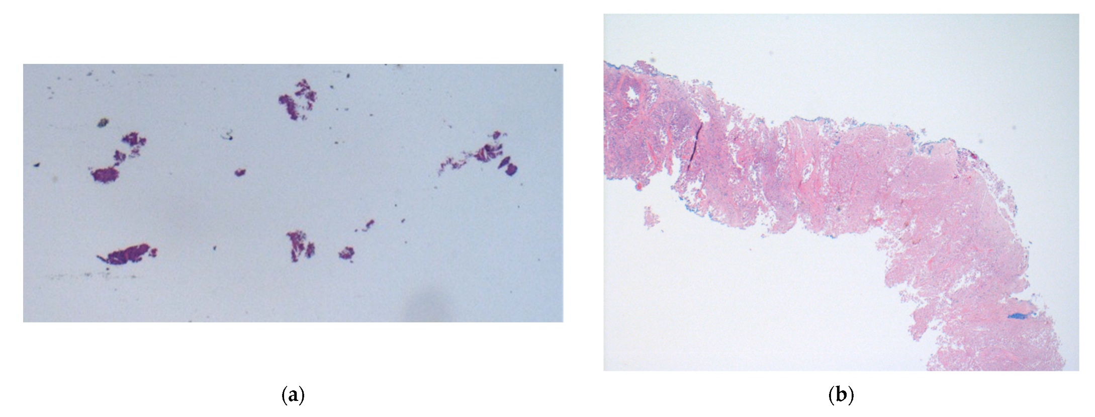
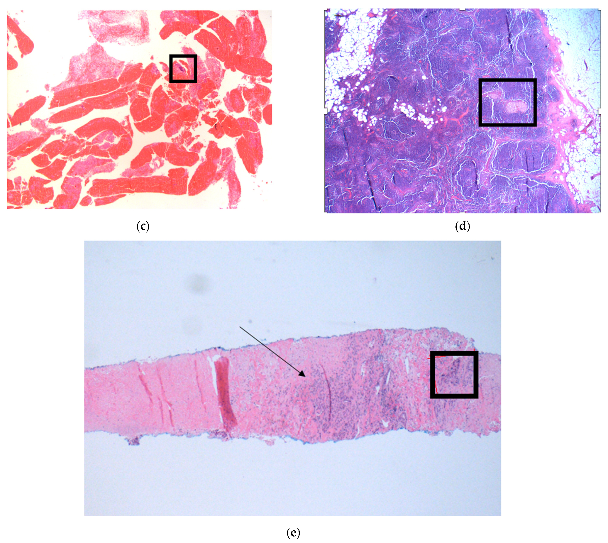
2.2. Pre-Analytical Considerations

Tissue specimens submitted to cellular pathology laboratories are fixed in formalin for between 6 and 72 h to meet standard operating procedures for quality in IHC techniques [36]. Formalin fixation causes the cross linking of proteins within tissue to prevent tissue breakdown in archives [37]. Formalin causes the direct degradation of RNA molecules and can also detrimentally interact with chemical agents used in RNA extraction [38]. RNA extraction is said to be more successful from fresh or frozen tissue samples rather than FFPE tissue samples, particularly if they have not been archived for long periods of time [39,40,41]. Clinicians may ask the question, “Why don’t we just move to using fresh tissue?”, which, on the face of it, seems to be a reasonable suggestion. That is, until one considers the huge logistical changes that would be required of histopathology laboratories to support such a change. It would involve change in practice by surgical theatre staff and porters. Some authors advocate using alternative fixation methods to preserve tissue, such as the PAXgene (BD Biosciences, Haryana, India) solution [42,43,44]. The morphological appearances of haematoxylin and eosin (H&E) tissue sections generated after fixation in PAXgene are excellent and comparable with FFPE [42,43]. However, the costs of such a change would be prohibitive in a public-funded, UK National Health Service (NHS) setting. Five litres of 10% formalin costs in the region of GBP 13, whereas 50 mL of PAXgene (servicing small specimens only) will cost in the region of GBP 150. The enhanced cost would also be compounded by a need to invest in new, dedicated tissue processors compatible for use with PAXgene in cellular pathology laboratories [45].

2.3. Timeliness of RNA Sequencing

The time taken to report RNA NGS from an external laboratory is the same as that for DNA sequencing in our experience. We have shown previously that a DNA sequencing report took 17 days from the time tissue was sent from our laboratory [46]. If one includes the time taken to cut tissue sections in the turnaround time calculation, NGS takes 23 calendar days to be reported [46]. As stated, more than a third of patients require fluorescence in situ hybridization (FISH) salvage testing as a result of the failure rate in RNA sequencing according to a recent audit. This means an average (mean) additional wait of 2 weeks for FISH reporting, which only includes NTRK, ALK1 and ROS1 biomarkers. The reasons for the length of time taken to report FISH at the referral laboratory are not clear, and many centres are able to report FISH in 2–3 days. We have already shown that patients with stage 4 disease are at risk of rapid clinical deterioration and that a third of this group are sadly deceased by the time DNA NGS reports are available [46]. The additional wait for FISH results in more than a third of samples is only likely to increase this proportion of deceased patients at the time of reporting. Further work is required to evidence this point.
Given that the majority of patients show a good objective response to tyrosine kinase inhibitors (TKIs) and that these drugs can give substantial increases in progression-free survival measured in years, it is imperative that we identify appropriate patients whilst they are alive and well enough to receive such treatments [47,48]. Furthermore, a recent retrospective observational study of stage 4 NSCLC outcomes has provided evidence that survival advantages are lost if patients are switched to TKIs after receiving standard chemotherapy +/− checkpoint inhibitors whilst waiting for somatic malignancy NGS to be completed [49]. Systems and processes designed to care for lung cancer patients, particularly those with stage 4 disease, need to factor in timeliness of reporting into their choice of testing methodology to ensure that patient care is not compromised and to avoid accusations of unethical clinical practice [50].

2.4. Tissue Consumption

Tissue consumption is a major issue in the care of lung cancer patients when only small amounts of diagnostic biopsy material are available for testing. This difficulty is compounded as the range of biomarkers required for lung adenocarcinoma expands [8,30,51]. Immunohistochemistry, though only available for use in a clinical diagnostic setting as a single-plex tool, offers the advantages of being fast, cheap, reliable and easy to perform on automated, large-throughput platforms. Tissue consumption for each antibody is 3–4 µm of FFPE tissue and offers the additional asset of marker assessment in a spatial context. That is to say, one can be sure that the protein biomarker of interest specifically relates to the malignant cells of interest and, thereby, enhances diagnostic confidence. That level of data granularity is lost in bulk sequencing assays using DNA or RNA extraction from tissue where there are nuclei with wild-type DNA and RNA species in connective tissue, inflammatory cells and normal background epithelium within the tissue section [52] (see Figure 1). Macro-dissection from the glass slide can help enrich samples for tumour nuclear content, but this may not be possible in centralized molecular laboratories with no resident cellular pathologist expertise.
NGS offers the advantage of multiplex detection of molecular biomarkers but requires a much greater input of tissue for assessment than some rapid polymerase chain reaction (PCR) assays available for clinical use. For example, the IdyllaTM Gene Fusion (Biocartis, Mechelen, Belgium) assay uses 5–15 µm of FFPE tissue as compared with at least 50 µm for each sequencing panel available to our patients [46]. When one considers that rescue FISH for failed RNA sequencing samples requires an additional 8 µm of tissue per marker, not using such a rapid assay becomes difficult to defend in a setting of a small amount of available tissue [46]. The IdyllaTM Gene Fusion cartridge covers all the actionable gene fusion events for detection that may guide treatment decisions in lung adenocarcinoma patients [53,54,55]. Interestingly, Sorber et al. found that RNA from fresh tissue was of poorer quality than that extracted from FFPE blocks [53]. Some of the fresh tissue samples used in the Sorber study were kept in frozen storage for as long as 9 years, and this may account for this difference.
The option to test for NTRK fusions also applies to all other solid malignancies where standard oncological options have failed [56,57]. It would be beneficial to utilize the infrastructure in place for NSCLC cases for the wider oncology patient community where appropriate. Beyond NSCLC, however, it may be more cost effective to screen all solid malignancies by using NTRK IHC before confirmation of positive findings using automated FISH rather than using the gene fusion cartridge by IdyllaTM (Biocartis) as a first-choice method [58,59]. The additional fusions of ALK1 ROS1, Met14 skipping and RET may not be indicated in malignancies other than NSCLC, so it may not be economically viable to use the IdyllaTM Gene Fusion cartridge in this setting. NTRK protein IHC is fast and cheap and consumes just 3–4 µm of FFPE tissue [59].

2.5. Further Consequences of Failed RNA NGS

The consequences of not having a report of gene fusion events in NSCLC at the time of outpatient appointment with an oncologist is a waste of a valuable appointment slot and the time of the hospital staff. There are additional knock-on effects to consider such as patient dissatisfaction, anxiety, staff morale and, most importantly, missed opportunities to start effective treatments in patients at risk of rapid clinical deterioration [60]. As discussed, TKI treatments should be started in the therapy-naïve setting [49]. In addition, if an opportunity to start TKI therapy is missed, the patient loses an opportunity to receive an oral therapy in the community, an option that can relieve some workload of secondary care. The consequence is that more patients wait for intravenous chemotherapy drug administration as day case patients in hospital facilities with limited capacity.

2.6. Why Does RNA Sequencing Fail So Frequently in Our Experience?

RNA sequencing can fail for many and varied reasons. Limited tissue and the impact of formalin fixation have been highlighted, but little discussed is the specimen exposure to environmentally ubiquitous RNA degradation enzymes [61]. There are RNA degradation enzymes in the air, on our hands and work surfaces that can cause the destruction of RNA within FFPE tissue sections. Indeed, it is surprising that RNA sequencing works at all given the nature of the processing occurring in the histopathology laboratory upstream of receipt by the molecular lab. FFPE tissue specimens are cut in a large, open room with no special sterile air flows or compartmentalization. Sections are floated on a water bath prior to mounting on glass slides with the inherent risk, though much guarded against, of contamination. Slides are then packed into a plastic, non-sterile slide mailer and standard packaging for postage to the external laboratory by courier. These are less than optimal conditions for handling and preserving RNA for sequencing and may account for why so many RNA NGS assays fail, in our experience.

2.7. Practical Alternatives to Centralized NGS Testing

Genomic technologies are advancing in capability and at a pace beyond which NHS cellular and molecular pathology laboratories can evolve. As such, it is understandable that some molecular laboratories have invested in NGS for the advantage of being able to expand the repertoire of gene variants reported in a quick and responsive way without additional capital expenditure. Automated technologies are now available that can reduce the turnaround time for reporting NGS samples with a 50-gene panel. Sheffield et al. showed they could generate biomarker NGS-based reports using the GenexusTM (Thermo Fisher Scientific, Waltham, MA, USA) platform in as little as three working days [62]. Using this technique may represent an opportunity for local histopathology laboratories to incorporate fully automated NGS reporting alongside morphological and IHC data in one step [63]. This assay requires a minimum of eight samples per run. A need for batching could have detrimental consequences for turnaround time in laboratories with small numbers of patient requests. This could be overcome by the use of automated rapid PCR systems such as the IdyllaTM (Biocartis) platform, which do not require batching [53,54]. The IdyllaTM platform provides the ability to give clinicians same-day biomarker results in urgent cases and could salvage outpatient appointments where NGS reports are not yet available [64]. The IdyllaTM Gene Fusion assay (Biocartis) has the added advantage of being a multiplex assay [53,54]. A recent multicentre European study of this assay obtained valid results in 98% of their patient in as little as 3 h with good sensitivity and specificity [54]. This compares very favourably to our current experience of valid results in just 65% of patients using RNA-based NGS as the first-line option.
Although fluorescence in situ hybridization (FISH) is single plex and potentially time consuming to conduct, this technique is well established and reliable for clinical use. There have been advances in FISH technologies in recent years with use of computer algorithms to count fluorescence signals with the effect of reducing the turnaround times and human resource requirements of traditional FISH. This could be a very valuable adjunct to the use of rapid PCR or IHC for screening out negative cases. IHC, being a rapid, cheap and easily automated technique, makes it an ideal starting point for screening for uncommon gene fusion events, given that it has a high negative predictive value in low-incidence settings. This may be a particularly attractive approach for histopathology laboratories with well-established expertise in IHC practice and sufficient case throughput to justify testing by this method in the majority of cases. Certainly, in resource-limited settings, such as the UK NHS, IHC should be considered as a robust option in NTRK testing for all solid malignancies.

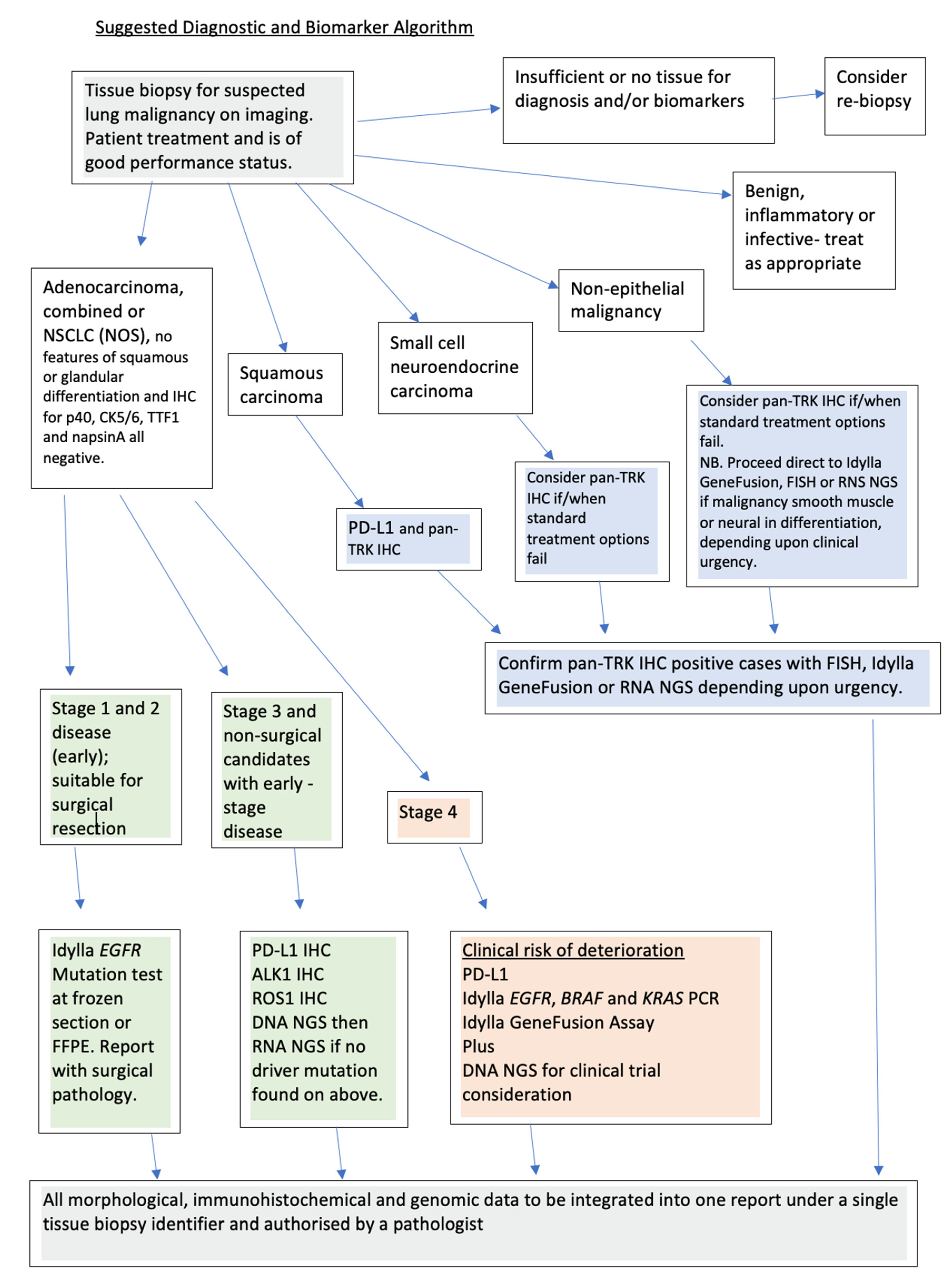
2.8. Suggestions for Improved Care Pathways Incorporating Molecular Biomarker Identification

We have previously described an actionable oncogenic driver identification pathway for NSCLC patients that uses rapid PCR for the identification of common, known somatic mutations in stage 4 patients to prevent missed opportunities for starting TKIs in treatment-naïve patients [46]. It may be best practice, however, to extend the use of rapid PCR to assess for gene fusion events in all cases where molecular testing is indicated on small biopsies (see Figure 2). This could prevent the waste of valuable tissue, given that, in our area, RNA-based NGS has a failure rate of 35% and the gene fusion cartridge has a much lower failure rate, in the region of 2% [54].

3. Conclusions

Cellular and molecular pathologists working in the public sector have a duty to consider the best use of often limited tissue samples to achieve maximum information for patient care. Pathologists, with the tissue morphology before them, are best suited to make the best choices regarding testing modality. Reflex requesting of biomarkers in NSCLC recognizes the role the pathologist can play in saving time for reporting of such biomarkers [65]. A recent audit of RNA sequencing reports a failure rate of 35%. There are a number of alternative testing strategies to consider that could improve biomarker identification in NSCLC patients in our region, including FISH, rapid PCR and fully automated rapid NGS workflows that could be harnessed in-house with the added benefit of integration alongside morphological and immunohistochemistry findings in one report. The timeliness of reporting both cellular and molecular pathology findings in tissue biopsies is of paramount importance in the care of our lung cancer patients. Rapid near-patient testing methods could positively impact up to a fifth of stage 4 patients and make a difference in overall progression-free survival [46]. This is of particular need in the UK, where we lag behind our European colleagues who have a wider range of molecular testing capabilities at their fingertips and greater control over choice of testing method based on individual patient needs [2,3,54]. However, whether the pathologist holds the key to closing the gap in survival outcome data for lung cancer patients in the UK remains to be seen. Pathologists should at least be given the opportunity to try.

Funding

This research received no external funding.

Institutional Review Board Statement

Not applicable. The study did not require ethical approval.

Informed Consent Statement

Not applicable.

Data Availability Statement

Available on request.

Conflicts of Interest

The author has received support from Biocartis to attend international conferences as a speaker. Biocartis had no role in the design of the study; in the collection, analyses or interpretation of data; in the writing of the manuscript or in the decision to publish.

References

  1. Arnold, M.; Rutherford, M.J.; Bardot, A.; Ferlay, J.; Andersson, T.M.; Myklebust, T.; Tervonen, H.; Thursfield, V.; Ransom, D.; Shacl, L.; et al. Progress in cancer survival, mortality, and incidence in seven high-income countries 1995–2014 (ICBP SURVMARK-2): A population-based study. Lancet Oncol. 2019, 20, 1493–1505. [Google Scholar] [CrossRef] [Green Version]
  2. Malvezzi, M.; Bosetti, C.; Rosso, T.; Bertuccio, P.; Chatenoud, L.; Levi, F.; Romano, C.; Negri, E.; La Vecchia, C. Lung cancer mortality in European men: Trends and predictions. Lung Cancer 2013, 80, 138–145. [Google Scholar] [CrossRef] [PubMed]
  3. Bosetti, C.; Malvezzi, M.; Rosso, T.; Bertuccio, P.; Gallus, S.; Chatenoud, L.; Levi, F.; Romano, C.; Negri, E.; La Vecchia, C. Lung cancer mortality in European women: Trends and predictions. Lung Cancer 2012, 78, 171–178. [Google Scholar] [CrossRef] [PubMed]
  4. Davidson, M.R.; Gazdar, A.F.; Clarke, B.E. The pivotal role of pathology in the management of lung cancer. J. Thorac. Dis. 2013, 5 (Suppl. 5), S463–S478. [Google Scholar] [PubMed]
  5. Giles, C. Having both patient advocates and patients at the MDT meeting might be useful. BMJ 2015, 351, h5285. [Google Scholar] [CrossRef] [PubMed]
  6. Garrido, P.; Conde, E.; de Castro, J.; Gómez-Román, J.J.; Felip, E.; Pijuan, L.; Isla, D.; Sanz, J.; Paz-Ares, L.; Lopez-Rios, F. Updated guidelines for predictive biomarker testing in advanced non-small-cell lung cancer: A National Consensus of the Spanish Society of Pathology and the Spanish Society of Medical Oncology. Clin. Transl. Oncol. 2020, 22, 989–1003. [Google Scholar] [CrossRef] [Green Version]
  7. Malapelle, U.; Pisapia, P.; Iaccarino, A.; Barberis, M.; Bellevicine, C.; Brunnström, H.; de Biase, H.; de Maglio, G.; Lindqvist, K.; Fassan, M.; et al. Predictive molecular pathology in the time of coronavirus disease (COVID-19) in Europe. J. Clin. Pathol. 2021, 74, 234–237. [Google Scholar] [CrossRef]
  8. Kerr, K.M.; Bibeau, F.; Thunnissen, E.; Botling, J.; Ryška, A.; Wolf, J. The evolving landscape of biomarker testing for non-small cell lung cancer in Europe. Lung Cancer 2021, 154, 161–175. [Google Scholar] [CrossRef]
  9. Lantuejoul, S.; Sound-Tsao, M.; Cooper, W.A.; Girard, N.; Hirsch, F.R.; Roden, A.C.; Lopez-Rios, F.; Jain, D.; Chou, T.J.; Motoi, N.; et al. PD-L1 Testing for Lung Cancer in 2019: Perspective From the IASLC Pathology Committee. J. Thorac. Oncol. 2020, 15, 499–519. [Google Scholar] [CrossRef]
  10. Lindeman, N.I.; Cagle, P.T.; Aisner, D.L.; Arcila, M.E.; Beasley, M.B.; Bernicker, E.H.; Colasacco, C.; Dacic, S.; Hirsch, F.R.; Kerr, K.; et al. Updated Molecular Testing Guideline for the Selection of Lung Cancer Patients for Treatment With Targeted Tyrosine Kinase Inhibitors: Guideline From the College of American Pathologists, the International Association for the Study of Lung Cancer, and the Association for Molecular Pathology. J. Mol. Diagn. 2018, 20, 129–159. [Google Scholar]
  11. López-Castro, R.; García-Peña, T.; Mielgo-Rubio, X.; Riudavets, M.; Teixidó, C.; Vilariño, N.; Vilariño, N.; Counago, T.; Mezquita, L. Targeting molecular alterations in non-small-cell lung cancer: What’s next? Per. Med. 2022, 19, 341–359. [Google Scholar] [CrossRef] [PubMed]
  12. Fernandes, M.G.O.; Jacob, M.; Martins, N.; Moura, C.S.; Guimarães, S.; Reis, J.P.; Justino, A.; Pina, M.J.; Cirnes, L.; Sousa, C.; et al. Targeted Gene Next-Generation Sequencing Panel in Patients with Advanced Lung Adenocarcinoma: Paving the Way for Clinical Implementation. Cancers 2019, 11, 1229. [Google Scholar] [CrossRef] [PubMed] [Green Version]
  13. Beckett, P.; Navani, N.; Harden, S.; Tweedie, J.; Tebay, R.; Rodgers, H. National Lung Cancer Audit: Spotlight on Molecular Testing in Advanced Lung Cancer; Healthcare Quality Improvement Partnership (HQIP): London, UK, 2020. [Google Scholar]
  14. Mino-Kenudson, M.; Le Stang, N.; Daigneault, J.B.; Nicholson, A.G.; Cooper, W.A.; Roden, A.C.; Moreira, A.L.; Thunnissen, E.; Papotti, M.; Pelosi, G.; et al. IASLC Global Survey on PD-L1 Testing for Non-Small Cell Lung Cancer. J. Thorac. Oncol. 2021, 16, 686. [Google Scholar] [CrossRef] [PubMed]
  15. Lamberti, G.; Andrini, E.; Sisi, M.; Rizzo, A.; Parisi, C.; Di Federico, A.; Gelsomino, F.; Ardizzoni, A. Beyond EGFR, ALK and ROS1: Current evidence and future perspectives on newly targetable oncogenic drivers in lung adenocarcinoma. Crit. Rev. Oncol. Hematol. 2020, 156, 103119. [Google Scholar] [CrossRef]
  16. De Luca, C.; Pepe, F.; Iaccarino, A.; Pisapia, P.; Righi, L.; Listì, A.; Greco, L.; Gragnano, G.; Campione, S.; Dominicis, G.; et al. RNA-Based Assay for Next-Generation Sequencing of Clinically Relevant Gene Fusions in Non-Small Cell Lung Cancer. Cancers 2021, 13, 139. [Google Scholar] [CrossRef]
  17. Zhao, R.; Han, Y.; Xiang, C.; Chen, S.; Zhao, J.; Guo, L.; Yu, A.; Shao, J.; Zhu, L.; Tian, Y.; et al. RNA sequencing effectively identifies gene fusions undetected by DNA sequencing in lung adenocarcinomas. J. Clin. Oncol. 2021, 39, 3052. [Google Scholar] [CrossRef]
  18. Deveson, I.W.; Brunck, M.E.; Blackburn, J.; Tseng, E.; Hon, T.; Clark, T.A.; Clark, M.B.; Crawford, J.; Dinger, M.E.; Nielsen, L.K.; et al. Universal Alternative Splicing of Noncoding Exons. Cell Syst. 2018, 6, 245–255.e5. [Google Scholar] [CrossRef] [Green Version]
  19. Kirchner, M.; Neumann, O.; Volckmar, A.L.; Stögbauer, F.; Allgäuer, M.; Kazdal, D.; Budczies, J.; Rempel, E.; Brandt, R.; Talla, S.B.; et al. RNA-Based Detection of Gene Fusions in Formalin-Fixed and Paraffin-Embedded Solid Cancer Samples. Cancers 2019, 11, 1309. [Google Scholar] [CrossRef] [Green Version]
  20. Matter, M.S.; Chijioke, O.; Savic, S.; Bubendorf, L. Narrative review of molecular pathways of kinase fusions and diagnostic approaches for their detection in non-small cell lung carcinomas. Transl. Lung Cancer Res. 2020, 9, 2645–2655. [Google Scholar] [CrossRef]
  21. Tsao, M.S.; Hirsch, F.R.; Yatabe, Y. IASLC Atlas of ALK and ROS1 Testing in Lung Cancer. IASLC. Available online: https://www.iaslc.org/Research-Education/Publications/IASLC-Atlases2016 (accessed on 27 October 2022).
  22. Zhao, R.; Yao, F.; Xiang, C.; Zhao, J.; Shang, Z.; Guo, L.; Ding, W.; Guo, L.; Ma, S.; Shao, J.; et al. Identification of NTRK gene fusions in lung adenocarcinomas in the Chinese population. J. Pathol. Clin. Res. 2021, 7, 375–384. [Google Scholar] [CrossRef]
  23. Doebele, R.C.; Drilon, A.; Paz-Ares, L.; Siena, S.; Shaw, A.T.; Farago, A.F.; Blakely, C.M.; Seto, T.; Cho, B.C.; Tosi, D.; et al. Entrectinib in patients with advanced or metastatic NTRK fusion-positive solid tumours: Integrated analysis of three phase 1–2 trials. Lancet Oncol. 2020, 21, 271–282. [Google Scholar] [CrossRef]
  24. Jalal, S.I.; Guo, A.; Ahmed, S.; Kelley, M.J. Analysis of actionable genetic alterations in lung carcinoma from the VA National Precision Oncology Program. Semin Oncol. 2022, 49, 265–274. [Google Scholar] [CrossRef] [PubMed]
  25. Sakai, H.; Morise, M.; Kato, T.; Matsumoto, S.; Sakamoto, T.; Kumagai, T.; Tokito, T.; Atagi, S.; Kozuki, S.; Tanaka, H.; et al. Tepotinib in patients with NSCLC harbouring MET exon 14 skipping: Japanese subset analysis from the Phase II VISION study. Jpn. J. Clin. Oncol. 2021, 51, 1261–1268. [Google Scholar] [CrossRef] [PubMed]
  26. Piton, N.; Lanic, M.D.; Marguet, F.; Lamy, A.; Blanchard, F.; Guisier, F.; Viennot, M.; Salaun, M.; Thiberville, L.; Jardin, F.; et al. An improved assay for detection of theranostic gene translocations and MET exon 14 skipping in thoracic oncology. Lab. Investig. 2021, 101, 648–660. [Google Scholar] [CrossRef] [PubMed]
  27. Drusbosky, L.M.; Rodriguez, E.; Dawar, R.; Ikpeazu, C.V. Therapeutic strategies in RET gene rearranged non-small cell lung cancer. J. Hematol. Oncol. 2021, 14, 50. [Google Scholar] [CrossRef] [PubMed]
  28. Mok, T.; Carbonne, D.; Hirsch, F. IASLC Atlas of EGFR Testing in Lung Cancer; International Association of the Study of Lung Cancer (IASLC): Denver, CO, USA, 2017. [Google Scholar]
  29. Yatabe, Y.; Dacic, S.; Borczuk, A.C.; Warth, A.; Russell, P.A.; Lantuejoul, S.; Beasley, M.B.; Thunnissen, E.; Pelosi, G.; Rekhtman, N.; et al. Best Practices Recommendations for Diagnostic Immunohistochemistry in Lung Cancer. J. Thorac. Oncol. 2019, 14, 377–407. [Google Scholar] [CrossRef] [Green Version]
  30. Ilie, M.; Hofman, P. Pitfalls in lung cancer molecular pathology: How to limit them in routine practice? Curr. Med. Chem. 2012, 19, 2638–2651. [Google Scholar] [CrossRef]
  31. Navani, N.; Brown, J.M.; Nankivell, M.; Woolhouse, I.; Harrison, R.N.; Jeebun, V.; Munavvar, M.; Ng, B.J.; Rassl, D.M.; Falzon, M.; et al. Suitability of endobronchial ultrasound-guided transbronchial needle aspiration specimens for subtyping and genotyping of non-small cell lung cancer: A multicenter study of 774 patients. Am. J. Respir Crit. Care Med. 2012, 185, 1316–1322. [Google Scholar] [CrossRef]
  32. Medford, A.R.; Agrawal, S.; Free, C.M.; Bennett, J.A. A performance and theoretical cost analysis of endobronchial ultrasound-guided transbronchial needle aspiration in a UK tertiary respiratory centre. QJM 2009, 102, 859–864. [Google Scholar] [CrossRef] [Green Version]
  33. Moore, D.A.; Benafif, S.; Poskitt, B.; Argue, S.; Lee, S.; Ahmad, T.; Papadatos-Pastos, D.; Jamal-Hanjani, M.; Bennett, P.; Forster, M.D. Optimising fusion detection through sequential DNA and RNA molecular profiling of non-small cell lung cancer. Lung Cancer 2021, 161, 55–59. [Google Scholar] [CrossRef]
  34. Faber, E.; Grosu, H.; Sabir, S.; San Lucas, F.A.; Barkoh, B.A.; Bassett, R.L.; Lthra, R.; Stwart, J.; Roy-Chowdhuri, S. Adequacy of small biopsy and cytology specimens for comprehensive genomic profiling of patients with non-small cell lung cancer to determine eligibility for immune checkpoint inhibitor and targeted therapy. J. Clin. Pathol. 2022, 75, 612–619. [Google Scholar] [CrossRef] [PubMed]
  35. Wei, S.; Talarchek, J.N.; Huang, M.; Gong, Y.; Du, F.; Ehya, H.; Flieder, D.B.; Patchefsky, A.S.; Wasik, M.A.; Pei, J. Cell block-based RNA next generation sequencing for detection of gene fusions in lung adenocarcinoma: An institutional experience. Cytopathology 2022, 1–7. [Google Scholar] [CrossRef] [PubMed]
  36. Shi, S.R.; Liu, C.; Taylor, C.R. Standardization of immunohistochemistry for formalin-fixed, paraffin-embedded tissue sections based on the antigen-retrieval technique: From experiments to hypothesis. J. Histochem Cytochem. 2007, 55, 105–109. [Google Scholar] [CrossRef] [PubMed] [Green Version]
  37. Thavarajah, R.; Mudimbaimannar, V.K.; Elizabeth, J.; Rao, U.K.; Ranganathan, K. Chemical and physical basics of routine formaldehyde fixation. J. Oral. Maxillofac. Pathol. 2012, 16, 400–405. [Google Scholar] [CrossRef] [Green Version]
  38. Ding, J.; Ichikawa, Y.; Ishikawa, T.; Shimada, H. Effect of formalin on extraction of mRNA from a formalin-fixed sample: A basic investigation. Scand. J. Clin. Lab. Investig. 2004, 64, 229–235. [Google Scholar] [CrossRef]
  39. Mullegama, S.V.; Alberti, M.O.; Au, C.; Li, Y.; Toy, T.; Tomasian, V.; Xian, R.R. Nucleic Acid Extraction from Human Biological Samples. Methods Mol. Biol. 2019, 1897, 359–383. [Google Scholar]
  40. Hedegaard, J.; Thorsen, K.; Lund, M.K.; Hein, A.M.; Hamilton-Dutoit, S.J.; Vang, S.; Nordentoft, I.; Birkenkamp-Demtroder, K.; Kruhoffer, M.; Hager, H.; et al. Next-generation sequencing of RNA and DNA isolated from paired fresh-frozen and formalin-fixed paraffin-embedded samples of human cancer and normal tissue. PLoS ONE 2014, 9, e98187. [Google Scholar] [CrossRef] [Green Version]
  41. Norton, N.; Sun, Z.; Asmann, Y.W.; Serie, D.J.; Necela, B.M.; Bhagwate, A.; Jen, J.; Eckloff, B.W.; Kalari, K.R.; Thompson, K.J.; et al. Gene expression, single nucleotide variant and fusion transcript discovery in archival material from breast tumors. PLoS ONE 2013, 8, e81925. [Google Scholar] [CrossRef]
  42. Kap, M.; Smedts, F.; Oosterhuis, W.; Winther, R.; Christensen, N.; Reischauer, B.; Viertler, C.; Groelz, D.; Becker, K.F.; Zatloual, K.; et al. Histological assessment of PAXgene tissue fixation and stabilization reagents. PLoS ONE 2011, 6, e27704. [Google Scholar] [CrossRef] [Green Version]
  43. Mathieson, W.; Marcon, N.; Antunes, L.; Ashford, D.A.; Betsou, F.; Frasquilho, S.G.; Kofanova, O.A.; McKay, S.C.; Pericleous, S.; Smith, C.; et al. A Critical Evaluation of the PAXgene Tissue Fixation System:  Morphology, Immunohistochemistry, Molecular Biology, and Proteomics. Am. J. Clin. Pathol. 2016, 146, 25–40. [Google Scholar] [CrossRef] [Green Version]
  44. Sanchez, I.; Betsou, F.; Culot, B.; Frasquilho, S.; McKay, S.C.; Pericleous, S.; Smith, C.; Thomas, G.; Mathieson, W. RNA and microRNA Stability in PAXgene-Fixed Paraffin-Embedded Tissue Blocks After Seven Years’ Storage. Am. J. Clin. Pathol. 2018, 149, 536–547. [Google Scholar] [CrossRef] [PubMed] [Green Version]
  45. Frasquilho, S.G.; Sanchez, I.; Yoo, C.; Antunes, L.; Bellora, C.; Mathieson, W. Do Tissues Fixed in a Non-crosslinking Fixative Require a Dedicated Formalin-free Processor? J. Histochem Cytochem. 2021, 69, 389–405. [Google Scholar] [CrossRef]
  46. Finall, A.; Davies, G.; Jones, T.; Emlyn, G.; Huey, P.; Mullard, A. Integration of rapid PCR testing as an adjunct to NGS in diagnostic pathology services within the UK: Evidence from a case series of non-squamous, non-small cell lung cancer (NSCLC) patients with follow-up. J. Clin. Pathol. 2022, 1–9. [Google Scholar] [CrossRef] [PubMed]
  47. Rybarczyk-Kasiuchnicz, A.; Ramlau, R.; Stencel, K. Treatment of Brain Metastases of Non-Small Cell Lung Carcinoma. Int. J. Mol. Sci. 2021, 22, 593. [Google Scholar] [CrossRef] [PubMed]
  48. Hardtstock, F.; Myers, D.; Li, T.; Cizova, D.; Maywald, U.; Wilke, T.; Griesinger, F. Real-world treatment and survival of patients with advanced non-small cell lung Cancer: A German retrospective data analysis. BMC Cancer 2020, 20, 260. [Google Scholar] [CrossRef] [Green Version]
  49. Smith, R.E.; Johnson, M.; Gordan, L.N.; Xue, M.; Varughese, P.; Dorrow, N.; Wang, B.; Vaidya, V.; Gart, M.; Gierman, H.J.; et al. Evaluation of outcomes in patients with stage 4 non-small cell lung cancer (NSCLC 4) harbouring actionable oncogenic drivers when treated prior to report of mutation without tyrosine kinase inhibitors: An Integra Connect Database retrospective observational study. J. Clin. Oncol. 2022, 40, 1530. [Google Scholar]
  50. Finall, A.; Jones, K. Applying bioethical principles for directing investment in precision medicine. Clin. Ethics 2020, 15, 23–28. [Google Scholar] [CrossRef] [Green Version]
  51. Melosky, B.; Wheatley-Price, P.; Juergens, R.A.; Sacher, A.; Leighl, N.B.; Tsao, M.S.; Cheema, P.; Snow, S.; Liu, G.; Card, P.; et al. The rapidly evolving landscape of novel targeted therapies in advanced non-small cell lung cancer. Lung Cancer 2021, 160, 136–151. [Google Scholar] [CrossRef]
  52. Ståhl, P.L.; Salmén, F.; Vickovic, S.; Lundmark, A.; Navarro, J.F.; Magnusson, J.; Giacomello, S.; Asp, M.; Westholm, J.O.; Huss, M.; et al. Visualization and analysis of gene expression in tissue sections by spatial transcriptomics. Science 2016, 353, 78–82. [Google Scholar] [CrossRef] [Green Version]
  53. Sorber, L.; Van Dorst, B.; Bellon, E.; Zwaenepoel, K.; Lambin, S.; De Winne, K.; Lardon, F.; Pauwels, P.; Siozopoulou, V. NTRK Gene Fusion Detection in a Pan-Cancer Setting Using the Idylla GeneFusion Assay. J. Mol. Diagn. 2022, 24, 750–759. [Google Scholar] [CrossRef]
  54. Depoilly, T.; Garinet, S.; van Kempen, L.C.; Schuuring, E.; Clavé, S.; Bellosillo, B.; Ercolani, C.; Buglioni, S.; Siemanowski, J.; Merkelbach-Bruse, S.; et al. Multicenter Evaluation of the Idylla GeneFusion in Non-Small-Cell Lung Cancer. J. Mol. Diagn. 2022, 24, 1021–1030. [Google Scholar] [CrossRef] [PubMed]
  55. Chu, Y.H.; Barbee, J.; Yang, S.R.; Chang, J.C.; Liang, P.; Mullaney, K.; Chan, R.; Salazar, P.; Benayed, R.; Offin, M.; et al. Clinical Utility and Performance of an Ultrarapid Multiplex RNA-Based Assay for Detection of ALK, ROS1, RET, and NTRK1/2/3 Rearrangements and MET Exon 14 Skipping Alterations. J. Mol. Diagn. 2022, 24, 642–654. [Google Scholar] [CrossRef] [PubMed]
  56. NICE. Entrectinib for Treating NTRK Fusion-Positive Solid Tumours. Available online: https://www.nice.org.uk/guidance/ta6442021 (accessed on 27 October 2022).
  57. NICE. Larotrectinib for Treating NTRK Fusion-Positive Solid Tumours. Available online: https://www.nice.org.uk/guidance/ta6302021 (accessed on 27 October 2022).
  58. Strohmeier, S.; Brcic, I.; Popper, H.; Liegl-Atzwanger, B.; Lindenmann, J.; Brcic, L. Applicability of pan-TRK immunohistochemistry for identification of NTRK fusions in lung carcinoma. Sci. Rep. 2021, 11, 9785. [Google Scholar] [CrossRef] [PubMed]
  59. Elfving, H.; Broström, E.; Moens, L.N.J.; Almlöf, J.; Cerjan, D.; Lauter, G.; Nord, H.; Mattsson, J.S.M.; Ullenhag, G.J.; Strell, C.; et al. Evaluation of NTRK immunohistochemistry as a screening method for NTRK gene fusion detection in non-small cell lung cancer. Lung Cancer 2021, 151, 53–59. [Google Scholar] [CrossRef]
  60. Yang, S.C.; Yeh, Y.C.; Chen, Y.L.; Chiu, C.H. Economic Analysis of Exclusionary EGFR Test Versus Up-Front NGS for Lung Adenocarcinoma in High EGFR Mutation Prevalence Areas. J. Natl. Compr. Canc. Netw. 2022, 20, 774–782.e4. [Google Scholar] [CrossRef]
  61. Houseley, J.; Tollervey, D. The many pathways of RNA degradation. Cell 2009, 136, 763–776. [Google Scholar] [CrossRef] [Green Version]
  62. Elliott, D.; Ladomery, M. Molecular Biology of RNA; Oxford University Press: Oxford, UK, 2011. [Google Scholar]
  63. Sheffield, B.S.; Beharry, A.; Diep, J.; Perdrizet, K.; Iafolla, M.A.J.; Raskin, W.; Dudani, S.; Brett, M.A.; Starova, B.; Olsen, B.; et al. Point of Care Molecular Testing: Community-Based Rapid Next-Generation Sequencing to Support Cancer Care. Curr. Oncol. 2022, 29, 30113. [Google Scholar] [CrossRef]
  64. Uguen, A.; Troncone, G. A review on the Idylla platform: Towards the assessment of actionable genomic alterations in one day. J. Clin. Pathol. 2018, 71, 757–762. [Google Scholar] [CrossRef]
  65. Adizie, J.B.; Tweedier, J.; Khakwani, A.; Peach, E.; Hubbard, R.; Wood, N.; Gosney, J.R.; Harden, S.V.; Beckett, P.; Popat, S.; et al. Biomarker Testing for People with Advanced Lung Cancer in England. J. Thorac. Oncol. Clin. Res. Rep. 2021, 2, 100176. [Google Scholar] [CrossRef]
Figure 1. Photomicrographic examples of histopathological tissue biopsies containing scarce or no malignant tissue. (a) Photomicrograph of bronchial biopsy (×4 magnification) showing the limited nature of some samples. This is small-cell neuroendocrine carcinoma (haematoxylin and eosin (H&E) stain). (b) Some CT-guided core biopsies may not contain malignancy but rather necrotic material, as in this example (×4, H&E). (c) Endobronchial ultrasound-guided (EBUS) fine needle aspiration cytology of mediastinal lymph nodes. EBUS samples often yield small amounts of tissue on a background of blood. A small fragment of carcinoma is highlighted in the black square (×4, H&E). (d) Photomicrograph illustrating a small deposit of metastatic carcinoma (black square) in a lymph node. Bulk RNA extraction from this section, without microdissection, is likely to yield large amounts of wild-type signal from lymphocyte nuclei. (e) An example of low-volume malignancy with a CT-guided core biopsy of lung highlighted in the black square. There is fibrosis and chronic inflammation (arrow) present in the background (×4, H&E).
Jmp 03 00026 g001aJmp 03 00026 g001b
Figure 2. Suggested algorithm for molecular analysis of somatic NSCLC tissue taking into consideration the real-world failure rate of RNA sequencing performed in a centralized service model. NSCLC, non-squamous, non-small-cell lung cancer; PD-L1, programmed death ligand 1; IHC, immunohistochemistry; EGFR, epidermal growth factor; BRAF, B-raf oncogene; KRAS, K-ras oncogene; NOS, not otherwise specified; NTRK, neurotrophin receptor kinase; Trk, tropomyosin receptor kinase (protein); FFPE, formalin-fixed paraffin-embedded tissue.
Jmp 03 00026 g002
Publisher’s Note: MDPI stays neutral with regard to jurisdictional claims in published maps and institutional affiliations.

