Brief Report

The Relationship between Mutations in Gene-Specific Domains of Salivary Fibronectin (cFn) and Dynamin-2 (Dynm-2) and the Development of Porphyromonas gingivalis-Initiated Periodontitis

by
Elena A. Oleinik
1,2,* and
Anna V. Goncharenko
2
1
Faculty of Dentistry, Pavlov First St. Petersburg State Medical University, 197022 St. Petersburg, Russia
2
Federal State Institution, Federal Research Center “Fundamental Foundations of Biotechnology” of the Russian Academy of Sciences, 119071 Moscow, Russia
*
Author to whom correspondence should be addressed.
J. Mol. Pathol. 2022, 3(3), 182-189; https://doi.org/10.3390/jmp3030015
Submission received: 30 July 2022 / Revised: 27 August 2022 / Accepted: 30 August 2022 / Published: 8 September 2022

Abstract

:
Periodontitis is a chronic inflammatory disease characterized by the destruction of the supporting structures of the teeth. Its high prevalence and negative effects on quality of life make it one of the current problems in dentistry. Porphyromonas gingivalis (P. gingivalis) is the predominant periodontal pathogen that expresses a number of virulence factors involved in the pathogenesis of periodontitis. P. gingivalis fimbriae are a critical factor in the interaction between the organism and the host tissue. They promote both bacterial adhesion and invasion into the target sites. Fimbriae are capable of binding to human saliva components, extracellular matrix proteins, and commensal bacteria, as well as firmly binding to the cellular integrin α5β1. After attachment to α5β1-integrin, P. gingivalis is captured by cellular pseudopodia, which makes invagination through an actin-mediated pathway possible. It has been proven that the invagination event also requires the participation of the host cell dynamin, actin fibers, microtubules and lipid rafts. Work has emerged investigating mutations in the proline-rich terminal domain (PRD) and their impact on disease development. Salivary antimicrobial peptides are early protective factors against microbial attack. Of great interest is fibronectin (FN) as the main competitor of P. gingivalis fimbriae. The FN can interact with cells in three different regions: the central cell-binding domain (CCBD), the COOH terminal heparin-binding domain (Hep2), and the type III connecting segment (IIICS), including the CS1 region (Yamada, 1991). CCBD is the major cell-adhesion domain of FN and contains an Arg–Gly–Asp (RGD) motif that is recognized by members of the cell adhesion receptor integrin family, including a5b1, which is the primary FN receptor in many cell types. The work focuses on identifying the relationship between the development of periodontitis and the presence of mutations in the adhesion domains of salivary proteins such as cellular fibronectin (cFN) and dynamin-2 (DYNM2).

1. Introduction

The global problems of modern dentistry are dental caries and periodontal diseases of inflammatory genesis. In order to solve them, it is necessary to develop new perspectives on the prevention of dental diseases.
Periodontitis is one of the leading causes of tooth loss worldwide. Periodontitis is thought to affect 20–50% of the world’s population and is the 11th most common disease worldwide [1]. Improved materials, precision technology, and standards of treatment used in dentistry have failed to fundamentally change the quality of periodontal treatment. The future lies in personalized dentistry. One of the main causative agents of periodontitis is P. gingivalis, which expresses various adhesive components: fimbriae, gingipains, haemagglutinin, and lipopolysaccharide. An important role in the interaction of a bacterium with host cells is played by fimbriae, the so-called adhesive pili [1,2,3,4,5,6]. P. gingivalis fimbriae bind to the cellular a5B1 integrin, which mediates bacterial adhesion to host cells [1,7,8,9]. Of scientific interest is type V, which is considered a key factor in the virulence of P. gingivalis. Studies by a team of researchers from the Okinawa Institute of Science and Technology, as well as Prof. Koji Nakayama’s group from Nagasaki University (2020), have established the structure of fimbriae and their assembly mechanism [7]. On the other hand, some components of saliva such as fibrinogen, histatin and fibronectin prevent P. gingivalis from attaching to periodontal tissues. Atsuo Amano (2007) studied the mechanism of invasion of P. gingivalis into the host cell. P. gingivalis fimbriae compete with FN for a5B1 integrin and inhibit fibronectin/integrin-regulating cellular functions [1,2,3,7]. A putative scheme of the P. gingivalis invasion mechanism, with respect to the host cell’s endocytic mechanism, cytoskeleton and lipid rafts, according to Atsuo Amano (2007), is as follows. P. gingivalis fimbriae adhere to α5β1-integrin, after which the bacterium is surrounded by α5β1-integrin and invaginated through an actin-mediated pathway controlled by phosphatidylinositol (PI) 3-kinase. The invasive event requires the involvement of dynamin, actin fibers, microtubules and host cell lipid rafts [1,2,10,11,12,13,14,15,16]. However, FN has an RGD-containing cell-binding domain in its structure, which binds integrins with high affinity [17]. Integrin receptors mediate cell adhesion to extracellular matrices and trigger signals that direct cell function. While many integrins bind to the arginine–glycine–aspartic acid (RGD) motif present in many extracellular proteins, the α5β1 integrin requires both the PHSRN synergy site in type 9 and the RGD synergy site in type 10 type III repeat FN. The attachment of α5β1 to FN is critical for many cellular processes [17]. Dynamins are multimodular proteins consisting of five highly conserved structural domains: a large N-terminal GTPase domain (G-domain), a middle domain, a PH domain-binding phosphoinositides, a GTPase effector domain (GED), and a C-terminal proline-rich domain (PRD) interacting with SH3-containing proteins [12,18,19,20]. DYNM2 performs several functions: it regulates Golgi apparatus function, cytoskeleton integrity, cellular pore expansion, and bacterial cell–cell adhesion [11,12,13,19]. However, its role in the pathogenesis of periodontal disease initiated by P. gingivalis is largely unexplored. Thus, it is of scientific interest to study mutations in the cFN, DYNM2 domains that may reveal mechanisms of susceptibility to periodontal disease initiated by P. gingivalis.
The aim of the study is to identify the presence of mutations in gene-specific domains (sites) of the salivary antimicrobial peptides (FN) and microtubule-associated proteins (DYNM2) responsible for the adhesion of P. gingivalis to host cells.
The aim of our experiment is to confirm the influence of mutations in the adhesive domains of FN and DYNM2 on the development of periodontitis.

2. Material and Methods of Study

The study of dental status was carried out in the Dentistry Clinic of the Pavlov First St. Petersburg State Medical University. Informed voluntary consent was obtained from the patients who participated in the study.
A total of 32 patients aged from 32 to 56 years were examined. General somatic status was not aggravated (absence of somatic pathology, or bad habits).
The American Academy of Periodontology 2018 classification was used to make a dental diagnosis.
Two groups were formed:
  • Group 1 (12 people)—patients with periodontal disease of severity II (periodontal pockets 3–4 mm, coronal third 15–33%);
  • Group 2 (20 people)—patients with healthy periodontal disease.
Clinical methods of investigation were used: collection of anamneses, examination, and evaluation of periodontal status, including RBL, R. Attstrom (1998).
RBL = % bone resorption ×100/age of patient.
R. Attstrom (1998): Criteria for the definition of periodontal ligament destruction of the teeth:
  • 1—Mild periodontal ligament destruction: loss of attachment by less than 1/4 of the tooth root length;
  • 2—Moderate destruction of periodontal ligaments: loss of attachment by 1/4 to 1/3 of the tooth root length;
  • 3—Severe destruction of the periodontal ligaments: loss of compliance for more than 1/3 of the tooth root length;
  • 4—Severe complicated destruction of periodontal ligaments: loss of compliance for more than 1/3 of the tooth root length, accompanied by intraosseous lesions, involvement in the pathological process of the root separation zone and/or increased mobility of I–II-degree teeth.
Based on the above criteria, the periodontal index is expressed as an indexogram:
  • The first digit is the age of the patient:
  • The second digit—the number of lost teeth out of 28;
  • The third digit—the number of teeth with mild periodontitis;
  • The fourth digit—number of teeth with moderate periodontitis;
  • The fifth figure—the number of teeth with severe periodontitis;
  • The sixth digit—the number of teeth with severe complicated periodontitis.
Identification of Porphyromonas Gingivalis in biomaterial was performed by real-time PCR. PCR fragments were then isolated and sequenced.
The method of biomaterial extraction: fluid sampling from the periodontal sulcus using a sterile absorber (standard protocol).
PCR fragment isolation and sequencing were performed at the “Syntol” (Moscow).

2.1. DNA Extraction Technique

DNA extraction was performed using the ExtractDNA Blood kit (Eurogen, Russia) according to the protocol. DNA quality and purity were assessed by gel electrophoresis using 2% agarose gel in TAE buffer. PCR was performed on a “Bio-Rad CFX96TM system” (Bio-Rad, Hercules, CA, USA) with HS SYBR qPCR mix (“Eurogen”, Russia). Initial denaturation was performed at 95 °C for 3 min, followed by 35 cycles: denaturation at 95 °C for 30 s; primer annealing at 59 °C for 30 s; elongation at 72 °C for 60 s. The primer sequences for PCR were as follows:
DNM2_F; 5′-GTGTCTCTCTATTGCGGTCCCT-3′;
DNM2_R; 5′-CCGGGTTCAAACGATTCTCC-3′;
FN_F; 5′-CATGCAATCCTGGGTTGAGGAGG-3′;
FN_R; 5′-TGGCTCAAAACTCTGTTCGCGC-3′;
The PCR product was then visualized by gel electrophoresis using 2% agarose gel in TAE buffer. The PCR product was eluted from the gel using a Cleanup Standard kit (Eurogen, Russia) according to the attached protocol. The samples were then sent for Sanger sequencing. The results were analyzed using the Bio Edit Sequence Alignment Editor software (version 7.0.4.1). “Clustal Omega” software was used for multiple sequence alignment (https://www.ebi.ac.uk/Tools/msa/clustalo/, 8 February 2022)

2.2. Statistical Research Methods

The Mann–Whitney U-test was used to determine the reliability of differences in the comparison groups (p < 0.08).

3. Results and Discussion

In Group 1, a clinical bone loss (CAL) of 4 mm was found in 10 patients (91%). The average value of the radiological bone loss index (RBL) in Group 1 patients was 38.6%. Grade II tooth mobility was diagnosed in 10 patients (91% of subjects). The Attstrom index was determined in all patients with periodontal disease.
Figure 1 (radiographic appearance) represents an orthopantomogram of a 37-year-old woman with periodontitis (degree II–III). There is extensive alveolar bone loss (usually 50–75% of the root length) affecting the entire tooth row, with an irregular pattern of bone loss.
Index R. Attsrom—37/02/00/05/20/0.
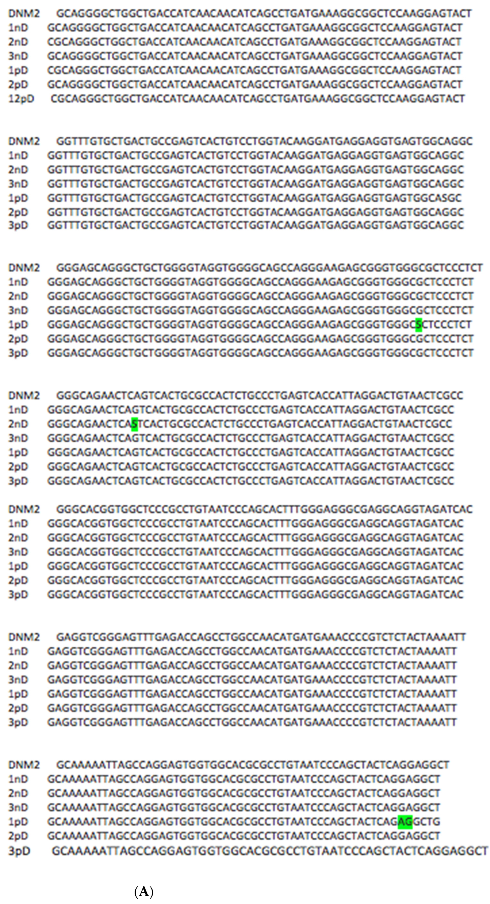
The nucleotide sequences of the dnm2 gene on chromosome 19 (site 10,812,275 to 10,812,684) were analyzed to determine SNPs. The analyzed site in the intron region revealed one SNP in the “normal” group and one SNP in the “pathology” group. In the “pathology” group, one GA transversion was detected in the intron region at position 10,812,682. (Figure 2A).
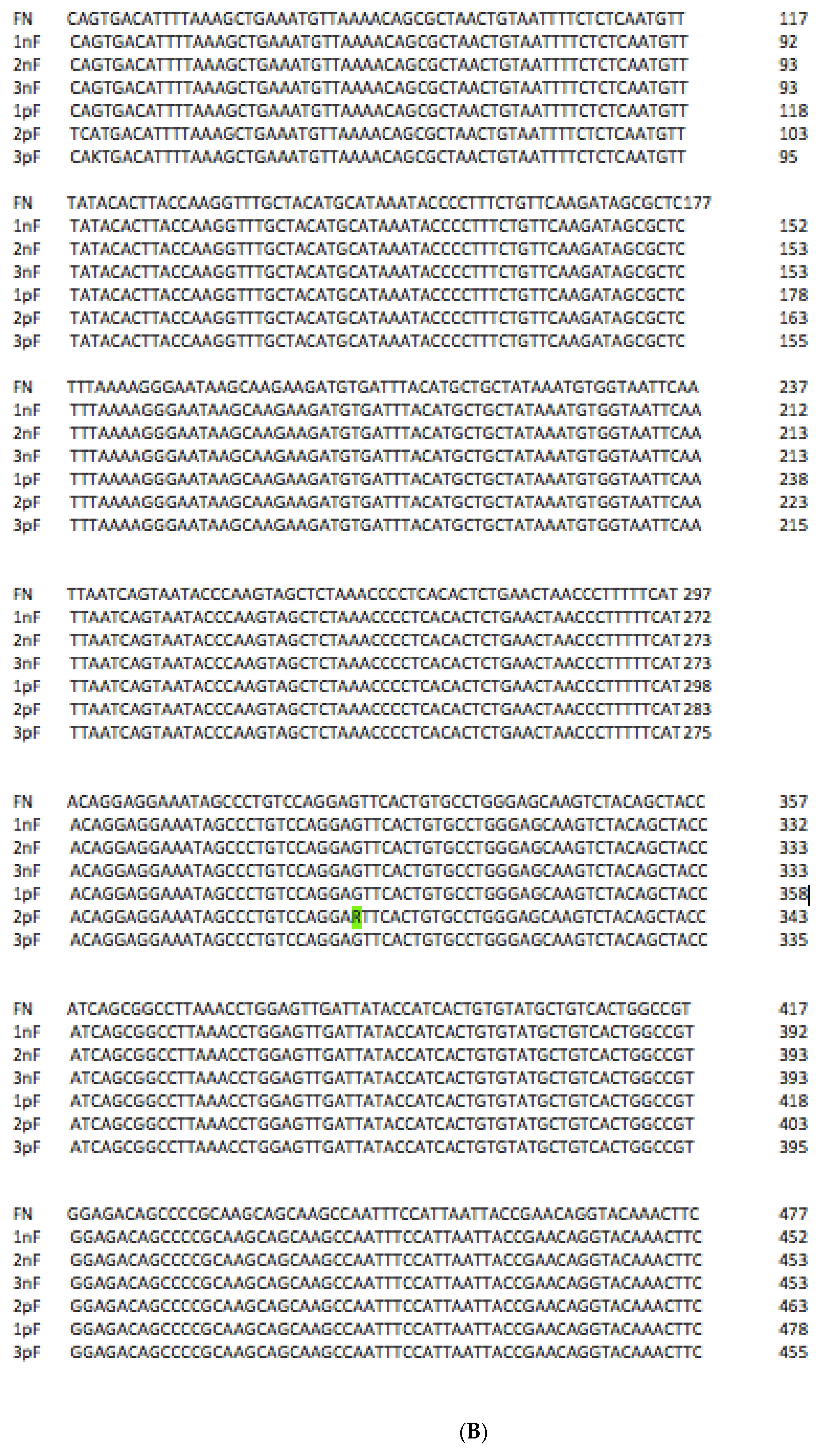
The nucleotide sequences of the fn1 gene on chromosome 2 (site 215,354,009 to 215,384,425) were also analyzed. No SNPs were detected in the “normal” group, whereas the “pathology” group showed an SNP in exon 30 of the fn1 gene at position 215,384,162. (Figure 2B).
Our findings support the fact that mutations in the adhesive domains of cFN can contribute to the development of periodontitis of inflammatory genesis. The detected transversion in the intron region of the DYNM2 gene can affect mRNA processing, which can affect the adhesive properties of DNM2 and contribute to the development of the pathological process.
DYNM2, whose role is underestimated and poorly understood, deserves special attention.

4. Conclusions

Thus, the topic of this study is interesting and requires further investigation. The development of a template of each patient’s anamnesis and more precise guidelines for patient selection are needed. To identify mutations and risk factors influencing the development of inflammatory periodontal disease, a full genome-wide association study (GWAS) on a significant number of patients is required.

Author Contributions

E.A.O. and A.V.G. contributed substantially to the conception, design, literature analysis, workshop discussion, drafting, and revision of the manuscript. All authors have read and agreed to the published version of the manuscript.

Funding

This study did not receive external funding.

Institutional Review Board Statement

The study was conducted in accordance with the Declaration of Helsinki and approved by the Pavlov First St.Petersburg State Medical University Ethics Committee (protocol code 114/22 of 24 February 2022).

Informed Consent Statement

Informed consent was obtained from all subjects participating in the study.

Data Availability Statement

Not applicable.

Conflicts of Interest

Authors declare no conflict of interest.

Abbreviations

FNFibronectin
DYNM2Dynamin-2
RGDArg-Gly-Asp motif
CCBDcentral cell-binding domain
PRD—Cterminal proline-rich domain (DYNM2)
CALclinical attachment loss
RBLradiological bone loss
SNPSingle Nucleotide Polymorphism

References

  1. Amano, A. Disruption of epithelial barrier and impairment of cellular function by Porphyromonas gingivalis. Front. Biosci. 2007, 12, 3965–3974. [Google Scholar] [CrossRef] [PubMed]
  2. Amano, A.I.; Nakagawa, N.; Okahashi, N. Hamada: Variations of Porphyromonas gingivalis fimbriae in relation to microbial pathogenesis. J. Periodontal Res. 2004, 39, 136–142. [Google Scholar] [CrossRef] [PubMed]
  3. Amaya Arbeláez, M.I.; de Paula ESilva, A.C.A.; Navegante, G.; Valente, V.; Barbugli, P.A.; Vergani, C.E. Proto-Oncogenes and Cell Cycle Gene Expression in Normal and Neoplastic Oral Epithelial Cells Stimulated With Soluble Factors From Single and Dual Biofilms of Candida albicans and Staphylococcus aureus. Front. Cell. Infect. Microbiol. 2021, 11, 627043. [Google Scholar] [CrossRef] [PubMed]
  4. Cecil, J.D.; O’Brien-Simpson, N.M.; Lenzo, J.C.; Holden, J.A.; Singleton, W.; Perez-Gonzalez, A.; Mansell, A.; Reynolds, E.C. Outer Membrane Vesicles Prime and Activate Macrophage Inflammasomes and Cytokine Secretion In Vitro and In Vivo. Front. Immunol. 2017, 8, 1017. [Google Scholar] [CrossRef] [PubMed]
  5. Shoji, M.; Shibata, S.; Naito, M.; Nakayama, K. Transport and Polimerization of Porphyromonas Gingivalis Type V Pili. Periodontal Pathog. 2021, 2210, 61–73. [Google Scholar] [CrossRef]
  6. Tsuda, K.; Amano, A.; Umebayashi, K.; Inaba, H.; Nakagawa, I.; Nakanishi, Y.; Yoshimori, T. Molecular dissection of internalization of Porphyromonas gingivalis by cells using fluorescent beads coated with bacterial membrane vesicle. Cell Struct. Funct. 2005, 30, 81–91. [Google Scholar] [CrossRef] [PubMed]
  7. Nakagawa, I.; Amano, A.H.; Inaba, S.K.; Hamada, S. Inhibitory effects of Porphyromonas gingivalis fimbriae on interactions between extracellular matrix proteins and cellular integrins. Microbes Infect. 2005, 7, 157–163. [Google Scholar] [CrossRef] [PubMed]
  8. Veith, P.D.; Luong, C.; Tan, K.H.; Dashper, S.G.; Reynolds, E.C. Outer Membrane Vesicle Proteome of Porphyromonas gingivalis Is Differentially Modulated Relative to the Outer Membrane in Response to Heme Availability. J. Proteome Res. 2018, 17, 2377–2389. [Google Scholar] [CrossRef] [PubMed]
  9. Zhang, Z.; Liu, D.; Liu, S.; Zhang, S.; Pan, Y. Porphyromonas gingivalis Outer Membrane Vesicles. Front. Cell. Infect. Microbiol. 2021, 10, 585917. [Google Scholar] [CrossRef] [PubMed]
  10. Cecil, J.D.; Sirisaengtaksin, N.; O’Brien-Simpson, N.M.; Krachler, A.M. Outer Membrane Vesicle-Host Cell Interactions. Microbiol. Spectr. 2019, 7, 201–214. [Google Scholar] [CrossRef] [PubMed] [Green Version]
  11. Inaba, H.S.; Kawai, T.; Kato, I.; Nakagawa, A. Amano: Association between epithelial cell death and invasion by microspheres conjugated to Porphyromonas gingivalis vesicles with different types of fimbriae. Infect. Immun. 2006, 74, 734–739. [Google Scholar] [CrossRef] [PubMed]
  12. Ishida, N.; Nakamura, Y.; Tanabe, K.; Li, S.; Takei, K. Dynamin 2 associates with microtubules at mitosis and regulates cell cycle progression. Cell Struct. Funct. 2011, 36, 145–154. [Google Scholar] [CrossRef] [PubMed]
  13. Khater, I.M.; Liu, Q.; Chou, K.C.; Hamarneh, G.; Nabi, I.R. Super-resolution modularity analysis shows polyhedral caveolin-1 oligomers combine to form scaffolds and caveolae. Sci. Rep. 2019, 9, 9888. [Google Scholar] [CrossRef] [PubMed]
  14. Kraehling, J.R.; Hao, Z.; Lee, M.Y.; Vinyard, D.J.; Velazquez, H.; Liu, X.; Stan, R.V.; Brudvig, G.W.; Sessa, W.C. Uncoupling Caveolae from intracellular signaling in vivo. Circ. Res. 2016, 118, 48–55. [Google Scholar] [CrossRef] [PubMed]
  15. Lafont, F.; van der Goot, F.G. Bacterial invasion via lipid rafts. Cell Microbiol. 2005, 7, 613–620. [Google Scholar] [CrossRef] [PubMed]
  16. Ludwig, A.; Nichols, B.J.; Sandin, S. Architecture of the caveolar coat complex. J. Cell Sci. 2016, 129, 3077–3083. [Google Scholar] [CrossRef] [PubMed]
  17. Manabe, R.; Ohe, N.; Maeda, T.; Fukuda, T.; Sekiguchi, K. Modulation of Fibronectin by EDA Segment. J. Cell Biol. 1997, 139, 295–306. [Google Scholar] [CrossRef] [PubMed]
  18. González-Jamett, A.M.; Momboisse, F.; Haro-Acuña, V.; Bevilacqua, J.A.; Caviedes, P.; Cárdenas, A.M. Dynamin-2 in the secretory pathway. Front. Endocrinol. 2013, 4, 126. [Google Scholar] [CrossRef] [PubMed]
  19. Kierdaszuk, B.; Berdynski, M.; Karolczak, J.; Redowicz, M.J.; Zekanowski, C.; Kaminska, A.M. A novel mutation in the DNM2 gene impairs dynamin 2 localization in skeletal muscle of a patient with late onset centronuclear myopathy. Neuromuscul. Disord. 2013, 23, 219–228. [Google Scholar] [CrossRef] [PubMed]
  20. Liu, Y.W.; Lukiyanchuk, V.; Schmid, S.L. Common membrane trafficking defects of disease-associated dynamin 2 mutations. Traffic 2011, 12, 1620–1633. [Google Scholar] [CrossRef] [PubMed] [Green Version]
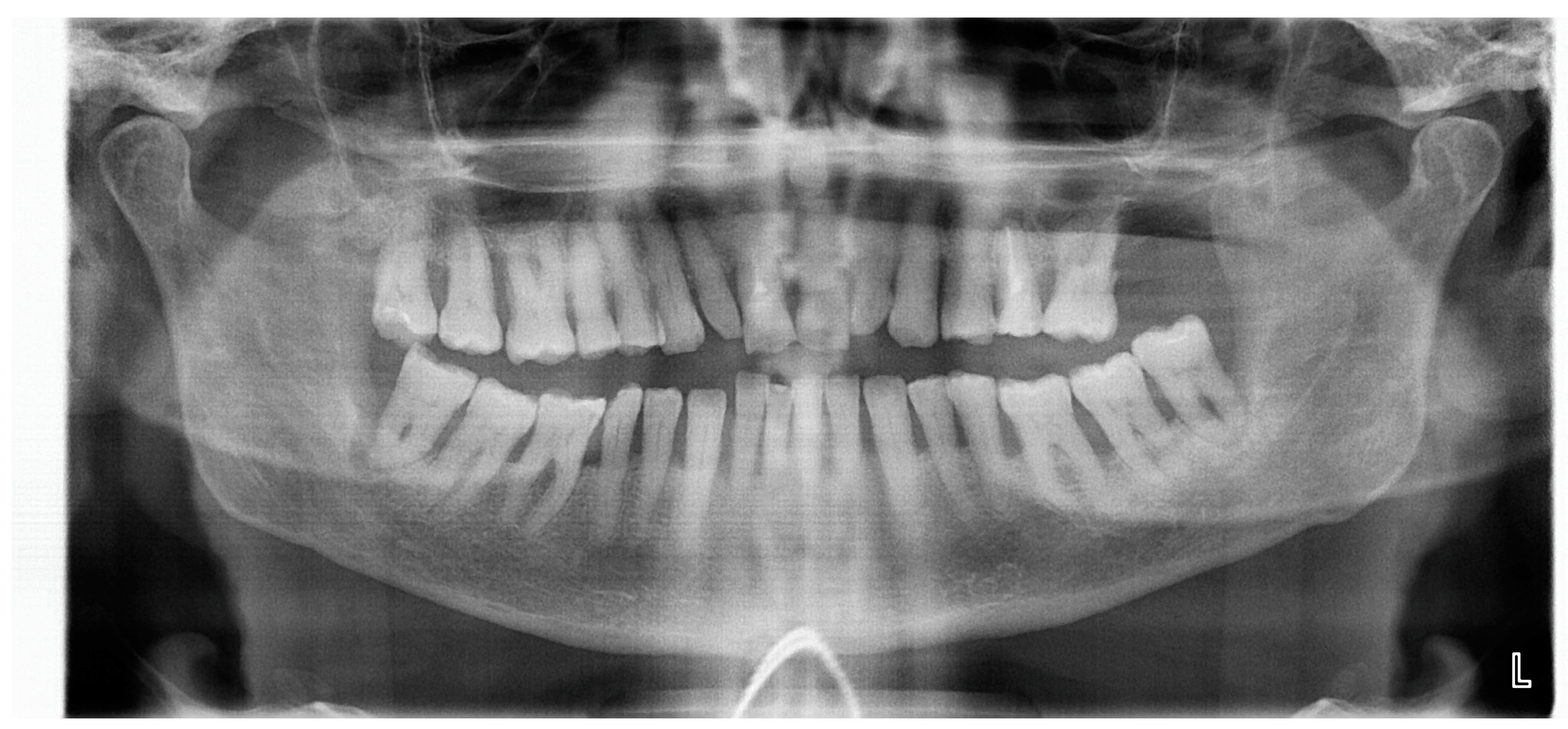
Figure 1. Periodontitis (radiographic appearance). A 37-year-old woman with generalized severe periodontitis (degree II–III). There is extensive alveolar bone loss (usually 50–75% of the root length) affecting the entire tooth row, with an irregular pattern of bone loss. Some teeth have lost almost all of their alveolar bone support as a result of the progression of periodontitis, such as the upper right molars and four lower incisors, all of which are mobile and held in the mouth only by soft tissue attachment (having lost over 85% of their bone support). Index R. Attsrom—37/02/00/05/20/0.
Jmp 03 00015 g001
Figure 2. (A) Sequencing result of the DNM2 gene fragment. Samples 1nD, 2nD, and 3nD are healthy patients; 1pD, 2pD, and 3pD are patients with pathology. SNPs and transversion are highlighted in green. (B) Sequencing result of the FN gene fragment. Samples 1nF, 2nF, 3nF are healthy patients; 1pF, 2pF, 3pF are patients with pathology. SNPs are highlighted in green.
Jmp 03 00015 g002aJmp 03 00015 g002b
Publisher’s Note: MDPI stays neutral with regard to jurisdictional claims in published maps and institutional affiliations.