Article

A Longitudinal Study of Bacteriophages as Indicators of Norovirus Contamination of Mussels (Mytilus edulis) and Their Overlying Waters

1
Environment and Public Health Research Group, School of Applied Sciences University of Brighton, Cockcroft Building, Lewes Rd, Brighton BN2 4GJ, UK
2
Nothe, Barrack Road, Weymouth, Dorset DT4 8UB, UK
*
Author to whom correspondence should be addressed.
This paper is a part of the PhD Thesis of Diogo Trajano Gomes da Silva, presented at University of Brighton (UK).
Pollutants 2022, 2(1), 66-81; https://doi.org/10.3390/pollutants2010008
Submission received: 30 November 2021 / Revised: 19 December 2021 / Accepted: 30 December 2021 / Published: 1 March 2022
(This article belongs to the Section Water Pollution)

Abstract

:
Sewage pollution leads to the contamination of bivalve shellfish by pathogenic microorganisms. Bacterial indicators support the management of risks associated with the consumption of shellfish; however, they often fail to indicate adequately the potential hazard to human health posed by certain human enteric viruses. Bacteriophages have been proposed as alternative indicators that may more effectively predict the presence of enteric viral pathogens. This study explored the relationships between bacterial indicators (Escherichia coli (E. coli), faecal coliforms (FC) and intestinal enterococci (IE)), phages (somatic (SOMPH), F-specific RNA (F + PH) and human-specific Bacteroides GB-124 phages (GB124PH)) and Norovirus (NoV) (GI/GII) in mussels (Mytilus edulis) and their overlying waters. The bioaccumulation of these indicators and Norovirus in shellfish matrices (e.g., flesh, digestive gland) was investigated bimonthly over a 12-month period in an English estuary. The findings revealed a marked seasonality in the distribution of all organisms, with the highest levels occurring during the autumn/winter months. The levels of all phages in shellfish and their overlying waters correlated better with the levels of Norovirus than with those of bacterial indicators. Somatic coliphages were the indicator that exhibited the strongest correlations with NoV (rho = 0.929). This study suggests that relatively low-cost culture-based phage enumeration appears to offer a more accurate indication of the likely presence of Norovirus in mussels than traditional bacterial indicators.

1. Introduction

Bivalve molluscs (shellfish) are filter feeders; thus, they may bioaccumulate pathogenic microorganisms originating from sewage pollution. During the twentieth century; advances in wastewater treatment, improvements to shellfish safety regulations, the implementation of methods for shellfish purification, and the adoption of bacterial indicators for the monitoring of shellfish all served to decrease the incidence of human excreta-borne illnesses, particularly those caused by pathogenic bacteria (e.g., typhoid fever) [1,2,3]. However, it has been suggested that the failure of bacterial indicators to identify accurately the potential risk to human health posed by enteric viral pathogens [4], which tend to be both more resistant to wastewater treatment processes [5,6] and more persistent during purification treatments than bacteria [7], explains why pathogenic viruses continue to pose a significant threat [8] to the health of shellfish consumers. Additionally, 359 disease outbreaks associated with the ingestion of bivalves that occurred in various parts of the world between 1980 and 2012 demonstrated a viral aetiology. Of these, Norovirus (NoV) was the principal disease-causing agent (responsible for 83.7% of cases), followed by the hepatitis A virus (HAV) (responsible for 12.8% of cases) [9].
It has been suggested [10] that long-term site-specific monitoring studies are needed to assess pathogen–indicator correlations, in order to provide a better estimation of contamination by specific pathogens within a particular water body, and thus to provide a useful assessment of potential risks to human health. Therefore, to address the problem of predicting and managing the viral contamination of surface waters and shellfisheries, several measures have been explored in recent years, including direct viral pathogen detection and the implementation of microbial source tracking (MST) techniques [11]. The ISO/TS 15216-1 method [12,13], which is based on real-time quantitative polymerase chain reaction (RT-PCR), allows for the enumeration of norovirus in bivalve shellfish, although it does not provide information on the infectivity of the pathogen [14]. However, a previous study suggested that high levels of NoV genome copies in molluscan shellfish detected by RT-PCR are indicative of a significantly elevated health risk [15]. The adoption of appropriate indicators of viral pathogens for use in shellfish and surface water surveillance has been proposed, with the most promising candidates being certain groups of enteric bacteriophages (phages) [16].
Bacteriophages (or ‘phages’) are viruses that infect the prokaryotic organisms (bacteria). To date, some groups of bacteriophages found among the gastrointestinal microbiota of mammals have been proposed as indicators of viral pathogens for use in shellfish and surface water surveillance. These include somatic coliphages (SOMPH) [17], F-specific RNA phages (F + PH) [18] and phages specific to human sources of faecal contamination, such as those infecting certain Bacteroides strains (e.g., HSP40) [19]. More recently strain GB-124 (GB124PH) has also been investigated [20,21]. The use of human-specific phages in shellfish hygiene monitoring is innovative and potentially important, as it has been suggested [22] that, although shellfish may also become contaminated by zoonotic pathogens originating from non-human faeces, pathogens associated with human faeces remain the main focus of public health concern.
In 2007, a US Environmental Protection Agency (EPA) scientific review of recreational water quality criteria suggested that the use of phages in health and epidemiological studies (alongside E. coli and intestinal enterococci (IE)) should be made a priority to support efforts to estimate the risks associated with a range of viral pathogens, including NoV [23]. Since then, the detection and enumeration of phages infecting the Bacteroides fragilis strain GB-124 has shown considerable promise, both for identifying the presence and source of faecally impacted surface waters in the UK [24] and in the USA [25].
In 2015, a further review of the extant evidence by the US EPA [26] concluded that while some knowledge gaps remain regarding the behaviour of coliphages in some environmental matrices, they are likely to be a better indicator of viruses from faecal contamination than currently used faecal indicator bacteria. Therefore, phages may not only offer a better indication of the likely presence and levels of human enteric viruses in shellfisheries than existing bacterial indicators, but they may also indirectly provide valuable information on the possible infectious state of the viral pathogens. Hence, the inclusion of phages as part of shellfish sanitary surveys may help support the more rational assessment of human health risks. The following study explores the bioaccumulation of, and the relationships between, traditional bacterial indicators and phage-based indicators in a range of mussel (Mytilus edulis) matrices (e.g., mussel flesh, intravalvular liquid, and digestive glands) and their overlying waters, within a tidal river estuary in southeast England (UK). These levels were then compared with the levels of NoV (GI and GII) present in the digestive glands of the mussels and for compliance with both EU and US shellfish legislation. It was hypothesised that the groups of bacteriophages would serve as a more efficacious indicator than traditionally utilised bacterial indicators for NoV contamination of shellfish.

2. Materials and Methods

2.1. The Study Site

The samples of mussels (Mytilus edulis) and their overlying waters were collected from a site (latitude = 50°48′595 N; longitude = 000°02′196 E) on the River Ouse (East Sussex, UK), located 4 km north of the coastal port town of Newhaven, where the river meets the English Channel. The R. Ouse watershed consists of approximately 290 km of river and tributaries [27] and more than 20 municipal wastewater treatment works (WWTW) and combined sewage overflows (CSO), which discharge partially treated wastewaters into the watershed; other sources of faecal pollution within the watershed include agricultural and wild inputs [24]. A map containing the sampling site and major wastewater discharge points to the watershed area can be found in Figure S1.

2.2. Sample Collection

A total of twenty-four (n = 24) grab samples of mussels and overlying waters were collected twice monthly from the study site. Three sample matrices, namely (a) overlying water, (b) mussel flesh including intravalvular liquid (mussel flesh), and (c) digestive gland (mussel gland), were analysed for the presence of six faecal indicators. These indicators were: Escherichia coli (E. coli); faecal coliforms (FC); intestinal enterococci (IE); somatic coliphages (SOMPH); F-specific RNA phages (F + PH); and phages infecting a human-specific Bacteroides fragilis strain (GB-124PH). Additionally, the detection and quantification of human NoV (GI and GII) in the mussel digestive glands was performed using RT-PCR. Samples were collected in the morning (08.00–10.00 a.m.) during the low tide, when the mussel beds were easily accessible. On each sampling occasion, 45 mussel samples were handpicked from a depth of approximately 20 cm below the water surface, and 1-L samples of overlying water were obtained. All samples were placed in sterile 1-L polyethylene containers (Fisher Scientific, Loughborough, UK) and transported on ice (within an hour) to the laboratory for immediate processing.

2.3. Sample Processing

Samples were processed in accordance with the standard methods, with some minor modifications, as described here. In brief, each batch of 45 mussels was separated into three sub-batches of approximately 15 mussels (equivalent to approximately 100 (±2) grams of flesh and intravalvular liquid). The mussels were scrubbed under running water to avoid cross-contamination from sediments and opened with a sterile shucking knife on sterile stainless-steel trays (Scientific Labs® Nottingham, UK). Flow charts detailing all the sampling processing steps can be found in the supporting information (Figures S2 and S3).
From the first batch of samples, the mussel flesh and intravalvular liquid were removed from the shells, placed in stomacher bags and blended (Seward® Stomacher400, Worthing, UK) at 230 rpm (paddle speed) for three minutes. The homogenate was then placed in a sterile Petri dish, weighed, and subdivided into two 50 (±1) gram homogenates. One of the resulting homogenates was then used to enumerate E. coli in accordance with the multiple tube technique [24]. The other homogenate was used for the enumeration of FC, IE and phages (SOMPH, GB124PH, F + PH). This homogenate was placed in another stomacher bag containing 50 mL of glycine buffer (pH10) and blended for an additional three minutes at 230 rpm. Subsequently, the homogenate was placed in a 200 mL Schott bottle, filled with an additional 50 mL of glycine buffer (pH10), resulting in a 1:3 (w/v) final solution.
From the second batch of mussels, 15 digestive glands (±0.4–0.5 g each) were extracted, placed in a sterile Petri dish, weighed, pooled and then finely chopped to homogenize using a sterile stainless-steel scalpel. This was followed by a 1:5 dilution with glycine buffer (pH10), producing a 42 (±2.5) mL final solution, which was subsequently used for the enumeration of FC, IE and phages (SOMPH, GB-124PH, F + PH). Finally, from the third batch of mussels, 15 digestive glands (0.4–0.5 g each) were extracted, placed in a sterile Petri dish, weighed, pooled and then finely chopped to homogenize using a sterile stainless-steel scalpel. The homogenate (7 ± 0.5 g) was then placed in a sterile 15 mL centrifuge tube and stored at −70 °C (for no more than 12 months). Approximately 2 g of the frozen homogenate were subsequently used for the detection of NoV (GI/GII).

2.3.1. Preparation of Mussel Homogenate and Overlying Water Samples for Phage Analysis

Before phage detection, the samples of mussel flesh and mussel gland solution (±30 to 40 mL) were poured into sterile 50 mL centrifuge tubes and clarified by centrifugation (Centaur® 2, Salford, UK) at 2000 G for 15 min. The supernatants and overlying waters were filtered using 0.22 µm syringe-driven polyether sulfone filters (Merck, Millipore®, Burlington, MA, USA), attached to 10 mL sterile plastic syringes (Plastipak®, Wrexham, UK) in order to remove background contamination. Filtered supernatants and overlying waters (approximately 20–30 mL each) were stored in 15 mL plastic sterile test tubes (Sterilin®, Thermo Scientific, Waltham, MA, USA) and kept in the dark at 4 °C before being assayed the following day, in accordance with standard methods.

2.3.2. Preparation of Mussel Homogenate for Bacterial Analysis

For each sampling event, 1 mL, 10 mL and 100 mL volumes of overlying waters were filtered through a 0.45 μm pore-size cellulose nitrate filter (Thermo Scientific, UK) and incubated on selective agar. Owing to the viscous nature of the shellfish homogenates, which tended to clog the membrane filters following laboratory trials (data not shown), 0.5 mL volumes of homogenate (diluted in 10 mL of ¼ strength Ringer’s solution) were filtered through a 0.45 μm pore-size cellulose nitrate filter onto selective agar. This volume showed the greatest filtration efficacy and was subsequently adopted for the entire study, as a modification of the standard method.

2.4. Detection and Enumeration of Bacterial Indicators

Enumeration of E. coli followed the EU/UK reference method [28] for the testing of shellfish; therefore, it was only undertaken for mussel flesh and intravalvular liquid, as prescribed. The results were expressed as Most Probable Number MPN/100 g of mussel flesh and intravalvular liquid. The enumeration of FC and IE in the overlying waters and shellfish homogenates followed ISO standard methods: 9308/1:2000 [29] and 7899/2:2000 [30], respectively. Triplicate filters of the overlying water, mussel flesh and mussel gland homogenates were placed onto either m-faecal coliform (mFC) or m-Enterococcus (mEnt) agar (Difco®, Lawrence, KS, USA) in Ø 55 mm Petri dishes. The results were expressed as colony-forming units (CFU) (e.g., CFU/100 g, CFU/100 mL or CFU/1 g). The detection limits for the overlying water, mussel flesh and mussel gland samples were 1 CFU/100 mL, 200 CFU/100 g and 4 CFU/g, respectively.

2.5. Detection and Enumeration of Phage-Based Indicators

Somatic coliphages (SOMPH) were enumerated in accordance with the ISO standard 10705-2 [31] and E. coli (WG5) was used as a host bacterium. F-specific RNA phages (F + PH) were enumerated according to the ISO standard 10705-1 [32], and Salmonella typhimurium (WG49) was used as a host. Phages infecting B. fragilis (GB124 PH) were enumerated according to the ISO standard 10705-4 [33] using the strain GB-124 as host. For overlying waters, 1 mL samples were processed; for mussel flesh, 1 mL samples from the 1:3 (w:v) dilution were processed; and for mussel flesh, 1 mL from the 1:5 (w:v) dilution was processed. All phage enumeration was carried out in triplicate and the results were expressed as plaque-forming units (PFU) (e.g., PFU/100 g, PFU/100 mL or PFU/1 g). The detection limits for the overlying water, mussel flesh and intravalvular liquid, and mussel gland samples were 33 PFU/100 mL, 100 PFU/100 g and 2 PFU/1 g, respectively. The MS2 F-RNA reference phage, Bacteroides fragilis B-124 reference phage, and naturally occurring somatic coliphages (previously isolated, propagated and stored at a known titre) were used on all occasions as positive controls throughout the study.

2.6. Detection and Enumeration of NoV

The concentrations (genome copies) of human NoV (GI and GII) were enumerated in the mussel gland samples at the laboratories of CEFAS (Centre for Environment, Fisheries and Aquaculture Science) in Weymouth (UK). NoV were extracted exclusively from the mussel glands according to a standardized (European Committee for Standardization, CEN) quantitative RT-PCR method reported elsewhere (ISO 15216-1:2017) [13]. The results were expressed as genome copies per gram (genome copies/g) of mussel gland digestive tissue, with a detection limit of 40 genome copies/g. Mengo virus was used as a process control for the detection of NoV. Quantification used a log dilution series (range 1 × 10⁵ to 1 × 101 copies/µL) of linear dsDNA molecules carrying the GI and GII target sequences and followed the principles outlined in the ISO 15216-1 [13]. For the mengo virus, this gave an indication of extraction efficiency and again is part of the CEN standard, where there is a criterion for the extraction efficiency to be greater than 10%, otherwise the sample is re-tested. No inhibition due to co-concentration was reported as part of the CEN method—this was not a duplex/multiplex PCR; therefore, no competition of qPCR reagents is expected.

2.7. Statistical Analysis

Statistical analysis was performed using SPSS statistical software. All data were tested to determine whether they demonstrated a normal distribution by applying the Kolmogorov–Smirnov (KS) Test and by analysing histograms and normal Q-Q plots and P-P plots of data. Parametric statistical tests were also used whenever the data followed a normal distribution, whereas equivalent non-parametric tests were employed when the data did not follow a normal distribution. For the non-parametric tests, the median, rather than the mean, was used to express the average levels more accurately [34]. The criterion of 95% confidence, or a 0.05 probability (p), was applied to test the significance of the various statistical tests. Prior to statistical testing samples from each matrix in which faecal indicators were not detected were identified (Table 1). The non-parametric Spearman’s rank correlation coefficient (ρ or Spearman’s rho) was then used for all correlational analyses and applied to the data to measure the significance and strength of the relationship between bacterial and phage-based indicators and NoV. Firstly on the entire ‘annual’ dataset (Table 2) and secondly by splitting the data into ‘spring/summer’ (April–September) and ‘autumn/winter’ (October–March) months (Table 3 and Table 4).
This somewhat arbitrary division of the data into two separate seasonal phases (autumn/winter and spring/summer) was performed to help identify specific temporal trends in the dataset associated with particular months; similarly, other divisions, e.g., wet and dry, cold and warm seasons or months, could also have been used to achieve this. This is supported by the work of Lowther et al. [35], wherein a ‘marked winter seasonality’ of NoV contamination was noted.
To visualise the contamination data, bacteria counts were logged, replacing left-censored values with 1.0. Therefore, values beneath the detection limits were assigned a value of zero. Those samples presenting 0 PFU, or CFU per 100 mL or 100 g were transformed to log10 (+1) PFU, or CFU per 100 mL or 100 g. The logged counts were then plotted as box-plot charts.

3. Results

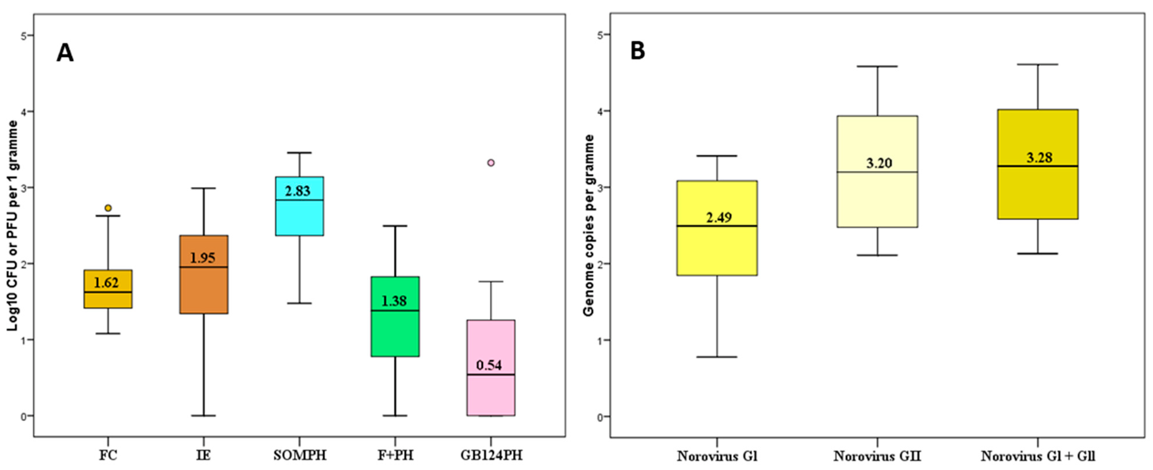
3.1. Levels of Faecal Pollution Indicators and Norovirus (NoV)

Table 1 displays the number and percentage of samples from each matrix in which faecal indicators were undetected during field-based research (including preliminary studies).
The results revealed that SOMPH were the most abundant faecal indicator and were detected in all samples of mussel flesh, digestive glands and in most samples (98%) of overlying water. Conversely, F + PH and GB124PH were more frequently detected in mussel flesh (88% and 57%, respectively) and digestive glands (91% and 57%, respectively), than in overlying waters (69% and 45%, respectively). Figure 1 displays the overall levels of faecal indicators in overlying waters (A) and in mussel flesh and intravalvular liquid matrices (B), while Figure 2 displays the overall levels of faecal indicator and NoV (genogroups I and II) in mussel digestive glands. A statistical summary of the annual levels of faecal indicators and NoV in all three sample matrices can be found in Table S1.
The levels of all faecal indicators in mussel flesh and intravalvular liquid were above those of the overlying waters. F + PH was the indicator that showed the greatest difference (1.98 Log10) between its levels in mussel flesh and intravalvular liquid, and its levels in the overlying waters, followed by IE (1.46 Log10); SOMPH (1.20 Log10); FC (0.88 Log10); and GB124PH (0.54 Log10). Combining the observed results from all three sample matrices (e.g., overlying water, flesh/intravalvular liquid, and digestive gland), the overall levels of SOMPH were found to be 1.46 and 1.72 log units higher than those of F + PH and GB-124PH, respectively. Levels of F + PH were also 0.26 log units higher than those of GB124PH phages. These proportions are in broad agreement with those observed in a research study from Spain [36], which compared the levels of phages infecting B. fragilis strains HSP-40 and RYC2056 with those of F + PH and SOMPH in mussels (Mytilus galloprovincialis).
Although the levels of human-specific GB124PH were generally lower than those of the other indicators (mean = 0.98 log10 PFU/100 mL in overlying waters), on two occasions they were detected at notably elevated levels (up to 4.24–4.93 log10 PFU/100 g; Table S2) in mussel flesh (i.e., at levels higher than those found in municipal wastewater effluents) [24]. Whilst specific discharge release data for nearby combined sewer overflows (CSO) was not available, it is interesting to note that these elevated levels of GB124PH coincided with storm events (>12 mm rainfall in 48 h). NoV was consistently detected throughout this study (24/24), although there were two sampling events at which NoV GI was undetected. Overall, the levels of NoV (median = 985 genome copies/g) observed in this study were relatively high, and similar to those observed in Ireland [37] and in England [38]. However, the levels of NoV reported in Scotland (50–169 genome copies/g) were much lower than those found during this study [35].

3.2. Relationship between Faecal Indicators and NoV in Mussels

Table 2 displays the annual Spearman’s rank correlation coefficient (ρ), measuring the strength and significance of relationships between bacterial and phage-based indicators and NoV (GI/GII). The strongest correlation coefficients per sample matrix are highlighted (shadowed).
The analyses revealed that the strongest correlations with NoV (up to ρ = 0.879; p = 0.01) were observed in phage-based indicators. The strongest NoV correlation observed in the overlying waters and in mussel flesh and intravalvular liquid was with SOMPH (ρ = 0.859; p = 0.0, and ρ = 0.761; p = 0.01, respectively), while the strongest NoV correlation observed in digestive gland was with F + PH (ρ = 0.879; p = 0.01). The levels of all other faecal indicators in mussel flesh were significantly, strongly and positively correlated with NoV, with the notable exception of FC. Interestingly, FC demonstrated the lowest correlation coefficient with NoV with respect to all mussel sample matrices, and it was the only faecal indicator that demonstrated a non-significant correlation with NoV levels.
The further breakdown of the samples into ‘spring/summer’ correlations (Table 3) and ‘autumn/winter’ correlations (Table 4) revealed that there were no significant correlations (p > 0.05) between any of the faecal indicators and NoV in overlying waters during the ‘spring/summer’ months. Furthermore, SOMPH and F + PH phages were the only faecal indicators to demonstrate any significant correlations (p > 0.05) with NoV in mussel flesh and digestive gland during this period. Interestingly, no significant correlation was observed between E. coli and FC during the ‘spring/summer’ months.
Conversely, the most significant and frequent correlations between NoV and faecal indicators were observed during the ‘autumn/winter’ months, with the strongest correlations again observed in phage-based indicators. In fact, SOMPH demonstrated the strongest observed correlation with NoV in overlying waters (ρ = 0.929; p = 0.01), F + PH demonstrated the strongest correlation with NoV observed in mussel flesh (ρ = 0.770; p = 0.05) and GB124PH demonstrated the strongest observed correlation with NoV in the digestive gland (ρ = 0.840; p = 0.01) during the ‘autumn/winter’ period. The strongest correlation between a bacterial indicator (E. coli) and NoV (ρ = 0.733; p = 0.05) was observed in mussel flesh during the ‘autumn/winter’ months.

4. Discussion

4.1. Compliance with EU Shellfish Classification Criteria

Although the study site is not an officially designated UK shellfish-harvesting area, an annual geometric mean of 930 Most Probable Number (MPN) E. coli/100 g in mussel flesh, combined with the fact that >90% of the test results were below the ‘Class C’ threshold, suggest that this site would most likely be classified as a ‘Class B’ harvesting site, according to the microbiological criteria of the European Regulation (EC 854/2004) [38]. According to a recent list (2020–2021) of designated shellfish areas in England and Wales, 8% are classed as ‘A’, 78% are classed as ‘B’ and 14% are classed as ‘C’ [39]. Therefore, the microbial loads observed at the study site may be regarded as broadly representative of the majority of shellfish production areas found in England and Wales.

4.2. Effect of Seasonality

The importance of gathering data over a 12-month period in a temperate climate, to encompass both ‘spring/summer’ and ‘autumn/winter’, is apparent from this study. For example, the levels of E. coli (the EU standard indicator organism for bivalve shellfish) exhibited a good correlation with NoV when the data for the entire study were combined, or particularly when the data from ‘autumn/winter’ alone were considered. However, in this study, the levels of E. coli did not exhibit any significant correlation with those of NoV when data from the ‘spring/summer’ period were treated independently (Table 4). This suggests that, although the levels of E. coli did not correlate as strongly with the levels of NoV as the phage-based indicators proposed here, this traditional indicator bacterium still has a role to play in protecting consumers of shellfish from the effects of pathogenic viruses.
Examining again the three seasonal groupings—namely ‘annual’, ‘spring/summer’ and ‘autumn/winter’—and the three sample matrices, eight combined parameters emerged as having the strongest correlations with NoV (Table 5).
Interestingly, all were phage-based approaches (four involving SOMPH, three involving F + PH and one involving GB-124PH). All three phage groups therefore demonstrated greater potential to predict the risk of NoV contamination of shellfish than did existing bacterial indicators. Therefore, these relatively low-cost methods could be employed alone, or as part of a ‘toolbox approach’, either during routine monitoring or as part of a focused sanitary investigation of shellfish harvesting areas. It is noteworthy that FC, the indicator prescribed by the US National Shellfish Sanitation Program (NSSP) [40] to classify shellfish growing areas, demonstrated the lowest significant correlations with NoV regardless of seasonal groupings. However, the latest revised version of the US NSSP [41] suggests that F + PH might be included as a tool during sanitary surveys of the microbiological quality of water in growing areas adjacent to wastewater discharges. Therefore, despite still not being used to classify the growing areas, this shows that the potential of using phages for shellfish safety is starting to be recognized.

4.3. Faecal Pollution Indicators and NoV in Mussel Matrices

The levels of all faecal indicators in mussel flesh and intravalvular liquid were above those of the overlying waters, suggesting bioaccumulation in these organisms. F + PH and SOMPH were the indicators that showed the highest bioaccumulation. SOMPH were observed to be the most abundant faecal pollution indicator observed in all sample matrices, including overlying waters, and its levels agreed with those observed in various species of shellfish in other parts of Europe [17]. Interestingly, these phages are normally recorded as being less abundant than FC (and often IE) in both local municipal wastewaters [24] and other aquatic matrices [42]. This increase in the relative proportion of SOMPH may suggest that these phages are replicating in the environment. However, this suggestion has been disputed, since the conditions needed for the replication of coliphages in the environment (i.e., the presence of a metabolically-active bacterial host at a sufficiently high cell density) are unlikely to be encountered in temperate climates; even when these conditions may occur, the impact on phage numbers is likely to be insignificant, given the relatively low levels of these phages in the natural environment [43,44]. Therefore, this relative increase in the abundance of SOMPH in the various matrices is more likely to be explained by the observation that this diverse group of phages appears to be more resilient to common wastewater treatment processes and to inactivation by natural environmental factors [45], and/or that they are more persistent in fresh, estuarine and marine waters than certain bacterial indicators [46]. SOMPH were the indicator that exhibited the strongest correlations with NoV (rho = 0.929). Another research study revealed that SOMPH demonstrated the most significant correlations with adenoviruses in mussels (ρ = 0.55) and their overlying waters (ρ = 0.66) [21]. SOMPH appear to be a useful phage group available as a generic indicator of the faecal pollution of shellfish and are a tool for predicting the likely presence of enteric viral pathogens, such as NoV. Key evidence to support this conclusion includes the observations that SOMPH: (1) exhibited high correlation coefficients with an important causative agent of shellfish-related food poisoning; (2) were highly abundant in faecally contaminated waters; and (3) were easily and rapidly quantified by a simple standardized culture-based phage-lysis method. Furthermore, our results suggest that a simple assay of overlying waters for SOMPH may be sufficient in more polluted areas; however, in less polluted areas, it is advisable to assay SOMPH in shellfish flesh and intravalvular liquid. Indeed, since shellfish may be harvested from water with lower levels of faecal contamination than are found in EU ‘Class B’ harvesting areas, further research should usefully consider whether the indicator–pathogen relationships described here are maintained in such waters.
The levels of F + PH and GB124PH were typically 1–2 logs lower than those of SOMPH, a finding that is in accordance with previous studies [17,36]. These phages were also frequently undetected in the overlying water and mussel flesh matrices, potentially limiting their suitability for routine monitoring in these matrices. However, the sensitivity of the mussel gland assay was greater, and consequently the detection rates for F + PH and GB124PH in this shellfish matrix were markedly higher. The reasons for these higher correlations of F + PH and GB124PH with NoV may include the following: F + PH were the least diverse phage group studied and their size, morphology and structure are the most similar to those of NoV [7,18,47,48], and GB124PH are human-specific [49] and theoretically they are more likely to co-present with human NoV than are phages that are found in a wider range of faecal sources.
The results from this study suggest that F + PH may be suitable indicators of the presence of NoV in mussels. However, as they were often recorded at low levels in overlying waters that presented with relatively high levels of faecal contamination, as is commonly observed in UK coastal waters (especially during the ‘autumn/winter’ period), it may be more appropriate in less-polluted waters to analyse the shellfish digestive gland. These observations support, to some extent, the work of other researchers who have proposed F + PH as an indicator of human viruses [18,50,51]. Furthermore, recent research [52,53] has highlighted the correlations between the F + PH subgroup II and NoV, suggesting that specific detection of infectious F + PH subgroup II could be regarded as an indication of the presence of infectious NoV.
During the ‘autumn/winter’ months, GB124PH phages exhibited the highest correlation coefficient with NoV within the mussel gland matrix. This was a highly significant finding, since the standard assay for quantifying levels of NoV uses this matrix. Moreover, it is during the ‘autumn/winter’ months that most NoV outbreaks associated with shellfish consumption occur in northern Europe [53]. A recent study [54] compared phages and NoV in wastewaters from five European countries and found a significant positive correlation between phages infecting the human-associated Bacteroides thetaiotaomicron strain GA17 and NoV, strengthening the findings of this research. GB124PH might be a suitable candidate for indicating NoV in shellfisheries, with the proviso that their use might be better focused on the investigation of more polluted waters, or when the origin of contamination is unclear. The human specificity of these phages makes them particularly useful for conducting initial sanitary surveys of proposed shellfish growing/harvesting areas, or in areas receiving intermittent human sources of faecal contamination.
Although, there have been recent advances in the study of NoV infectivity [55], such as the use of propidium monoazide compounds to indicate viral infectivity [56,57,58], the absence of robust, reliable and easy-to-use capsid integrity methods to solve the limitation of qPCR suggests that phages may have an important role to play in the assessment of health risks associated with the presence of enteric viruses in such matrices. This proposed application of phage technology is reiterated by Lowther et al. [59]. This research suggests that qPCR alone overestimates viral infectivity; however, combining qPCR with F + PH phage testing has the potential to better predict the presence of NoV.
It is worth mentioning that research has demonstrated that the reduction in levels of NoV, measured by q-PCR, during wastewater treatment [37], shellfish heat-treatment [60] and shellfish depuration [61] do not appear to follow the same patterns as those of infectious phages. Given that it is a requisite that shellfish harvested from ‘Class B’ areas must be purified (depurated) prior to sale and consumption, it is therefore necessary to assess the depuration efficiency.
Finally, although there is growing evidence for a dose−response relationship between the amount of NoV genome ingested and the probability of illness [11], molecular-based approaches remain technologically unfeasible and/or prohibitively expensive for routine monitoring in many parts of the world.

5. Conclusions

During this study, the levels of NoV and of all faecal indicators demonstrated a marked seasonality, with higher levels occurring during the colder and darker months of autumn and winter. Mussel digestive gland was shown to be the most useful and effective matrix for assessing levels of faecal indicators, being easier to assay than the mussel flesh and intravalvular liquid. Furthermore, as the presence of human enteric viruses is measured in shellfish digestive glands, a focus on this matrix when enumerating pathogen surrogates is recommended. Of all the microbial species investigated, SOMPH demonstrated the greatest potential for predicting levels of NoV in mussels and their overlying waters. The findings of this study suggest that existing ways to assess the microbiological safety of shellfish, as prescribed by US and EU legislation (such as the use of FC as an indicator organism, or the analysis of mussel flesh and intravalvular liquid), may not adequately indicate risk to the health of shellfish consumers of enteric viral pathogens, particularly NoV. Therefore, the enumeration of phages using relatively low-cost, culture-based approaches potentially offers a more accurate indication of the likely presence of NoV in mussels than does the use of traditional bacterial indicators, while also providing information on both the probable source and the infectious state of the pathogen. Therefore, the enumeration of SOMPH and F + PH may offer a better indication of NoV contamination of mussel harvesting areas than the enumeration of traditional bacterial indicators, and the enumeration of GB124PH in mussel matrices may offer a better indication of human faecal pollution impacts. Although this study used norovirus as a readout for the viral contamination of shellfish, future work may also focus on other relevant pathogenic viruses. It will be of interest to analyse the cross-species efficacy of bacteriophages as indicators of shellfish safety. In conclusion, given the aforementioned findings, continued research into the application of bacteriophages as indicators of shellfish safety is of vital importance.

Supplementary Materials

The following are available online at https://www.mdpi.com/article/10.3390/pollutants2010008/s1, Figure S1: Map of the sampling site catchment including major sewage discharge points; Figure S2: Flow chart describing processing of mussel flesh and intravalvular liquid samples; Figure S3: Flow chart describing processing of mussel digestive gland; Table S1: Statistics summary of the annual levels of faecal indicators and norovirus in all mussel matrices; Table S2: Raw data of faecal indicators and norovirus levels during the whole field-based research.

Author Contributions

Conceptualization, D.T.G.d.S., H.T., J.E. and C.B.-A.; methodology, D.T.G.d.S.; validation, H.T., J.E. and C.B.-A.; formal analysis, D.T.G.d.S. and D.D.; investigation, D.T.G.d.S. and D.D.; resources H.T. and J.E.; data curation, D.T.G.d.S.; writing—original draft preparation, D.T.G.d.S.; writing—review and editing, H.T. and J.E.; visualization, D.T.G.d.S.; supervision, H.T., J.E. and C.B.-A.; project administration, D.T.G.d.S., H.T. and J.E.; funding acquisition, H.T. and J.E. All authors have read and agreed to the published version of the manuscript.

Funding

This work was partly funded by the European Regional Development Fund Interreg IVA Programme as part of the collaborative project AquaManche (Aquatic Management of Catchments and Coasts for Health and Environment), with additional support provided to first author by the Society for Applied Microbiology (SfAM).

Data Availability Statement

All primary data can be found in the supplementary information file Table S2: Raw data of faecal indicators and norovirus levels during the whole field-based research.

Acknowledgments

The authors would also like to acknowledge the support of the Ouse & Adur Rivers Trust (OART).

Conflicts of Interest

The authors declare no conflict of interest.

Abbreviations

CFUColony-Forming Units
CSOCombined sewer overflow
FCFaecal coliforms
F + PHF-specific RNA phages
G I/IINorovirus genogroup I and II
GB124PHPhages infecting Bacteroides fragilis strain GB-124
IEIntestinal enterococci
NoVNorovirus
NSSPNational Shellfish Sanitation Program
PFUPlaque-forming units
RT-PCRReal-time polymerase chain reaction
SOMPHSomatic coliphages
WWTWWastewater treatment works

References

  1. Crane, S.; Moore, J. A Management Strategy to Reduce Bacterial Pollution in Shellfish Areas: A Case Study. Environ. Manag. 1986, 10, 41–51. [Google Scholar] [CrossRef]
  2. Schaeffer, J.; Treguier, C.; Piquet, J.; Gachelin, S.; Cochennec-Laureau, N.; Le Saux, J.; Garry, P.; Le Guyader, F. Improving the Efficacy of Sewage Treatment Decreases Norovirus Contamination in Oysters. Int. J. Food Microbiol. 2018, 286, 1–5. [Google Scholar] [CrossRef] [Green Version]
  3. Martinez-Albores, A.; Lopez-Santamarina, A.; Rodriguez, J.; Ibarra, I.; Mondragón, A.; Miranda, J.; Lamas, A.; Cepeda, A. Complementary Methods to Improve the Depuration of Bivalves: A Review. Foods 2020, 9, 129. [Google Scholar] [CrossRef] [Green Version]
  4. Ang, L.H. An outbreak of viral gastroenteritis associated with eating raw oysters. Commun. Dis. Public Health 1998, 1, 38–40. [Google Scholar]
  5. Gerba, C.; Wallis, C.; Melnick, J. Viruses in Water: The problem, some solutions. Environ. Sci. Technol. 1975, 9, 1122–1126. [Google Scholar] [CrossRef]
  6. McMinn, B.; Ashbolt, N.; Korajkic, A. Bacteriophages as Indicators of Faecal Pollution and Enteric Virus Removal. Lett. Appl. Microbiol. 2017, 65, 11–26. [Google Scholar] [CrossRef] [Green Version]
  7. Love, D.C.; Lovelace, G.L.; Sobsey, M.D. Removal of Escherichia coli, Enterococcus fecalis, coliphage MS-2, poliovirus, and hepatitis A virus from oysters (Crassostrea virginica) and hard-shell clams (Mercinaria mercinaria) by depuration. Int. J. Food Microbiol. 2010, 143, 211–217. [Google Scholar] [CrossRef]
  8. Iwamoto, M.; Ayers, T.; Mahon, B.; Swerdlow, D. Epidemiology of Seafood-Associated Infections in the United States. Clinic. Microbiol. Rev. 2010, 23, 399–411. [Google Scholar] [CrossRef] [Green Version]
  9. Bellou, M.; Kokkinos, P.; Vantarakis, A. Shellfish-Borne Viral Outbreaks: A Systematic Review. Food Environ. Virol. 2012, 5, 13–23. [Google Scholar] [CrossRef]
  10. Wu, J.; Long, S.C.; Das, D.; Dorner, S.M. Are microbial indicators and pathogens correlated? A statistical analysis of 40 years of research. J. Water Health 2011, 9, 265–278. [Google Scholar] [CrossRef] [Green Version]
  11. Simpson, J.M.; Santo Domingo, J.W.; Reasoner, D.J. Microbial Source Tracking:  State of the Science. Environ. Sci. Technol. 2002, 36, 5279–5288. [Google Scholar] [CrossRef]
  12. Lees, D.; CEN WG6 TAG4. International standardisation of a method for detection of human pathogenic viruses in molluscan shellfish. Food Environ. Virol. 2010, 2, 146–155. [Google Scholar] [CrossRef]
  13. Anon. ISO 15216-1:2017; Microbiology of the Food Chain—Horizontal Method for Determination of Hepatitis A Virus and Norovirus Using Real-Time RT-PCR—Part 1: Method for Quantification. International Organization for Standardization: Geneva, Switzerland, 2017.
  14. Girones, R.; Ferrús, M.A.; Alonso, J.L.; Rodriguez-Manzano, J.; Calgua, B.; de Abreu Corrêa, A.; Hundesa, A.; Carratala, A.; Bofill-Mas, S. Molecular detection of pathogens in water—The pros and cons of molecular techniques. Water Res. 2010, 44, 4325–4339. [Google Scholar] [CrossRef]
  15. Lowther, J.; Avant, J.; Gizynski, K.; Rangdale, R.; Lees, D. Comparison between Quantitative Real-Time Reverse Transcription PCR Results for Norovirus in Oysters and Self-Reported Gastroenteric Illness in Restaurant Customers. J. Food Prot. 2010, 73, 305–311. [Google Scholar] [CrossRef] [PubMed]
  16. Hodgson, K.; Torok, V.; Turnbull, A. Bacteriophages as Enteric Viral Indicators in Bivalve Mollusc Management. Food Microbiol. 2017, 65, 284–293. [Google Scholar] [CrossRef]
  17. Formiga-Cruz, M.; Allard, A.K.; Conden-Hansson, A.C.; Henshilwood, K.; Hernroth, B.E.; Jofre, J.; Lees, D.N.; Lucena, F.; Papapetropoulou, M.; Rangdale, R.E.; et al. Evaluation of potential indicators of viral contamination in shellfish and their applicability to diverse geographical areas. Appl. Environ. Microbiol. 2003, 69, 1556–1563. [Google Scholar] [CrossRef] [Green Version]
  18. Doré, W.J.; Henshilwood, K.; Lees, D.N. Evaluation of F-specific RNA bacteriophage as a candidate human enteric virus indicator for bivalve molluscan shellfish. Appl. Environ. Microbiol. 2000, 66, 1280–1285. [Google Scholar] [CrossRef] [Green Version]
  19. Lucena, F.; Lasobras, J.; Mcintosh, D.; Forcadell, M.; Jofre, J. Effect of distance from the polluting focus on relative concentrations of Bacteroides fragilis phages and coliphages in mussels. Appl. Environ. Microbiol. 1994, 60, 2272–2277. [Google Scholar] [CrossRef] [Green Version]
  20. Da Silva, D.T.G. Bacteriophages as Indicators of Human Enteric Viruses in Mussels. Ph.D. Thesis, University of Brighton, Brighton, UK, 2013. [Google Scholar]
  21. Olalemi, A.; Purnell, S.; Caplin, J.; Ebdon, J.; Taylor, H. The application of phage-based faecal pollution markers to predict the concentration of adenoviruses in mussels (Mytilus edulis) and their overlying waters. J. Appl. Microbiol. 2016, 121, 1152–1162. [Google Scholar] [CrossRef] [Green Version]
  22. Potasman, I.; Paz, A.; Odeh, M. Infectious outbreaks associated with bivalve shellfish consumption: A worldwide perspective. Clin. Infect. Dis. 2002, 35, 921–928. [Google Scholar] [CrossRef] [Green Version]
  23. Ashbolt, N.; Fujioka, R.; Glymph, T.; McGee, C.; Schaub, S.; Sobsey, M.; Toranzos, G. Pathogens, pathogen indicators, and indicators of fecal contamination. In Report of the Experts Scientific Workshop on Critical Research Needs for the Development of New or Revised Recreational Water Quality Criteria; EPA 823-R-07-006; Environmental Protection Agency Office of Water Office of Research and Development: Warrenton, VA, USA, 2007; Volume 147, pp. 35–52. [Google Scholar]
  24. Ebdon, J.E.; Sellwood, J.; Shore, J.; Taylor, H. Phages of Bacteroides (GB-124): A novel tool for viral waterborne disease control? Environm. Sci. Technol. 2012, 46, 1163–1169. [Google Scholar] [CrossRef]
  25. McMinn, B.R.; Korajkic, A.; Ashbolt, H.J. Evaluation of Bacteroides fragilis GB-124 bacteriophages as novel human-associated fecal indicators in the United States. Lett. Appl. Microbiol. 2014, 59, 115–121. [Google Scholar] [CrossRef]
  26. USEPA Office of Water. Review of Coliphages as Possible Indicators of Fecal Contamination for Ambient Water Quality; 820-R-15-098; United States Environment Protection Agency, Office of Science and Technology, Office of Water: Washington, DC, USA, 2015. Available online: http://water.epa.gov/scitech/swguidance/standards/criteria/health/microbial/upload/coliphages-literature-review-report-2015.pdf (accessed on 25 July 2021).
  27. OART. The Sussex River Ouse Corridor. Available online: https://oart.org.uk/rivers-new/river-ouse/ (accessed on 2 November 2021).
  28. Anon. ISO/TS 16649-3; Microbiology of Food and Animal Feeding Stuffs. Horizontal Method for the Enumeration of Beta-Glucoronidase-positive Escherichia coli. Part 3: Most Probable Number Technique Using 5-Bromo-4-Chloro-3-Indolyl-Beta-D-Glucuro-Nide. International Organization for Standardization: Geneva, Switzerland, 2005.
  29. Anon. ISO 9308-1:2000; Water Quality. Detection and Enumeration of Escherichia coli and Coliform Bacteria Part 1: Membrane Filtration Method. International Organization for Standardization: Geneva, Switzerland, 2000.
  30. Anon. ISO 7899-2:2000; Water Quality: Detection and Enumeration of Intestinal Enterococci Part 2: Membrane Filtration Method. International Organization for Standardization: Geneva, Switzerland, 2000.
  31. Anon. ISO 10705-2; Water Quality—Detection and Enumeration of Bacteriophages—Part 2: Enumeration of Somatic Coliphages. International Organization for Standardization: Geneva, Switzerland, 2001.
  32. Anon. ISO 10705-1; Water Quality—Detection and Enumeration of Bacteriophages—Part 1: Enumeration of F-Specific RNA Bacteriophages. International Organisation for Standardization: Geneva, Switzerland, 2001.
  33. Anon. ISO 10705-4; Water Quality—Detection and Enumeration of Bacteriophages—Part 4: Enumeration of Bacteriophages Infecting Bacteroides fragilis. International Organisation for Standardization: Geneva, Switzerland, 2001.
  34. Helsel, D.R. Advantages of nonparametric procedures for analysis of water-quality data. Hydrol. Sci. J. 1987, 32, 179–190. [Google Scholar] [CrossRef] [Green Version]
  35. Lowther, J.; Gustar, N.; Powell, A.; Hartnell, R.; Lees, D. Two-Year Systematic Study to Assess Norovirus Contamination in Oysters from Commercial Harvesting Areas in the United Kingdom. Appl. Environ. Microbiol. 2012, 78, 5812–5817. [Google Scholar] [CrossRef] [Green Version]
  36. Muniain-Mujika, I.; Calvo, M.; Lucena, F.; Girones, R. Comparative analysis of viral pathogens and potential indicators in shellfish. Int. J. Food Microbiol. 2003, 83, 75–85. [Google Scholar] [CrossRef]
  37. Flannery, J.; Keaveney, S.; Rajko-Nenow, P.; O’Flaherty, V.; Dore, W. NoV and FRNA bacteriophage determined by RT-qPCR and infectious FRNA bacteriophage in wastewater and oysters. Water Res. 2013, 47, 5222–5231. [Google Scholar] [CrossRef]
  38. EC Regulation No 854/2004 of the European Parliament and of the Council of 29 April 2004 Laying Down Specific Rules for the Organization of Official Controls on Products of Animal Origin Intended for Human Consumption. Off. J. Eur. Union 2004, L226, 83–127.
  39. Food Standards Agency (FSA). Shellfish Harvesting Classifications England and Wales: 2020–2021. Available online: https://www.food.gov.uk/sites/default/files/media/document/shellfish-classifications-2020-2021-enw.pdf (accessed on 17 January 2021).
  40. NSSP. National Shellfish Sanitation Program. In Guide for the Control of Molluscan Shellfish: 2013 Revision. Available online: http://wayback.archive-it.org/7993/20180126093259/https://www.fda.gov/downloads/Food/GuidanceRegulation/FederalStateFoodPrograms/UCM415522.pdf (accessed on 5 March 2015).
  41. NSSP. National Shellfish Sanitation Program. In Guide for the Control of Molluscan Shellfish: 2019 Revision. Available online: https://www.fda.gov/media/143238/download (accessed on 22 November 2021).
  42. Contreras-Coll, N.; Lucena, F.; Mooijman, K.; Havelaar, A.; Pierzo, V.; Boque, M.; Gawler, A.; Holler, C.; Lambiri, M.; Mirolo, G.; et al. Occurrence and levels of indicator bacteriophages in bathing waters throughout Europe. Water Res. 2002, 36, 4963–4974. [Google Scholar] [CrossRef]
  43. Wiggins, B.A.; Alexander, M. Minimum bacterial density for bacteriophage replication—Implications for significance of bacteriophages in natural ecosystems. Appl. Environ. Microbiol. 1985, 49, 19–23. [Google Scholar] [CrossRef] [PubMed] [Green Version]
  44. Jofre, J. Is the replication of somatic coliphages in water environments significant? J. Appl. Microbiol. 2009, 106, 1059–1069. [Google Scholar] [CrossRef] [PubMed]
  45. Sinton, L.W.; Finlay, R.K.; Lynch, P.A. Sunlight inactivation of fecal bacteriophages and bacteria in sewage-polluted seawater. Appl. Environ. Microbiol. 1999, 65, 3605–3613. [Google Scholar] [CrossRef] [PubMed] [Green Version]
  46. Mocé-Llivina, L.; Lucena, F.; Jofre, J. Enteroviruses and Bacteriophages in Bathing Waters. Appl. Environ. Microbiol. 2005, 71, 6838–6844. [Google Scholar] [CrossRef] [Green Version]
  47. Leclerc, H.; Edberg, S.; Pierzo, V.; Delattre, J.M. Bacteriophages as indicators of enteric viruses and public health risk in groundwaters. J. Appl. Microbiol. 2000, 88, 5–21. [Google Scholar] [CrossRef] [Green Version]
  48. Grabow, W.O.K. Bacteriophages: Update on application as models for viruses in water. Water SA 2001, 27, 251–268. [Google Scholar] [CrossRef] [Green Version]
  49. Payan, A.; Ebdon, J.; Taylor, H.; Gantzer, C.; Ottoson, J.; Papageorgiou, G.T.; Blanch, A.R.; Lucena, F.; Jofre, J.; Muniesa, M. Method for isolation of Bacteroides bacteriophage host strains suitable for tracking sources of fecal pollution in water. Appl. Environ. Microbiol. 2005, 71, 5659–5662. [Google Scholar] [CrossRef] [Green Version]
  50. Flannery, J.; Keaveney, S.; Dore, W. Use of F-RNA bacteriophages to indicate the risk of NoV contamination in Irish oysters. J. Food Prot. 2009, 72, 2358–2362. [Google Scholar] [CrossRef] [PubMed]
  51. Hartard, C.; Banas, S.; Loutreul, J.; Rincé, A.; Benoit, F.; Boudaud, N.; Gantzer, C. Relevance of F-Specific RNA Bacteriophages in Assessing Human Norovirus Risk in Shellfish and Environmental Waters. Appl. Environ. Microbiol. 2016, 82, 5709–5719. [Google Scholar] [CrossRef] [PubMed] [Green Version]
  52. Gyawali, P.; Devane, M.; Scholes, P.; Hewitt, J. Application of Crassphage, F-RNA Phage and Pepper Mild Mottle Virus as Indicators of Human Faecal and Norovirus Contamination in Shellfish. Sci. Total Environ. 2021, 783, 146848. [Google Scholar] [CrossRef] [PubMed]
  53. Westrell, T.; Dusch, V.; Ethelberg, S.; Harris, J.; Hjertqvist, M.; Jourdan-Da Silva, N.; Koller, A.; Lenglet, A.; Lisby, M.; Vold, L. NoV outbreaks linked to oyster consumption in the United Kingdom, Norway, France, Sweden and Denmark, 2010. Eurosurveillance 2010, 15, 8–11. [Google Scholar] [CrossRef]
  54. Ballesté, E.; Blanch, A.; Mendez, J.; Sala-Comorera, L.; Maunula, L.; Monteiro, S.; Farnleitner, A.; Tiehm, A.; Jofre, J.; García-Aljaro, C. Bacteriophages Are Good Estimators of Human Viruses Present in Water. Front. Microbiol. 2021, 12, 973. [Google Scholar] [CrossRef] [PubMed]
  55. Jones, M.K.; Watanabe, M.; Zhu, S.; Graves, C.L.; Keyes, L.R.; Grau, K.R.; .Gonzalez-Hernandez, M.B.; Iovine, N.M.; Wobus, C.E.; Vinjé, J.; et al. Enteric bacteria promote human and mouse NoV infection of B cells. Science 2014, 6210, 755–759. [Google Scholar] [CrossRef] [Green Version]
  56. Kim, S.Y.; Ko, G. Using propidium monoazide to distinguish between viable and nonviable bacteria, MS2 and murine norovirus. Lett. Appl. Microbiol. 2012, 55, 182–188. [Google Scholar] [CrossRef] [PubMed]
  57. Jeon, E.; Choi, M.; Kim, J.; Ha, K.; Kwon, J.; Jeong, S.; Lee, H.; Jung, Y.; Ha, J.; Park, S. Characterizing the Effects of Thermal Treatment on Human Norovirus GII.4 Viability Using Propidium Monoazide Combined with RT-Qpcr and Quality Assessments in Mussels. Food Control 2020, 109, 106954. [Google Scholar] [CrossRef]
  58. Quijada, N.; Fongaro, G.; Barardi, C.; Hernández, M.; Rodríguez-Lázaro, D. Propidium Monoazide Integrated with Qpcr Enables the Detection and Enumeration of Infectious Enteric RNA and DNA Viruses in Clam and Fermented Sausages. Front. Microbiol. 2016, 7, 2008. [Google Scholar] [CrossRef] [PubMed] [Green Version]
  59. Lowther, J.; Cross, L.; Stapleton, T.; Gustar, N.; Walker, D.; Sills, M.; Treagus, S.; Pollington, V.; Lees, D. Use of F-Specific RNA Bacteriophage to Estimate Infectious Norovirus Levels in Oysters. Food Environ. Virol. 2019, 11, 247–258. [Google Scholar] [CrossRef]
  60. Flannery, J.; Rajko-Nenow, P.; Winterbourn, J.B.; Malham, S.K.; Jones, D.L. Effectiveness of cooking to reduce Norovirus and infectious F-specific RNA bacteriophage concentrations in Mytilus edulis. J. Appl. Microbiol. 2014, 117, 564–571. [Google Scholar] [CrossRef]
  61. Polo, D.; Alvarez, C.; Diez, J.; Darriba, S.; Longa, A.; Romalde, J.L. Viral elimination during commercial depuration of shellfish. Food Control 2014, 43, 206–212. [Google Scholar] [CrossRef]
Figure 1. Box plots of faecal indicator concentrations in overlying waters (A) and in mussel flesh and intravalvular liquid matrices (B) (n = 24). Values beneath the detection limits were assigned a value of zero. Those samples presenting 0 Plaque Forming Units (PFU), or Colony Forming Units (CFU) per 100 mL or 100 g were transformed to log10 (+1) PFU, or CFU per 100 mL or 100 g. (FC: faecal coliforms; IE: intestinal enterococci; SOMPH: somatic coliphages; F + PH: F-specific RNA phages; GB124PH: Bacteroides fragilis GB-124 phages). Coloured circles represent mild outliers; stars (*) represent extreme outliers.
Pollutants 02 00008 g001
Figure 2. Box plots of faecal indicator (A) and NoV (B) (genogroups I and II) concentrations in mussel digestive glands (n = 24). Values beneath the detection limits were assigned a value of zero. Those samples presenting 0 PFU, CFU or genome copies/g were transformed to log10 (+1) PFU, CFU or genome copies/g. (FC: faecal coliforms; IE: intestinal enterococci; SOMPH: somatic coliphages; F + PH: F-specific RNA phages; GB124PH: Bacteroides fragilis GB-124 phages). Coloured circles represent mild outliers.
Pollutants 02 00008 g002
Table 1. Number (n) and percentage (%) of samples from each sample matrix in which faecal indicators were undetected during field-based research (including preliminary studies).
IndicatorSample Matrix(n = 42)
n%
Overlying waters00
Faecal coliforms (FC)Mussel flesh24.8
Mussel gland00
Overlying waters24.8
Intestinal enterococci (IE)Mussel flesh37.1
Mussel gland24.8
Overlying waters12.4
Somatic coliphages (SOMPH)Mussel flesh00
Mussel gland00
Overlying waters1331
F-specific RNA phages (F + PH) Mussel flesh511.9
Mussel gland39.1
Overlying waters2354.8
Bacteroides fragilis phages (GB124PH)Mussel flesh1842.9
Mussel gland1842.9
Mean (%) Undetected Samples Per Matrix
Overlying waters7.818.5
Mussel flesh5.613.3
Mussel gland4.610.9
Table 2. Annual correlation coefficients (Spearman’s ρ) between levels of faecal indicators and Norovirus with respect to mussel sample matrix.
FCIESOMPHF + PHGB124PHNoV
Mussel overlying Waters
FC1.0000.848 **0.601 **0.417 *0.532 **0.577 **
IE 1.0000.506 *0.481 *0.439 *0.615 **
SOMPH 1.0000.577 **0.720 **0.859 **
F + PH 1.0000.471 *0.601 **
GB124PH 1.0000.596 **
Mussel flesh and intravalvular liquid
E. coli0.600 **0.843 **0.723 **0.678 **0.725 **0.646 **
FC1.0000.601 **0.518 **0.2670.461 *0.386
IE 1.0000.753 **0.699 **0.699 **0.656 **
SOMPH 1.0000.706 **0.821 **0.761 **
F + PH 1.0000.718 **0.658 **
GB124PH 1.0000.661 **
Mussel digestive gland
FC1.0000.603 **0.529 **0.534 **0.545 **0.466 **
IE 1.0000.694 **0.755 **0.778 **0.689 **
SOMPH 1.0000.755 **0.764 **0.684 **
F + PH 1.0000.780 **0.879 **
GB124PH 1.0000.734 **
* Correlation is significant at the 0.05 level (two-tailed). ** Correlation is significant at the 0.01 level (two-tailed). The strongest correlation coefficients per sample matrix are shadowed.
Table 3. ‘Spring/summer’ correlation coefficients (Spearman’s ρ) between faecal indicators and Norovirus with respect to mussel sample matrix.
FCIESOMPHF + PHGB124PHNoV
Mussel Overlying Waters
FC1.0000.5910.4150.3350.2560.336
IE 1.0000.0270.156−0.1350.418
SOMPH 1.0000.3590.0810.506
F + PH 1.0000.1890.480
GB124PH 1.000−0.243
Mussel Flesh and Intravalvular Liquid
E. coli0.1670.4990.1150.4850.703 *−0.238
FC1.0000.5260.639 *0.2620.672 *0.415
IE 1.0000.4370.4700.608 *0.056
SOMPH 1.0000.3140.620 *0.610 *
F + PH 1.0000.4280.182
GB124PH 1.0000.035
Mussel Digestive Gland
FC1.0000.3270.4920.5500.4260.395
IE 1.0000.629 *0.3720.4500.009
SOMPH 1.0000.656 *0.3360.683 *
F + PH 1.0000.1600.723 *
GB124PH 1.0000.035
* Correlation is significant at the 0.05 level (two-tailed). The strongest correlation coefficients per sample matrix are shadowed.
Table 4. ‘Autumn/winter’ correlation coefficients (Spearman’s ρ) between faecal indicators and Norovirus with respect to mussel sample matrix.
FCIESOMPHF + PHGB124PHNoV
Mussel Overlying Waters
FC1.0000.786 **0.2640.2400.4080.236
IE 1.0000.3350.612 *0.4250.451
SOMPH 1.0000.657 *0.764 **0.929 **
F + PH 1.0000.5170.746 **
GB124PH 1.0000.708 **
Mussel Flesh and Intravalvular Liquid
E. coli0.768 **0.719 **0.598 *0.575 *0.563 *0.733 *
FC1.0000.623 *0.5010.2010.4700.476
IE 1.0000.4150.656 *0.553 *0.641 *
SOMPH 1.0000.629 *0.684 **0.764 **
F + PH 1.0000.599 **0.770 *
GB124PH 1.0000.759 **
Mussel Digestive Gland
FC1.0000.617 *0.3100.2490.4660.340
IE 1.0000.2530.629 *0.769 **0.725 **
SOMPH 1.0000.4410.5010.495
F + PH 1.0000.744 **0.767 **
GB124PH 1.0000.840 **
* Correlation is significant at the 0.05 level (two-tailed). ** Correlation is significant at the 0.01 level (two-tailed). The strongest correlation coefficients per sample matrix are shadowed.
Table 5. Indicators demonstrating greatest NoV predictive capacity for mussels (Mytilus edulis).
PeriodSample Matrices
Overlying WaterMussel FleshMussel Gland
AnnualSOMPHSOMPHF + PH
(rho = 0.859)(rho = 0.761)(rho = 0.879)
Spring/SummerNoneSOMPHF+PH
(rho = 0.610)(rho = 0.723)
NoneSOMPHF + PHGB124PH
(rho = 0.929)(rho = 0.770)(rho = 0.840)
Publisher’s Note: MDPI stays neutral with regard to jurisdictional claims in published maps and institutional affiliations.

