1. Introduction
Nickel nanoparticles (NiNPs) are used in catalysts for hydrogenation of dietary fats, in cosmetics, insecticides, preparations for theranostics in medicine [
1,
2,
3,
4], and can also expose people occupationally in the metallurgy and mining industry [
5]. According to multiple studies, in vitro NiNPs possess pro-oxidant, pro-inflammatory, genotoxic and carcinogenic activity [
6,
7,
8]. At inhalation or intratracheal instillation in experimental animals, different adverse effects of Ni-containing NPs have been detected including general toxicity, immunotoxicity and reproductive toxicity [
9]. However, consequences of a prolonged administration of these nanomaterials with food at low doses are not well understood. The aim of the study was to evaluate the toxic effects of NiNPs in their prolonged oral administration to male Wistar rats
2. Materials and Methods
Ten groups of 12 male Wistar rats received a balanced semisynthetic diet (AIN93M) either without additions (Control group 1), or supplemented with Ni carbonate basic salt (Ni salt) at doses of 0.1, 1 and 10 mg/kg b.w. as Ni (groups 2–4), NiNPs preparation 1 (NiNP1) (groups 5–7) or NiNPs preparation 2 (NiNP2) (groups 8–10) at the same doses as Ni, respectively, for 92 days. At the end of the feeding period, biochemical, immunological and morphological endpoints were studied.
3. Results
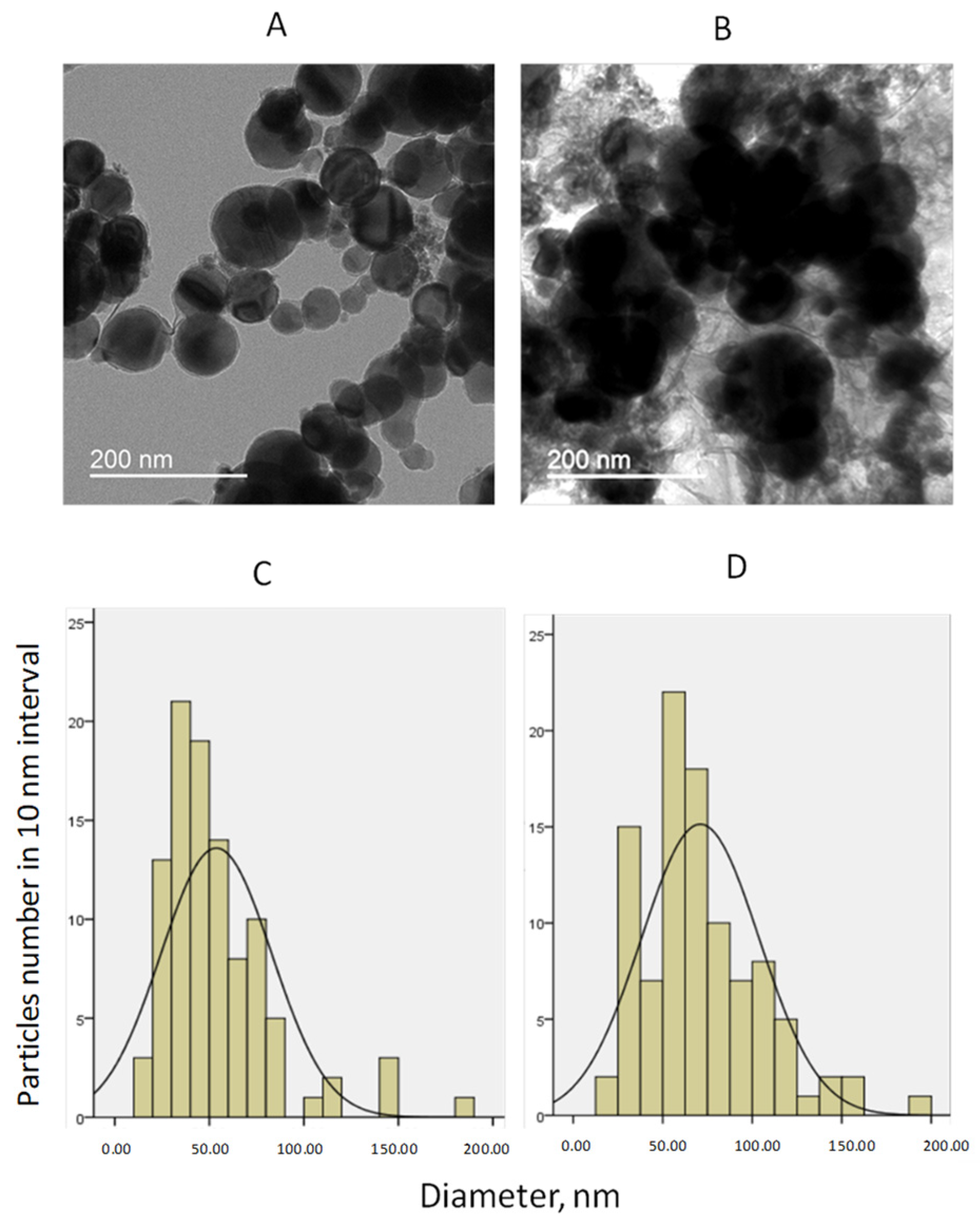
As may be seen from
Figure 1, NiNPs 1 and 2 preparation contained spherical particles with more than 99% Ni (according to study by electron energy dispersion spectrometry). Mean diameters of the particles in the preparations were equal to 53.7 ± 2.9 nm and 70.9 ± 3.3 nm; 55.5% of the particles in the NiNP1 and 24.0% in NiNP2 were less than 50 nm in diameter.
A blood biochemistry study showed that, as a result of oral exposure of animals to NiNP1, an increase in glucose, LDL, serum albumin and globulin fraction and a decrease in uric acid were noticed. The main change produced by Ni salt included a rise in triglycerides level. Corresponding changes in rats receiving NiNP2 were absent or less pronounced with the exception of protein fraction levels. Serum AlAT and AsAT activities in experimental groups stayed mainly within normal range (the results are presented in details in the poster presentation).
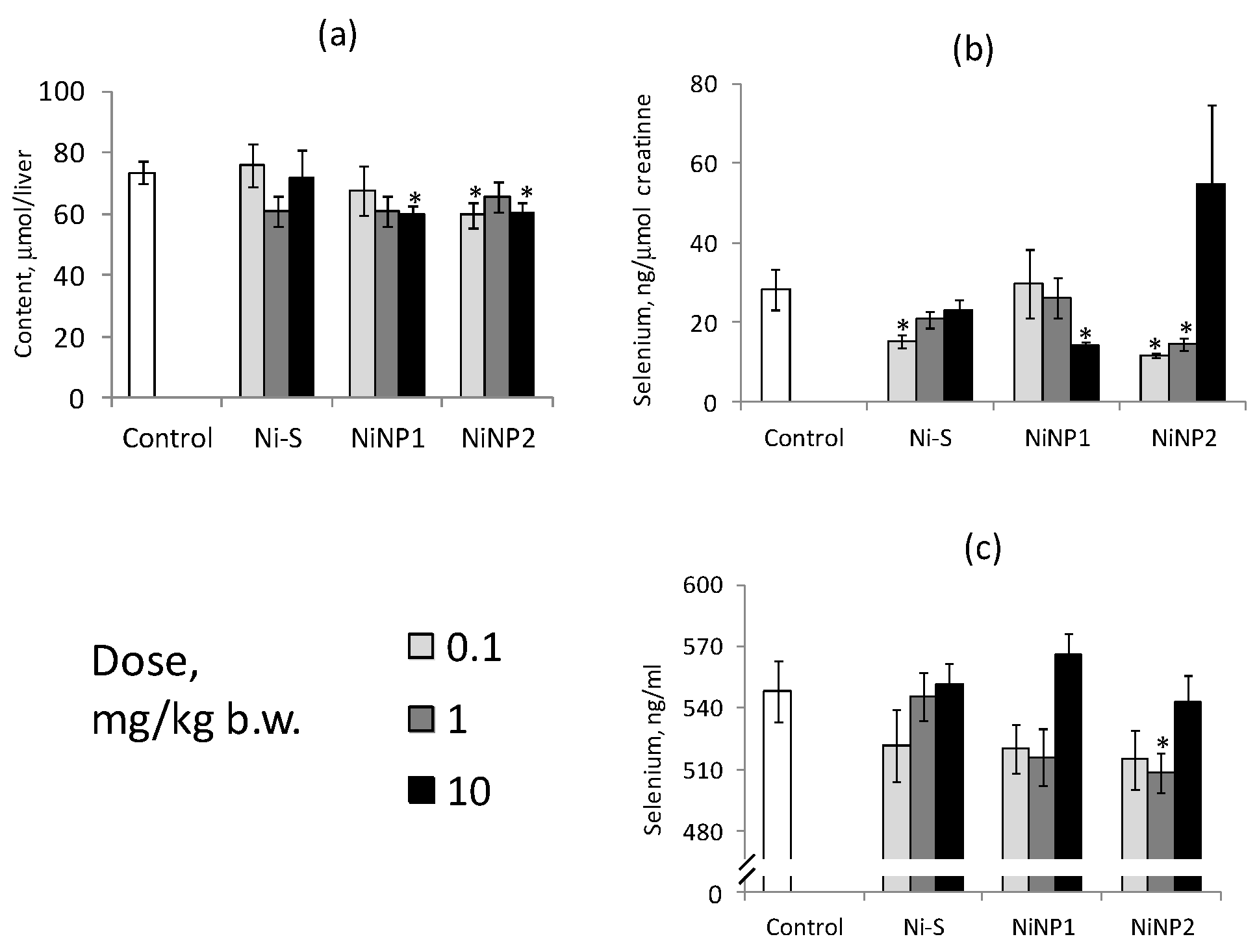
As may be seen from
Figure 2, the stores of reduced glutathione in liver and selenium reserves in rats subjected to NiNPs were significantly depleted and said effects were in some degree more pronounced than in Ni-salt-exposed animals.
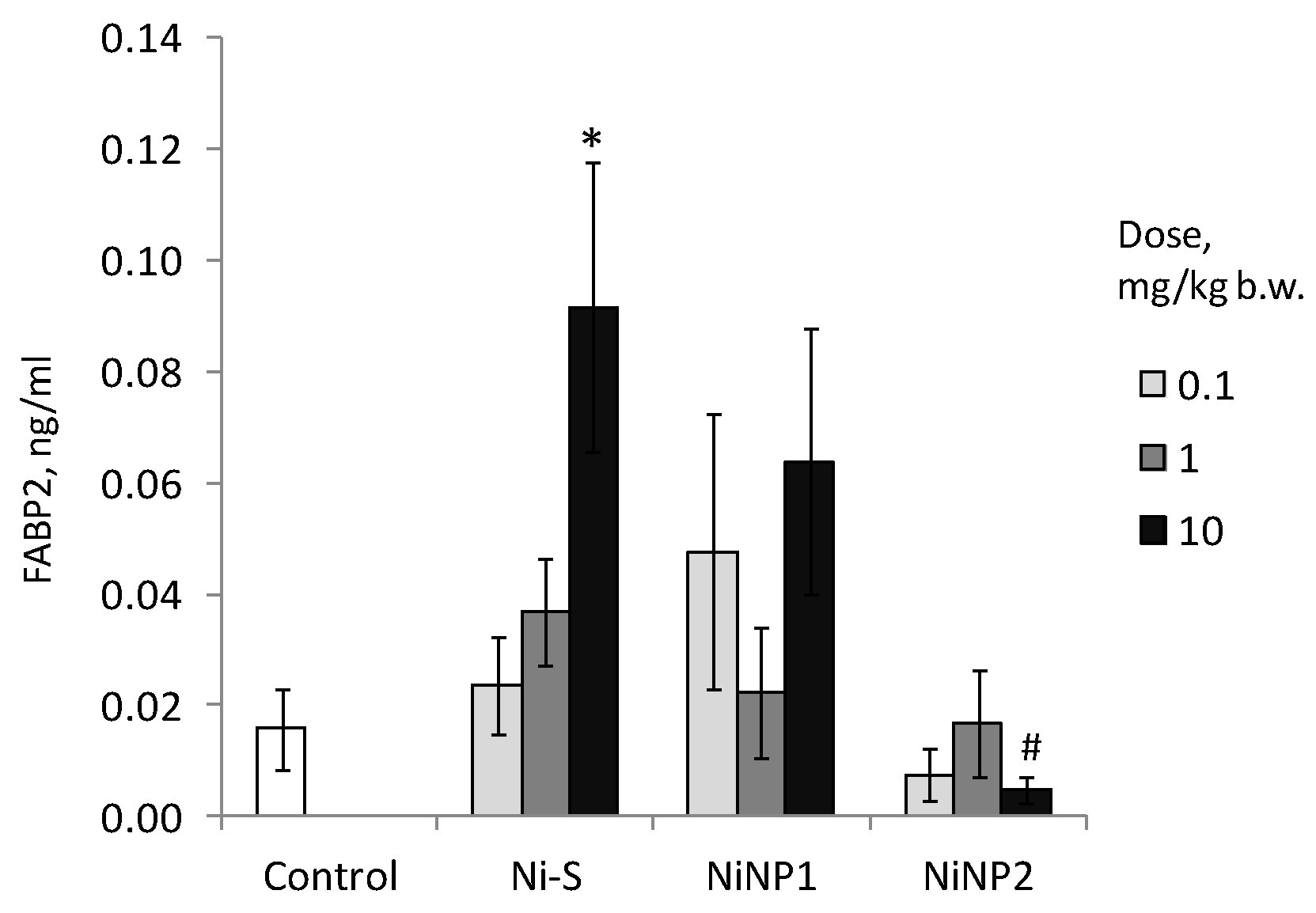
The increase of fatty acids binding protein FABP2 (
Figure 3) levels in Ni-salt-exposed animals suggests the presence of intestinal mucosal barrier violations in these animals. Corresponding effects were less pronounced in NiNP1-exposed rats and were eventually absent in NiNP2-exposed rats.
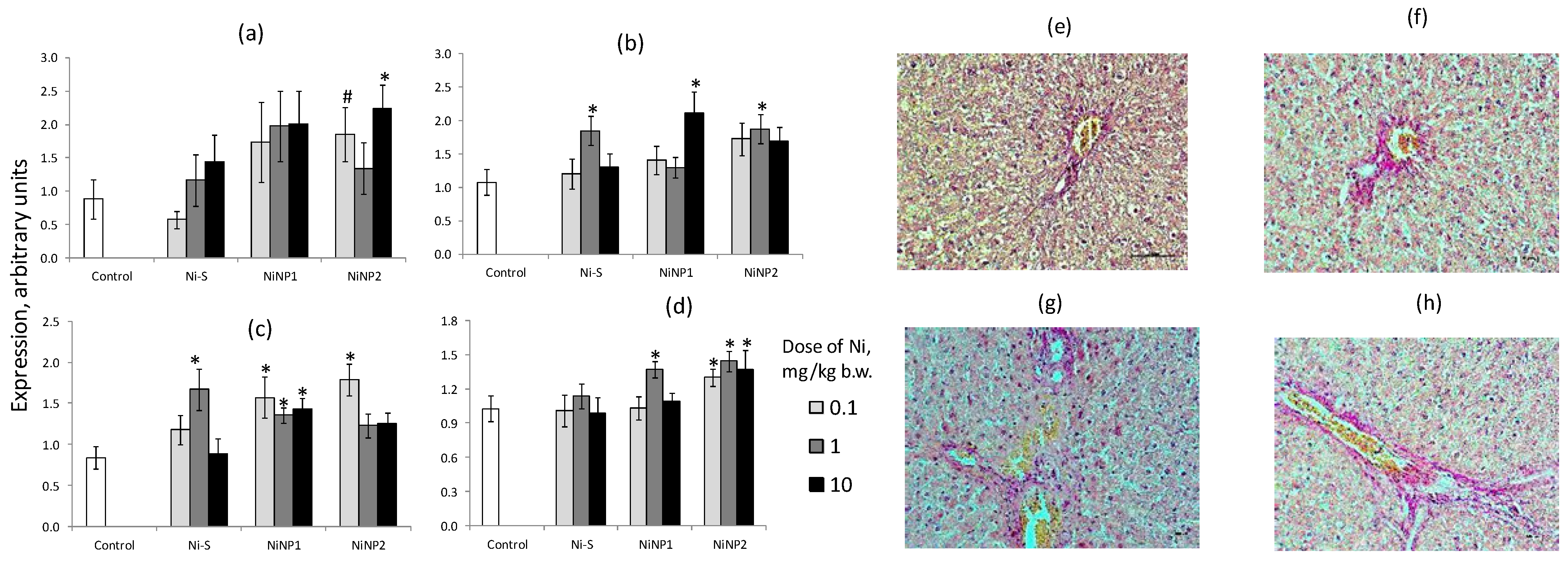
Oral exposure of rats to NiNP1 and NiNP2 resulted in an increase in fibrosis and apoptosis marker expression (
Figure 4). In rats receiving Ni salt, these changes were in some degree less pronounced. A light microscopy study of liver (van Gieson staining) revealed the accumulation of collagen elements in perivascular areas of tissue that was somewhat more pronounced in rats receiving Ni salt and especially NiNP1 and NiNP2.
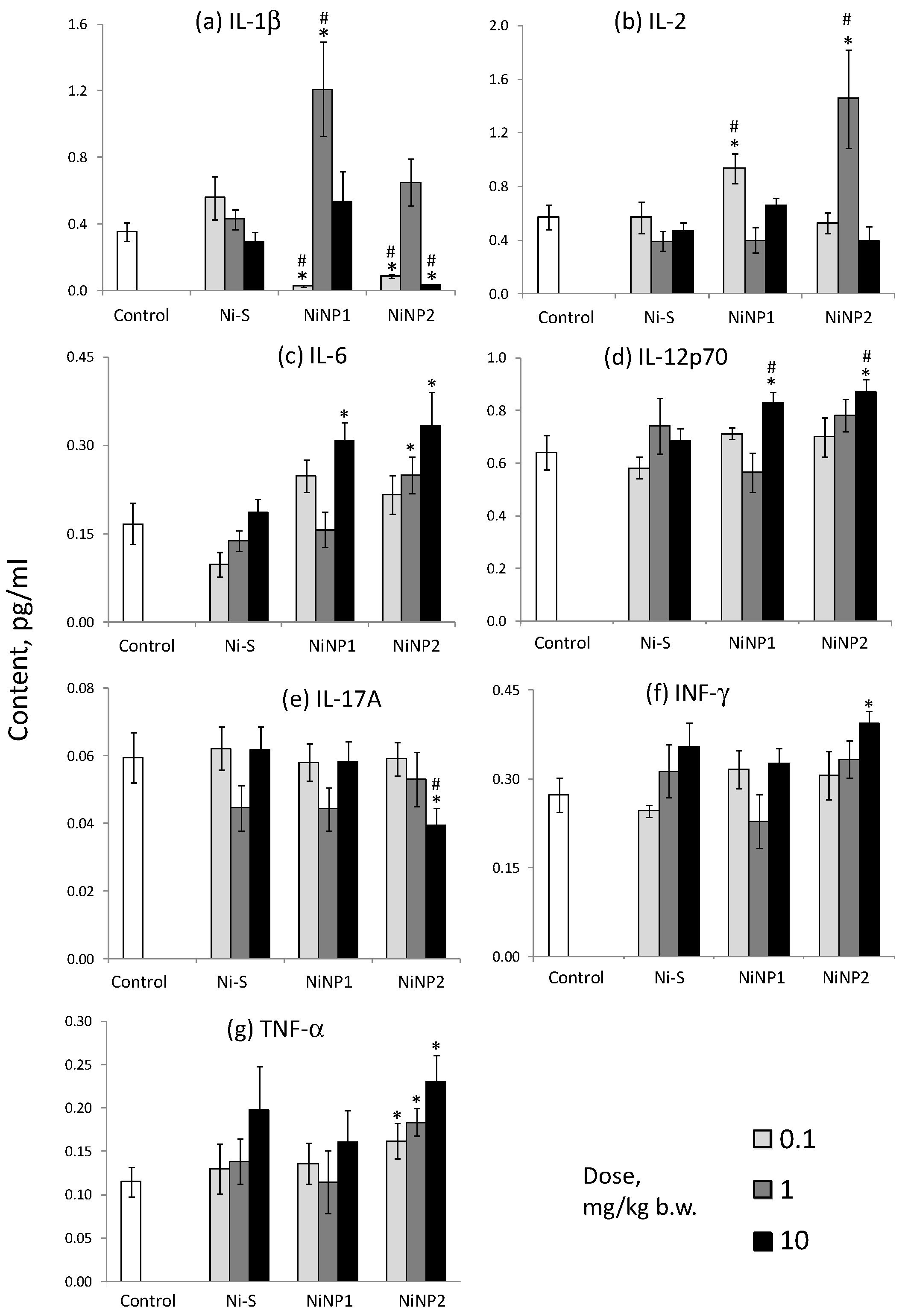
The study showed the complex nature of the response of cytokine levels to nickel NPs consumed by rats (
Figure 5). The maximum levels of IL-1β were achieved at the intermediate dose of Ni-NP1, and IL-2 at the smallest dose of Ni-NP1 and the intermediate dose of Ni-NP2. At the highest doses of both types of NPs, elevated levels of the pro-inflammatory cytokines IL-6 and IL-12p70 were observed, and in the case of Ni-NP2, also IFN-γ and TNF-α. The latter was accompanied by a decrease in the levels of IL-1β and IL-17A. Corresponding changes under the influence of an equivalent dose of nickel salt were absent or were less pronounced. The data obtained indicate the formation of a pro-inflammatory profile of cytokines in rats exposed to nickel NPs.
Morphological study of liver, small intestine and kidney of rats demonstrated changes in liver (increase of binucleated cells and cells with disintegrated nuclei, decrease in nucleus diameter), ileal mucosa (change in villous length and villous to crypt length ratio) and kidney (glomerular edema) of rats exposed to both Ni salt and two preparations of Ni-NPs (data on the morphological examination of organs are presented in the poster presentation).
4. Conclusions
The experimental data showed that the severity of the toxic effects of NiNPs depended on their size and, in some cases, were more pronounced at low or intermediate doses than at the highest one. Some manifestations of the toxic effects of NiNPs, including immunological endpoints (cytokine levels), blood biochemistry, and Timp3 and Tp53 expression, were absent or less pronounced in animals that were subjected to a soluble Ni salt in a metal-equivalent dose. We may conclude that the toxic action of NiNPs is mediated in general by the emission of Ni++ ions in biological environments, but in some degree, this may be influenced by the kinetic peculiarities of NP penetration through biological barriers compared to soluble Ni salt. The estimate for the LOAEL of NiNPs is less than 0.1 mg/kg of body weight according to the endpoints studied. This indicates the need to regulate the content of nickel in nanoforms in various types of products and the environment.
Author Contributions
Conceptualization, I.V.G., S.A.K. and V.A.S.; methodology, V.A.S.; software, I.V.G.; validation, I.V.G.; formal analysis, I.V.G.; investigation, N.A.R., N.V.T., A.S.B. and G.V.G.; resources, V.A.S.; data curation, I.V.G.; writing—original draft preparation, I.V.G.; writing—review and editing, S.A.K.; visualization, V.A.S.; supervision, S.A.K.; project administration, V.A.S.; funding acquisition, S.A.K. All authors have read and agreed to the published version of the manuscript.
Funding
The work was performed at the expense of subsidies from the Program of Basic Scientific Research (project of the Ministry of Science and Higher Education of the Russian Federation No. 0410-2022-0003).
Institutional Review Board Statement
The design of the experiment was approved by the Ethics Committee of the Federal Research Centre of Nutrition, Biotechnology and Food Safety (protocol No. 7 of 17 September 2021).
Informed Consent Statement
Not applicable.
Data Availability Statement
Data available on request due to restrictions, e.g., privacy or ethical.
Acknowledgments
The authors are grateful to Masyutin A.G. for TEM imaging of Ni-NPs.
Conflicts of Interest
The authors declare no conflict of interest.
References
- O’Brien, R.D. Fats and Oils: Formulating and Processing for Applications, 3rd ed.; CRC Press: Boca Raton, FL, USA, 2008; 680p. [Google Scholar]
- Ban, I.; Drofenik, S.J.; Makovec, M.D. Synthesis of copper-nickel nanoparticles prepared by mechanical milling for use in magnetic hyperthermia. J. Magn. Magn. Mater. 2011, 323, 2254–2258. [Google Scholar] [CrossRef]
- Elango, G.; Roopan, S.M.; Dhamodaran, K.I.; Elumalai, K.; Al-Dhabi, N.A.; Arasu, M.V. Spectroscopic investigation of biosynthesized nickel nanoparticles and its larvicidal, pesticidal activities. J. Photochem. Photobiol. B Biol. 2016, 162, 162–167. [Google Scholar] [CrossRef] [PubMed]
- Borowska, S.; Brzóska, M.M. Metals in cosmetics, implications for human health. J. Appl. Toxicol. 2015, 35, 551–752. [Google Scholar] [CrossRef]
- Katsnelson, B.A.; Privalova, L.I.; Sutunkova, M.P.; Gurvich, V.B.; Loginova, N.V.; Minigalieva, I.A.; Kireyeva, E.P.; Shur, V.Y.; Shishkina, E.V.; Beikin, Y.B.; et al. Some inferences from in vivo experiments with metal and metal oxide nanoparticles: The pulmonary phagocytosis response, subchronic systemic toxicity and genotoxicity, regulatory proposals, searching for bioprotectors, a self-overview. Int. J. Nanomed. 2015, 10, 3013–3029. [Google Scholar] [CrossRef] [PubMed] [Green Version]
- Siddiqui, M.A.; Ahamed, M.; Ahmad, J.; Khan, M.M.; Musarrat, J.; Al-Khedhairy, A.A.; Alrokayan, S.A. Nickel oxide nanoparticles induce cytotoxicity, oxidative stress and apoptosis in cultured human cells that is abrogated by the dietary antioxidant curcumin. Food Chem. Toxicol. 2012, 50, 641–647. [Google Scholar] [CrossRef] [PubMed]
- Saquib, Q.; Siddiqui, M.A.; Ahmad, J.; Ansari, S.M.; Faisal, M.; Wahab, R.; Alatar, A.A.; Al-Khedhairy, A.A.; Musarrat, J. Nickel oxide nanoparticles induced transcriptomic alterations in HEPG2 cells. Adv. Exp. Med. Biol. 2018, 1048, 163–174. [Google Scholar] [PubMed]
- Magaye, R.; Zhao, J. Recent progress in studies of metallic nickel and nickel-based nanoparticles’ genotoxicity and carcinogenicity. Environ. Toxicol. Pharmacol. 2012, 34, 644–650. [Google Scholar] [CrossRef] [PubMed]
- Nishi, K.I.; Kadoya, C.; Ogami, A.; Oyabu, T.; Morimoto, Y.; Ueno, S.; Myojo, T. Changes over time in pulmonary inflammatory response in rat lungs after intratracheal instillation of nickel oxide nanoparticles. J. Occup. Health 2020, 62, e12162. [Google Scholar] [CrossRef] [PubMed]
| Publisher’s Note: MDPI stays neutral with regard to jurisdictional claims in published maps and institutional affiliations. |
© 2022 by the authors. Licensee MDPI, Basel, Switzerland. This article is an open access article distributed under the terms and conditions of the Creative Commons Attribution (CC BY) license (https://creativecommons.org/licenses/by/4.0/).


 ,
, 


