Proceeding Paper

Synthesis, Characterization, and Functionalization of Graphene Oxide-Based Nanoplatforms for Gene Delivery †

1
Department of Chemical Engineering, Universidad de Los Andes, Cra. 1E No. 19a-40, Bogotá, 111711, Colombia
2
Department of Biomedical Engineering, Universidad de Los Andes, Cra. 1E No. 19a-40, Bogotá, 111711, Colombia
3
School of Chemical Engineering and Advanced Materials, The University of Adelaide, Adelaide 5005, Australia
*
Author to whom correspondence should be addressed.
Presented at the 2nd International Online-Conference on Nanomaterials, 15–30 November 2020; Available online: https://iocn2020.sciforum.net/.
Mater. Proc. 2021, 4(1), 23; https://doi.org/10.3390/IOCN2020-07925
Published: 11 November 2020
(This article belongs to the Proceedings of The 2nd International Online-Conference on Nanomaterials)

Abstract

:
Gene therapy has been considered a promising strategy for treating several inherited diseases and acquired complex disorders. One crucial challenge yet to be solved to ensure the nanomaterials’ success in delivering gene therapies is their ability to escape from endosomes. To address this issue, we previously developed magnetite nanoparticles conjugated with the antimicrobial peptide Buforin II, which showed potent translocating and endosomal escape abilities in several cell lines. In this work, we propose developing new cell-penetrating nanoplatforms by interfacing graphene oxide (GO) with powerful translocating peptides to take advantage of already tested and unique peptides as well as the distinctive interactions of GO with the phospholipids of membranes and endosomes. GO was prepared by the modified Hummers’ method through the oxidation of graphite sheets. Next, the functionalization of GO was carried out by rendering pendant amine groups to the GO surface. Thermogravimetric analysis (TGA) and Fourier-transform infrared spectroscopy (FTIR) were used to corroborate the successful functionalization of the nanoplatform. FTIR analysis exhibited the peaks related to the distinct carboxyl groups of GO and the Si–O bonds after silanization. TGA allowed us to estimate a silanization efficiency of 38%. Future work will be focused on conjugating Buforin II and assessing translocation efficiency by conducting uptake assays in liposomes and various cell lines. Additionally, endosomal escape will be determined via confocal microscopy by labeling the peptide with fluorescent molecules and examining colocalization with the fluorescent marker of endosomes, LysoTracker. By taking advantage of the exceptional qualities in terms of the physicochemical, electrical, and optical properties of GO, this study might provide novel strategies to overcome limitations commonly faced, such as low stability of the translocating biomolecules and endosomal entrapment.

1. Introduction

Gene therapy has been projected as a promising strategy to treat several genetic disorders and acquired diseases such as autosomal or X-linked recessive single-gene disorders, including cystic fibrosis (C.F.), adenosine deaminase deficiency (ADA), emphysema, retinitis pigmentosa, sickle cell anemia, phenylketonuria, hemophilia, and Duchenne muscular dystrophy (DMD) [1]. Apart from such severe conditions, recent studies have suggested promising treatments for some autosomal dominant disorders, polygenic disorders, different forms of cancers, vascular diseases, neurodegenerative disorders, and inflammatory conditions, among many others [2]. Gene therapy consists of replacing a distorted gene with a healthy one, or completing a missing gene to express the required functional protein [3].
Despite potential findings for both in vivo and ex vivo, a significant challenge that remains to be overcome is the efficient delivery of the therapies. Only a small part of the delivered genetic cargo reaches the target site [4]. Therefore, there is a need to develop efficient delivery systems. In line with this, viral and non-viral vectors are current approaches to address the issue related to efficient delivery [1,2]. Viral vectors have demonstrated superior efficiencies for the delivery of genetic material, but their failure to comply with biosafety requirements has been considered problematic by regulatory bodies [5]. As a result, non-viral vectors, particularly nanostructured systems, have gained more attention during the last few years due to their lower immune responses, inexpensive preparation, and advantageous properties for assembling customizable and modular gene delivery nanovehicles [6]. A significant hurdle yet to be solved for these systems is their ability to escape from endosomes, which are cell compartments that entrap therapeutic molecules after their internalization and eventually degrade them due to the reduction in pH and the action of several enzymes [7].
Recently, we have conjugated magnetic nanoparticles with the antimicrobial peptide Buforin II. Interestingly, the obtained nanobioconjugates exhibited potent translocating and endosomal escape abilities in several cell lines [8]. Thus, the purpose of this work is to extend this approach to other families of nanomaterials to ultimately obtain an ampler variety of platforms available for a rational design of cell-penetrating vehicles to address the need for a more comprehensive set of tissues and diseases. Carbon-based nanomaterials have been considered as an exciting family to be explored [9]. Mainly, graphene oxide (GO) has recently emerged as an attractive material for gene and drug delivery because of its high surface area and electrical and optical properties [10]. In addition to these benefits, recent reports have demonstrated important advances in the large-scale and high-throughput production of GO with controlled thickness [11,12,13]. Consequently, this work pursues to put forward a new family of cell-penetrating systems based on GO-based nanoplatforms by interfacing GO with potent translocating peptides.

2. Materials and Methods

GO was synthesized by following the modified Hummers’ method [14], which involves the oxidation of graphite sheets as schematized in Figure 1A. Such oxidation was conducted by mixing 0.75 g of graphite sheets and 4.5 g of KMnO4 in a solution containing H2SO4 (90 mL) and H3PO4 (10 mL). The reaction mixture was stirred for 12 h at 50 °C. The oxidation was then quenched by rapidly cooling down the mixture to room temperature, followed by the addition of H2O2 for filtering later. Next, exfoliation of GO was carried out to obtain a solid that was cyclically centrifugated at 4000 rpm for 4 h, resuspended in an aqueous solution of HCl and ethanol, and filtered out. Finally, the as-prepared GO was lyophilized and stored at 4 °C until further use.
The functionalization process was conducted by mixing 100 mg of an aqueous solution of GO with 2 mL of tetramethylammonium hydroxide (TMAH) 25% (v/v), 50 µL of pure acetic acid, and 1 mL of (3-Aminopropyl) triethoxysilane (APTES) 10% (v/v). This strategy was carried out to render free amine groups on the GO’s surface, as shown in Figure 1B. Next, to remove unused reagents, the resulting mixture was centrifugated and resuspended in distilled water. Finally, functionalized GO (fGO) was lyophilized and stored at 4 °C until further use.
Fourier-transform infrared spectroscopy (FTIR) and thermogravimetric analysis (TGA) were used to confirm surface modification effectiveness.

3. Results

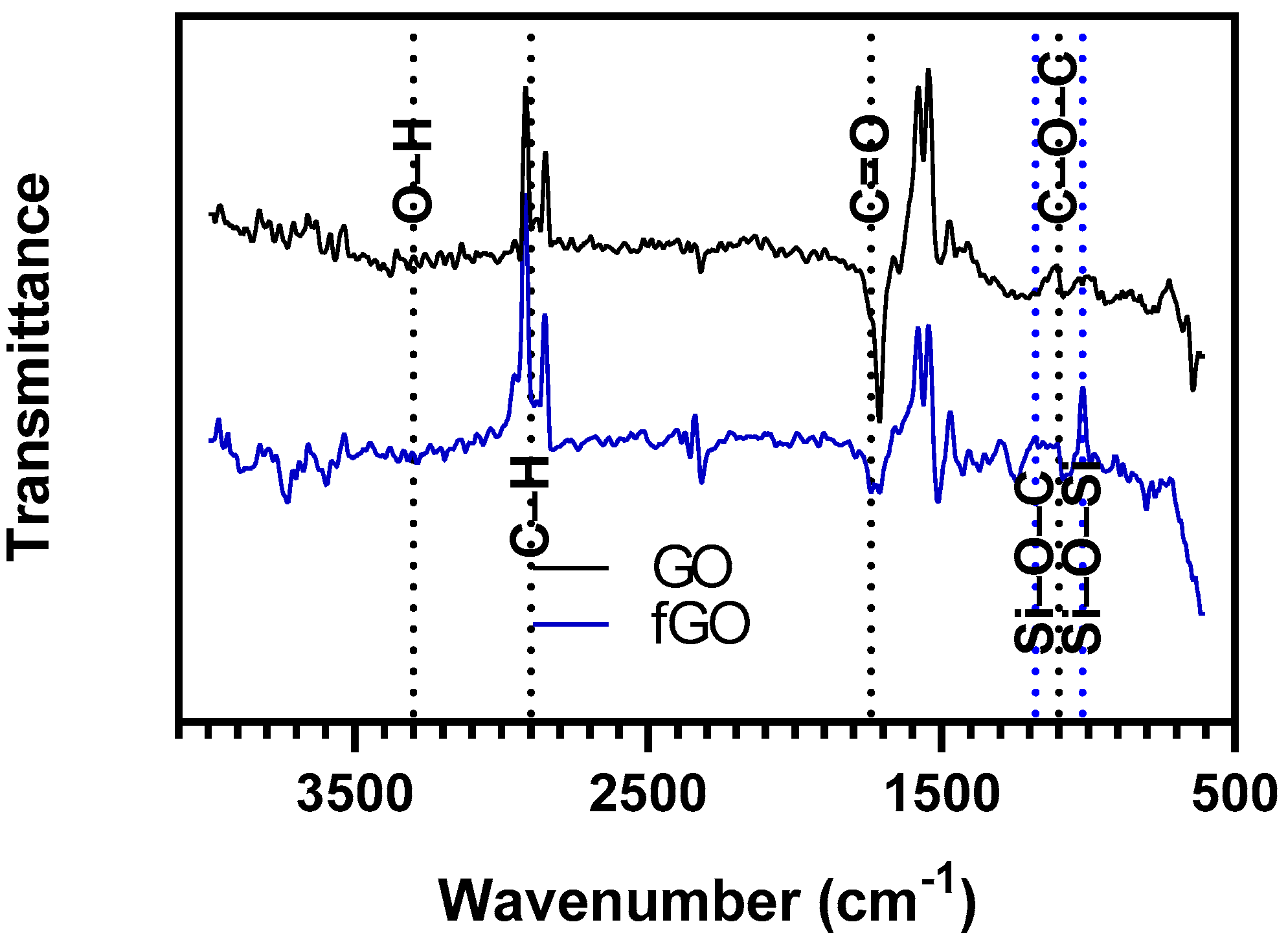
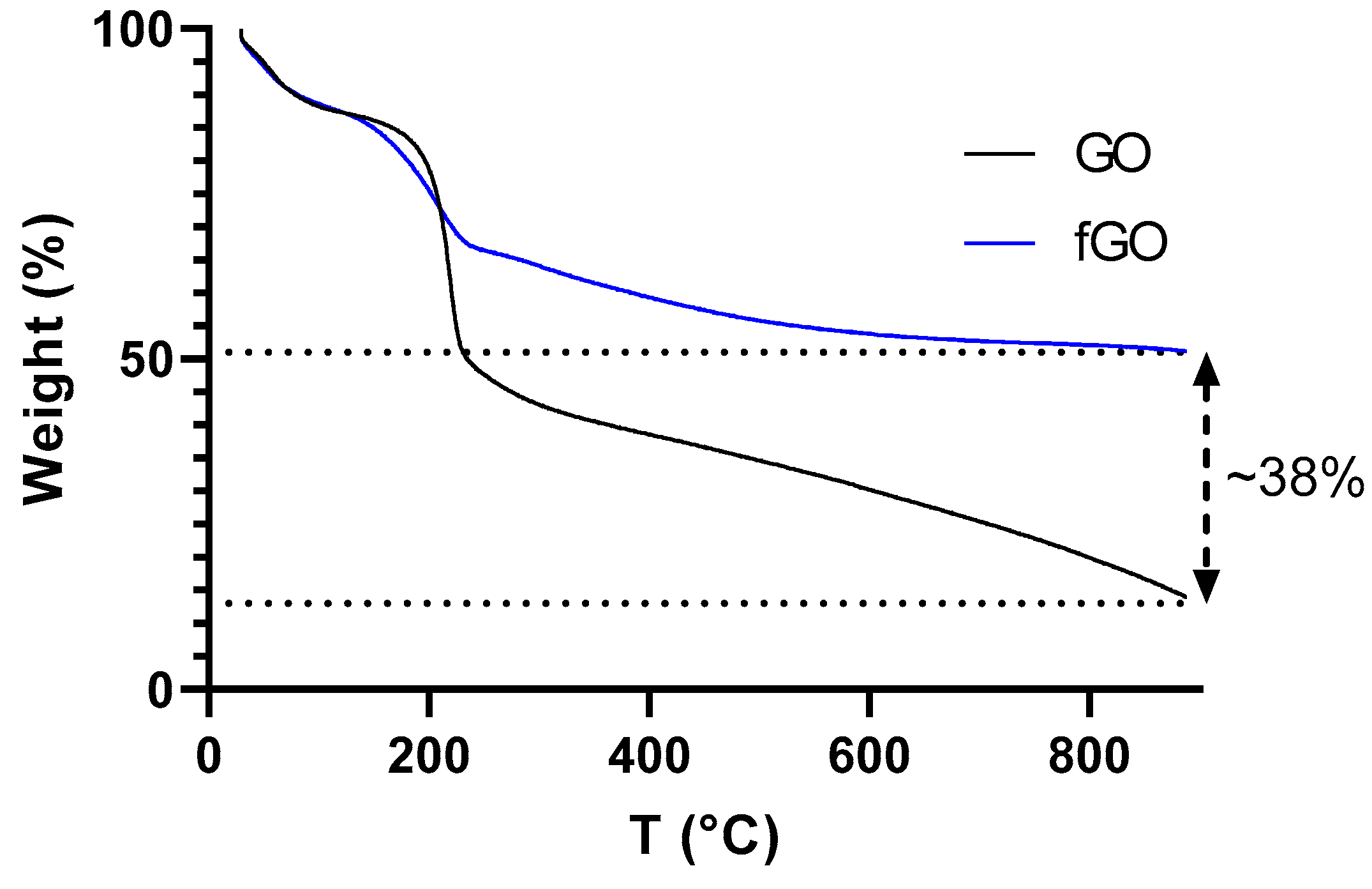
To demonstrate the process of rendering amine groups on the surface of GO, infrared spectra were obtained for GO (black line) and fGO (blue line) samples, as showed in Figure 2. The characteristic groups for GO were detected: the peak detected at about 3300 cm−1 is attributed to the O–H stretching vibrations of hydroxide groups [15]. Moreover, the bands observed at about 1100 cm−1 and 1750 cm−1 correspond to the stretching vibration of epoxy and the stretching vibration of carbonyl groups, respectively [16,17]. After functionalization, it was possible to observe bands at about 1020, 1180 and 1460 cm−1, attributed to the stretching vibration of Si–O–Si, stretching vibration of Si–O–C and stretching vibration of C–N bonds, respectively [16,18]. Furthermore, the efficiency of functionalization with APTES was estimated at 38% through TGA. Profiles of such analyses are shown in Figure 3.

4. Discussion

To achieve the successful delivery of cargoes, nanomaterials have often been superficially modified to improve their endosomal escape abilities. In this work, the GO surface has been interfaced with the organosilane APTES to render free amine groups. This was confirmed qualitatively through FTIR analysis. Furthermore, it was possible to estimate the functionalization efficiency by comparing the TGA profiles for GO and fGO. This efficiency is comparable to previous reports pursuing different applications such as lead sequestration [19] and protection from corrosive environments [20].
Since we are particularly interested in delivering gene therapies, future work will focus on conjugating Buforin II and evaluating translocation efficiency by conducting uptake experiments in liposomes and various cell lines. We will also determine endosomal escape via confocal microscopy by labeling the peptide with fluorescent molecules and examining colocalization with the fluorescent probe of endosome compartments, LysoTracker.
This work is expected to provide an avenue for developing gene delivery systems based on GO-based nanoplatforms. Moreover, by taking advantage of the great qualities in terms of the physicochemical, electrical, and optical properties of GO, this study might provide novel strategies to overcome limitations commonly faced by drug delivery nanocarriers, such as low stability of the translocating biomolecules and endosomal entrapment.

Author Contributions

Conceptualization, J.C.C. and L.H.R.; methodology, data curation, and data analysis J.D.T.; formal analysis and investigation, J.D.T.; validation, J.C.C. and L.H.R.; writing—original draft preparation, J.D.T.; writing—review and editing, J.C.C. and L.H.R.; supervision, J.C.C. and L.H.R. All authors have read and agreed to the published version of the manuscript.

Funding

This work was funded by the Colombian Ministry of Science, Technology, and Innovation (Minciencias) Grant ID 120380763212–PPTA # 8352.

Institutional Review Board Statement

Not applicable

Informed Consent Statement

Not applicable

Data Availability Statement

No new data were created or analyzed in this study. Data sharing is notapplicable to this article.

Acknowledgments

The authors would like to thank the Department of Biomedical Engineering, Food and Chemical Engineering and the Department of Electrical & Electronics Engineering at Universidad de los Andes for the financial and technical support.

Conflicts of Interest

The authors declare no conflict of interest.

References

  1. Nayerossadat, N.; Ali, P.A.; Maedeh, T. Viral and nonviral delivery systems for gene delivery. Adv. Biomed. Res. 2012, 1, 27. [Google Scholar] [CrossRef] [PubMed]
  2. Ibraheem, D.; Elaissari, A.; Fessi, H. Gene therapy and DNA delivery systems. Int. J. Pharm. 2014, 459, 70–83. [Google Scholar] [CrossRef] [PubMed]
  3. Ebara, M.; Uto, K. Gold Nanomaterials for Gene Therapy; Elsevier Ltd.: Amsterdam, The Netherlands, 2016. [Google Scholar]
  4. Mady, M.M. Cationic liposomes as gene delivery system. Afr. J. Pharm. Pharmacol. 2011, 5, 2007–2012. [Google Scholar]
  5. Shirley, J.L.; de Jong, Y.P.; Terhorst, C.; Herzog, R.W. Immune Responses to Viral Gene Therapy Vectors. Mol. Ther. 2020, 28, 709–722. [Google Scholar] [CrossRef] [PubMed]
  6. Malmsten, M. Inorganic nanomaterials as delivery systems for proteins, peptides, DNA, and siRNA. Curr. Opin. Colloid Interface Sci. 2013, 18, 468–480. [Google Scholar] [CrossRef]
  7. Hillaireau, H.; Couvreur, P. Nanocarriers’ entry into the cell: Relevance to drug delivery. Cell. Mol. Life Sci. 2009, 66, 2873–2896. [Google Scholar] [CrossRef] [PubMed]
  8. Cuellar, M.; Cifuentes, J.; Perez, J.; Suarez-Arnedo, A.; Serna, J.A.; Groot, H.; Muñoz-Camargo, C.; Cruz, J.C. Novel BUF2-magnetite nanobioconjugates with cell-penetrating abilities. Int. J. Nanomed. 2018, 13, 8087–8094. [Google Scholar] [CrossRef] [PubMed]
  9. Yin, F.; Gu, B.; Lin, Y.; Panwar, N.; Tjin, S.C.; Qu, J.; Lau, S.P.; Yong, K.-T. Functionalized 2D nanomaterials for gene delivery applications. Coord. Chem. Rev. 2017, 347, 77–97. [Google Scholar] [CrossRef]
  10. Rahmanian, N.; Eskandani, M.; Barar, J.; Omidi, Y. Recent trends in targeted therapy of cancer using graphene oxide-modified multifunctional nanomedicines. J. Drug Target. 2016, 25, 202–215. [Google Scholar] [CrossRef] [PubMed]
  11. De Melo-Diogo, D.; Lima-Sousa, R.; Alves, C.G.; Costa, E.C.; Louro, R.O.; Correia, I.J. Functionalization of graphene family nanomaterials for application in cancer therapy. Colloids Surf. B Biointerfaces 2018, 171, 260–275. [Google Scholar] [CrossRef] [PubMed]
  12. Liu, P.; Wang, S.; Liu, X.; Ding, J.; Zhou, W. Platinated graphene oxide: A nanoplatform for efficient gene-chemo combination cancer therapy. Eur. J. Pharm. Sci. 2018, 121, 319–329. [Google Scholar] [CrossRef] [PubMed]
  13. Marcano, D.C.; Kosynkin, D.V.; Berlin, J.M.; Sinitskii, A.; Sun, Z.; Slesarev, A.; Alemany, L.B.; Lu, W.; Tour, J.M. Improved synthesis of graphene oxide. ACS Nano 2010, 4, 4806–4814. [Google Scholar] [CrossRef] [PubMed]
  14. Zawisza, B.; Baranik, A.; Malicka, E.; Talik, E.; Sitko, R. Preconcentration of Fe(III), Co(II), Ni(II), Cu(II), Zn(II) and Pb(II) with ethylenediamine-modified graphene oxide. Microchim. Acta 2016, 183, 231–240. [Google Scholar] [CrossRef] [PubMed]
  15. Bouazizi, N.; Vieillard, J.; Bargougui, R.; Couvrat, N.; Thoumire, O.; Morin, S.; Ladam, G.; Mofaddel, N.; Brun, N.; Azzouz, A.; et al. Entrapment and stabilization of iron nanoparticles within APTES modified graphene oxide sheets for catalytic activity improvement. J. Alloys Compd. 2019, 771, 1090–1102. [Google Scholar] [CrossRef]
  16. Leaper, S.; Abdel-Karim, A.; Faki, B.; Luque-Alled, J.M.; Alberto, M.; Vijayaraghavan, A.; Holmes, S.M.; Szekely, G.; Badawy, M.I.; Shokri, N.; et al. Flux-enhanced PVDF mixed matrix membranes incorporating APTS-functionalized graphene oxide for membrane distillation. J. Memb. Sci. 2018, 554, 309–323. [Google Scholar] [CrossRef]
  17. Shahidi, F.; Arachchi, J.K.V.; Jeon, Y.J. Food applications of chitin and chitosans. Trends Food Sci. Technol. 1999, 10, 37–51. [Google Scholar] [CrossRef]
  18. Sun, W.; Wang, L.; Wu, T.; Wang, M.; Yang, Z.; Pan, Y.; Liu, G. Inhibiting the corrosion-promotion activity of graphene. Chem. Mater. 2015, 27, 2367–2373. [Google Scholar] [CrossRef]
  19. Diagboya, P.N.; Mmako, H.K.; Dikio, E.D.; Mtunzi, F.M. Synthesis of amine and thiol dual functionalized graphene oxide for aqueous sequestration of lead. J. Environ. Chem. Eng. 2019, 7, 103461. [Google Scholar] [CrossRef]
  20. Ramezanzadeh, B.; Ahmadi, A.; Mahdavian, M. Enhancement of the corrosion protection performance and cathodic delamination resistance of epoxy coating through treatment of steel substrate by a novel nanometric sol-gel based silane composite film filled with functionalized graphene oxide nanosheets. Corros. Sci. 2016, 109, 182–205. [Google Scholar] [CrossRef]
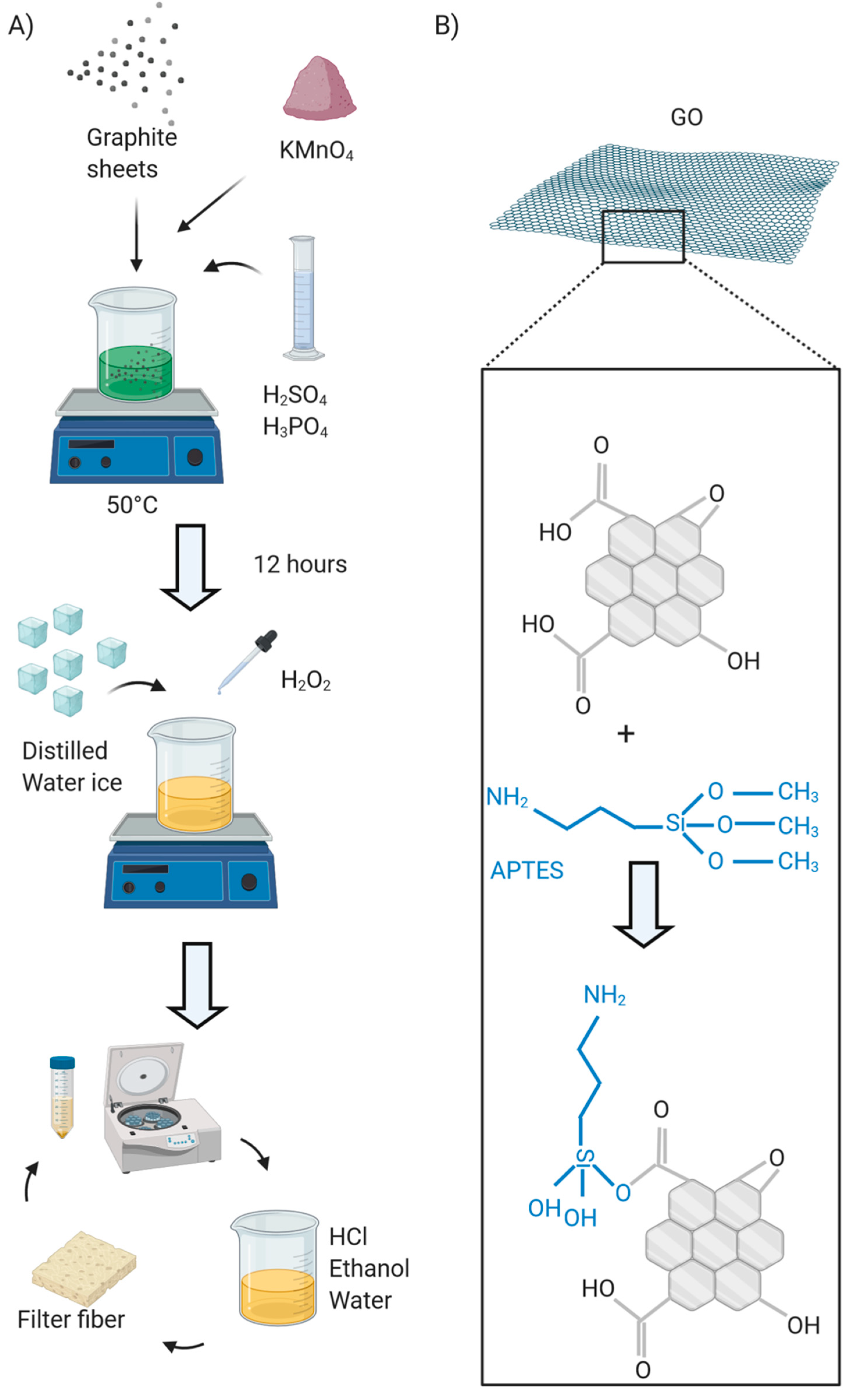
Figure 1. Scheme of the methodology conducted for the following: (A) The synthesis of graphene oxide (GO): The oxidation of graphite sheets was carried out through the action of KMnO4, H2SO4 and H3PO4. Once the oxidation step was quenched by cooling down the resulting mixture to room temperature followed by the addition of H2O2 for later filtering, the mixture was centrifugated to obtain a solid. Such solid was exfoliated by resuspending it in an aqueous solution of HCl and ethanol. Next, the exfoliation step was cyclically repeated by centrifuging, resuspending, and filtering. The protocol was based on the Hummer’s synthesis [14]; (B) functionalization of GO: GO as-synthesized was dissolved in an aqueous solution containing tetramethylammonium hydroxide (TMAH) (25% v/v), acetic acid, and (3-Aminopropyl) triethoxysilane (APTES) (10% v/v). The solution was stirred for one hour at room temperature. The protocol was based on recent studies conducted for magnetic nanoparticles [8].
Materproc 04 00023 g001
Figure 2. Infrared spectra for GO (black line) and functionalized graphene oxide (fGO) (blue line). After rendering amine groups on the GO’s surface, it was possible to observe characteristic peaks at 1020, 1180 and 1460 cm−1 attributed to expected bonds such as Si–O–Si, Si–O–C and C–N, respectively.
Materproc 04 00023 g002
Figure 3. Thermogravimetric analysis (TGA) profiles for GO (black line) and fGO (blue line). Within this temperature range, GO lost over 85% of its mass, while fGO lost about 50% of its mass. This indicates that the functionalization process increased the density-per-weight unit of the functionalized GO. In addition, the difference between both curves gives a measurement of how efficient the functionalization process was.
Materproc 04 00023 g003
Publisher’s Note: MDPI stays neutral with regard to jurisdictional claims in published maps and institutional affiliations.