1. Introduction
Physical, chemical, and biological methods were used for the synthesis of metal nanoparticles (MtNP). Physical and chemical methods for MtNP synthesis have many drawbacks, including the use of expensive equipment, high energy consumption, and the use of toxic chemicals, which pose an environmental problem [
1,
2,
3]. There has been a need for an environmentally friendly alternative to synthesizing MtNPs, the focus of which is the green synthesis of MtNPs using plants, microorganisms, enzymes, polysaccharides, and biodegradable polymers [
3,
4]. In the synthesis of nanoparticles, organism extracts can act as reducing agents as well as stabilizers. Innovative and diverse applications of MtNP in different fields of medical science, environmental science, and agriculture led to the accelerated development of different methods of synthesizing these compounds during recent years [
1,
5].
A silver nanoparticle (Ag NP), a stable, colloidal dispersion in water or organic solvents, is most prepared by chemical reduction in organic solvents or water. A plant extract can be used as a reducing agent and as a stabilizer (to prevent unwanted agglomeration of colloids) during nanoparticle synthesis [
6]. Nanoparticles of silver have unique physical, chemical, and biological properties. There is significant catalytic and antibacterial activity in these nanoparticles, as well as good potential for nanobiotechnological applications [
7].
Agrimonia eupatoria L. (common name:
agrimony) belongs to the family
Rosaceae (Tribe:
Sanguisorbeae). The plant is known for being used as a raw material for the extraction of medicinal ingredients or the production of medicines in the pharmaceutical industry. The plant not only has antioxidant and antibacterial properties but also anti-inflammatory, neuroprotective, antidiabetic, hepatoprotective, and anticancer properties [
8]. As part of our earlier research, silver nitrate, and the acetone extract of
A. eupatoria L. were used for the synthesis of silver nanoparticles [
9]. Our study examined the best conditions for the synthesis of silver nanoparticles from
A. eupatoria L. aqueous extracts.
2. Materials and Methods
Silver nitrate, sodium hydroxide, and nitric acid used were from Sigma Aldrich, USA. All solutions were prepared in distilled water. The aqueous extract of the plant was prepared according to a previously published procedure [
8]. Dried, crushed plant material was extracted in distilled water by maceration. In brief, 60 g of the plant material was soaked in 800 mL of the solvent. The plant material was macerated three times at room temperature using a fresh solvent every 24 h. After every 24 h, the samples were filtered through a filter paper, and the filtrates were collected and evaporated to dryness using a rotary evaporator (DLAB, RE 100 S) at 40 °C. All nanoparticle synthesis reactions were performed on a magnetic stirrer (MAGE 12/17) under controlled conditions. Monitoring the synthesis of AgNPs within the wavelength range of 200–800 nm was carried out using a Perkin Elmer Lambda365 spectrophotometer. A microcentrifuge DM0412 from Scilogek|Laboratory was used to centrifuge the suspension for 20 min at 4500 rpm. The data were analyzed using OriginPro 2019b-64bit software.
The optimal conditions for this green synthesis were examined: the concentration of starting substances, pH value, and temperature [
10]. Silver nitrate was dissolved in concentrations of 5 mM, 10 mM, and 20 mM. The pH of the reaction mixtures was adjusted to 4, 6, and 8 using solutions of 0.1 M NaOH and 0.1 M HNO
3. The reaction mixture was heated to 25 °C and 50 °C on a magnetic stirrer under controlled conditions. Visual color change (from light yellow to dark brown) and UV-Vis spectrophotometry were used to monitor the process of AgNP formation. The suspensions were centrifuged for 20 min at 4500 rpm after AgNPs synthesis. After centrifugation, the residue was resuspended in demineralized water and centrifuged again. Precipitated nanoparticles were then dried in a hot air oven (40 °C) and stored at 4 °C in the fridge.
3. Results
In this research study, we used silver nitrate and an aqueous A. eupatoria L. extract for the synthesis of silver nanoparticles (AgNPs).
3.1. UV-Vis Spectral Analysis
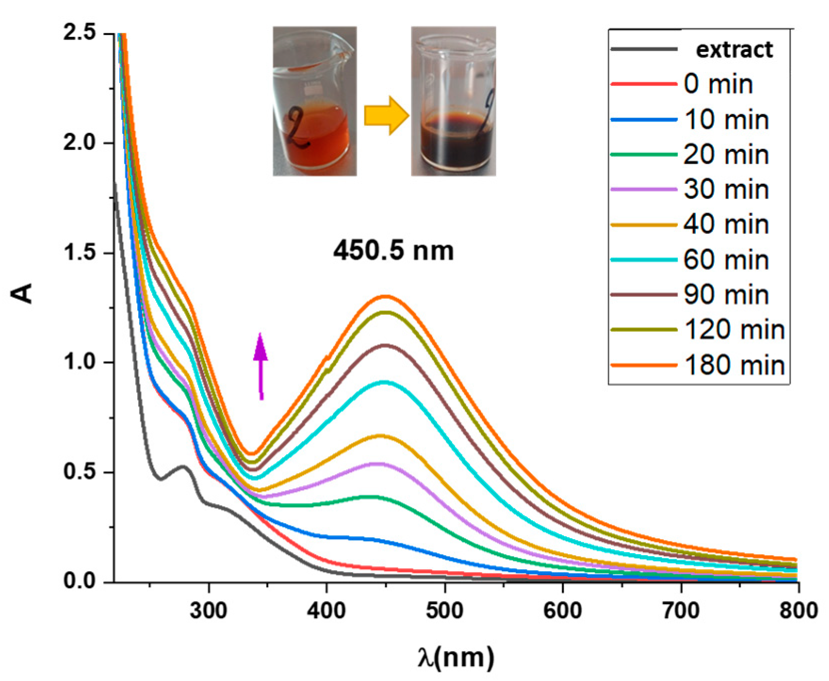
The generation of AgNPs in a solution during their synthesis using extracts was monitored spectrophotometrically. The color change in solutions from light yellow to dark brown is a characteristic indicator of the synthesized AgNPs. The color change is caused by surface plasmon resonance (SPR). We recorded the UV-Vis absorption spectra of formed nanoparticles at 200 to 800 nm. There were peaks within 425–475 nm (typical peak for AgNPs), indicating that AgNPs were formed. (
Figure 1). At 3 h, the maximum absorption values were obtained, and thereafter, there was no increase in absorption, indicating the end of the synthesis process. The effects of AgNO
3, temperature, and pH on the biosynthesis of nanoparticles using
A. eupatoria aqueous extracts were evaluated. Initially, both types of nanoparticles were synthesized at 5 mM AgNO
3, 25 °C and 1% extract concentration without adjusting pH values (pH ≈ 6).
3.2. Influence of Temperature
The starting point for testing the temperature sensitivity during the biosynthesis of nanoparticles was a concentration of 5 mM AgNO3, 1% of the concentrated plant extract without additional adjustment of the pH value. The reaction mixture was heated to 25 °C and 50 °C on a magnetic stirrer under controlled conditions. When the temperature increases to 50 °C, the rate of the formation of nanoparticles increases significantly.
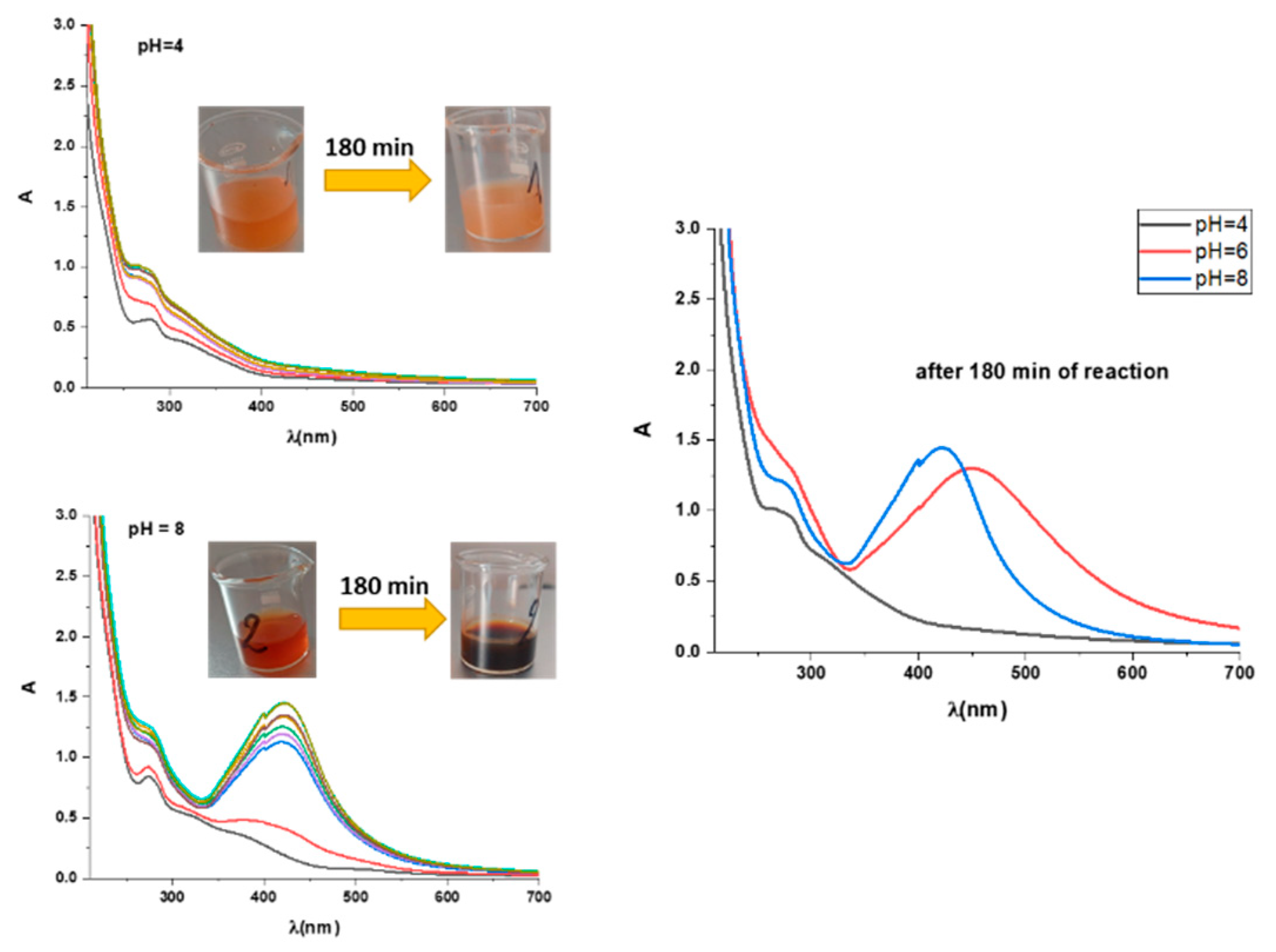
3.3. Influence of pH
The initial conditions for testing pH sensitivity were a concentration of 5 mM AgNO
3, a temperature of the reaction mixture of 25 °C, and a 1% concentration of the aqueous extract of the plant. To adjust the pH = 4, a few drops of 0.1 M HNO
3 solution were added to the mixture of extract solution and AgNO
3. The solution’s pH = 6 value was obtained by mixing a solution of extract and silver nitrate. To adjust pH = 8, a few drops of 0.1M NaOH solution were added.
Figure 2 shows the UV-Vis absorption spectra of AgNP biosynthesis depending on the change in the f value. Based on the obtained results, it can be concluded that pH = 6 is the most optimal value for the synthesis of AgNPs with the help of this plant.
3.4. Influence of Concentration
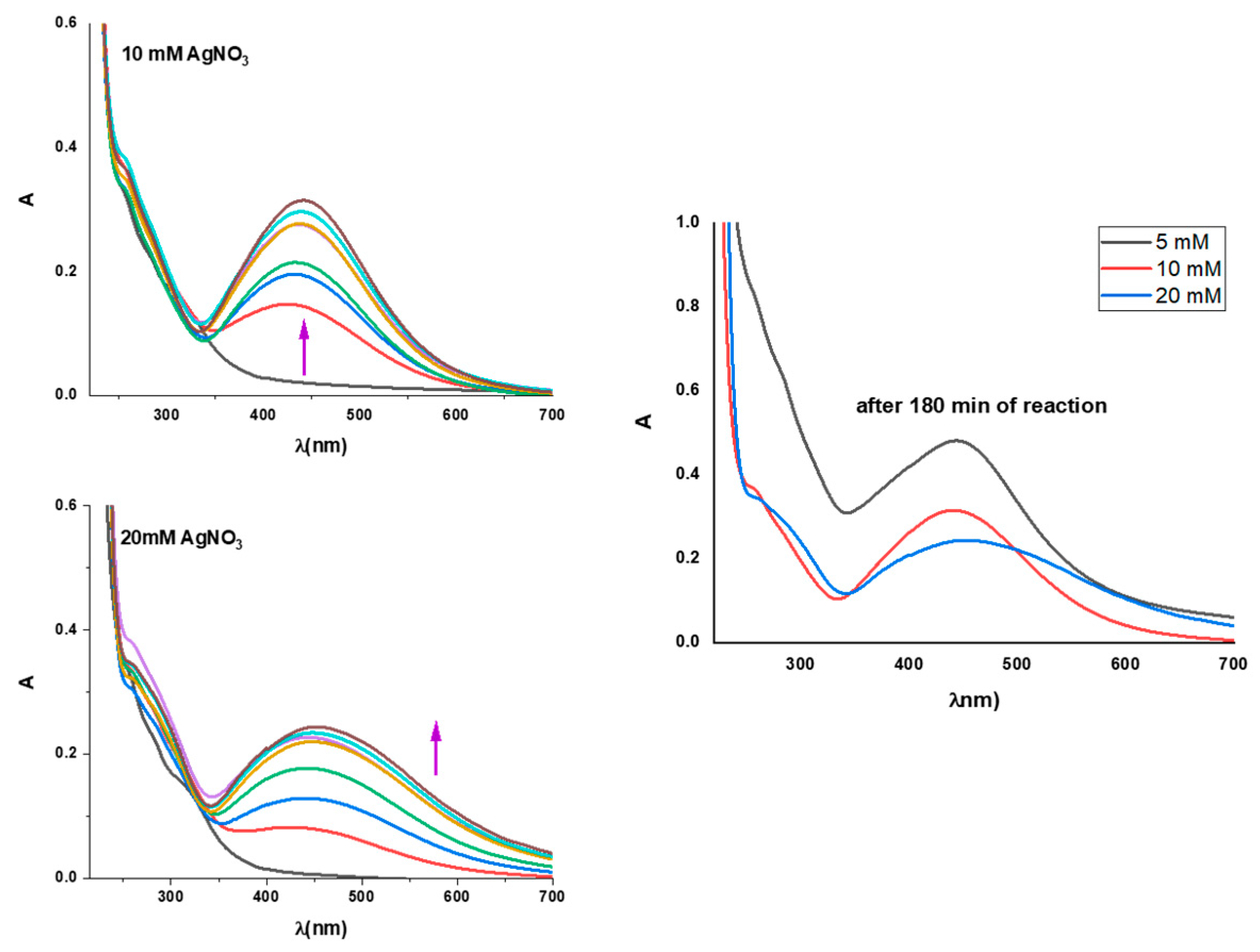
Initially, 25 °C, 1% extract concentration, and pH = 6 conditions were used to examine the dependence on the AgNO
3 concentration. Three extract solutions were prepared, to which AgNO
3 was added in concentrations of 5 mm, 10 mm, and 20 mm. As observed in
Figure 3, 5 mm AgNO
3 is the optimal concentration for AgNP synthesis.
4. Conclusions
According to the results of this research work, A. eupatoria is a good reducing agent and therefore a suitable plant for the green synthesis of silver nanoparticles. The plant’s aqueous extract is an effective reducer and stabilizer of nanoparticles. Silver nanoparticles are gradually synthesized, which is confirmed by the change in the color of the reaction solution from light yellow to dark brown and the change in the appearance of UV-Vis absorption spectra over time. When the temperature of the reaction mixture increases, the rate of biosynthesis of silver nanoparticles increases drastically. Based on the research on optimal pH values for this biosynthesis, it could be concluded that an acidic environment is more suitable, and the most optimal pH value is 6, which is achieved by simply mixing the starting substances. Based on the examination of this biosynthesis at different concentrations of the starting AgNO3 salt, it was concluded that the best concentration of AgNO3 is 5 mM. Finally, it can be concluded that the best conditions for obtaining the highest yield of AgNPs are as follows: AgNO3 initial salt concentration of 5 mM, a temperature of the reaction mixture of 25 °C, pH = 6, and duration of the nanoparticle biosynthesis reaction of 3 h.
Author Contributions
Conceptualization, Z.M. and A.K.; methodology, K.M.; software, M.G.; validation, Z.M., A.K. and K.M.; formal analysis, M.G.; investigation, A.K.; resources, K.M.; data curation, A.K.; writing—original draft preparation, A.K.; writing—review and editing, M.G.; visualization, A.K.; supervision, Z.M.; project administration, K.M.; funding acquisition, Z.M. All authors have read and agreed to the published version of the manuscript.
Funding
This research was funded by the Minister of Science, Technological Development, and Innovation of the Republic of Serbia (Agreement No. 451-03-47/2023-01/200378).
Institutional Review Board Statement
Not applicable.
Informed Consent Statement
Not applicable.
Data Availability Statement
Not applicable.
Acknowledgments
The authors would like to thank the Ministry of Science, Technological Development, and Innovation of the Republic of Serbia (Contract No. 451-03-47/2023-01/200378), the Institute for Information Technologies, and the University of Kragujevac for their support during the research.
Conflicts of Interest
The authors declare no conflict of interest.
References
- Gahlawat, G.; Choudhury, A.R. A review on the biosynthesis of metal and metal salt nanoparticles by microbes. RSC Adv. 2019, 9, 12944–12967. [Google Scholar] [CrossRef] [PubMed]
- Soni, M.; Mehta, P.; Soni, A.; Goswami, G.K. Green nanoparticles: Synthesis and applications. J. Biotechnol. Biochem. 2018, 4, 78–83. [Google Scholar]
- Pal, G.; Rai, P.; Pandey, A. Green synthesis of nanoparticles: A greener approach for a cleaner future. In Green Synthesis, Characterization and Applications of Nanoparticles; Elsevier: Amsterdam, The Netherlands, 2019; pp. 1–26. [Google Scholar]
- Roychoudhury, A. Yeast-mediated green synthesis of nanoparticles for biological applications. Indian J. Pharm. Biol. Res. 2020, 8, 26–31. [Google Scholar]
- Bahrulolum, H.; Nooraei, S.; Javanshir, N.; Tarrahimofrad, H.; Mirbagheri, V.S.; Easton, A.J.; Ahmadian, G. Green synthesis of metal nanoparticles using microorganisms and their application in the agrifood sector. J. Nanobiotechnol. 2021, 19, 86–126. [Google Scholar] [CrossRef] [PubMed]
- Sharma, V.K.; Yngard, R.A.; Lin, Y. Silver nanoparticles: Green synthesis and their antimicrobial activities. Adv. Colloid Interface 2009, 45, 83–96. [Google Scholar] [CrossRef] [PubMed]
- Srikar, S.K.; Giri1, D.D.; Pal1, D.B.; Mishra1, P.K.; Upadhyay, S.N. Green and Sustainable Chemistry. Green Sustain. Chem. 2016, 6, 34–56. [Google Scholar] [CrossRef]
- Muruzović, M.Ž.; Mladenović, G.K.; Stefanović, O.D.; Vasić, S.M.; Čomić, L.J.R. Extracts of Agrimonia eupatoria L. as sources of biologically active compounds and evaluation of their antioxidant, antimicrobial, and antibiofilm activities. J. Food Drug Anal. 2016, 24, 539–547. [Google Scholar] [CrossRef] [PubMed]
- Marković, G.K.; Grujović, Ž.M.; Kesić, S.A.; Markovic, S.Z. Green Synthesis of Silver Nanoparticles Using A. eupatoria extract in certain conditions. In Proceedings of the 8th International Electronic Conferences on Medicinal Chemistry (ECMC 2022), Online, 1–30 November 2022. [Google Scholar]
- Srećković, N.Z.; Nedić, Z.P.; Liberti, D.; Monti, D.M.; Mihailović, N.R.; Katanić Stanković, J.S.; Dimitrijević, S.; Mihailović, V.B. Application potential of biogenically synthesized silver nanoparticles using Lythrum salicaria L. extracts as pharmaceuticals and catalysts for organic pollutant degradation. RSC Adv. 2021, 11, 35585–35599. [Google Scholar] [CrossRef] [PubMed]
| Disclaimer/Publisher’s Note: The statements, opinions and data contained in all publications are solely those of the individual author(s) and contributor(s) and not of MDPI and/or the editor(s). MDPI and/or the editor(s) disclaim responsibility for any injury to people or property resulting from any ideas, methods, instructions or products referred to in the content. |
© 2023 by the authors. Licensee MDPI, Basel, Switzerland. This article is an open access article distributed under the terms and conditions of the Creative Commons Attribution (CC BY) license (https://creativecommons.org/licenses/by/4.0/).


