Proceeding Paper

A Novel Deep Learning Technique for Brain Tumor Detection and Classification Using Parallel CNN with Support Vector Machine †

Department of Electronics and Telecommunication Engineering, Chittagong University of Engineering and Technology (CUET), Chittagong 4349, Bangladesh
*
Authors to whom correspondence should be addressed.
Presented at the 11th International Electronic Conference on Sensors and Applications (ECSA-11), 26–28 November 2024; Available online: https://sciforum.net/event/ecsa-11.
Eng. Proc. 2024, 82(1), 101; https://doi.org/10.3390/ecsa-11-20505
Published: 26 November 2024

Abstract

Brain tumors (BT) are also known as intracranial diseases, which occur due to uncontrolled cell growth in the brain. Detecting and classifying the brain tumors at the initial stage is crucial to saving the patient’s life. A radiologist uses MRI scans to identify and classify the various types of BT using a manual approach. However, it is inaccurate and time-consuming because of the many images. In machine learning, convolutional neural networks (CNN) are one significant algorithm that can extract features automatically with high accuracy. The drawback of this algorithm is that it can extract features without knowing micro and macro features. The proposed architecture of parallel CNN (PCNN) can extract the features by knowing the micro and macro features from two separate window sizes and, at first, augmenting the normalized data using geometric transformation to enhance the number of images. Then, micro and macro features are extracted using the proposed architecture, PCNN, alongside batch normalization to reduce the overfitting problem. Finally, three kinds of tumors—glioma, meningioma, pituitary—and a no tumor condition are classified using various classifiers like Softmax, KNN, and SVM. The proposed PCNN-SVM obtained the best accuracy of 96.1% with the special features compared with the other pertained model.

1. Introduction

Of all the cancers, brain tumors are among the most deadly, with a high second-order fatality rate [1]. According to the Global Cancer Registry’s 2018 results, 29,681 of the 18,078,957 cancer patients with cancer had brain cancer [2]. There are two types of brain tumors: benign and malignant, depending on where they originate. Benign tumors begin in the brain and grow extremely slowly. Surgery is used to remove this kind of brain tumor. Malignant brain tumors, on the other hand, start elsewhere in the body and spread to the brain. Chemotherapy is the only treatment for this extremely severe condition [3,4].
Patients with brain tumors may experience headaches, seizures, visual nausea, vomiting, and cognitive and neurological abnormalities. Various methods depend on the images used to detect the brain tumor. Computed tomography (CT), positron emission tomography (PETR), magnetic resonance spectroscopy (MRS), magnetic resonance spectroscopy (MRS), and functional magnetic resonance imaging (fMRI) are the most often used imaging modalities [5]. The knowledge and proficiency of the radiologist are essential to all imaging modalities. Another diagnostic method for determining the presence of a brain tumor is a biopsy, which requires surgery [6]. These days, different forms of brain tumors can be recognized and categorized using machine learning algorithms.
Ahmadi et al. introduced a machine learning method that operates a convolutional neural network (CNN) and robust principal component analysis (PCA) built on brain tumor segmentation [7,8]. Ahmed Wasif Reza proposed a VGG-16 CNN architecture without augmented data, providing sufficient accuracy [9]. Mantripragada et al. presented a hybrid model built on deep neural networks (DNN) and adaptive fuzzy deformable fusion (AFDM) [10,11]. Pitcha et al. established a novel model using FKM and ANN based on the segmentation of brain tumors [12]. Mohsen et al. suggested an architecture where brain tumors are classified by deep neural networks (DNNs). Features are extracted by utilizing discrete wavelet transformation (DWT) and principal component analysis (PCA) with 66 MRIs of brain tumors [8,13]. Ahmed Saleh suggested a model where different types of brain tumors are detected and classified by AI algorithms, CNN, and deep learning. The datasets are trained by pre-trained models such as Xception, ResNet50, InceptionV3, VGG16, and MobileNet [14,15]. Yoon Kim et al. developed a word-level CNN-based model for sentence classification that used pre-trained word embeddings [16].
In machine learning, the lightweight CNN can extract features with a high degree of accuracy, but it cannot extract features that are aware of micro and macro features. This paper suggests a model, PCNN, to address this issue. It can extract features with high accuracy, recognize micro and macro features, and classify different kinds of brain tumors (BT) using multi-classifiers.
This paper presents the performance analysis in the methods and materials section in Section 2. The outcome of the proposed paper and comparison with other papers are discussed in the result analysis and discussion parts, which are presented in Section 3, and the conclusion of this architecture is discussed in the conclusion part, which is presented in Section 4.

2. Methods and Materials

In this paper, the various kinds of BT are classified using the proposed architecture, PCNN-SVM, as shown in Figure 1. The MRI images are divided into two parts: training (80%) and testing (20%). Depending on window widths, the feature extraction stages are divided into two sections: the micro feature selection pathway and the macro feature selection pathway. Classifiers are employed to categorize the many kinds of BT, like glioma, meningioma, and pituitary, as well as no tumor.

2.1. Dataset Description

We used the publicly accessible “Kaggle” dataset to determine the effectiveness of the suggested strategy [17]. Three types of BT—glioma, meningioma, and pituitary—as well as a no tumor condition are present in the dataset. The collection has 2870 pictures in total, of which 395 are of glioma, 826 are of meningioma, 822 are of no tumor, and 827 are of the pituitary.

2.2. Pre-Processing

The pre-processing step is divided into two sections: one is image reshaping, and another is image augmentation.

2.2.1. Image Reshaping

Before training the proposed technique, resizing images in the dataset is a common pre-processing step. Since the original images have different dimensions, scaling an image is necessary to enhance efficiency and remove computational constraints. The dataset’s images are downsized from (256 × 256) to (60 × 60) dimensions without losing any content in order to produce a simple calculation.

2.2.2. Image Augmentation

Image augmentation in machine learning uses geometric transformation techniques, including scaling, translation, and shear, to expand the number of images in the dataset to avoid overfitting issues. Data augmentation is used to reduce misclassification and overfitting problems.

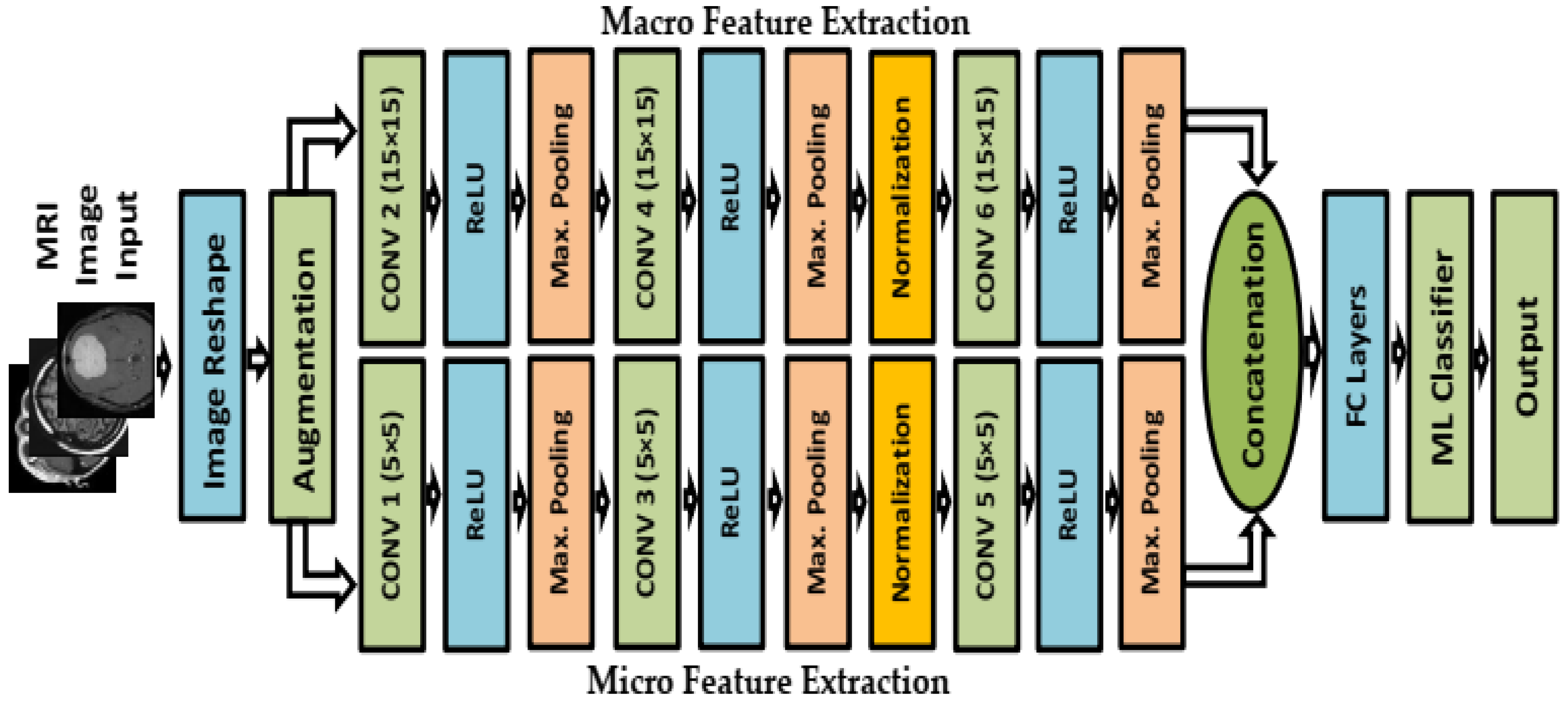
2.3. Feature Extraction Step

Currently, one of the most important machine learning techniques is convolutional neural networks (CNN). CNN can automatically extract features from MRI scans and classify them with outstanding performance. One significant drawback of the technique is CNN’s inability to extract unknown micro and macro features at random. To solve the issue, this study suggests using a lightweight deep parallel convolutional neural network (PCNN) with a two-pathway CNN to extract known micro and macro features to identify and categorize BT images. PCNN is composed of two pathways: a micro feature selection pathway and a macro feature selection pathway with 26 layers. The micro feature selection pathway has the same kernel size (5 × 5) with a 2D convolution layer. On the other hand, the macro feature selection pathway has the same kernel size (15 × 15) with a 2D convolutional layer, and the same stride and padding are used. A process of activation called ReLU is frequently used after the convolutional layer to improve efficiency and decrease gradient diffusion. Batch normalization is used to improve accuracy, decrease overfitting issues, speed up training, and increase stability. After that, maxpooling is used for downsampling the feature map. Two pathways are connected by a concatenation layer, and then a fully connected layer is used. Finally, ML classifiers are used to classify various types of BT, and PCNN_SVM provides the most prominent accuracy.

2.4. Classifier

One of the most popular supervised learning algorithms, the Support Vector Machine (SVM), can solve both regression and classification problems. The goal of the SVM algorithm is to determine the optimal line or decision boundary that can separate data points from different classes in the future very quickly. A hyperplane is the name of this best decision boundary [18]. The following Equation 1 defines the hyperplane function that can be used to separate the data. Where xi, represents the features from the dataset, ai is the Lagrange multiplier, and yi represents the dataset’s target class. The most often used kernel function of the algorithm is the linear kernel [19].
f x = s i g n ( i = 1 n a i y i K x , x i + b )

3. Experimental RESULT and Analysis

Brain tumors were detected and classified using modified PCNN using machine learning classifiers such as Softmax, KNN, and SVM. The studies were conducted using MATLAB 2023, and the lightweight PCNN-SVM model yielded the best results with epoch 30, learning rate.002, and optimizer sgdm. The specifications of the system include Windows 10 Home 64-bit, AMD Ryzen 5, and 8 GB of RAM.

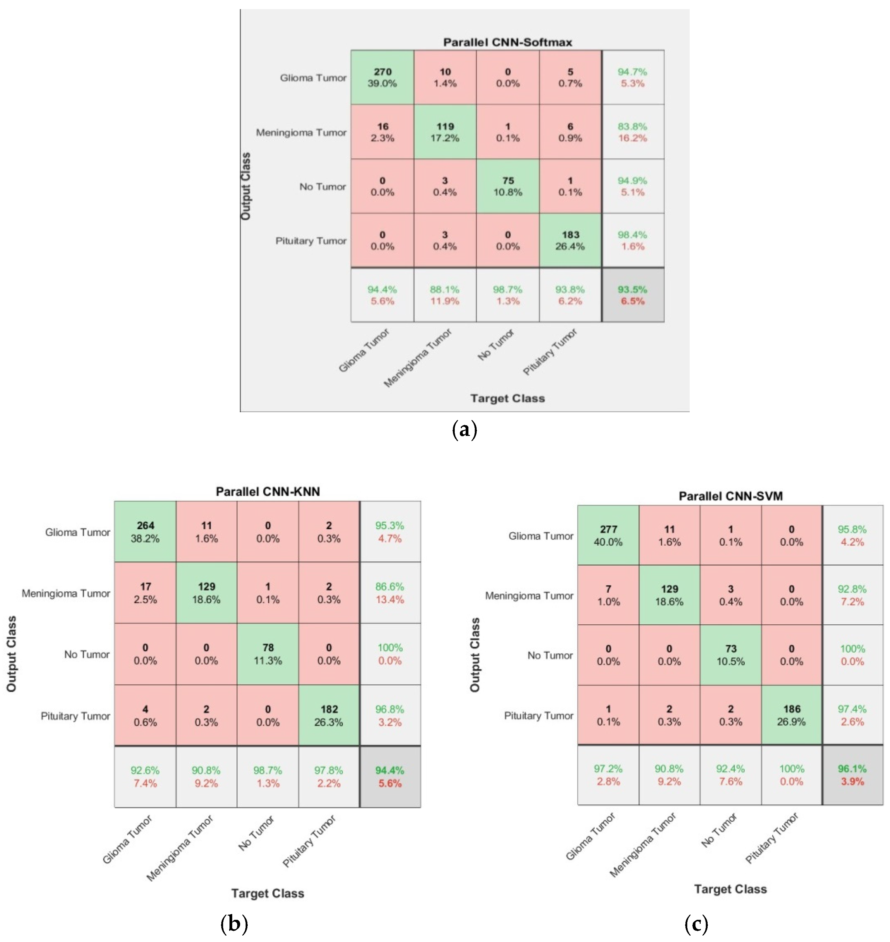
3.1. Confusion Matrix

A confusion matrix is a table that is operated to summarize the performance of a classification algorithm. A confusion matrix provides the values of true positive (TP), true negative (TN), false positive (FP), and false negative (FN). The aim of calculating a model is to increase the values of TP and TN and decrease the values of FP and FN by using a confusion matrix. The target classes appear on the X axis; on the other hand, the output classes appear on the Y axis. The proposed architecture provides the confusion matrix of PCNN-Softmax, PCNN-KNN, and PCNN-SVM, respectively, which are shown in Figure 2.

3.2. Performance Metrics

The essential metrics were computed from true positive, true negative, false positive, and false negative and comprise accuracy, sensitivity, specificity, and precision. The following are the formulas for precision, sensitivity, specificity, and accuracy:
A c c u r a c y = T r u e   p o s i t i v e + T r u e   n e g e t i v e T r u e   p o s i t i v e + T r u e   n e g e t i v e + F a l s e   p o s i t i v e + F a l s e   n e g e t i v e
S p e c i f i c i t y = T r u e   n e g e t i v e T r u e   n e g e t i v e + F a l s e   p o s i t i v e
S e n s i t i v i t y = T r u e   p o s i t i v e T r u e   p o s i t i v e + F a l s e   n e g e t i v e
P r e c i s i o n = T r u e   p o s i t i v e T r u e   p o s i t i v e + f a l s e   p o s i t i v e
F 1 S c o r e = 2 × T r u e   p o s i t i v e 2 × T r u e   p o s i t i v e + F a l s e   p o s i t i v e + F a l s e   n e g e t i v e
In Table 1, accuracy, specificity, sensitivity, and precision have been obtained using the TP, TN, FP, and FN values from the confusion matrix of PCNN-Softmax, PCNN-KNN, and PCNN-SVM. Among them, PCNN-SVM provides the best performance. PCNN-SVM provides 98.10% for accuracy, 98.20% for specificity, 96.51% for sensitivity, 95.38% for precision, and 91.97% for F1-score.

3.3. Pre-Trained Models

The present research utilized some pre-trained models, including Google Net, VGG16, VGG19, ResNet18, and ResNet50, by changing the final three layers: the output layer, fully connected layer, and softmax layer, providing the accuracies of 93.90%, 94.80%, 95.30%, 95.30%, and 95.80% accordingly. In comparison, the suggested model, PCNN-SVM, offers the best accuracy at 96.1%, which is shown in Table 2.

3.4. Comparison with Existing Research

Sajjad M., Khan S., and Muhammad K. used CNN for extracting the features, and VGG-19 was used for classifying the various types of BT and achieved an accuracy of 87.39%. Afshar and P. Plataniotis used the Capsule network for classifying the BT with an accuracy 90.89%. P. Thejaswini and MB. Bhat utilized ARKFCM for segmenting the MRI scans; ANN was used to extract the features; and SVM was used to classify the various types of BT with 91.4% accuracy. Alternatively, the proposed model PCNN was used to extract the macro and micro characteristics, while SVM was employed to categorize the various kinds of BT with the greatest accuracy of 96.1%, which is shown in Table 3.

4. Conclusions

Fatal diseases can be avoided by accurately identifying and classifying various types of BT as early as possible. Considering these problems, a lightweight PCNN was designed to accurately identify and categorize various types of BT, such as pituitary tumors, gliomas, meningiomas, and no tumors. Firstly, geometric transformation operators are utilized to augment the reshaped data. Next, the PCNN operates on the augmented data to extract the known micro and macro features. Finally, various classifiers, including Softmax, KNN, and SVM, are employed to classify the data. PCNN-Softmax, PCNN-KNN, and PCNN-SVM have respective accuracy values of 93.5%, 94.4%, and 96.1%. Of these, the lightweight PCNN-SVM has the best accuracy of 96.1%, which is compared with other pre-trained models.

Author Contributions

All authors equally contributed. All authors have read and agreed to the published version of the manuscript.

Funding

This research received no external funding.

Institutional Review Board Statement

Not applicable.

Informed Consent Statement

Not applicable.

Data Availability Statement

Kaggle dataset [Accessed: 28-October-2021].

Conflicts of Interest

The authors declare no conflicts of interest.

References

  1. Shanjida, S.; Islam, S.; Mohiuddin, M. Hybrid model-based Brain Tumor detection and classification using Deep CNN-SVM. In Proceedings of the 2024 6th International Conference on Electrical Engineering and Information & Communication Technology (ICEEICT), Dhaka, Bangladesh, 2–4 May 2024. [Google Scholar]
  2. Khazaei, Z.; Goodarzi, E.; Borhaninejad, V.; Iranmanesh, F.; Mirshekarpour, H.; Mirzaei, B.; Naemi, H.; Bechashk, S.M.; Darvishi, I.; Sarabi, R.E.; et al. The association between incidence and mortality of brain cancer and human development index (HDI). BMC Public Health 2020, 20, 1696. [Google Scholar] [CrossRef] [PubMed]
  3. Kalpana, R.; Chandrasekar, P. An optimized technique for brain tumor classification and detection with radiation dosage calculation in MR image. Microprocess. Microsyst. 2019, 72, 102903. [Google Scholar] [CrossRef]
  4. Ullah, U.; Ahmad, M.; Anwar, S.; Khattak, M.I. An Intelligent Hybrid Approach for brain tumor detection. Pak. J. Eng. Technol. 2023, 6, 42–50. [Google Scholar] [CrossRef]
  5. Isin, A.; Direko, C.; Sah, M. Review of MRI-Based Brain Tumor Image Segmentation Using Deep Learning Methods. Procedia Comput. Sci. 2016, 102, 317–324. [Google Scholar] [CrossRef]
  6. Rogers, L.; Barani, I.; Chamberlain, M.; Kaley, T.J.; McDermott, M.; Raizer, J.; Schiff, D.; Weber, D.C.; Wen, P.Y.; Vogelbaum, M.A. Meningiomas: Knowledge base, treatment outcomes, and uncertainties. A RANO review. J. Neurosurg. 2015, 122, 4–23. [Google Scholar] [CrossRef] [PubMed]
  7. Ahmadi, M.; Sharifi, A.; Fard, M.J.; Soleimani, N. Detection of brain lesion location in MRI images using convolutional neural network and robust PCA. Int. J. Neurosci. 2021, 133, 55–66. [Google Scholar] [CrossRef]
  8. Mohsen, H.; El-Dahshan, E.S.A.; El-Horbaty, E.S.M.; Salem, A.B.M. Classification using deep learning neural networks for brain tumors. Future Comput. Inform. J. 2018, 3, 68–71. [Google Scholar] [CrossRef]
  9. Reza, A.W.; Hossain, M.S.; Wardiful, M.A.; Farzana, M.; Ahmad, S.; Alam, F.; Nandi, R.N.; Siddique, N. A CNN-Based strategy to classify MRI—Based Brain Tumors Using Deep Convolutional Network. Appl. Sci. 2023, 13, 312. [Google Scholar] [CrossRef]
  10. Garg, G.; Garg, R. Brain tumor detection and classification based on hybrid ensemble classifier. arXiv 2021, arXiv:2101.00216. [Google Scholar]
  11. Kabir, M.H.; Islam, M.S.; Mohiuddin, M.; Mortuza, M.G. Fuzzy-Based Smart Path Guidance System for Visually Impaired and Deaf Person. In Proceedings of the 2021 IEEE 4th International Conference on Computing, Power and Communication Technologies (GUCON), Kuala Lumpur, Malaysia, 24–26 September 2021; pp. 1–5. [Google Scholar] [CrossRef]
  12. Seetha, J.; Raja, S.S. Brain Tumor Classification Using Convolutional Neural Networks. Biomed. Pharmacol. J. 2018, 11, 1457–1461. [Google Scholar] [CrossRef]
  13. Mohiuddin, M.; Islam, M.S. Rolling Element Bearing Faults Detection and Classification Technique Using Vibration Signals. Eng. Proc. 2022, 27, 53. [Google Scholar] [CrossRef]
  14. Saleh, A.; Sukaik, R.; Abu-Naser, S.S. Brain Tumor Classification Using Deep Learning. In Proceedings of the 2020 International Conference on Assistive and Rehabilitation Technologies (iCareTech), Gaza, Palestine, 28–29 August 2020. [Google Scholar]
  15. Mohiuddin, M.; Islam, S.; Uddin, J. Feature Optimization for Machine Learning Based Bearing Fault Classification. Indones. J. Electr. Eng. Inform. (IJEEI) 2024, 12, 610–624. [Google Scholar] [CrossRef]
  16. Kim, Y. Convolutional neural networks for sentence classification. In Proceedings of the 2014 Conference on Empirical Methods in Natural, Language Processing, EMNLP 2014. Doha, Qatar, 25–29 October 2014; pp. 1746–1755. [Google Scholar]
  17. Available online: https://www.kaggle.com/sartajbhuvaji/brain-tumor-classification-mri?select=Training (accessed on 28 October 2021).
  18. Wasule, V.; Sonar, P. Classifiacation of Brain MRI Using SVM and KNN Classifier. In Proceedings of the IEEE 3rd International Conference on Sensing, Signal Processing and Security (ICSSS), Chennai, India, 4–5 May 2017. [Google Scholar]
  19. Mohiuddin, M.; Islam, M.S.; Islam, S.; Miah, M.S.; Niu, M.-B. Intelligent Fault Diagnosis of Rolling Element Bearings Based on Modified AlexNet. Sensors 2023, 23, 7764. [Google Scholar] [CrossRef] [PubMed]
  20. Sajjad, M.; Khan, S.; Muhammad, K.; Wu, W.; Ullah, A.; Baik, S.W. Multi-grade brain tumor classification using deep CNN with extensive data augmentation. J. Comput. Sci. 2019, 30, 174–182. [Google Scholar] [CrossRef]
  21. Afshar, P.; Plataniotis, K.N.; Mohammadi, A. Capsule Networks for Brain Tumor Classification Based on MRI Images and Coarse Tumor Boundaries. In Proceedings of the ICASSP 2019—2019 IEEE International Conference on Acoustics, Speech and Signal Processing (ICASSP), Brighton, UK, 12–17 May 2019; pp. 1368–1372. [Google Scholar]
  22. Thejaswini, P.; Bhat, M.B.; Prakash, M.K. Detection and classification of tumor in brain MRI. Int. J. Eng. Manuf. (IJEM) 2019, 9, 11–20. [Google Scholar]
Figure 1. Flow diagram of the proposed architecture.
Engproc 82 00101 g001
Figure 2. Confusion matrices: (a) parallel CNN-softmax, (b) parallel CNN-KNN, (c) parallel CNN-SVM.
Engproc 82 00101 g002
Table 1. Classification performance analysis using PCNN with multi-classifiers.
ModelAccuarySpecificitySensitivityPrecisionF1-Score
PCNN-Softmax96.67%97.57%92.96%93.80%87.80%
PCNN-KNN97.26%97.86%94.93%95.11%90.44%
PCNN_SVM98.10%98.20%96.51%95.38%91.97%
Table 2. Comparing the pre-trained model with the suggested model.
ModelGoogle NetVGG16VGG19ResNet18ResNet50PCNN-SVM
Accuracy93.90%94.80%95.30%,95.30%95.80%96.1%
Table 3. Comparison with Existing Research.
AuthorsModelsAccuracy
Sajjad M, Khan S, Muhammad K [20]CNN, VGG-1987.39%
Afshar, P. Plataniotis [21]Capsule network90.89%
P.Thejaswini, MB. Bhat [22]ARKFCM, ANN, SVM91.4%
Proposed modelAugmentation, PCNN, SVM96.1%
Disclaimer/Publisher’s Note: The statements, opinions and data contained in all publications are solely those of the individual author(s) and contributor(s) and not of MDPI and/or the editor(s). MDPI and/or the editor(s) disclaim responsibility for any injury to people or property resulting from any ideas, methods, instructions or products referred to in the content.

