1. Introduction
The advantage of developed surface morphology is associated with a high ratio of surface area to volume and enhanced roughness. These features can be applied when choosing ways to obtain sensors for various gases. Complicating the surface structure increases the sensitivity of the sensor, which is accompanied by the formation of particles with an uncompensated charge [
1,
2,
3].
Since silicon is frequently used in semiconducting devices, zinc oxide nanostructures (Ncs) synthesized on Si-based substrates are of great interest in recent years. Nevertheless, the discrepancy between the coefficients of thermal expansion and the crystal lattice between ZnO and Si substrate precludes the ZnO layer from being formed or deposited directly on the substrate. In contrast, porous silicon (PS) has a spongy structure, a high internal surface-to-volume ratio, and controllable roughness that decreases mismatches between Si substrates and ZnO Ncs. It provides a suitable substrate for the formation of ZnO nanostructures without the metal catalysts [
4,
5,
6,
7].
Nevertheless, in order to reduce the degradation of devices, it is necessary to enhance the stability of porous silicon. There are several methods to solve this problem, and a metal oxide coating on PS is the most promising [
8].
One of the basic defects of zinc oxide is the vacancy of oxygen. Current models of methanol synthesis on the ZnO surface, as well as new theoretical calculations, show that when methanol synthesis begins, oxygen vacancies synthesized on the surface of ZnO crystals may be the active centers for CO chemisorption [
9].
Simultaneously, if oxygen vacancies catch a charge, the process of particle adsorption changes. Moreover, when the vacancy concentration increases, the sample’s sorption capacity increases.
Vacancies are also involved in the processes of radiative recombination of particles, for example, when catching an electron or a hole.
The role of vacancies in the process of spatial separation of charges is attractive. This process is necessary to save the charges and prevent their recombination. Nearby vacancies of different types (one cationic and the other anionic) capture an electron and a hole, and as a result, these particles lose mobility and autolocalize. However, the energy connection between them is preserved.
In our previous work, using the method of spectroscopy electron paramagnetic resonance (EPR) confirmed the possibility of the accumulation of charges of the same type with the growth of nanostructures of ZnO deposited on the surface of PS by spray pyrolysis. An important feature of this process is the spatial localization of paramagnetic centers on fragments or clusters localized at the interface of the walls of the surface macropores and the main surface, as well as in micropores [
10].
The effectiveness of the formation of nanostructures on the surface of a substance is associated with the presence of particles with an uncompensated charge on it. The formation of a hierarchical porous surface is interesting because there is a synthesis of structures with different sizes and properties.
The novelty of these studies is the formation of the sample structure, which includes five levels of the pores hierarchy and clusters of matter.
The purpose of these studies is to determine the mechanisms of formation of light-emitting ZnO/SiO2/Si structures of various scales with increased photoluminescence intensity.
2. Materials and Methods
2.1. Materials and Synthesis
2.1.1. Synthesis of Porous Silicon
By electrochemically etching n-Si (100), having a resistance of 12 Ω⸱sm, porous silicon layers were produced. The electrolyte was an aqueous alcohol solution of hydrogen fluoride. Hydrofluoric acid 45.00%. Isopropanol SSPIRT-9805.F01080. For 10 min, there was a 10 mA/cm2 anodizing current density.
2.1.2. Synthesis and Deposition of a ZnO Coating on a Substrate
Sol-Gel Method
The film-forming solution was synthesized using the sol-gel technique. The sol solution was produced by mixing 0.1 M zinc acetate dihydrate (Zn(CH3COO)2·2H2O) with 9 mL of isopropanol (C3H7OH) as a solvent and 1 mL of monoethanolamine (C2H7NO) as a stabilizing agent. Within an hour of the synthesis of the solutions, ZnO films started to be deposited on the substrate.
Deposition of the Film Layers
Substrates on the heating element are sprayed with liquid solutions using a pneumatic airbrush. The distance between the airbrush nozzle and the substrates ranged between 20 and 30 cm. The spray solution flow was most consistent when the pressure was adjusted to 1.4 bar.
The substrate’s temperature ranged from 350 to 400 °C. In this case, spherical and hexagonal zinc oxide films were produced.
Based on the fact that in the spray pyrolysis technology, one layer of ZnO is extremely thin, it was assumed that when 20 and 25 layers are deposited on a porous surface, volumetric nanostructures are formed.
2.2. Characterization Methods
Scanning electron microscope (SEM) JSM-6490LA (“JEOL”, Akishima, Japan). The take-off angle for the JSM-6490LA is 35°, with an analytical working distance of 10 mm. The microscope has a high resolution of 3.0 nm.
EPR spectrometer “JEOL” (JES-FA200, Japan). Measurements in the ranges ~9.4 GHz (X-Band) and ~35 GHz (Q-Band). Microwave frequency stability~10−6. Sensitivity—7 × 10⁹/10−4 Tl. Resolution—2.35 μT. Output power—from 200 mW to 0.1 μW, quality factor (Q—factor) 18,000.
Atomic force microscope (AFM) (Japan, “JEOL” JSPM-5200). The operating temperature range is −100 °C to 500 °C, and the vacuum depth is up to 10−6 mm Hg.
The spectrophotometer Cary Eclipse (Agilent, Santa Clara, CA, USA) was used to detect photoluminescence (PL) from 200 to 800 nm. The spectral width of the slit in this device is changeable, ranging from 0.5 to 2.4 nm. As a radiation source, a tungsten–halogen lamp is used for visible measurements, and a deuterium lamp is used for UV measurements.
3. Results and Discussion
3.1. Investigation of Surface Morphology Using Scanning Electron Microscopy
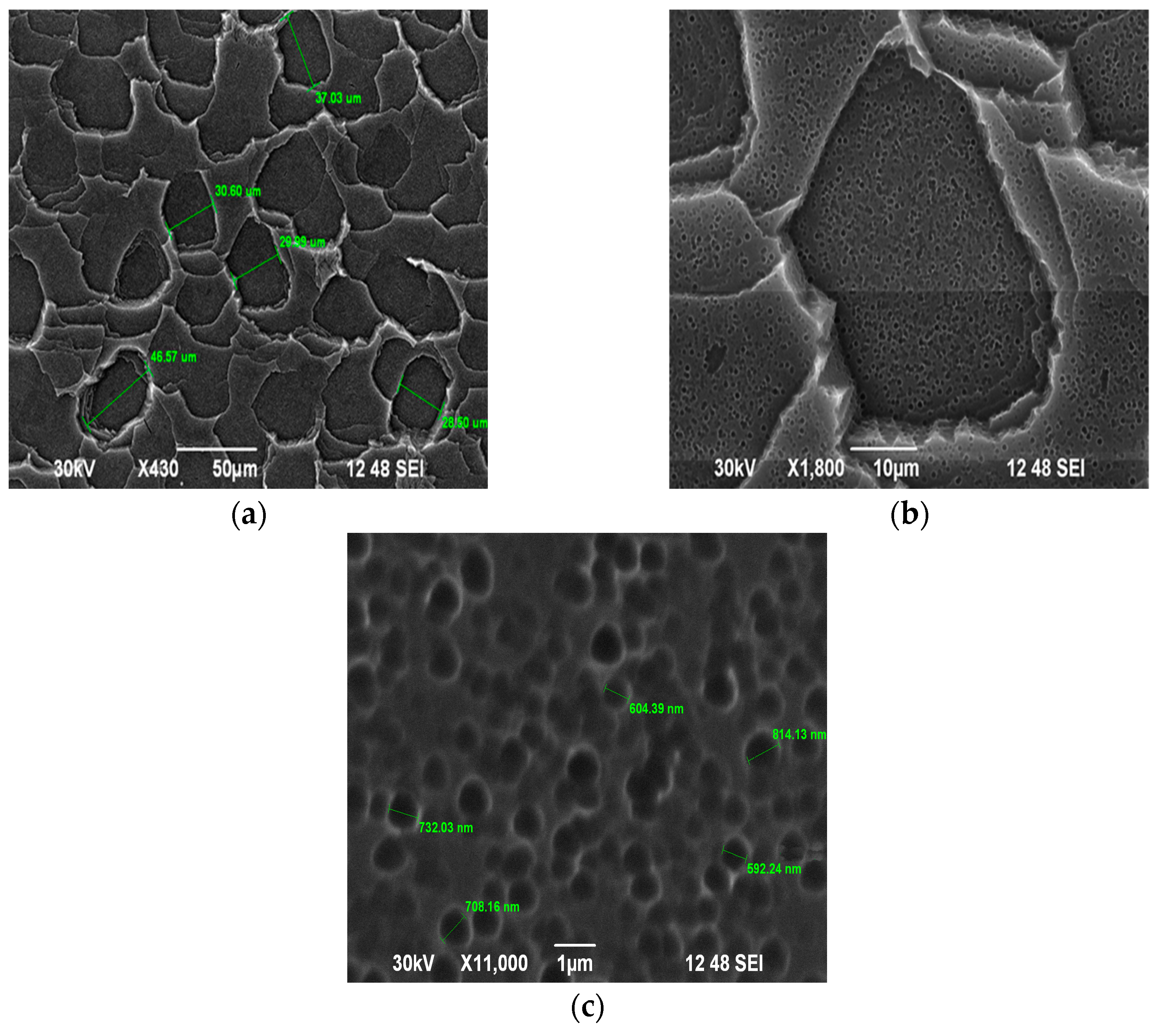
To study the process of surface structure formation, images of the initial sample and samples coated with ZnO were obtained using a scanning electron microscope. The macroporous structure of the samples is clearly represented in the images at a magnification of 430 times (
Figure 1a).
Figure 1b,c show the results of the SEM study of surface morphology at a higher resolution (at magnification in ×430 and in ×11,000). The images show that the surface structures have a step-like nature of porosity, from macro- to micro dimensional. It is also possible to determine the developed structure of the pore walls.
The deposition of coating layers led to a decrease in the diameter of the macropores. Thus, with an increase in the number of layers, the surface became more finely porous and at the same time the porosity is more uniform in size. This is because the coating is localized at the boundaries of the pores. Moreover, with an increase in the number of coating layers, the walls of the macropores became smoother, and the size of the micropores decreased.
The more developed the structure of the samples, the more particles with uncompensated charge they contain. This is due to the high chemical activity of the surface. When zinc oxide is deposited to PS, the mechanical stability of the structures increases and light-emitting particles appear.
3.2. Investigation of Surface Morphology Using Atomic Force Microscopy
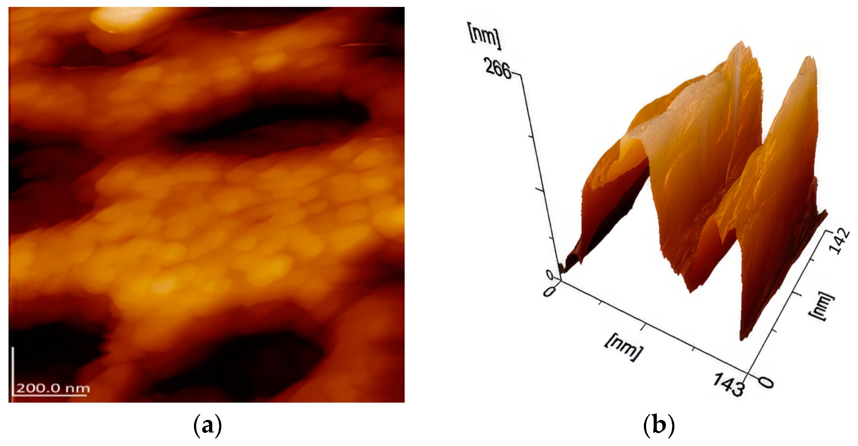
For a detailed study of the structures inside the macropores, atomic force microscopy images were obtained.
Figure 2a shows the structure between the micropores. It is clearly seen that in addition to regularly spaced pores, the clusters of matter are noticeable. The clusters have similar shape and the distances between them are identical.
The following
Figure 2b shows the structure of a micro pore. The structure inside and outside the valley is noticeably developed, which is associated with the penetration of the film-forming solution in different directions. This is important when using a porous surface as a substrate. ZnO crystals could be formed due to porous silicon substrates, providing nucleation centers.
Thus, a hierarchical porous structure of the sample surface was formed, including macro and micro pores, nanocrystals between micro pores and ridges on the outer and inner sides of micro pores. The morphology of the surface is complicated by the developed structure of the walls of macropores. The complexity was increased through the synthesis of multilayer ZnO/SiO2/Si.
3.3. Photoluminescence Studies
At the same time, the smaller the size of the structures, the more effective photoluminescence they have.
Figure 3 shows the PL spectra recorded at room temperature, for the initial sample and coated samples (excitation at 320 nm).
The spectrum of the initial sample has a low intensity, which disappears after the films are deposited. This is due to the passivation of the sample surface, that is, a decrease in the number of particles with broken bonds.
The complex spectrum of PL after coating is associated with the presence of structures with different sizes. The most characteristic peak for zinc oxide crystals at 380 nm is noticeable on the spectrum of a sample with 25 layers. Its nature is related to the recombination of excitons. The band at 400 nm is caused by radiative transitions near the edge of fundamental absorption. The broadband of ~500 nm is associated with recombination of charges on oxygen vacancies during the formation of nanocrystalline ZnO films. The boundaries of nanocrystals have the structure of a gradual transition from one type of particle to another.
3.4. EPR Studies
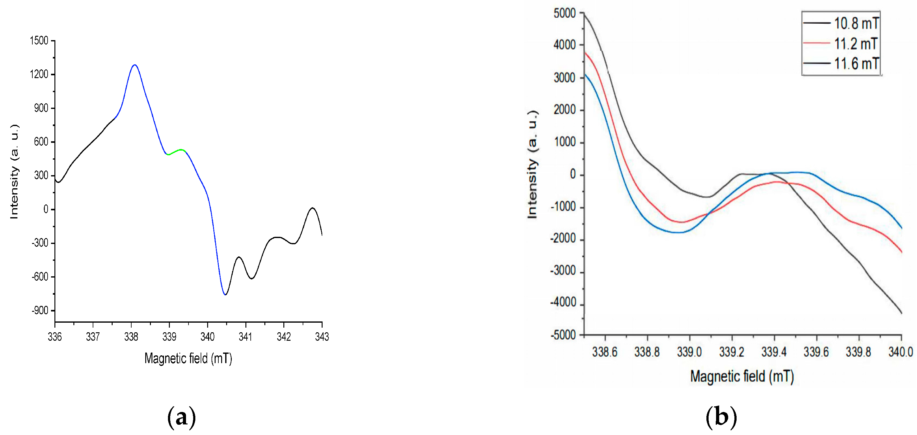
To clarify the mechanisms of interaction of particles with uncompensated charge during the formation of nanostructures, EPR studies were carried out.
Figure 4a,b shows the EPR spectrum of a sample with 25 coating layers. In the central part of the spectrum (highlighted in blue), a signal with a G-factor of ~1.99 is observed. Noticeably, its shape differs from the graph of the first derivative. This is due to the presence of a weak anisotropy of a signal, which can be seen when the EPR signal is saturated (
Figure 4b). Its nature is associated with trapped electrons on oxygen vacancies, probably at the boundaries of micropores, possibly F-centers in a small concentration.


 ,
, 