Proceeding Paper

Morphological Study of Insect Mechanoreceptors to Develop Artificial Bio-Inspired Mechanosensors †

1
Faculty of Mechanical Engineering and Design, Kaunas University of Technology, Kaunas LT-51424, Lithuania
2
Institute of Insect Science, Zhejiang University, Hangzhou 310058, China
3
Institute of Data Science and Digital Technologies, Vilnius University, Vilnius LT-08412, Lithuania
*
Author to whom correspondence should be addressed.
Presented at the 7th International Electronic Conference on Sensors and Applications, 15–30 November 2020; Available online: https://ecsa-7.sciforum.net/.
Eng. Proc. 2020, 2(1), 70; https://doi.org/10.3390/ecsa-7-08199
Published: 14 November 2020
(This article belongs to the Proceedings of 7th International Electronic Conference on Sensors and Applications)

Abstract

:
Mechanoreceptors of the insect play a vital role for the insect to sense and monitor the environmental parameters, like flow, tactile pressure, etc. This paper presents the studies made on the morphology of the mechanoreceptor of the insect Blattella asahinai (scientific name of cockroach) that is a hair-like structure known as trichoid sensilla, by scanning electron microscope and confocal laser microscope. The scanned images show the details of sensilla components in which the hair is embedded in the sockets, which are connected with the cuticle and joint membrane, where the dendrite touches at the base of the hair passing through the cuticle layers. The images also show that the tubular bodies and microtubules are tightly compacted inside the dendrite. This paper presents the details of how the sensilla work when an external stimulus act on them. The hair deflects with the disturbance of the cuticle and joint membrane, and this deformed hair leans on the dendrite, which is attached at the base of the hair that in turn presses the tubular bodies and microtubules, which develop negative ions passing down through the dendrite to the neuron, which provides information as an electric signal to the brain of the insect so that it responds for necessary action. Based on the morphological studies, sensing mechanism, material properties of the components, and design principles will be evolved for the development of an artificial bio-inspired sensor. A solid works model of the sensilla is also presented.

1. Introduction

Insects are ultrasensitive in sensing the environmental changes mainly by mechanical stimuli like air flow, vibration, pressure changes, etc., by using their specialized sensory organs, which are known as mechanoreceptors. These are hair-like structures technically called sensilla. These mechanoreceptors spread all over the body and mostly on the exoskeleton surfaces, which are projected from the cuticle. Forces due to external disturbances around the hair of the insect will develop stresses inside the walls of the cuticle. These mechanoreceptors present on the cuticle surface will have different sizes and shapes and sensitivity based on their location on the body of the insect. Trichoid sensilla are one amongst them which exist on all body parts of the insect. They help in detecting the forces developed internally and externally when the hair deflects [1]. Antennae are the foremost sensory parts in the insect on which the trichoid sensilla are abundantly found. These sensilla have a simple external and complex internal construction in contrast to the chemosensillar. The external part of the trichoid sensilla senses the changes in air flow and contact with other surfaces. Usually the hairs are flexible, but some are stiff. When the hair bends, it presses against the joint membrane, the dendrite, and the tubular bodies and sends signal to the nerve cells [2].
The external and internal morphological studies of these mechanoreceptors are performed by different electron microscopes. Due to their high resolution and magnification, the studies made through scanning electron microscope (SEM) and transmission electron microscope (TEM) on the mechanoreceptors of the insects by many researchers have contributed to the vast knowledge on their external and internal morphology of sensilla [3].
The present studies on the morphology of sensilla and their sensing behavior will help in replicating the features for developing the sensors by using MEMS technology. Several researchers have worked on the artificial hair [4,5,6] through bio-inspired insect mechanoreceptors, by modelling them as an inverted pendulum. Some have developed biomimetic flow sensitive sensors, biomimetic accelerometers, etc. These bio-inspired mechanoreceptors have been fabricated by using surface machining, 3D printing, photolithography, and MEMS technologies [7,8,9].
The external and internal structure and details of functioning of the trichoid sensilla of the Blattella asahinai insect are studied during an external stimulus, by SEM and TEM, and are presented in this paper.

2. Materials and Methods

2.1. Insects

The Blattella asahinai insects are collected from the bank of Musi River, in Hyderabad, India, and preserved in the laboratory of Biological Regional Centre of Zoological Survey of India, Hyderabad, India. These insects are reared at a temperature of 22 °C, till they develop their cerci and full length of antennas. The insects of identical growth are selected for the studies.

2.2. Preparation of Samples

The selected cockroach is anesthetized by keeping it on an ice block for about a minute. The antennal segments are separated from the surface of the body, using scissors and tweezers, and are kept in 1N potassium hydroxide solution, prepared by dissolving 10 mg of KOH pellets in H2O. This KOH solution dissolves the unwanted soft tissue, leaving behind the hard cuticle, which is then washed with 60% and 70% ethanol, at room temperature, for about 5 min. The antennal tissues are laterally dissected and transferred into C8H8O3 methyl salicylate solution, to make the sample more transparent, to get clear images under the microscope. These samples are dried well by placing them over filter paper.

2.3. Samples under SEM and TEM

Dried antennae tissues are placed over the surface of aluminum stubs by sticking them with a carbon tape. A slight gold coating is done on the tissue, with a sputter coater (LEICA EM ACE 200). The morphology of the sensilla on each antenna is examined under FE-SEM (Quanta FEG 250, LEICA EM TP), at an electron beam voltage of 5 KV. To observe the internal morphology, the mechanosensilla are removed from the antennae of the insect and embedded into the Epon blocks and then placed under Ultramicrotome (LEICA EM UC7), to make the ultrathin sections. Ultrathin sections of the samples are observed under TEM (FEI Technai TF20), with magnification of 25× to 2,000,000×. The images captured from both SEM and TEM are edited by using Adobe Photoshop.

3. Results

3.1. Sensilla on Antennal Segments External MORPHOLOGY

The sensilla on the antennal segments are more in number on the distal end of the segment, when compared with other segments. There are several types of sensilla present on the surface of the antennal segment; amongst those, the trichoid sensilla are large in number. Sensilla of different types are distributed over the surface shown in Figure 1. Trichoid sensilla are one of the types of sensilla that have different shapes. Based on their length and shape, they are classified into two groups, namely s.t.I and s.t.II. The trichoid sensilla, s.t.I, have a pointed apex. Trichoid sensilla, s.t.II, are another type which are abundant on the segmental surface. They have curved shape with blunt tips, as shown in Figure 1a Sensilla basiconica (s.b.) are s.b.I type of sensilla, with blunt tips and stout pegs. The s.b.II type of sensilla are very short, with a bulgy base. The s.b III type sensilla are longer, with sharp tips which rise from side walls of antennal surface, as shown in Figure 1b. In Figure 1c, another type of sensilla with a blunt tip and stout pegs, called sensilla chaetica (s.c.), are present over the length of the antennae.

3.2. Internal Morphology of the Sensilla

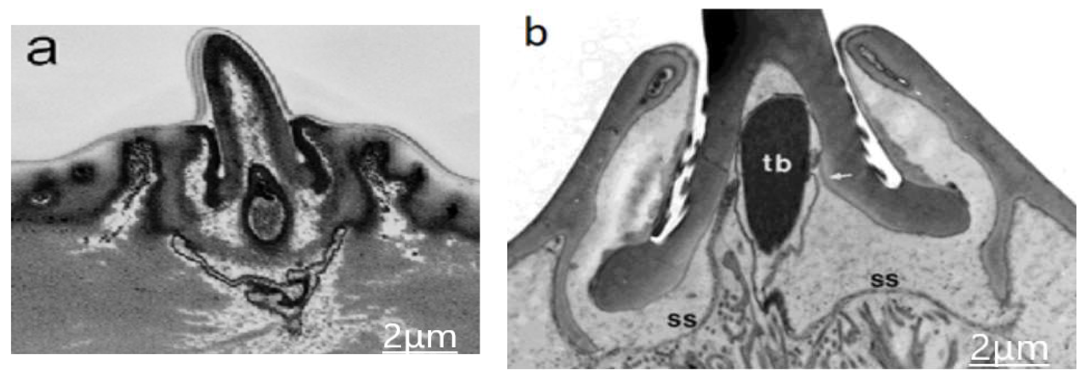
The red arrow in Figure 1a indicates the sensilla trochoid for which the internal morphology are indicated in Figure 2. The hollow hair is seen to be rising from the cuticle, with the base of the hair being supported with the joint membrane and socket septum. They are connected with few suspension fibers which restrict the extent of hair base movement [10,11].

4. Discussion

In Blattella asahinai, sensilla on antennas are of various shapes and sizes, and they varies from one insect to another; however, the internal morphology of all types of sensilla are the same for all insects [12].

4.1. Development of the Sensilla

The development of sensilla starts with the division of cells which are attached bellow the cuticle in the sensory mother cell (SMC) and trichogen and tormogen mother cell (TMC). The SMC further divides into sensory neuron (SC) and thecogen (Th), whereas TMC divides into trichogen cell (Tr) and tormogen cells (To). The dendrite and neuron are developed from the thecogen cell (Th) and sensory neuron (Sc). The other parts of the sensilla, like the hair base, joint membrane, socket, and socket septum, are developed by the TMC cells mutation [13,14,15].
In Figure 2, the socket appears on the cuticle surface from which the hollow hair rises. The joint membrane is a hollow elastic cylinder, with one end connected to the base of the hair and the other end connected to the socket. The base of the hair is supported by the joint membrane and socket septum, along with a few suspension fibers, which are helpful in hair movements due to their elastic nature. The distal end of the dendrite is slightly in touch with the hair base, where the dendrite is covered with a dendrite sheath [16]. The main sensory part of the sensilla is the dendrite which is circular in cross-section. The tubular bodies are embedded in the dendrites, which are tightly packed with the electron-dense material known as microtubules.

4.2. Functioning of Sensilla

Whenever an external disturbance occurs in the air, it flows over the hair, and then the hair deflects slightly. As the dendrite is in touch at the base of the hollow hair, this deflection of hair will press the dendrite, as well as the tubular body, and microtubules result in electrical charging and downward flow of ions in the microtubules from the tubular body to the neurons, sending the information to the brain of the insect, for taking necessary action [12,16,17].

4.3. Design of Artificial Mechanosensors

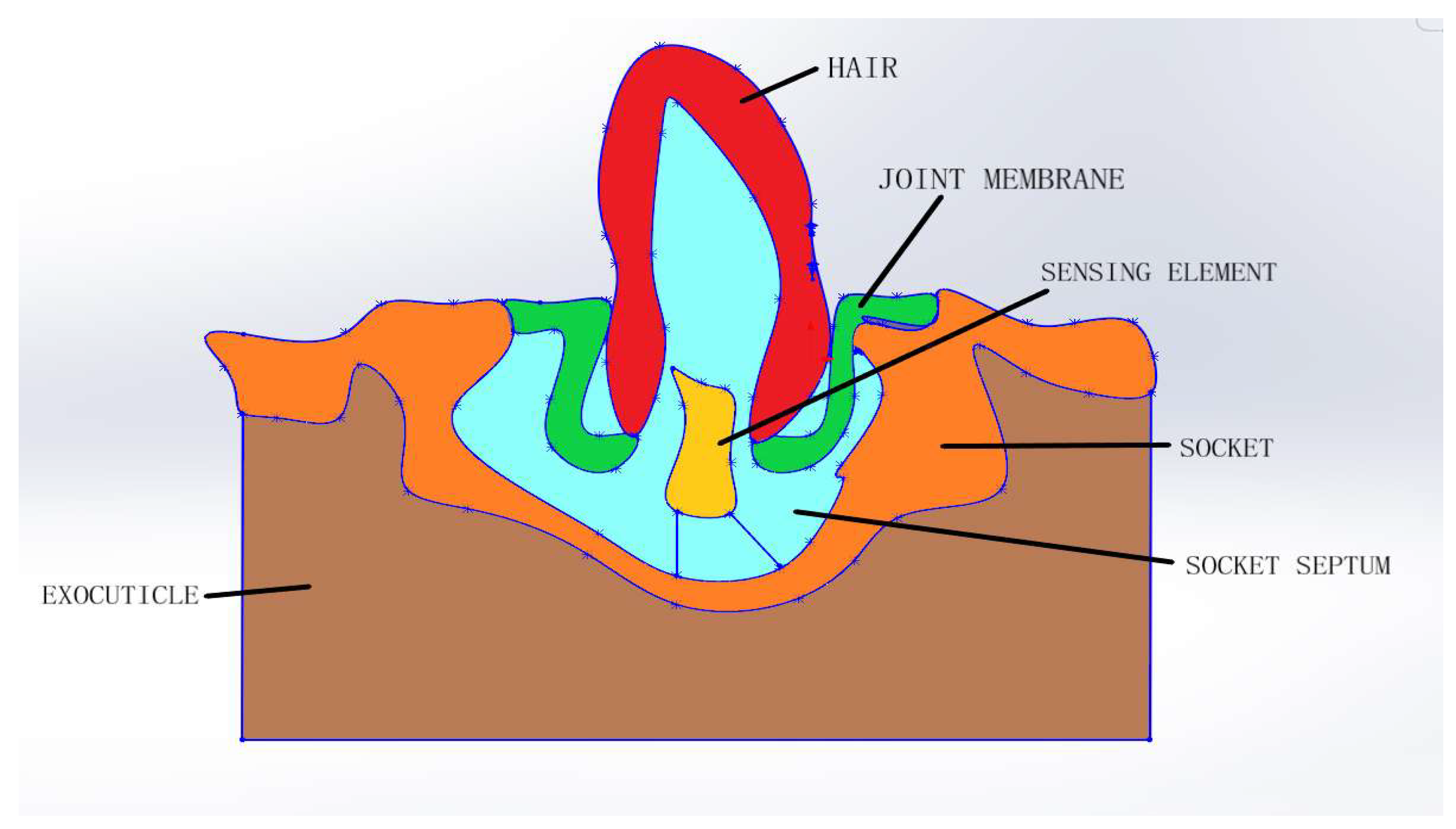
The dimensions of the sensilla were measured by using the SEM and TEM and found that the long axis of the socket is about 5 ± 0.5 µm, the short axis of the socket is about 4 ± 0.5 µm, the length of the hair is 15 ± 2 µm, and the diameter is 900 ± 150 nm; the dendrite cross-section is round and has a diameter of about 600 nm, the tubular body inside this dendrite is about the length of 850 nm, and the diameter is of 500 nm; and microtubules are of 50 nm each in diameter, and they are tightly packed in the tubular body. By considering the morphology, functioning, and dimensions of the sensilla, the artificial mechanosensor is designed as shown in Figure 3. The material of the hair is keratin, which is a protein nanofiber containing sulfur. The mechanical properties of the hair are a density of 1100 kg/m3, a low elastic modulus of 6 Gpa, a high elastic modulus of 9 Gpa, and a Poisson’s ratio of 0.38. Joint membrane and socket are the viscoelastic materials that have a Young’s modulus of 50 Mpa and a Poisson’s ratio of 0.4. Suspension fibers are fibrous materials which are elastic in nature [18]. The dendrite is covered with dendrite sheath, and the tubular body contains microtubules, which are the sensing elements that are of piezoelectric material.

5. Conclusions

In this paper, the morphology and physiology of sensilla of Blattella asahinai the and its functioning are explained when an external stimulus acts on it. The model of the bio-inspired artificial mechanosensor is presented. The mathematical model and simulation studies on designed artificial hair sensor for flow and tactile parameters will be made in the future.

References

  1. George, A.; Nagy, B.A.L. Morphology, distribution, and ultrastructural differences of sensilla trichodea and basiconica on the antennae of the oriental fruit moth, Grapholitha molesta (Busck) (Lepidoptera: Tortricidae). Int. J. Insect Morphol. Embryol. 1984, 13, 157–170. [Google Scholar] [CrossRef]
  2. Shimozawa, T.; Kanou, M. The aerodynamics and sensory physiology of range fractionation in the cereal filiform sensilla of the cricket Gryllus bimaculatus. J. Comp. Physiol. 1984, 155, 495–505. [Google Scholar] [CrossRef]
  3. Dey, S.; Hooroo, R.N.; Wankhar, D. Scanning electron microscopic studies of the external morphology of sensilla on the legs of a butterfly, Graphium sarpedon (Lepidoptera—Papillionidae). Micron 1995, 26, 367–376. [Google Scholar] [CrossRef]
  4. Dijkstra, M.; van Baar, J.J.; Wiegerink, R.J.; Lammerink, T.S.J.; de Boer, J.H.; Krijnen, G.J.M. Artificial sensory hairs based on the flow sensitive receptor hairs of crickets. J. Micromech. Microeng. 2005, 15, S132–S138. [Google Scholar] [CrossRef]
  5. Dagamseh, A.M.K. Estimation of squeeze film damping in artificial hair-sensor towards the detection-limit of crickets’ hairs. Microsyst. Technol. 2014, 20, 963–970. [Google Scholar] [CrossRef]
  6. Krijnen, G.J.M.; Droogendijk, H.; Dagamseh, A.M.K.; Jaganatharaja, R.K.; Casas, J. Crickets as Bio-Inspiration for MEMS-Based Flow-Sensing. In Flow Sensing in Air and Water; Bleckmann, H., Mogdans, J., Coombs, S., Eds.; Springer: Berlin/Heidelberg, Germany, 2014; pp. 459–488. [Google Scholar] [CrossRef]
  7. Krijnen, G.J.M.; Lammerink, T.; Wiegerink, R. Learning from crickets: Artificial hair-sensor array developments. In Proceedings of the SENSORS 2010 IEEE, Kona, HI, USA, 1–4 November 2010; pp. 2218–2223. [Google Scholar] [CrossRef]
  8. Krijnen, G.; Lammerink, T.; Wiegerink, R.; Casas, J. Cricket Inspired Flow-Sensor Arrays. In Proceedings of the SENSORS 2007 IEEE, Atlanta, GA, USA, 28–31 October 2007; pp. 539–546. [Google Scholar] [CrossRef]
  9. Droogendijk, H.; Casas, J.; Steinmann, T.; Krijnen, G.J. Performance assessment of bio-inspired systems: Flow sensing MEMS hairs. Bioinspir. Biomim. 2014, 10, 016001. [Google Scholar] [CrossRef] [PubMed]
  10. Droogendijk, H. Bio-Inspired MEMS Flow and Inertial Sensors; University of Twente: Enschede, The Netherlands, 2014. [Google Scholar] [CrossRef]
  11. di Giulio, A.; Maurizi, E.; Valerio, M.; Stacconi, R.; Romani, R. Functional structure of antennal sensilla in the myrmecophilous beetle Paussus favieri (Coleoptera, Carabidae, Paussini). Micron 2012, 43, 705–719. [Google Scholar] [CrossRef] [PubMed]
  12. Keil, T.A. Functional morphology of insect mechanoreceptors. Microsc. Res. Tech. 1997, 39, 506–531. [Google Scholar] [CrossRef]
  13. Letourneau, P.C.; Condic, M.L.; Snow, D.M. Interactions of developing neurons with the extracellular matrix. J. Neurosci. 1994, 14, 915–928. [Google Scholar] [CrossRef] [PubMed]
  14. Orgogozo, V.; Grueber, W.B. FlyPNS, A database of the Drosophila embryonic and larval peripheral nervous system. BMC Dev. Biol. 2005, 5, 4. [Google Scholar] [CrossRef] [PubMed]
  15. Giangrande, A.; Palka, J. Genes involved in the development of the peripheral nervous system of Drosophila. Semin. Cell Biol. 1990, 1, 197–209. [Google Scholar] [PubMed]
  16. Gnatzy, W.; Weber, K.M. Tormogen cell and receptor-lymph space in insect olfactory sensilla. Cell Tissue Res. 1978, 189, 549–554. [Google Scholar] [CrossRef] [PubMed]
  17. Fred, T.; Hartley, D.A. Inhibition of cell fate in Drosophila by Enhancer of split genes. Mech. Dev. 1995, 51, 305–315. [Google Scholar]
  18. Barth, F.G. Spider mechanoreceptors. Curr. Opin. Neurobiol. 2004, 4, 415–422. [Google Scholar] [CrossRef]
Figure 1. SEM images of External morphology of sensilla on antenna and its types. (a) Conical projection of the insect Blattella asahinai antennal segment. Sensilla trichoid (s.t.). Indicated by short arrow, s.t.I is a type of trichoid with pointed apex; long arrow, s.t.II, are trichoid sensilla which are curved and have blunt tips. (b) Apical antenna segment shows the sensilla trichoid I and II and also another type of sensilla which are distributed on the surface, i.e., sensilla basiconica, (s.b.), s.b.I, s.b.II, and s.b.III. (c) Black arrow shows another type of sensilla, i.e., sensilla chaetica (s.c.), which are distributed along the axis of the antennal segment with some angle.
Engproc 02 00070 g001
Figure 2. TEM images of internal morphology of the trichoid sensilla. (a) Cross-sectional view of the sensillum. (b) Sectional view of the tubular body and socket septum. Scale line: 2 µm.
Engproc 02 00070 g002
Figure 3. Artificial sensilla model.
Engproc 02 00070 g003
Publisher’s Note: MDPI stays neutral with regard to jurisdictional claims in published maps and institutional affiliations.