Proceeding Paper

Development of a Biosensor for the Early Detection of Tuberculous Meningitis in Infants †

1
Department of Electrical and Electronic Engineering, Faculty of Engineering, Stellenbosch University, Private Bag X1, Stellenbosch 7602, South Africa
2
South African Medical Research Council Centre for Tuberculosis Research, Division of Immunology, Department of Biomedical Sciences, Faculty of Medicine and Health Sciences, Stellenbosch University, P.O. Box 241, Cape Town 8000, South Africa
*
Author to whom correspondence should be addressed.
Presented at the Micro Manufacturing Convergence Conference, Stellenbosch, South Africa, 7–9 July 2024.
Eng. Proc. 2025, 109(1), 12; https://doi.org/10.3390/engproc2025109012
Published: 15 September 2025

Abstract

Tuberculous meningitis (TBM) is a severe illness that is predominantly observed in countries with a high burden of tuberculosis. It is primarily found in infants and human immunodeficiency virus (HIV)-infected adults, and, if left untreated, causes irreversible damage to the host’s nerve and brain tissue, often leading to mortality. Current methods of TBM detection relies on cerebrospinal fluid (CSF) culture, which may only yield results in up to 6 weeks, is not very sensitive, and requires a biological safety level III laboratory to conduct. Other detection methods are equally not very sensitive and laborious. This research investigates the detection of interferon-gamma (IFN-γ) protein biomarker using fluoroimmunoassay with an optical biosensor and a custom-manufactured chip. The glass-surface of the chip was treated with 3-aminopropyltriethoxysilane (APTES) and incubated with glutaraldehyde to prepare for immobilization, after which a sandwich ELISA format was used to perform a dilution series by immobilizing the capture antibody, IFN-γ protein, and fluorescein isothiocyanate (FITC)-stained detection antibody onto the chip. The optical biosensor excited the FITC-stained antibodies to capture the emission light at multiple exposures, which were then merged to create a high dynamic range (HDR) image for image processing. The results from the optical biosensor were verified with a Zeiss LSM780 confocal microscope (Carl Zeiss (Pty) Limited, Cape Town, South Africa). The system demonstrated the capability to rapidly identify the biomarker, detect the binding sites, and quantify IFN-γ in blood serum. This fluorescent optical sensor proposes a possible approach for the development of a point-of-care system for TBM, providing a quicker and simpler method for the early detection of TBM.

1. Introduction

Tuberculous meningitis (TBM), an infection that largely targets infants and adults who are infected with human immunodeficiency virus (HIV), is a common disease in high TB-burdened developing countries [1]. TBM is caused by the same bacterium that causes TB of the lungs (pulmonary TB). Following the inhalation of the causative organism (Mycobacterium tuberculosis) into the lungs, the bacterium then disseminates from the lungs and infects the brain of the host [2]. TBM is the most severe form of TB, and up to 50% of individuals that are afflicted with the disease die or suffer from lifelong neurological complications if successfully treated [3].
TBM is a treatable disease. The reason for its high mortality is the lack of effective diagnostic tools, and this is compounded by the locations where the disease commonly manifests. The majority of TBM cases are recorded to be in developing countries located in Southeast Asia and Africa [4]. In these areas, it is unlikely for infants to have regular health checkups at clinics. Also, even when symptoms do start showing, guardians might overlook the infant’s discomfort until the symptoms worsen, which might already be too late for complete recovery or even an option of treatment for the patient [1]. Even where health care services seem adequate, compared to other lower- and middle-income countries such as the Western Cape Province of South Africa, children who are under the age of 5 years still require a median of four visits (up to six) before admission at tertiary hospital and eventual diagnosis of TBM [5]. This implies that diagnostic delay may be the most important reason for the high mortality rates of TBM, especially in children.
Presently there are no point-of-care tests available for TBM; instead, the most accepted method, which is also the reference standard diagnostic test, is cerebrospinal fluid (CSF) culture [1,6]. The biggest flaw of CSF culture is the time it takes for the bacterium that causes the disease to grow. These tests can take up to 6 weeks and require biological safety level III laboratories, which are not common in developing countries due to their high costs. The test is also not very sensitive, owing to the low numbers of TB-causing bacteria in the CSF [7]. Recently developed TB tests, such as the GeneXpert test (Cepheid, Sunnyvale, CA, USA), have shown promise but have several implementation challenges in addition to high costs [7].
Recent studies have identified host protein biomarkers as potentially useful diagnostic candidates for TBM. The use of such biomarkers in point-of-care tests is highly recommended by the World Health Organisation [8]. Vascular endothelial growth factor (VEGF), interferon-gamma (IFN-γ), and myeloperoxidase (MPO) were one group of biomarkers that was identified [9] and validated [10] in recent studies. When evaluated in a multiplexed assay, the biomarkers exhibited a sensitivity of 91.3% and a specificity of up to 100%, depending on the cut-off value selected [9]. Since these tests were performed in a laboratory with complex instruments, the application of these biomarkers in a point-of-care device was the next step towards developing a potentially useful TBM diagnostic test.
In this research, one host protein biomarker, IFN-γ, was used to develop a fluorescent optical biosensor to capture the fluorescence emitted from the fluorescein isothiocyanate (FITC) tagged detection antibody. The captured fluorescent measurements were quantified to determine the concentration of IFN-γ present within the sample.

2. Materials and Methodology

2.1. Biosensor Fabrication

The biosensor was designed and fabricated with the following components: A Raspberry Pi 4 (PiShop, Johannesburg, South Africa) was selected as the microcontroller for the control of hardware responsible for image capture and processing of the captured data; an HQ camera module (RS Components, Cape Town, South Africa) with a Sony IMX477R sensor with a colour channel bit depth of 8 bits was chosen, and a 465 nm wavelength excitation Kingbright3.3 V Blue LED (RS Components, Cape Town, South Africa) was chosen to match the FITC excitation range. The camera lens was shielded with a SYBR green filter (ThermoFisher, Johannesburg, South Africa) to filter out the excitation light. The components were assembled using joints and mounts manufactured with a 3D printer. The chip on which the fluoroimmunoassay was performed was manufactured from a microscope slide and laser-cut wells.

2.2. Chip Functionalization and Immobilization

The chip served as the immobilization site for the antibodies responsible for capturing the target IFN-γ biomarker. A microscope slide was initially prepared by rinsing with 75% ethanol, followed by deionized (DI) water. The chip was then activated using an ultraviolet ozone (UVO) cleaner to enhance the reactivity of the glass surface for functionalization with 3-aminopropyltriethoxysilane (APTES) (Merck, Johannesburg, South Africa). Subsequently, the chip was bathed in an APTES solution (4% v/v in ethanol-95%) for 30 min. After rinsing with ethanol-95%, the chip was incubated with glutaraldehyde (Merck, Johannesburg, South Africa) (2% v/v in DI water) for 1 h, followed by a rinse with phosphate-buffered saline (PBS). A sandwich ELISA protocol was adapted to immobilize the IFN-γ protein and antibodies. The chip was incubated overnight with the capture antibody (Mouse Anti-Human IFN-γ-UNLB (A35), SouthernBiotech, Birmingham, AL, USA) (50 µg/mL) at 4 °C [11]. It was then washed with PBS-tween and incubated with the test sample spiked with IFN-γ recombinant protein (Human IFN-gamma/IFNG/γ-IFN Protein (11725-HNAS), Sino Biological, Beijing, China) for 2 h at room temperature [12]. Finally, the chip was washed with PBS-tween and incubated with FITC-tagged detection antibody (Mouse Anti-Human IFN-γ-FITC (B27), SouthernBiotech, Birmingham, AL, USA) (10 µg/mL) at 4 °C overnight and washed with PBS-tween and imaged [13].

2.3. Image Capturing and Processing

The optical biosensor excited the FITC-tagged antibodies bound to the immobilized IFN-γ protein with the excitation LEDs (465 nm). The camera module captured the emission light from the FITC by sampling the images at multiple exposures. The multiple exposures were then merged using the Devevec high dynamic range (HDR) imaging technique to create a singular HDR image with an enhanced colour depth. The resulting HDR image was processed using Hough Circle detection to detect the wells where the immobilization took place and filtered to eliminate any noise generated externally or internally from the camera sensor. Lastly, each well was measured by interpreting the pixel mean intensity, and the results were plotted accordingly.

3. Results and Discussion

3.1. Immobilization Verification

To verify the immobilization of IFN-γ antibodies and protein, a Zeiss LSM780 confocal microscope was utilized to image the chip. With the configuration to capture the emission spectrum of FITC with a peak of 525 nm, the confocal microscope captured the images exhibited in Figure 1. Figure 1a depicts the chip incubated with an IFN-γ protein concentration of 20 µg/mL, while Figure 1c was incubated only with PBS to serve as a negative control. These images confirmed the capture of IFN-γ protein and binding of the IFN-γ detection antibody. Figure 1b exhibits dark patches in the FITC fluorescence, which was the result of scraping of the chip surface after the incubation of capture anti-body. The dark patches validate that there was no non-specific binding of IFN-γ protein and detection antibody on the chip surface.

3.2. Optical Biosensor Verification and Image Processing

After the incubation of the chip was confirmed, a dilution series was performed and imaged with the optical biosensor. The dilution series involved spiking PBS with IFN-γ protein to achieve concentrations in the range of 20 µg/mL to 0.00244 µg/mL (or 2.44 ng/mL), decreasing with a logarithmic factor of 2. Each concentration was tested in triplicate.
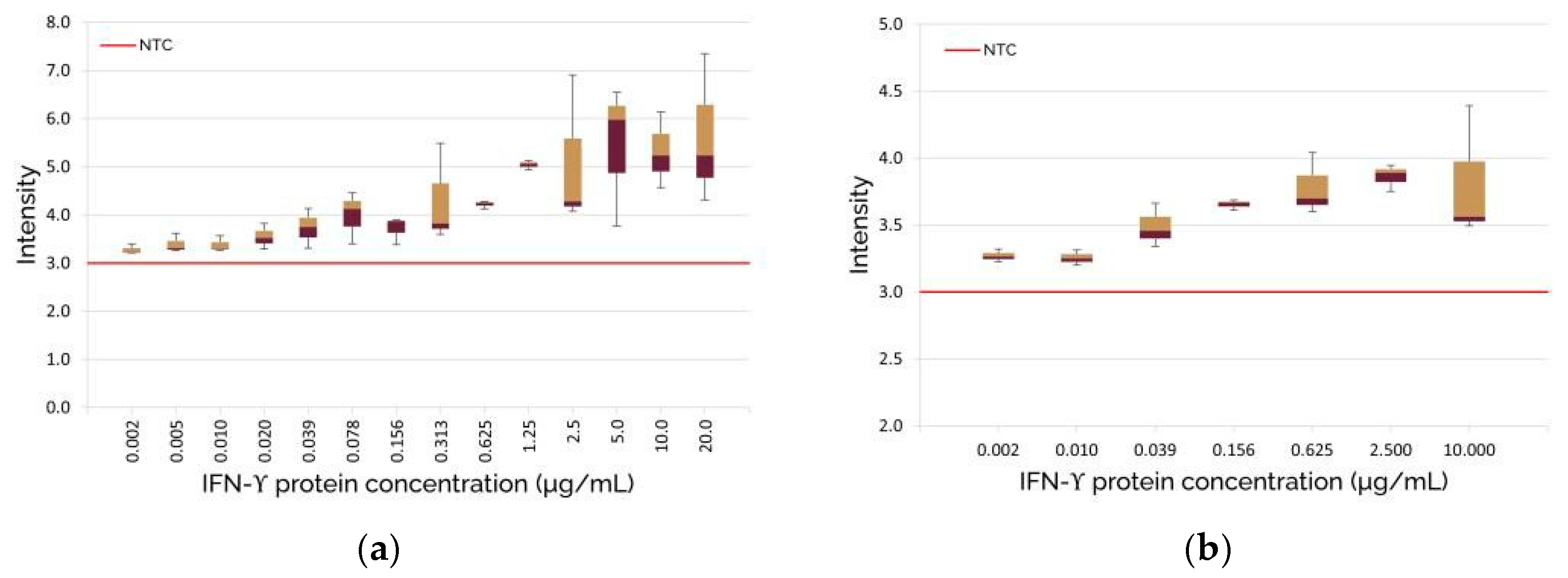
After the capturing of images with the optical biosensor, the images were processed, and each IFN-γ protein concentration was quantified as average pixel intensity and plotted in Figure 2a. The results demonstrate that the optical biosensor could distinguish the difference in fluorescence resulting from the varying protein concentrations. It was also observed that the intensity of the fluorescence saturated around 5 µg/mL, and measurements below 2 ng/mL were hard to distinguish from the negative test control (non-spiked PBS).
The verification of the optical biosensor enabled the testing of blood serum from a non-TBM patient. The serum was spiked with IFN-γ protein to simulate that of a TBM patient. Similarly, in the test in Figure 2a, a dilution series was performed, with concentrations ranging from 10 µg/mL to 0.00244 µg/mL (or 2.44 ng/mL), decreasing with a logarithmic factor of 4. The concentration of the dilution series was adjusted, as minute differences in the dilution range showed minimal difference. Each concentration in the dilution series was tested in triplicate. The resulting images were processed, and the outcome was plotted in Figure 2b. In Figure 2b, the dilution series of the blood serum displays an increasing trend with higher IFN-γ protein concentrations. However, the fluorescence intensity of the spiked serum showed a much lower value in comparison to the spiked PBS. The fluorescence intensity of the spiked serum saturated around 3.5–4, whereas the spiked PBS saturated around 5–6, as seen in Figure 2. The discrepancy between the two plots is likely due to the non-specific particulates found within the serum interfering with the immobilization of proteins to the capture antibodies on the chip surface, which would lead to fewer FITC-tagged antibodies binding to the protein and emitting an overall dimmer fluorescence.
Furthermore, given that the serum sample tested was from an individual who was not infected with TB and thus had to be spiked with IFN-γ protein to imitate the elevated IFN-γ protein levels in a TBM patient, it is difficult to conclude that the system is capable of detecting IFN-γ protein levels in a TBM patient. Further investigation in samples from confirmed TBM patients and individuals without TBM would be required to ascertain the performance of the developed biosensor. That notwithstanding, our results show that we successfully developed a biosensor capable of detecting IFN-γ in biological specimens by capturing the IFN-γ protein in the specimen, binding a FITC-tagged detection antibody to the protein, and measuring the emitted fluorescence to determine IFN-γ protein concentration in the specimen.

4. Conclusions

In conclusion, the development of a fluorescent optical biosensor for the detection of tuberculous meningitis in infants represents an innovative advancement of point-of-care devices. The use of a fluorescent optical biosensor was successful in utilizing FITC-stained IFN-γ antibodies to identify the biomarker, detect the binding sites, and quantify the IFN-γ protein concentration in both spiked PBS and blood serum samples. With current diagnosis taking up to 6 weeks to identify TBM, the synergizing effect of both the optical biosensor and host protein biomarkers offers a rapid detection process for TBM biomarkers. This research holds significant promise in the advancement of point-of-care systems, enabling early detection of TBM in infants, particularly benefitting those living in limited resourced settings.
This research contributed to the exploration of the use of glass surface chips to perform sandwich ELISA of IFN-γ antibodies and protein in a fluorescent optical biosensor format, which could be further explored for a potential adaptation in a point-of-care system for the diagnosis of TBM. Future work will further develop the optical biosensor to test MPO and VEGF alongside IFN-γ host protein biomarkers in a multiplex assay. This includes exploring the use of nanobeads to increase the surface area for immobilization, achieving higher light intensity, and assessing different detection dyes to improve the sensitivity of the biosensor. Subsequent steps will involve clinical evaluation using samples collected from children diagnosed with and without TBM.

Author Contributions

Conceptualization, D.K., W.J.P. and N.N.C.; methodology, D.K., W.J.P. and N.N.C.; software, D.K.; validation, D.K., W.J.P. and N.N.C.; investigation, D.K., W.J.P. and N.N.C.; resources, W.J.P. and N.N.C.; data curation, D.K., W.J.P. and N.N.C.; writing—original draft preparation, D.K.; writing—review and editing, W.J.P. and N.N.C.; supervision, W.J.P. and N.N.C. All authors have read and agreed to the published version of the manuscript.

Funding

This work was carried out as part of an EDCTP2 project, supported by the European Union (grant number TMA2018SF-2470-TBMBIOMARKERS). The views and opinions of authors expressed herein do not necessarily state or reflect those of the EDCTP.

Institutional Review Board Statement

The study was conducted in accordance with the Declaration of Helsinki, and approved by the Health Research Ethics Committee (HREC) of Stellenbosch University (Ethics Ref no: N16/11/142).

Informed Consent Statement

Written informed consent was obtained from parents or legal guardians of all children enrolled into the study. Assent was obtained if children were older than 7 years, and if they had a normal level of consciousness; that is, a Glasgow Coma Score (GCS) of 15/15.

Data Availability Statement

The raw data supporting the conclusions of this article will be made available by the authors on request.

Conflicts of Interest

The authors declare no conflicts of interest.

References

  1. Slane, V.H.; Unakal, C.G. Tuberculous Meningitis. In StatPearls [Internet]; StatPearls Publishing: Treasure Island, FL, USA, 2024. Available online: https://www.ncbi.nlm.nih.gov/books/NBK541015/ (accessed on 2 August 2024).
  2. Rich, A.R.; McCordick, H.A. The pathogenesis of tuberculous meningitis. Bull. John Hopkins Hosp. 1933, 52, 5–37. [Google Scholar]
  3. du Preez, K.; Jenkins, H.E.; Donald, P.R.; Solomons, R.S.; Graham, S.M.; Schaaf, H.S.; Starke, J.R.; Hesseling, A.C.; Seddon, J.A. Tuberculous Meningitis in Children: A Forgotten Public Health Emergency. Front. Neurol. 2022, 13, 751133. [Google Scholar] [CrossRef] [PubMed]
  4. Dodd, P.J.; Osman, M.; Cresswell, F.V.; Stadelman, A.M.; Lan, N.H.; Thuong, N.T.T.; Muzyamba, M.; Glaser, L.; Dlamini, S.S.; Seddon, J.A. The global burden of tuberculous meningitis in adults: A modelling study. PLoS Glob. Public Health 2021, 1, e0000069. [Google Scholar] [CrossRef] [PubMed]
  5. Solomons, R.; Grantham, M.; Marais, B.J.; van Toorn, R. IMCI indicators of childhood TBM at primary health care level in the Western Cape Province of South Africa. Int. J. Tuberc. Lung Dis. 2016, 20, 1309–1313. [Google Scholar] [CrossRef] [PubMed]
  6. Marx, G.E.; Chan, E.D. Tuberculous meningitis: Diagnosis and treatment overview. Tuberc. Res. Treat. 2011, 2011, 798764. [Google Scholar] [CrossRef] [PubMed]
  7. Nalugwa, T.; Shete, P.B.; Nantale, M.; Farr, K.; Ojok, C.; Ochom, E.; Mugabe, F.; Joloba, M.; Dowdy, D.W.; Moore, D.A.J.; et al. Challenges with scale-up of GeneXpert MTB/RIF® in Uganda: A health systems perspective. BMC Health Serv. Res. 2020, 20, 162. [Google Scholar] [CrossRef] [PubMed]
  8. World Health Organisation. Meeting Report: High-Priority Target Product Profiles for New Tuberculosis Diagnostics: Report of a Consensus Meeting; World Health Organisation: Geneva, Switzerland, 2014. [Google Scholar]
  9. Manyelo, C.M.; Solomons, R.S.; Snyders, C.I.; Manngo, P.M.; Mutavhatsindi, H.; Kriel, B.; Stanley, K.; Walzl, G.; Chegou, N.N. Application of Cerebrospinal Fluid Host Protein Biosignatures in the Diagnosis of Tuberculous Meningitis in Children from a High Burden Setting. Mediat. Inflamm. 2019, 2019, 7582948. [Google Scholar] [CrossRef] [PubMed]
  10. Manyelo, C.M.; Solomons, R.S.; Snyders, C.I.; Kidd, M.; Kooblal, Y.; Leukes, V.N.; Claassen, C.; Roos, K.; Stanley, K.; Walzl, G.; et al. Validation of host cerebrospinal fluid protein biomarkers for early diagnosis of tuberculous meningitis in children: A replication and new biosignature discovery study. Biomarkers 2022, 27, 549–561. [Google Scholar] [CrossRef] [PubMed]
  11. Mouse Anti-Human IFN-γ-UNLB (A35). Available online: https://www.southernbiotech.com/mouse-anti-human-ifn-g-unlb-a35-10113-01 (accessed on 29 January 2025).
  12. Recombinant Human IFN Gamma Protein. Available online: https://www.sinobiological.com/recombinant-proteins/human-interferon-gamma-11725-hnas (accessed on 29 January 2025).
  13. Mouse Anti-Human IFN-γ-FITC (B27). Available online: https://www.southernbiotech.com/mouse-anti-human-ifn-g-fitc-b27-10114-02 (accessed on 29 January 2025).
Figure 1. Fluorescent images of the chip, captured with a confocal microscope: (a) chip-well image of positive control (the bright-green spherical outline shows the well walls); (b) magnified view of the red square seen in (a); (c) chip-well image of negative control.
Engproc 109 00012 g001
Figure 2. Plots of results obtained after processing of images captured by the optical biosensor: (a) Box and whiskers plot with error bars of the dilution series performed with spiked PBS; (b) Box and whiskers plot with error bars of the dilution series performed with spiked serum.
Engproc 109 00012 g002
Disclaimer/Publisher’s Note: The statements, opinions and data contained in all publications are solely those of the individual author(s) and contributor(s) and not of MDPI and/or the editor(s). MDPI and/or the editor(s) disclaim responsibility for any injury to people or property resulting from any ideas, methods, instructions or products referred to in the content.