
Identification, Quantification, and Method Validation of Anthocyanins †

 ,
,  , ,
, ,  ,
,  ,
,  , ,
, ,  ,
,  ,
,  ,
,  and
and 
Abstract
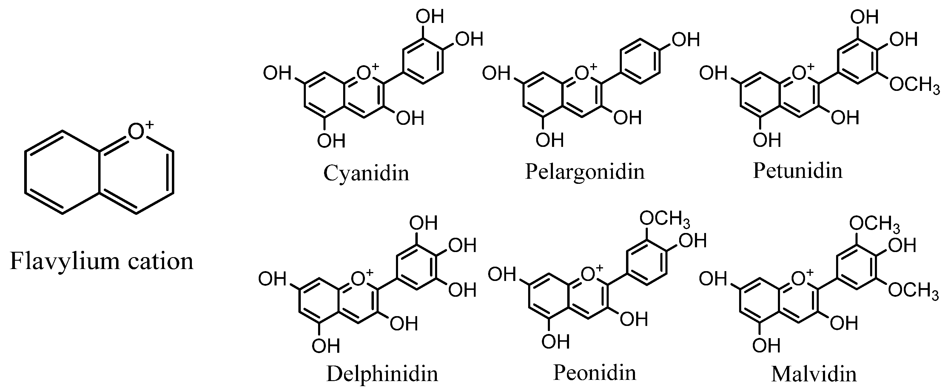
:1. Introduction
2. Identification and Quantification Techniques
3. Validation of Methods
3.1. Selectivity
3.2. Linearity, Limit of Detection (LOD), and Quantification (LOQ)
3.3. Accuracy and Precision
3.4. Stability and Robustness
4. Conclusions
Author Contributions
Funding
Acknowledgments
Conflicts of Interest
References
- Cavalcanti, R.N.; Santos, D.T.; Meireles, M.A.A. Non-thermal stabilization mechanisms of anthocyanins in model and food systems-An overview. Food Res. Int. 2011, 44, 499–509. [Google Scholar] [CrossRef]
- Ge, Q.; Ma, X. Composition and antioxidant activity of anthocyanins isolated from Yunnan edible rose (An ning). Food Sci. Hum. Wellness 2013, 2, 68–74. [Google Scholar] [CrossRef] [Green Version]
- Sasaki, R.; Nishimura, N.; Hoshino, H.; Isa, Y.; Kadowaki, M.; Ichi, T.; Tanaka, A.; Nishiumi, S.; Fukuda, I.; Ashida, H.; et al. Cyanidin 3-glucoside ameliorates hyperglycemia and insulin sensitivity due to downregulation of retinol binding protein 4 expression in diabetic mice. Biochem. Pharmacol. 2007, 74, 1619–1627. [Google Scholar] [CrossRef]
- Tsuda, T.; Horio, F.; Uchida, K.; Aoki, H.; Osawa, T. Dietary cyanidin 3-O-beta-D-glucoside-rich purple corn color prevents obesity and ameliorates hyperglycemia in mice. J. Nutr. 2003, 133, 2125–2130. [Google Scholar] [CrossRef] [PubMed]
- Xu, J.W.; Ikeda, K.; Yamori, Y. Upregulation of endothelial nitric oxide synthase by cyanidin-3-glucoside, a typical anthocyanin pigment. Hypertension 2004, 44, 217–222. [Google Scholar] [CrossRef] [Green Version]
- Grace, M.H.; Xiong, J.; Esposito, D.; Ehlenfeldt, M.; Lila, M.A. Simultaneous LC-MS quantification of anthocyanins and non-anthocyanin phenolics from blueberries with widely divergent profiles and biological activities. Food Chem. 2019, 277, 336–346. [Google Scholar] [CrossRef]
- Kamiya, H.; Yanase, E.; Nakatsuka, S. Novel oxidation products of cyanidin 3-O-glucoside with 2,2′-azobis-(2,4-dimethyl)valeronitrile and evaluation of anthocyanin content and its oxidation in black rice. Food Chem. 2014, 155, 221–226. [Google Scholar] [CrossRef]
- Tatsuzawa, F.; Saito, N.; Seki, H.; Yokoi, M.; Yukawa, T.; Shinoda, K.; Honda, T. Acylated anthocyanins in the flowers of Vanda (Orchidaceae). Biochem. Syst. Ecol. Biochem. SYST ECOL 2004, 32, 651–664. [Google Scholar] [CrossRef]
- Keppler, K.; Humpf, H.U. Metabolism of anthocyanins and their phenolic degradation products by the intestinal microflora. Bioorgan. Med. Chem. 2005, 13, 5195–5205. [Google Scholar] [CrossRef] [PubMed]
- Jang, Y.P.; Zhou, J.; Nakanishi, K.; Sparrow, J.R. Anthocyanins protect against A2E photooxidation and membrane permeabilization in retinal pigment epithelial cells. Photochem. Photobiol. 2005, 81, 529–536. [Google Scholar] [CrossRef] [PubMed]
- Acevedo De la Cruz, A.; Hilbert, G.; Rivière, C.; Mengin, V.; Ollat, N.; Bordenave, L.; Decroocq, S.; Delaunay, J.C.; Delrot, S.; Mérillon, J.M.; et al. Anthocyanin identification and composition of wild Vitis spp. accessions by using LC-MS and LC-NMR. Anal. Chim. Acta 2012, 732, 145–152. [Google Scholar] [CrossRef] [PubMed]
- Kirby, C.W.; Wu, T.; Tsao, R.; McCallum, J.L. Isolation and structural characterization of unusual pyranoanthocyanins and related anthocyanins from Staghorn sumac (Rhus typhina L.) via UPLC-ESI-MS, 1H, 13C, and 2D NMR spectroscopy. Phytochemistry 2013, 94, 284–293. [Google Scholar] [CrossRef]
- Brauch, J.E.; Reuter, L.; Conrad, J.; Vogel, H.; Schweiggert, R.M.; Carle, R. Characterization of anthocyanins in novel Chilean maqui berry clones by HPLC–DAD–ESI/MSn and NMR-spectroscopy. J. Food Compos. Anal. 2017, 58, 16–22. [Google Scholar] [CrossRef]
- Zhang, J.L.; Luo, C.L.; Zhou, Q.; Zhang, Z.C. Isolation and identification of two major acylated anthocyanins from purple sweet potato (Ipomoea batatas L. cultivar Eshu No. 8) by UPLC-QTOF-MS/MS and NMR. Int. J. Food Sci. Technol. 2018, 53, 1932–1941. [Google Scholar] [CrossRef]
- Zielińska, A.; Siudem, P.; Paradowska, K.; Gralec, M.; Kaźmierski, S.; Wawer, I. Aronia melanocarpa fruits as a rich dietary source of chlorogenic acids and anthocyanins: 1H-NMR, HPLC-DAD, and chemometric studies. Molecules 2020, 25, 3234. [Google Scholar] [CrossRef]
- Müller-Maatsch, J.; Gurtner, K.; Carle, R.; Björn Steingass, C. Investigation into the removal of glucosinolates and volatiles from anthocyanin-rich extracts of red cabbage. Food Chem. 2019, 278, 406–414. [Google Scholar] [CrossRef]
- Grajeda-Iglesias, C.; Salas, E.; Barouh, N.; Baréa, B.; Figueroa-Espinoza, M.C. Lipophilization and MS characterization of the main anthocyanins purified from hibiscus flowers. Food Chem. 2017, 230, 189–194. [Google Scholar] [CrossRef]
- Wang, Y.; Johnson-Cicalese, J.; Singh, A.P.; Vorsa, N. Characterization and quantification of flavonoids and organic acids over fruit development in American cranberry (Vaccinium macrocarpon) cultivars using HPLC and APCI-MS/MS. Plant Sci. 2017. [Google Scholar] [CrossRef]
- Karaaslan, N.M.; Yaman, M. Anthocyanin profile of strawberry fruit as affected by extraction conditions. Int. J. Food Prop. 2018, 20, S2313–S2322. [Google Scholar] [CrossRef] [Green Version]
- Hohnová, B.; Šťavíková, L.; Karásek, P. Determination of anthocyanins in red grape skin by pressurised fluid extraction and HPLC. Czech J. Food Sci. 2008, 26, 39–41. [Google Scholar] [CrossRef] [Green Version]
- Benmeziane, F.; Cadot, Y.; Djamai, R.; Djermoun, L. Determination of major anthocyanin pigments and flavonols in red grape skin of some table grape varieties (vitis vinifera SP.) by high-performance liquid chromatography-photodiode array detection (HPLC-DAD). OENO One 2016, 50, 125–135. [Google Scholar] [CrossRef] [Green Version]
- Lopes-Da-Silva, F.; De Pascual-Teresa, S.; Rivas-Gonzalo, J.; Santos-Buelga, C. Identification of anthocyanin pigments in strawberry (cv Camarosa) by LC using DAD and ESI-MS detection. Eur. Food Res. Technol. 2002, 214, 248–253. [Google Scholar] [CrossRef]
- Gras, C.C.; Carle, R.; Schweiggert, R.M. Determination of anthocyanins from black carrots by UHPLC-PDA after ultrasound-assisted extraction. J. Food Compos. Anal. 2015, 44, 170–177. [Google Scholar] [CrossRef]
- Vieira, G.S.; Marques, A.S.F.; Machado, M.T.C.; Silva, V.M.; Hubinger, M.D. Determination of anthocyanins and non-anthocyanin polyphenols by ultra performance liquid chromatography/electrospray ionization mass spectrometry (UPLC/ESI–MS) in jussara (Euterpe edulis) extracts. J. Food Sci. Technol. 2017, 54, 2135–2144. [Google Scholar] [CrossRef]
- Huang, Z.; Wang, B.; Williams, P.; Pace, R.D. Identification of anthocyanins in muscadine grapes with HPLC-ESI-MS. LWT Food Sci. Technol. 2009, 42, 819–824. [Google Scholar] [CrossRef]
- Fibigr, J.; Šatínský, D.; Solich, P. A UHPLC method for the rapid separation and quantification of anthocyanins in acai berry and dry blueberry extracts. J. Pharm. Biomed. Anal. 2017, 143, 204–213. [Google Scholar] [CrossRef] [PubMed]
- Chen, S.; Xiang, Y.; Deng, J.; Liu, Y.; Li, S. Simultaneous Analysis of Anthocyanin and Non-Anthocyanin Flavonoid in Various Tissues of Different Lotus (Nelumbo) Cultivars by HPLC-DAD-ESI-MS n. PLoS ONE 2013, 8, e62291. [Google Scholar] [CrossRef]
- Gonçalves, G.A.; Soares, A.A.; Correa, R.C.G.; Barros, L.; Haminiuk, C.W.I.; Peralta, R.M.; Ferreira, I.C.F.R.; Bracht, A. Merlot grape pomace hydroalcoholic extract improves the oxidative and inflammatory states of rats with adjuvant-induced arthritis. J. Funct. Foods 2017, 33, 408–418. [Google Scholar] [CrossRef]
- Li, X.; Hilgers, M.; Cunningham, M.; Chen, Z.; Trzoss, M.; Zhang, J.; Kohnen, L.; Lam, T.; Creighton, C.; Gc, K.; et al. Structure-based design of new DHFR-based antibacterial agents: 7-aryl-2,4-diaminoquinazolines. Bioorgan. Med. Chem. Lett. 2011, 21, 5171–5176. [Google Scholar] [CrossRef]
- Made, I.; Wirasuta, I.M.A.G.; Kadek, N.; Suryani, A.; Made, P.; Armitasari, N.; Mirah, P.; Dewi, K. Validation Assay of Total Flavonoids Content in Ipomoea batatas L., as Rutin Equivalent, by Using Thin Layer Spectrophotodensitometry. Int. J. Pharm. Sci. Rev. Res. 2019, 53, 30–33. [Google Scholar]
- Bordonaba, J.G.; Crespo, P.; Terry, L.A. A new acetonitrile-free mobile phase for HPLC-DAD determination of individual anthocyanins in blackcurrant and strawberry fruits: A comparison and validation study. Food Chem. 2011, 129, 1265–1273. [Google Scholar] [CrossRef]
- Canuto, G.A.B.; Oliveira, D.R.; Da Conceição, L.S.M.; Farah, J.P.S.; Tavares, M.F.M. Development and validation of a liquid chromatography method for anthocyanins in strawberry (Fragaria spp.) and complementary studies on stability, kinetics and antioxidant power. Food Chem. 2016, 192, 566–574. [Google Scholar] [CrossRef]
- Brown, P.N.; Shipley, P.R. Determination of anthocyanins in cranberry fruit and cranberry fruit products by high-performance liquid chromatography with ultraviolet detection: Single-laboratory validation. J. AOAC Int. 2011, 94, 459–466. [Google Scholar] [CrossRef] [Green Version]


| Identification Technique | Source | Compounds | Ref. |
|---|---|---|---|
| MS; EI-MS; FAB-MS | Black rice, orchids, bilberries | DEL, CYA, PET, and MAL derivatives | [7,8,9,10] |
| NMR | Maqui berries, grapes, sumac, black currant, blue flowers, sweet potato, chokeberry | DEL, CYA, PET, MAL, and PEO derivatives | [11,12,13,14,15] |
| HPLC; HPLC-DAD; HPLC-MS/MS; HPLC-ESI/MS | Blueberries, hibiscus, red cabbage, cranberry, strawberry, grapefruits, grape skin, Euterpe oleracea | DEL, CYA, PET, PEO, and MAL derivatives | [6,16,17,18,19,20,21,22] |
Publisher’s Note: MDPI stays neutral with regard to jurisdictional claims in published maps and institutional affiliations. |
© 2021 by the authors. Licensee MDPI, Basel, Switzerland. This article is an open access article distributed under the terms and conditions of the Creative Commons Attribution (CC BY) license (https://creativecommons.org/licenses/by/4.0/).
Share and Cite
Garcia-Oliveira, P.; Pereira, A.G.; Fraga-Corral, M.; Lourenço-Lopes, C.; Chamorro, F.; Silva, A.; Garcia-Perez, P.; Barroso, F.; Barros, L.; Ferreira, I.C.F.R.; et al. Identification, Quantification, and Method Validation of Anthocyanins. Chem. Proc. 2021, 5, 43. https://doi.org/10.3390/CSAC2021-10680
Garcia-Oliveira P, Pereira AG, Fraga-Corral M, Lourenço-Lopes C, Chamorro F, Silva A, Garcia-Perez P, Barroso F, Barros L, Ferreira ICFR, et al. Identification, Quantification, and Method Validation of Anthocyanins. Chemistry Proceedings. 2021; 5(1):43. https://doi.org/10.3390/CSAC2021-10680
Chicago/Turabian StyleGarcia-Oliveira, Paula, Antia G. Pereira, Maria Fraga-Corral, Catarina Lourenço-Lopes, Franklin Chamorro, Aurora Silva, Pascual Garcia-Perez, Fatima Barroso, Lillian Barros, Isabel C. F. R. Ferreira, and et al. 2021. "Identification, Quantification, and Method Validation of Anthocyanins" Chemistry Proceedings 5, no. 1: 43. https://doi.org/10.3390/CSAC2021-10680
APA StyleGarcia-Oliveira, P., Pereira, A. G., Fraga-Corral, M., Lourenço-Lopes, C., Chamorro, F., Silva, A., Garcia-Perez, P., Barroso, F., Barros, L., Ferreira, I. C. F. R., Simal-Gandara, J., & Prieto, M. A. (2021). Identification, Quantification, and Method Validation of Anthocyanins. Chemistry Proceedings, 5(1), 43. https://doi.org/10.3390/CSAC2021-10680