Proceeding Paper

Thermal Desorption of Explosives Vapour from Organic Fluorescent Sensors †

by
Edward B. Ogugu
,
Ross N. Gillanders
and
Graham A. Turnbull
*
Organic Semiconductor Centre, School of Physics & Astronomy, SUPA, University of St Andrews, Fife KY16 9SS, UK
*
Author to whom correspondence should be addressed.
Presented at the 1st International Electronic Conference on Chemical Sensors and Analytical Chemistry, 1–15 July 2021; Available online: https://csac2021.sciforum.net/.
Chem. Proc. 2021, 5(1), 11; https://doi.org/10.3390/CSAC2021-10559
Published: 1 July 2021

Abstract

:
Organic semiconductors can be used as highly sensitive fluorescent sensors for the detection of trace-level vapours of nitroaromatic explosives. This involves fluorescence quenching of the sensors and indicates the presence of explosives in the surrounding environment. However, for many organic fluorescent sensors, the quenching of fluorescence is irreversible and imposes a limitation in terms of the reusability of the sensors. Here, we present a study of thermal desorption of 2,4-DNT from thin-film explosives sensors made from the commercial fluorescent polymers Super Yellow and poly(9-vinyl carbazole). Thermal cycling of the sensor results in recovery of fluorescence, thereby making them reusable.

1. Introduction

Detection of explosives is critical for homeland security, humanitarian demining, and military operations. Various detection technologies [1] are being used complementarily to mitigate explosive hazards which may arise from terrorist attacks. For example, X-ray [2] can be used in conjunction with other sensing technologies, such as ion mobility spectroscopy [3], canines for airport security [4], or radar imaging together with metal detectors for demining operations [5].
Most of these technologies are either expensive and bulky, or complex, requiring a well-experienced operator for optimum performance. Organic semiconductors are promising when used as explosives sensors [6,7,8], as they can be used to design thin film explosive sensors that be easily fabricated by solution processing. Interestingly, the sensors are comparably portable, cheaper [9,10], and can detect trace vapours of explosive molecules with high sensitivity and fast response time [11,12].
When trace-level vapours of explosives encounter an organic fluorescent sensor, molecules from the vapour are absorbed into the film and modify its light-emitting properties [13,14]. Specifically, an electron is transferred from a photogenerated exciton in the sensor to a sorbed nitroaromatic molecule, which results in fluorescence quenching and indicates the presence of explosives in the surrounding environment. The response to ppb levels of explosives is very rapid; however, for many organic fluorescent sensors, the quenching of fluorescence is irreversible or has slow reversibility [15], which makes them single-use sensors and imposes a limitation in terms of reusability.
It is crucial to have sensors that are reusable, especially in security or military operations. This implies the sensors can recover their fluorescence after exposure to analytes, thereby minimising the number of sensors that would be carried out for operations. There are limited data in the literature showing fluorescence recovery of polymer films after quenching due to explosives vapours [10,12,16]. Zhoa and Swager [16] showed that poly (p-phenylene ethynylene) (PPEs) films can rapidly recover their fluorescence after exposure to DNT and TNT vapours. The measurement was made using a commercially available explosives trace detector called FIDO [11]. In the same experiment, sensory films made from poly(p-phenylenebutadiynylene)s (PPDs) showed much slower recovery, which was attributed to strong binding interactions between the analytes and polymer films.
A promising approach to obtain a reusable organic fluorescent sensor is the application of heat to the fluorescent film, which can thermally desorb the sorbed analytes and result in recovery of the fluorescence [17,18]. Tang et al. [18] showed the thermal desorption of various analytes from carbazole dendrimers films. The heating of the films from 40 °C to 80 °C under the flow of nitrogen resulted in almost full recovery of the sensors’ fluorescence.
Here, we present a study of thermal desorption of 2,4-DNT from thin-film explosives sensors made from the commercial fluorescent polymer Super Yellow (SY). Thermal cycling of the sensor results in a recovery of fluorescence, thereby making them reusable, while additionally providing a route to confirm that the fluorescence quenching arises from the analyte response. To optimise the performance of the sensors in terms of reusability, Super Yellow sensors and blended Super Yellow–Poly(9-vinyl carbazole) (PVK) sensors were fabricated. The sensors were exposed to 2,4-DNT vapour in a custom-made chamber while monitoring their fluorescence, before being heated to desorb the DNT molecules from the sensors. Finally, an improvement of the sensors made from the polymer blend and the effect of temperature on these sensors are discussed. This method can be applied to other organic fluorescent sensors, removing the limitation of single-use sensors.

2. Materials and Methods

The fluorescent polymers Super Yellow (SY) and poly(9-vinyl carbazole) (PVK), and the solvents used were purchased from Sigma Aldrich (Merck) and were used without further purification. See Figure 1 for the chemical structure of the polymers.

2.1. Sample Preparation

Thin films for sensing were fabricated by first preparing SY and PVK solutions by dissolving in chlorobenzene at 6.5 mg/mL and 20 mg/mL, respectively. The solutions were left on a magnetic stirrer for 24 h at room temperature. For the polymer blend, appropriate mass ratios were measured from the prepared polymer solutions and left for another 24 h on the stirrer for proper mixing of the blend. Thin films for sensing were made by spin-coating the prepared solutions on 25 mm2 glass substrates at 2000 rpm for 60 s, both for the SY sensor and the blend of SY–PVK. Before spin-coating, the substrates were ultrasonically cleaned in Hellmanex III solution, DI-water three times, acetone and isopropanol for 20, 5 × 3, 10, and 10 min, respectively, dried in a nitrogen stream, and then plasma ashed in a 100% oxygen plasma (Plasma Technology MiniFlecto) for 3 min. For optical absorbance measurements, fused quartz windows of 12 mm diameter (from UQG optics) were used as substrates.

2.2. Film Characterisation

Optical absorption measurements of the thin films were made using a Cary 300 UV-vis spectrophotometer, and an Edinburgh Instruments FLS980 fluorimeter was used the measure emission spectra. Photoluminescence Quantum Yield (PLQY) measurements were performed in an integrating sphere [19], using a Hamamatsu Photonics C9920-02 measurement system with 444 nm and 340 nm excitation wavelengths. The thickness measurement was made using an Ellipsometer (J. A Woollam M2000U).

2.3. Explosives Vapours Sensing

A custom design vacuum-tight chamber (made of stainless steel) was used for the sensing experiment. The setup for the experiment is the same as that used in our previous work [20], with a slight modification—the sensors were placed in contact with the heater. Excitation of the films was performed using 405 nm continuous wave laser light from a diode laser (Photonic Solutions) after attenuation of the power to 16 µW. Photoluminescence from the sensors was measured using a fibre-coupled CCD spectrometer, taking a measurement every 3 s for 300 s. Explosives vapours were generated using the setup as shown in [21] and nitrogen was used as the carrier gas, and at a flow rate of 6 mL/min. To check for fluorescence recovery, nitrogen gas was used to flush the analyte-exposed sensors, or the sensors heated to 90 °C, followed by a flow of nitrogen gas to flush out the desorbed analytes from the chamber.

3. Results and Discussion

3.1. Photophysical Characterisation

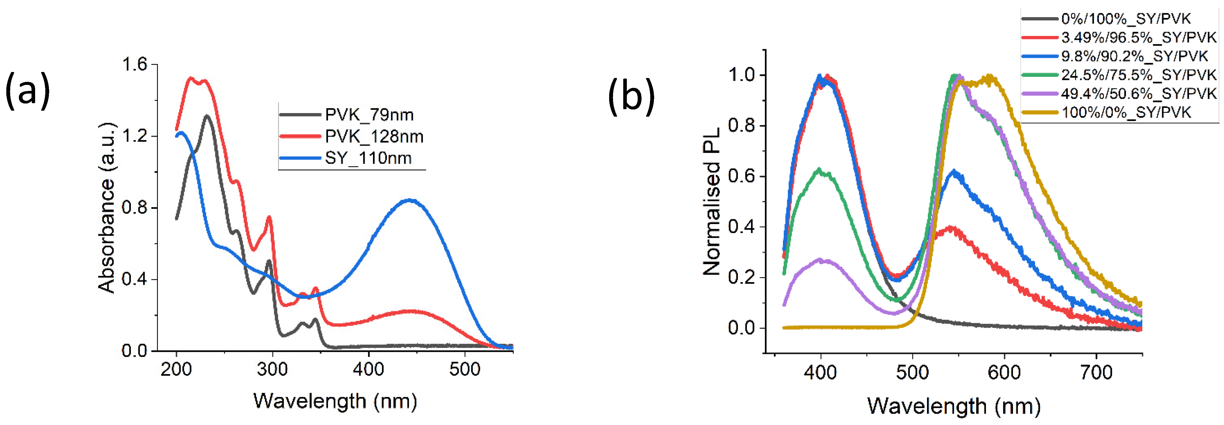
Figure 2a shows the optical absorption of SY, PVK, and a blend of SY and PVK at a 25% to 75% mass ratio. PVK is transparent in the visible wavelength region, SY absorbance peaks at about 443 nm, while the blend retains the properties of both SY and PVK. To understand whether there is a transfer of energy from the PVK to SY molecules in the polymer blend or the blend films phase-separated into regions of SY and PVK, PL measurements of the films were measured (Figure 2b). The films were excited at 340 nm, in a region where both PVK and SY absorb light. PL emissions from both SY and PVK in the blends indicate that there is incomplete transfer of energy between PVK and SY, and there is some level of phase separation of regions of SY and PVK. A blue shift is seen in the peak emission of SY in the blend, which can be attributed to a separation of the SY molecules by the PVK matrix [22].

3.2. Fluorescence Quenching and Thermal Release of DNT from Sensors

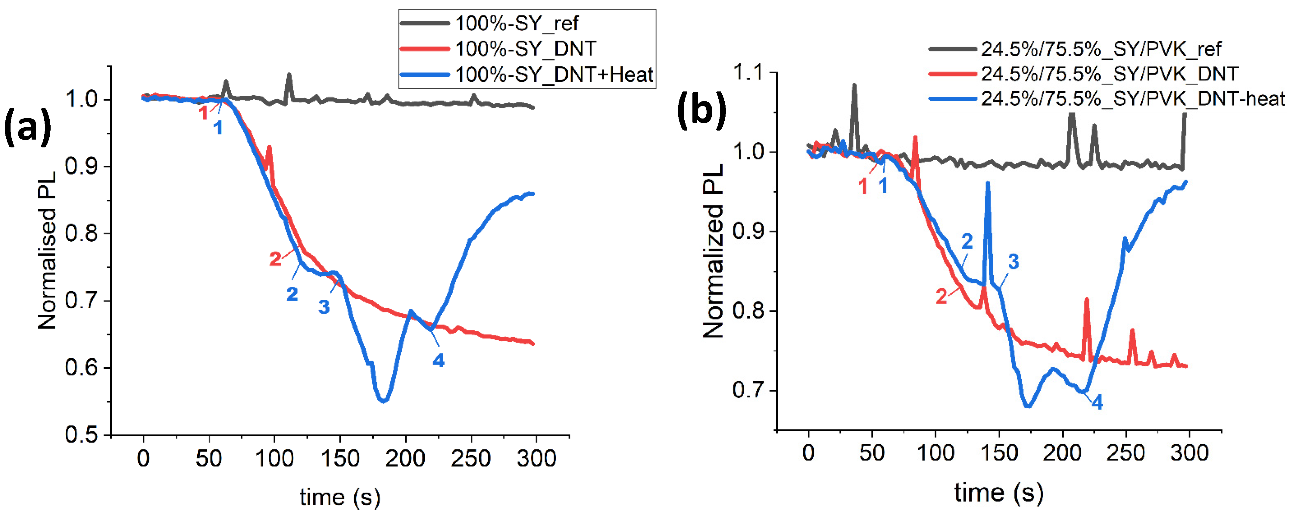
Sensing of DNT vapour using 90 nm thin films of SY and thermal desorption to recover the fluorescence of the quenched SY sensors was demonstrated (Figure 3a). For reference (black line), the PL of the sensor (when only exposed to clean nitrogen gas) remains stable throughout the 300 s. The red line shows the fluorescence from the film when exposed to DNT vapour; fluorescence was collected in clean nitrogen for 60 s, then DNT vapour was introduced in a nitrogen stream (point 1) at 6 mL/min and left to flow for 60 s before cutting off the flow (point 2).
Although the source of the DNT vapour was turned off and the system flushed with clean nitrogen, the fluorescence continued to decrease, suggesting that there was a continuing diffusion of DNT vapour into the film [14,21] and that there was a strong binding interaction between the DNT molecules and the SY film [17]. An increase in temperature can weaken the binding strength and release the DNT molecules from the thin film. The blue line shows the thermal desorption of DNT molecules from SY films after fluorescence quenching due to DNT vapour exposure. At point 3, the nitrogen gas flow was temporarily stopped and a heating element was turned on (point 3), which ramped up the temperature of the film from room temperature to 90 °C (point 4). This resulted in an initial thermal degradation [23], then a turning point at around 180 s. We attribute this change to the point when DNT molecules started desorbing from the film, as fluorescence recovery can be seen. When the nitrogen flow was turned back on (point 4), a further increase in the fluorescence was observed. However, the fluorescence could not reach the reference baseline, this may be due either to thermal degradation or there might be some residual DNT molecules left in the film [17].
To optimise the sensor in terms of thermal stability, films fabricated using a blend of SY and PVK were used for similar DNT vapour sensing—see Figure 3b. PVK was chosen because it has a high glass transition temperature and may likely improve the thermal stability of SY sensors [24]. Thermal desorption of the sorbed DNT molecules (point 3 to 4) resulted in a much higher recovery of fluorescence, almost reaching the reference baseline. An investigation of the complex processes during the temperature ramp is in progress.

4. Conclusions

We have shown that commercially available conjugated polymer Super Yellow (SY) can be used as a highly sensitive and reusable sensor for nitroaromatic explosives. An increase in the temperature of the sensors weakens the analyte binding interaction and allows the sorbed analytes to diffuse out of the thin film, which results in the recovery of the PL of the sensors. To optimise the sensors, the high thermally stable polymer PVK was blended with SY, which showed an improvement during thermal desorption of the analytes when resetting the sensors. This method can be applied to other organic fluorescent sensors, removing the limitation of single-use sensors.

Supplementary Materials

The following are available online at https://www.mdpi.com/article/10.3390/CSAC2021-10559/s1.

Author Contributions

Conceptualisation E.B.O., R.N.G., and G.A.T.; methodology, E.B.O., R.N.G., and G.A.T.; software, E.B.O.; formal analysis, E.B.O., R.N.G., and G.A.T.; investigation, E.B.O.; data curation, E.B.O.; writing—original draft preparation, E.B.O.; visualization, E.B.O.; writing—review and editing, R.N.G. and G.A.T.; funding acquisition, R.N.G. and G.A.T.; supervision, G.A.T.; project administration, G.A.T. All authors have read and agreed to the published version of the manuscript.

Funding

This research was funded by the Commonwealth Scholarships Commision, and the NATO Science for Peace & Security Programme under grant agreement MYP G5355.

Institutional Review Board Statement

Not applicable.

Informed Consent Statement

Not applicable.

Data Availability Statement

The research data underpinning this publication can be accessed at https://doi.org/10.17630/b573f7de-553f-4ec4-b907-bfdb62584fc9.

Acknowledgments

We would like to acknowledge useful scientific discussions with I. D. W. Samuel.

Conflicts of Interest

The authors declare no conflict of interest.

References

  1. Moore, D.S. Instrumentation for trace detection of high explosives. Rev. Sci. Instrum. 2004, 75, 2499–2512. [Google Scholar] [CrossRef]
  2. Wells, K.; Bradley, D.A. A review of X-ray explosives detection techniques for checked baggage. Appl. Radiat. Isot. 2012, 70, 1729–1746. [Google Scholar] [CrossRef] [Green Version]
  3. Perr, J.M.; Furton, K.G.; Almirall, J.R. Solid phase microextraction ion mobility spectrometer interface for explosive and taggant detection. J. Sep. Sci. 2005, 28, 177–183. [Google Scholar] [CrossRef]
  4. Singh, S.; Singh, M. Explosives detection systems (EDS) for aviation security. Signal Process. 2003, 83, 31–55. [Google Scholar] [CrossRef]
  5. Daniels, D.J. A review of GPR for landmine detection. Sens. Imaging Int. J. 2006, 7, 90–123. [Google Scholar] [CrossRef]
  6. Thomas, S.W.; Joly, G.D.; Swager, T.M. Chemical Sensors Based on Amplifying Fluorescent Conjugated Polymers. Chem. Rev. 2007, 107, 1339–1386. [Google Scholar] [CrossRef] [PubMed]
  7. Swager, T.M.; Wosnick, J.H. Self-Amplifying Semiconducting Polymers for Chemical Sensors. MRS Bull. 2002, 27, 446–450. [Google Scholar] [CrossRef]
  8. Cumming, C.; Aker, C.; Fisher, M.; Fok, M.; La Grone, M.; Reust, D.; Rockley, M.; Swager, T.; Towers, E.; Williams, V. Using novel fluorescent polymers as sensory materials for above-ground sensing of chemical signature compounds emanating from buried landmines. IEEE Trans. Geosci. Remote. Sens. 2001, 39, 1119–1128. [Google Scholar] [CrossRef] [Green Version]
  9. Gillanders, R.N.; Samuel, I.D.; Turnbull, G.A. A low-cost, portable optical explosive-vapour sensor. Sens. Actuators B Chem. 2017, 245, 334–340. [Google Scholar] [CrossRef] [Green Version]
  10. Caron, T.; Guillemot, M.; Montméat, P.; Veignal, F.; Perraut, F.; Prené, P.; Serein-Spirau, F. Ultra trace detection of explosives in air: Development of a portable fluorescent detector. Talanta 2010, 81, 543–548. [Google Scholar] [CrossRef]
  11. Rose, A.; Zhu, Z.; Madigan, C.F.; Swager, T.M.; Bulovic, V. Chemosensory lasing action for detection of TNT and other analytes. Organic Light Emitting Materials and Devices X. Proc. SPIE 2006, 6333, 63330Y. [Google Scholar] [CrossRef]
  12. Iii, S.W.T.; Amara, J.P.; Bjork, R.E.; Swager, T.M. Amplifying fluorescent polymer sensors for the explosives taggant 2,3-dimethyl-2,3-dinitrobutane (DMNB). Chem. Commun. 2005, 2005, 4572–4574. [Google Scholar] [CrossRef]
  13. Shaw, P.E.; Burn, P.L. Real-time fluorescence quenching-based detection of nitro-containing explosive vapours: What are the key processes? Phys. Chem. Chem. Phys. 2017, 19, 29714–29730. [Google Scholar] [CrossRef] [PubMed]
  14. Ali, M.A.; Geng, Y.; Cavaye, H.; Burn, P.L.; Gentle, I.R.; Meredith, P.; Shaw, P.E. Molecular versus exciton diffusion in fluorescence-based explosive vapour sensors. Chem. Commun. 2015, 51, 17406–17409. [Google Scholar] [CrossRef]
  15. Bolse, N.; Eckstein, R.; Schend, M.; Habermehl, A.; Eschenbaum, C.; Hernandez-Sosa, G.; Lemmer, U. A digitally printed optoelectronic nose for the selective trace detection of nitroaromatic explosive vapours using fluorescence quenching. Flex. Print. Electron. 2017, 2, 024001. [Google Scholar] [CrossRef]
  16. Zhao, D.; Swager, T.M. Sensory Responses in Solution vs Solid State: A Fluorescence Quenching Study of Poly(iptycenebutadiynylene)s. Macromolecules 2005, 38, 9377–9384. [Google Scholar] [CrossRef]
  17. Shaw, P.E.; Cavaye, H.; Chen, S.S.Y.; James, M.; Gentle, I.R.; Burn, P.L. The binding and fluorescence quenching efficiency of nitroaromatic (explosive) vapors in fluorescent carbazole dendrimer thin films. Phys. Chem. Chem. Phys. 2013, 15, 9845–9853. [Google Scholar] [CrossRef]
  18. Tang, G.; Chen, S.S.Y.; Shaw, P.; Hegedűs, K.; Wang, X.; Burn, P.L.; Meredith, P. Fluorescent carbazole dendrimers for the detection of explosives. Polym. Chem. 2011, 2, 2360–2368. [Google Scholar] [CrossRef]
  19. Greenham, N.; Samuel, I.; Hayes, G.; Phillips, R.; Kessener, Y.; Moratti, S.; Holmes, A.; Friend, R. Measurement of absolute photoluminescence quantum efficiencies in conjugated polymers. Chem. Phys. Lett. 1995, 241, 89–96. [Google Scholar] [CrossRef]
  20. Gillanders, R.N.; Glackin, J.M.; Filipi, J.; Kezic, N.; Samuel, I.D.; Turnbull, G.A. Preconcentration techniques for trace explosive sensing. Sci. Total. Environ. 2019, 658, 650–658. [Google Scholar] [CrossRef] [Green Version]
  21. Campbell, I.A.; Turnbull, G.A. A kinetic model of thin-film fluorescent sensors for strategies to enhance chemical selectivity. Phys. Chem. Chem. Phys. 2021, 23, 10791–10798. [Google Scholar] [CrossRef]
  22. Summers, M.A.; Buratto, S.K.; Edman, L. Morphology and environment-dependent fluorescence in blends containing a phenylenevinylene-conjugated polymer. Thin Solid Films 2007, 515, 8412–8418. [Google Scholar] [CrossRef]
  23. Burns, S.; MacLeod, J.; Do, T.T.; Sonar, P.; Yambem, S.D. Effect of thermal annealing Super Yellow emissive layer on efficiency of OLEDs. Sci. Rep. 2017, 7, srep40805. [Google Scholar] [CrossRef] [Green Version]
  24. Gumyusenge, A.; Tran, D.T.; Luo, X.; Pitch, G.M.; Zhao, Y.; Jenkins, K.A.; Dunn, T.J.; Ayzner, A.L.; Savoie, B.M.; Mei, J. Semiconducting polymer blends that exhibit stable charge transport at high temperatures. Science 2018, 362, 1131–1134. [Google Scholar] [CrossRef] [Green Version]
Figure 1. Chemical structures of (a) Super Yellow (SY) and (b) Poly (9-vinyl carbazole) PVK.
Chemproc 05 00011 g001
Figure 2. (a) Optical absorption of SY, PVK, and a blend of SY and PVK at 25% to 75% mass ratio, respectively; (b) Fluorescence spectra of various concentrations of SY–PVK blend at 340 nm excitation wavelength.
Chemproc 05 00011 g002
Figure 3. Fluorescence quenching due to DNT vapour and thermal desorption of DNT from sensors made of (a) SY film, and (b) blend of SY and PVK at 24.5% and 75.5% mass ratio, respectively. Key (Red): 1 = DNT flow on; 2 = DNT flow off and clean nitrogen flow on. Key (Blue): 1 = DNT flow on; 2 = DNT flow off; 3 = heat on; 4 = heat off and clean nitrogen flow on.
Chemproc 05 00011 g003
Publisher’s Note: MDPI stays neutral with regard to jurisdictional claims in published maps and institutional affiliations.