
Stokesia laevis Ethanolic Extract Activity on the Normal and Malignant Murine Cell Line Viability L969 and B16 †

 ,
,  and
and
Abstract
:1. Introduction
2. Materials and Methods
3. Results
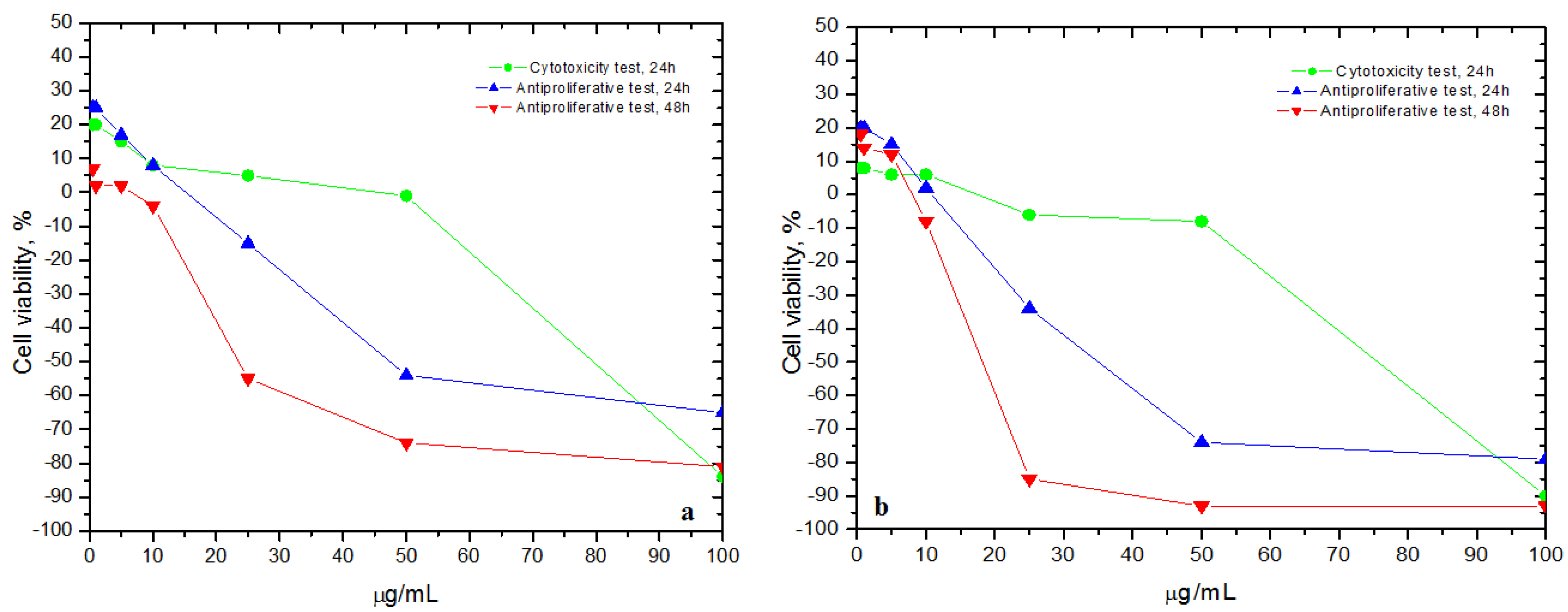
3.1. Cytotoxicity and Anti-Proliferative Assays
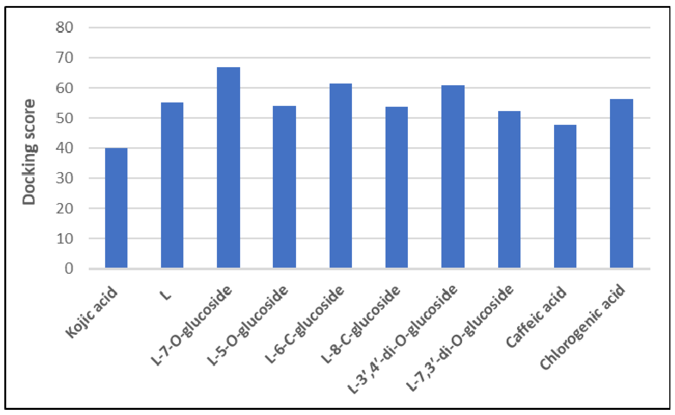
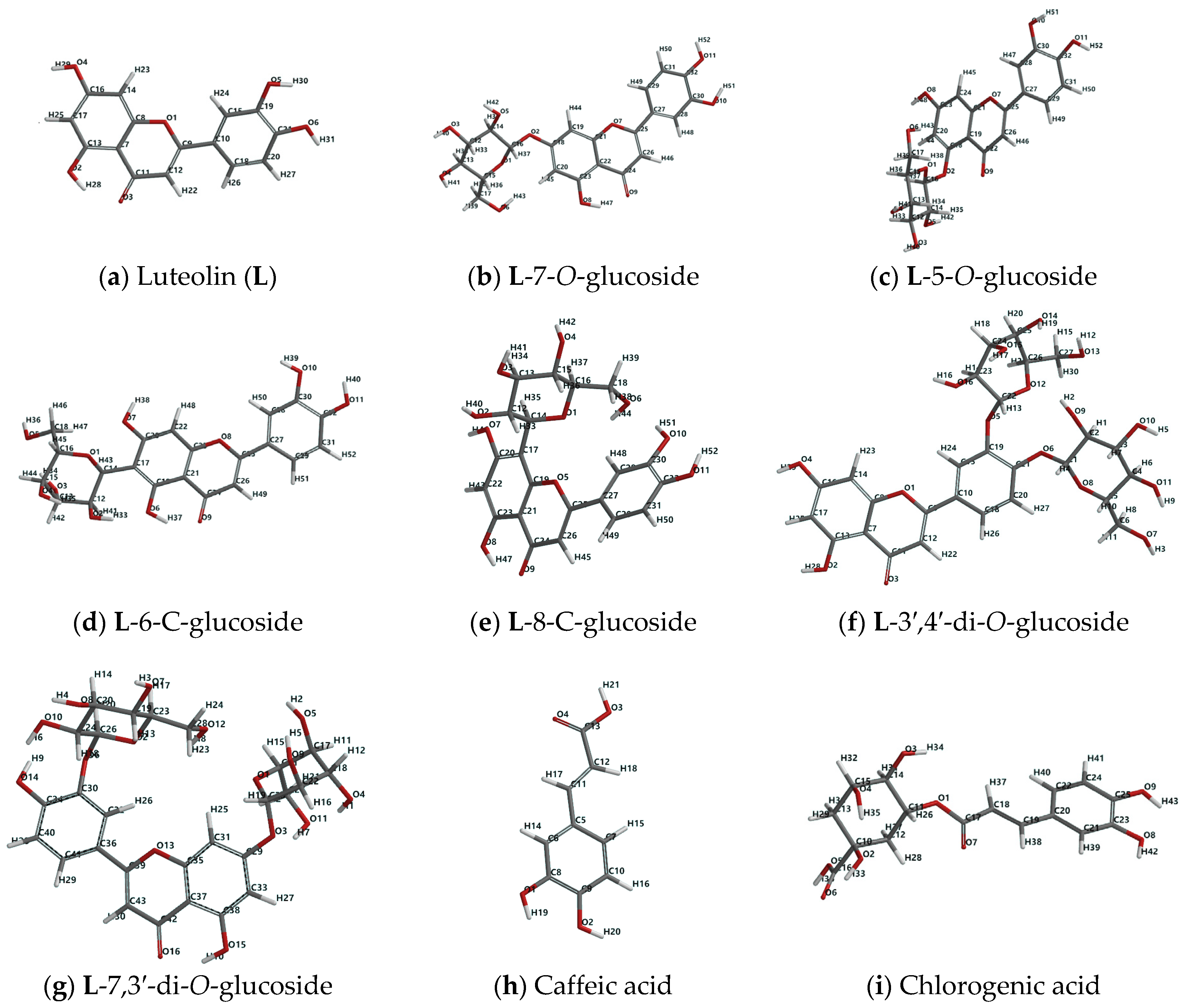
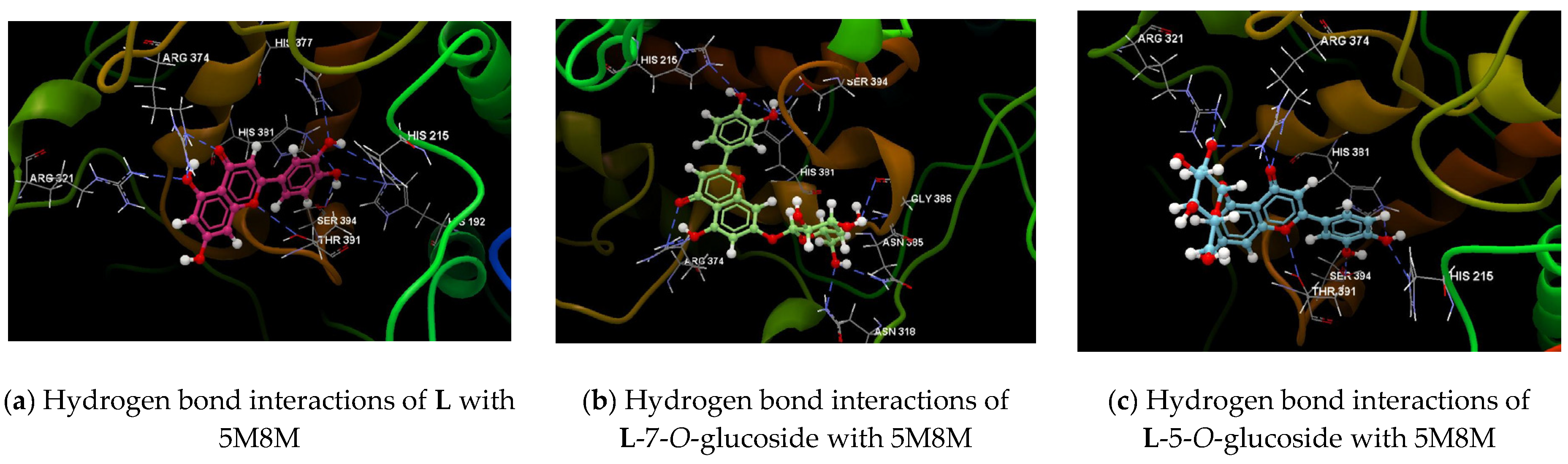
3.2. Molecular Docking Results
4. Discussion
5. Conclusions
Author Contributions
Funding
Institutional Review Board Statement
Informed Consent Statement
Conflicts of Interest
References
- Domingues, B.; Lopes, J.M.; Soares, P.; Populo, H. Melanoma treatment in review. Immunotargets Ther. 2018, 7, 35–49. [Google Scholar] [CrossRef] [PubMed]
- Skin Cancer Statistics. Available online: https://www.wcrf.org/dietandcancer/cancer-trends/skin-cancer-statistics (accessed on 24 October 2020).
- Kalal, B.S.; Upadhya, D.; Pai, V.R. Chemotherapy resistance mechanisms in advanced skin cancer. Oncol. Rev. 2017, 11, 326. [Google Scholar] [CrossRef] [PubMed]
- Chinembiri, N.T.; du Plessis, L.H.; Gerber, M.; Hamman, J.H.; du Plessis, J. Review of natural compounds for potential skin cancer treatment. Molecules 2014, 19, 11679–11721. [Google Scholar] [CrossRef] [PubMed]
- Pirvu, L.; Neagu, G.; Terchescu, I.; Albu, B.; Stefaniu, A. Comparative studies of two vegetal extracts from Stokesia laevis and Geranium pratense: Polyphenol profile, cytotoxic effect and antiproliferative activity. Open Chem. 2020, 18, 488–502. [Google Scholar] [CrossRef]
- Lai, X.; Wichers, H.J.; Soler-Lopez, M.; Dijkstra, B.W. Structure of human tyrosinase related protein 1 reveals a binuclear zinc active site important for melanogenesis. Angew. Chem. Int. Ed. 2017, 56, 9812–9815. [Google Scholar] [CrossRef] [PubMed]
- Lai, X.; Soler-Lopez, M.; Wichers, H.J.; Dijkstra, B.W. Crystal Structure of Human Tyrosinase Related Protein 1 in Complex with Kojic Acid; PDB ID: 5M8M. Deposited on: 2016-10-29. Available online: https://www.rcsb.org/structure/5M8M (accessed on 5 November 2020).
- Shao, Y.; Molnar, L.F.; Jung, Y.; Kussmann, J.; Ochsenfeld, C.; Gilbert, A.T.B.; Slipchenko, L.V.; Levchenko, S.V.; O’Neill, D.P.; DiStasio, R.A., Jr.; et al. Advances in methods and algorithms in a modern quantum chemistry program package. Phys. Chem. Chem. Phys. 2006, 8, 3172–3191. [Google Scholar] [CrossRef] [PubMed]
- Kinjo, J.; Nakano, D.; Fujioka, T.; Okabe, H. Screening of promising chemotherapeutic candidates from plants extracts. J. Nat. Med. 2016, 70, 335–360. [Google Scholar] [CrossRef] [PubMed]


| Ligand | Score | RMSD | Interacting Group (Chain A) | Hydrogen Bond | Length (Å) |
|---|---|---|---|---|---|
| KOJ A514 (grey) | −39.79 | 0.06 | HIS215, HIS192, THR391, PRO395, SER394, PHE400, GLN390, GLY388, GLY389, LEU382, HIS381, ARG374, LEE379, ASN378, HIS377, TYR362 | Osp3 (O2)-Osp3 SER394 | 2.889 |
| Osp2 (O3)-Nsp2 HIS215 | 3.203 | ||||
| L (magenta rose) | −55.02 | 0.02 | ARG374, TYR362, HIS377, HIS401, PHE220, HIS224, ARG321, LEU382, ASN378, HIS381, PHE400, HIS215, HIS192, PRO395, SER394, GLY388, GLY389, THR391, GLN390, HIS392 | Osp2 (O3)-Nsp2 ARG374 | 2.969 |
| Osp3 (O2)-Nsp2 ARG374 | 2.846 | ||||
| Osp 3(O2)-Nsp2 ARG374 | 2.885 | ||||
| Osp3 (O2)-Nsp2 ARG321 | 3.174 | ||||
| Osp2 (O1)-Osp3 THR391 | 3.166 | ||||
| Osp3 (O5)-Nsp2 HIS377 | 2.982 | ||||
| Osp3 (O5)-Nsp2 HIS215 | 3.161 | ||||
| Osp3 (O6)-Nsp2 HIS192 | 3.134 | ||||
| Osp3 (O6)-Osp3 SER394 | 2.968 | ||||
| Osp3 (O6)-Nsp2 HIS381 | 3.387 | ||||
| L-7-O-glucoside (green) | −66.76 | 1.27 | HIS192, HIS224, PHE220, HIS215, HIS404, THR391, PHE400, SER394, GLU360, GLN390, GLY388, THR387, HIS377, ASN378, HIS381, GLY389, TYR362, ARG374, GLY386, LEU384, ASN385, LEU382, PHE383, ASN318, ARG321 | Osp2 (O9)-Nsp2 ARG374 | 2.825 |
| Osp3 (O8)-Nsp2 ARG374 | 2.552 | ||||
| Osp3 (O6)-Nsp2 ASN318 | 2.967 | ||||
| Osp3 (O6)-Nsp2 ASN385 | 3.103 | ||||
| Osp3 (O4)-Nsp2 GLY386 | 3.039 | ||||
| Osp3 (O4)-Osp2 GLY386 | 3.050 | ||||
| Osp3 (O10)-Nsp2 HIS381 | 3.173 | ||||
| Osp3 (O10)-Osp3 SER394 | 2.906 | ||||
| Osp3 (O11)-Nsp2 HIS215 | 3.985 | ||||
| Osp3 (O11)-Nsp2 HIS381 | 3.084 | ||||
| L-5-O-glucoside (light blue) | −54.21 | 0.13 | ARG321, ARG374, LEU382, TYR362, ASN378, HIS381, HIS377, GLU360, HIS404, PHE220, HIS224, HIS192, HIS215, PHE400, THR391, SER394, GLN390, GLY388, GLY389 | Osp2 (O9)-Nsp2ARG374 | 3.038 |
| Osp2 (O9)-Nsp2ARG374 | 3.030 | ||||
| Osp3(O5)-Nsp2ARG374 | 2.820 | ||||
| Osp3(O5)-Nsp2ARG321 | 2.713 | ||||
| Osp2(O7)-Osp3THR391 | 2.744 | ||||
| Osp3(O11)-Nsp2HIS215 | 3.103 | ||||
| Osp3(O11)-Nsp2HIS381 | 3.225 | ||||
| Osp3(O10)-Osp3SER394 | 2.642 | ||||
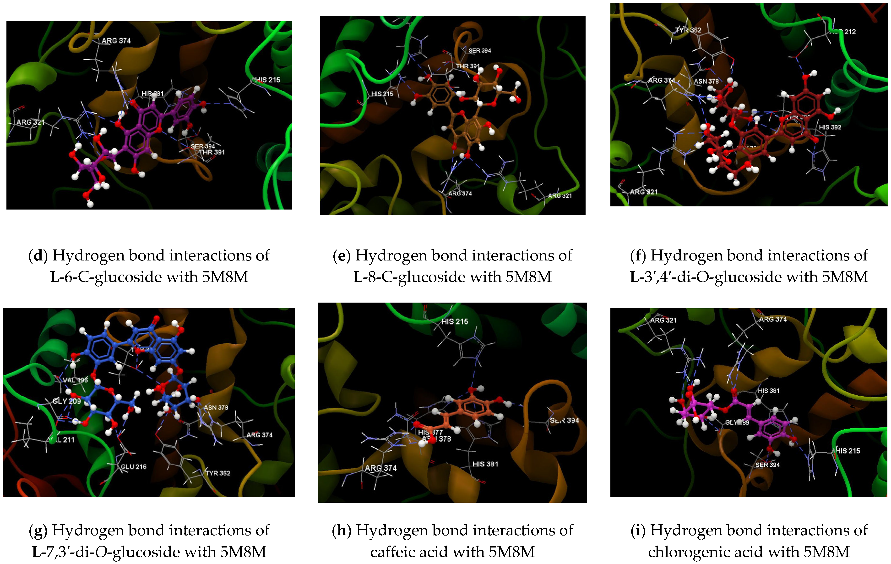
| L-6-C-glucoside (purple) | −61.55 | 0.15 | ARG374, TYR362, GLU360, HIS377, ASN378, HIS404, PHE220, HIS224, HIS215, HIS192, HIS392, THR391, SER394, GLN390, GLY388, GLY389, HIS381, LEU382, ARG321, ASN318 | Osp3(O9)-Nsp2 ARG374 | 2.423 |
| Osp3(O9)-Nsp2 ARG374 | 3.156 | ||||
| Osp3(O6)-Nsp2 ARG374 | 3.110 | ||||
| Osp3(O6)-Nsp2 ARG321 | 3.115 | ||||
| Osp3(O2)-Nsp2 ARG321 | 2.990 | ||||
| Osp3(O2)-Nsp2 ARG321 | 2.901 | ||||
| Osp3(O1)-Nsp2 ARG321 | 2.967 | ||||
| Osp3(O4)-Nsp2 ARG321 | 2.831 | ||||
| Osp3(O10)-Osp3 SER394 | 2.502 | ||||
| Osp3(O11)-Nsp2 HIS381 | 3.248 | ||||
| Osp3(O11)-Nsp2HIS215 | 3.113 | ||||
| Osp2(O8)-Osp3THR391 | 3.180 | ||||
| L-8-C-glucoside (brown) | −53.81 | 0.03 | HIS192, HIS392, SER394, PHE400, THR391, GLN390, GLY388, GLY389, HIS381, LEU382, ARG321, ARG374, TYR362, ASN378, HIS377, GLU360, PHE220, HIS215, HIS204, HIS224 | Osp2 (O9)- Nsp2ARG374 | 2.884 |
| Osp2 (O8)-Nsp2ARG374 | 2.569 | ||||
| Osp2 (O8)-Nsp2ARG374 | 2.592 | ||||
| Osp2 (O8)-Nsp2 ARG321 | 3.083 | ||||
| Osp3 (O10)-Nsp2 HIS215 | 2.751 | ||||
| Osp3 (O11)-Nsp2 HIS192 | 3.134 | ||||
| Osp3 (O11)-Osp3 SER394 | 3.060 | ||||
| Osp3 (O2)-Osp3 THR391 | 2.451 | ||||
| L-3′,4′-di-O-glucoside (red brown) | −60.91 | 2.68 | GLU216, HIS215, ASP212, VAL211, VAL196, GLY209, LYS198, LYS197, LEU293, HIS392, THR391, GLN390, GLY388, GLY389, ARG321, LEU382, HIS381, LEU379, ASN378, ARG374, HIS377, TYR362 | Osp2 (O3)-Nsp2 HIS392 | 2.789 |
| Osp3 (O4)-Osp3 ASP212 | 3.001 | ||||
| Osp2 (O1)-Osp3 THR391 | 2.707 | ||||
| Osp3 (O12)-Osp3 THR391 | 3.271 | ||||
| Osp3 (O13)-Osp3 THR391 | 2.906 | ||||
| Osp3 (O13)-Nsp2 THR391 | 3.050 | ||||
| Osp3 (O13)-Osp2 GLY389 | 2.856 | ||||
| Osp3 (O14)-Osp2 ASN378 | 3.046 | ||||
| Osp3 (O15)-Osp3 TYR362 | 2.656 | ||||
| Osp3 (O16)-Nsp2 ARG374 | 3.307 | ||||
| Osp3 (O16)-Nsp2 ARG374 | 2.671 | ||||
| Osp3 (O9)-Nsp2 ARG321 | 3.022 | ||||
| Osp3 (O9)-Nsp2 ARG321 | 2.909 | ||||
| Osp3 (O10)-Nsp2 ARG374 | 3.073 | ||||
| L-7,3′-di-O-glucoside (blue) | −52.44 | 2.43 | LEU293, HIS392, THR391, GLN390, GLY389, HIS381, LEU382, ARG321, ASN378, HIS377, ARG374, GLU360, TYR362, TYR348, GLU216, HIS215, GLU210, VAL211, ASP212, GLY209, VAL196, LYS198, LYS197 | Osp2 (O13)-Osp3 THR391 | 2.836 |
| Osp2 (O13)-Nsp2 THR391 | 3.192 | ||||
| Osp3 (O14)-Osp2 VAL196 | 2.698 | ||||
| Osp3 (O10)-Osp2 VAL196 | 3.094 | ||||
| Osp3 (O10)-Osp2 VAL211 | 2.913 | ||||
| Osp3 (O10)-Osp2 GLY209 | 3.240 | ||||
| Osp3 (O8)-Osp2 VAL211 | 2.867 | ||||
| Osp3 (O8)-Osp2 GLY209 | 2.823 | ||||
| Osp3 (O7)-Osp3 GLU216 | 2.674 | ||||
| Osp3 (O12)-Osp3 GLU216 | 3.054 | ||||
| Osp3 (O1)-Osp3 THR391 | 3.157 | ||||
| Osp3 (O5)-Osp3 TYR362 | 2.920 | ||||
| Osp3 (O5)-Osp2 ASN378 | 3.089 | ||||
| Osp3 (O4)-Nsp2 ARG374 | 3.148 | ||||
| Osp3 (O4)-Nsp2 ARG374 | 2.554 | ||||
| Caffeic acid (orange brown) | −47.63 | 0.05 | PHE220, HIS215, HIS224, HIS192, THR391, PRO395, HIS404, SER394, PHE400, GLN390, GLY388, GLY389, LEU382, HIS381, ASN378, HIS307, ARG374, TYR362, GLU360 | Osp3 (O3)-Nsp2 ARG374 | 3.146 |
| Osp2 (O4)-Nsp2 ARG374 | 2.860 | ||||
| Osp2 (O4)-Nsp2 ASN378 | 3.034 | ||||
| Osp3 (O1)-Nsp2 HIS377 | 3.195 | ||||
| Osp3 (O1)-Nsp2 HIS215 | 3.019 | ||||
| Osp3 (O1)-Nsp2 HIS381 | 3.374 | ||||
| Osp3 (O2)-Osp3 SER394 | 2.424 | ||||
| Chlorogenic acid (light purple) | −56.08 | 2.05 | ARG321, ARG374, TYR362, LEU382, ASN378, HIS377, GLU360, HIS381, HIS404, PHE220, HIS224, HIS215, PHE400, GLY389, GLY388, GLN390, SER394, THR391, PRO395, HIS192 | Osp3 (O9)-Nsp2 HIS381 | 3.289 |
| Osp3 (O9)-Nsp2 HIS215 | 3.031 | ||||
| Osp3 (O8)-Osp3 SER394 | 2.523 | ||||
| Osp2 (O7)-Nsp2 ARG374 | 2.854 | ||||
| Osp2 (O7)-Nsp2 ARG374 | 3.072 | ||||
| Osp3 (O2)-Nsp2 ARG321 | 3.107 | ||||
| Osp3 (O2)-Nsp2 ARG321 | 2.738 | ||||
| Osp3 (O4)-Nsp2 ARG321 | 2.862 | ||||
| Osp3 (O3)-Osp2 GLY389 | 2.817 |
Publisher’s Note: MDPI stays neutral with regard to jurisdictional claims in published maps and institutional affiliations. |
© 2020 by the authors. Licensee MDPI, Basel, Switzerland. This article is an open access article distributed under the terms and conditions of the Creative Commons Attribution (CC BY) license (https://creativecommons.org/licenses/by/4.0/).
Share and Cite
Neagu, G.; Stefaniu, A.; Albu, B.; Terchescu, I.; Pintilie, L.; Pirvu, L.C. Stokesia laevis Ethanolic Extract Activity on the Normal and Malignant Murine Cell Line Viability L969 and B16. Chem. Proc. 2021, 3, 42. https://doi.org/10.3390/ecsoc-24-08318
Neagu G, Stefaniu A, Albu B, Terchescu I, Pintilie L, Pirvu LC. Stokesia laevis Ethanolic Extract Activity on the Normal and Malignant Murine Cell Line Viability L969 and B16. Chemistry Proceedings. 2021; 3(1):42. https://doi.org/10.3390/ecsoc-24-08318
Chicago/Turabian StyleNeagu, Georgeta, Amalia Stefaniu, Bujor Albu, Iulian Terchescu, Lucia Pintilie, and Lucia Camelia Pirvu. 2021. "Stokesia laevis Ethanolic Extract Activity on the Normal and Malignant Murine Cell Line Viability L969 and B16" Chemistry Proceedings 3, no. 1: 42. https://doi.org/10.3390/ecsoc-24-08318
APA StyleNeagu, G., Stefaniu, A., Albu, B., Terchescu, I., Pintilie, L., & Pirvu, L. C. (2021). Stokesia laevis Ethanolic Extract Activity on the Normal and Malignant Murine Cell Line Viability L969 and B16. Chemistry Proceedings, 3(1), 42. https://doi.org/10.3390/ecsoc-24-08318