Abstract
Background: Schizophrenia manifests through behavioral abnormalities, suicidal ideation, and neuropsychological deficits. Hence, this study focused on 5-hydroxytryptamine (5-HT 2C) which influenced the modulation of the series of events that lead to schizophrenia. Methodology: Based on the computational study, the potential 5-HT 2C inhibitors such as Ephemeranthoquinone from Arundina graminifolia and Actinodaphnine from Litsea polyantha were determined. The candidate ligands were optimized using the Gaussian 16 software package and the DFT 6-31g (d,p) basis set. The interaction between the ligands and proteins was examined with PyRx 0.8. Additionally, pharmacokinetics was assessed using SwissADME, and Protox II for toxicity prediction. The network pharmacology study was examined by using the STRING database and the Cytoscape 3.10.1 tool. Moreover, a 100-nanosecond molecular dynamics simulation analysis using Desmond to ensure the stability of these two compounds was carried out. Result: This computational research observed that ephemeranthoquinone and actinodaphnine are the most selective 5-HT 2C inhibitors due to their docking score, optimization, and molecular dynamics simulation results. Conclusions: These compounds are required to be studied further to develop a useful 5-HT 2C inhibitors for the treatment of schizophrenia.
1. Introduction
As a complex mental illness with lifetime prevalence, schizophrenia (SCZ) impacts about 24 million people globally, equating to 0.32% of the population [1]. Unfortunately, currently available therapies have also failed to tackle SCZ on the molecular level and have come with significant adverse effects which can exacerbate the patient’s condition [2]. Given these challenges, natural compounds from medicinal plants present a promising alternative for treating SCZ due to their enrichment in secondary metabolites which have minimal side effects [3].
Our target, the 5-HT 2C receptor at Xq24, belongs to the G-protein-coupled receptor (GPCR) superfamily and is mainly associated with serotonin neurotransmission via the HT2CR in the cortico-limbic circuitry pathway that is relevant to SCZ [4]. Additionally, the hypo-glutamatergic basis for certain SCZ symptoms may involve HTR2C, which is present in GABAergic interneurons [5]. Considering the mechanism, computational studies in drug design aim to develop potent antipsychotics from medicinal plants that treat SCZ. Consequently, in this study, molecular docking and simulations were used to understand binding interactions and optimize ligand stability. Further, this approach highlights the potential of network pharmacology and natural compounds in SCZ treatment, hoping for effective drug development through further preclinical studies.
2. Methodology
2.1. Preparation of Protein and Ligands
The RCSB protein databank provided the 3D structure of the 5-HT 2C protein (PDB ID: 6BQH), which was produced using Discovery Studio 2020 by eliminating co-factors and stabilized using SWISS PDB 4.10. Approximately, sixty CNS-Penetrant compounds were chosen from the IMPPAT database and retrieved from PubChem in SDF format. Subsequently, the compound library was prepared using OpenBabel 3.1.1 software.
2.2. ADMET Analysis
After screening plants, the pharmacokinetics (PK) and ADME properties of chosen compounds were estimated using SwissADME along with the Protox-II web tool, which was utilized to analyze the toxicity of the compounds we found.
2.3. Molecular Docking and Network Pharmacology Study
The best binding configuration of the target protein with ligands was found using the PyRx 0.8 tool. Also, the protein–ligand complex’s binding pose was observed using Pymol 2.5.2. and the Discovery Studio 2021 BIOVIA visualizer. The potential interaction between 5-HT 2C and other proteins was investigated in the STRING database and the Cytoscape 3.10.1 tool to understand the connection between the top two ligands, the targeted protein, and linked diseases.
2.4. Optimization
The DFT theoretical computations were performed in the gas phase using the 631-G d,p (+,+) basis set integrating into Gaussian 9 software package to observe the stability of medicines’ softness (S) and hardness (η) by using the following formula:
2.5. Molecular Dynamic Simulation
To assure the stability of protein–ligand complex, molecular dynamic simulation was run in the Desmond Dynamics module. It is available in the Schrödinger suite, for 100 picoseconds at energy of 1.2, with a simple point-charge (SPC) water model assigned with an orthorhombic periodic boundary box at a distance of (10 × 10 × 10 A3), concentration of salt at 0.15 M, Na+ and Cl− ions, OPLS3e force field, a temperature of 300.0 K and bar pressure of 1.01325, by calculating the root mean square deviation (RMSD), root mean square fluctuation (RMSF), solvent-accessible surface area (SASA) values, and radius of gyration (rRg).
3. Results and Discussion
3.1. Molecular Docking
After performing the docking study, we considered only two compounds with the highest binding affinity for further study; these are displayed in Table 1. The protein–ligand interaction is presented in Figure 1.
Table 1.
A list of ligand names and binding affinities with the RMSD values of the top two compounds.
Figure 1.
Protein–ligand binding interaction of top two compounds based on binding score: (a) ephemeranthoquinone and (b) actinodaphnine.
As for the hydrogen bond in the protein–ligand interaction, the donor and acceptor pairs should be at a distance of 2.7–3.3 Å. Ephemeranthoquinone (CID 10038025) and actinodaphnine (CID 160502) have different hydrogen bond distances in this investigation, as illustrated in Table 2.
Table 2.
The highest-ranking protein–ligand complex and the non-bonding interaction of the top two compounds with amino acid residues of 5-HT 2C.
3.2. ADMET Analysis
The pharmacokinetic parameters and toxicological characteristics of the top two compounds are listed in Table 3 and Table 4.
Table 3.
ADME analysis of the top two compounds showing the molecular weight, lipophilicity (XLOGP3), water solubility (Log S (ESOL)), GI absorption, BBB permeant, and Lipinski rule of five.
Table 4.
The toxicity profile of the top two compounds.
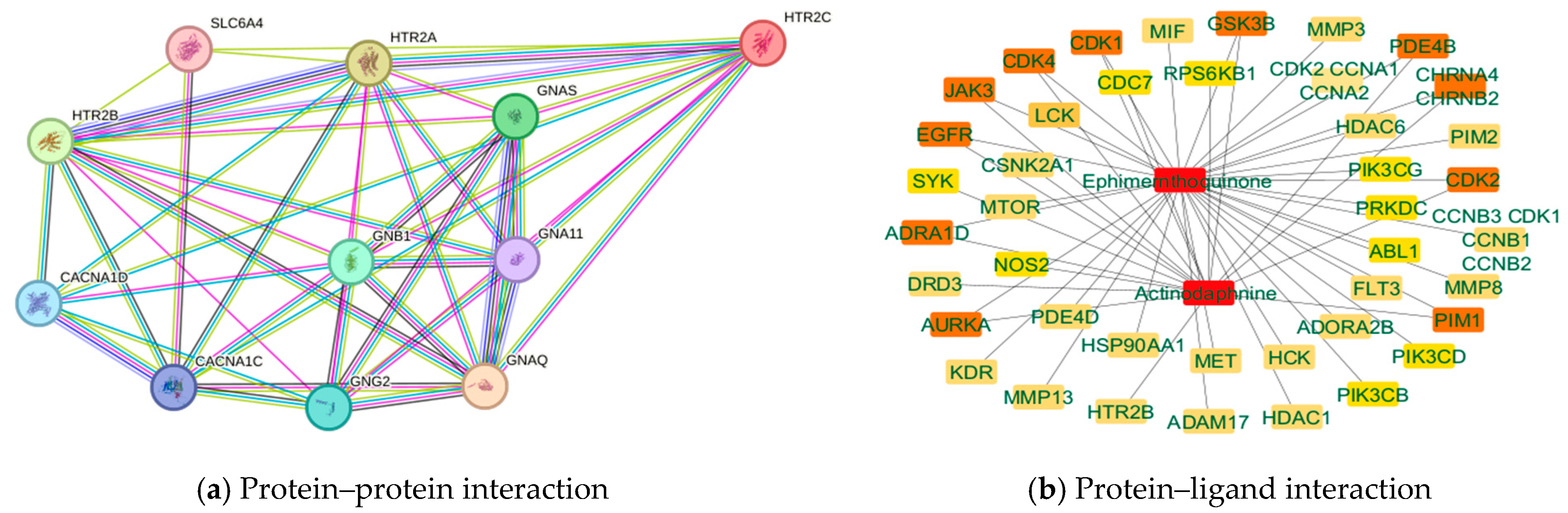
3.3. Network Pharmacology
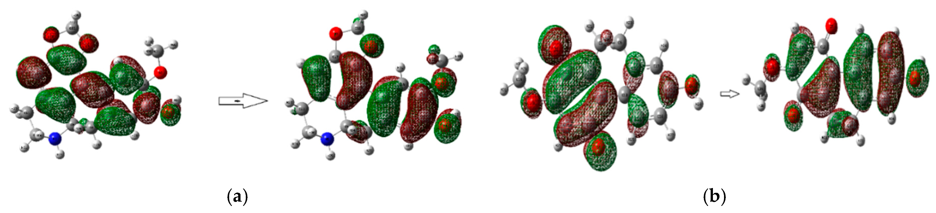
The network diagram data of the targeted protein with other proteins and the top two compounds with other protein interactions are shown in Figure 2a, whereas the interacted protein HTR2A is also responsible for SCZ. Conversely, Figure 2b shows that candidate compounds primarily influenced the genes PIM1, GSK3B, and EGFR.
Figure 2.
Network pharmacology analysis of 5-HT 2C protein (a) and top two compounds (b).
3.4. Optimization
The two global chemical descriptors (softness and hardness) and the orbital energies for the two compounds are shown in Table 5. Ephemeranthoquinone has the highest softness with the lowest HOMO-LUMO gap and hardness, indicating a more reactive molecule overall. In contrast, actinodaphnine is less soft than ephemeranthoqunine and has a somewhat higher hardness and HOMO-LUGO gap. Moreover, Table 6 presents the compounds’ stoichiometry, enthalpy, Gibbs free energy, electronic energy, and dipole moment. Figure 3 shows the optimized structures, where actinodaphnine has the highest energy, enthalpy, and Gibbs free energy, along with the largest dipole moment of 2.220016 Debye, indicating a high polarity in real life.
Table 5.
The energy of HOMO and LUGO, and the gap, hardness, and softness (all units are in hartree) of ephemeranthoquinone and actinodaphnine.
Table 6.
The stereochemistry, electronic energy, enthalpy, Gibbs free energy (in hartree), and dipole moment (Debye) of ephemeranthoquinone and actinodaphnine.
Figure 3.
The optimization structure of the top two compounds, (a) Actinodaphnine and (b) Ephemeranthoquinone.
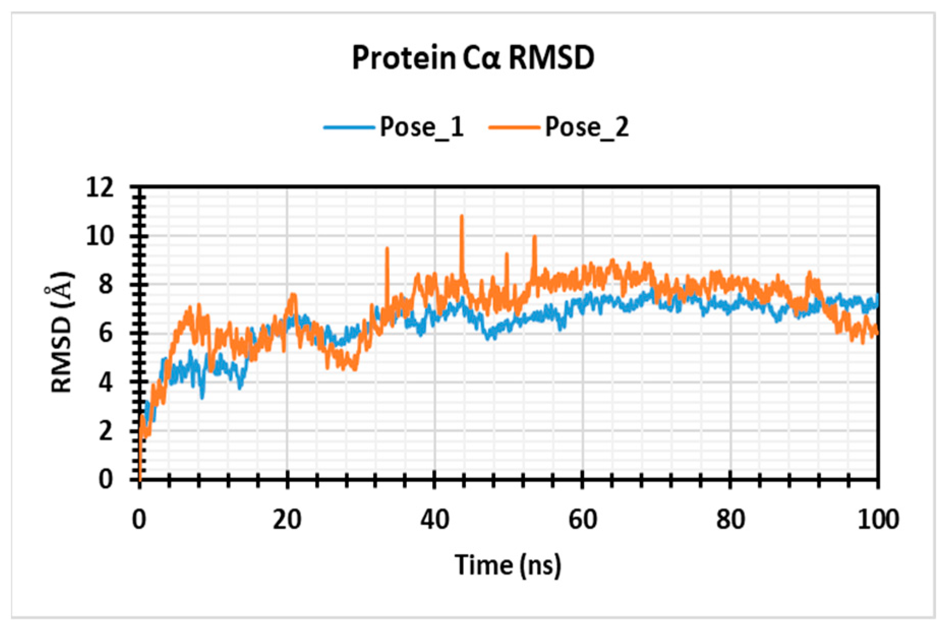
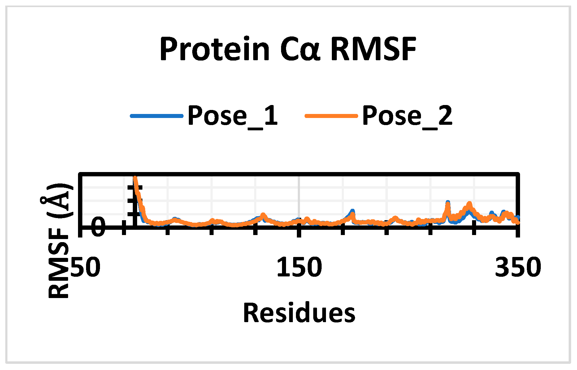
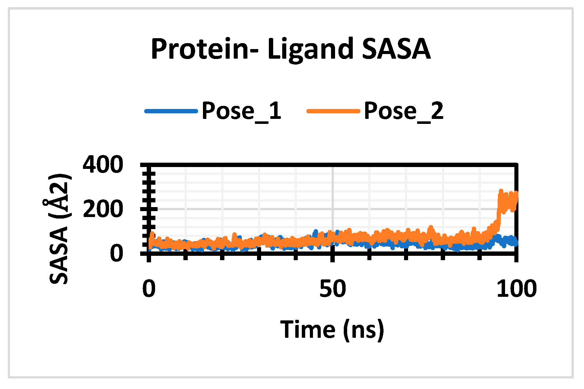
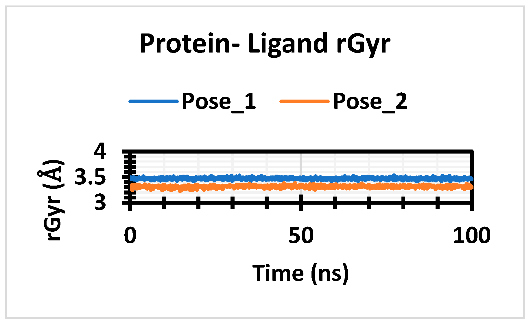
3.5. Molecular Dynamic Simulation
In this experiment, a 100 ns MD simulation was used to obtain a better knowledge of the conformational changes of the protein with a particular ligand by examining the SASA, the rGyr, RMSF, and RMSD. Of the two most highly selected compounds, CID 160502 had average RMSD values of 6.39 Å and exhibited reduced fluctuations. Conversely, the average RMSD value of the CID 10038025 compound was 6.97 Å. It exhibited poorer stability with large fluctuations across the simulation time of 34 to 54 ns, as demonstrated in Figure 4. Again, from Figure 5, it is clear that a maximum deviation of 14.878 Å is seen between residues in the PHE 46 control 5HT 2C instance. Greater fluctuations are observed twice for the first compound (CID_160502) between the residues PHE 46 and LYS 47; these are approximately 14.768 Å and 12.335 Å, respectively. The second compound (CID_10038025) yields a maximum variation of 14.878 Å in PHE 46 and 13.172 Å in LYS 47. The average value of the first compound (CID_ 160502) is 45.91 Å, and the average value of the second compound (CID_10038025) is 69.41 Å, as shown in Figure 5. The complex system’s average SASA value, ranging from 80 Å to 195 Å, indicated that the compounds that were selected were subjected to high quantities of amino acid residues, as depicted in Figure 6. In Figure 7, the stability of the target protein complexes of CID_160502 and CID_10038025 was also examined in terms of rGyr. The average rGyr for the compounds with CID_160502 and CID_10038025 was 3.47 Å and 3.31 Å, respectively.
Figure 4.
RMSD values of top 2 compounds.
Figure 5.
RMSF value of top 2 compounds.
Figure 6.
SASA value of top 2 compounds.
Figure 7.
rGyr value of top 2 compounds.
4. Conclusions
In conclusion, because of the exceptional pharmacokinetic properties, good bioavailability characteristics, and noteworthy biochemical interactions of ephemeranthoquinone and actinodaphnine against 5-HT 2C receptors, further research using animal models and preclinical studies should be conducted to examine these two naturally occurring chemicals as latent 5-HT 2C inhibitors in order to produce antipsychotic medications to treat SCZ.
Author Contributions
All authors have equally contributed to this work. All authors have read and agreed to the published version of the manuscript.
Funding
This research received no external funding.
Data Availability Statement
The data presented in this study are available on request from the corresponding author due to privacy concern of further research.
Acknowledgments
This research is supported by the Bioinformatics and Molecular Dynamics Simulation Laboratory, Department of Pharmacy, University of Science and Technology Chittagong, Bangladesh.
Conflicts of Interest
The authors declare no conflicts of interest.
References
- Bhalala, O.G.; Nath, A.P.; Consortium, U.B.E.; Inouye, M.; Sibley, C.R. Identification of expression quantitative trait loci associated with schizophrenia and affective disorders in normal brain tissue. PLoS Genet. 2018, 14, e1007607. [Google Scholar] [CrossRef] [PubMed]
- Blokhin, I.O.; Khorkova, O.; Saveanu, R.V.; Wahlestedt, C. Molecular mechanisms of psychiatric diseases. Neurobiol. Dis. 2020, 146, 105136. [Google Scholar] [CrossRef] [PubMed]
- Munawar, N.; Ahsan, K.; Ahmad, A. Natural molecules in the treatment of schizophrenia. In Natural Molecules in Neuroprotection and Neurotoxicity; Elsevier: Amsterdam, The Netherlands, 2024; pp. 259–280. [Google Scholar]
- Stępnicki, P.; Kondej, M.; Kaczor, A.A. Current concepts and treatments of schizophrenia. Molecules 2018, 23, 2087. [Google Scholar] [CrossRef] [PubMed]
- Cacabelos, R.; Martínez-Bouza, R. Genomics and pharmacogenomics of schizophrenia. CNS Neurosci. Ther. 2011, 17, 541–565. [Google Scholar] [CrossRef]
Disclaimer/Publisher’s Note: The statements, opinions and data contained in all publications are solely those of the individual author(s) and contributor(s) and not of MDPI and/or the editor(s). MDPI and/or the editor(s) disclaim responsibility for any injury to people or property resulting from any ideas, methods, instructions or products referred to in the content. |
© 2025 by the authors. Licensee MDPI, Basel, Switzerland. This article is an open access article distributed under the terms and conditions of the Creative Commons Attribution (CC BY) license (https://creativecommons.org/licenses/by/4.0/).


