Proceeding Paper

Cyclodextrin-Based Host–Guest Supramolecular Nanofibrous Composite for Biomedical Applications †

1
School of Nano-Tech and Nano-Bionics, University of Science and Technology of China, Hefei 230026, China
2
Suzhou Institute of Nano-Tech and Nano-Bionics, Chinese Academy of Sciences, Suzhou 215123, China
*
Authors to whom correspondence should be addressed.
Presented at the 26th International Electronic Conference on Synthetic Organic Chemistry, 15–30 November 2022; Available online: https://sciforum.net/event/ecsoc-26.
Chem. Proc. 2022, 12(1), 60; https://doi.org/10.3390/ecsoc-26-13523
Published: 14 November 2022

Abstract

:
Cyclodextrins (CDs) are macrocyclic oligosaccharides, containing between six and eight alpha(1 → 4)-linked glucopyranoses. CDs have a hydrophobic cone-shaped internal cavity and a hydrophilic exterior surface. They form non-covalent inclusion complexes (ICs) with various drugs by trapping the full or partial inclusions in their cavity. Supramolecular ICs have gained attention in engineering entrapped drug performance field due to their potential to protect and modify the physicochemical properties of entrapped lipophilic and volatile drugs. However, the poor structural and mechanical properties of pure CD-ICs could restrict their application and the need for a suitable carrier system. Electrospun nanofibers have been the center of attention for biomedical applications due to their tunable physicochemical properties. Recent studies have highlighted that the entrapment of drug/CD-based ICs into nanofibers is an active research area since it facilitates high encapsulation, it modulates the release profile of the guest, integrates multi-type drugs, and leads to a synergistic effect. This mini-review first summarizes the potential benefits and shortcomings of drug/CD-ICs and nanofibers, and then, we discuss the advancements in the fabrication and characteristics of CD-ICs embedded nanofibers, along with some practical suggestions for potential biomedical applications.

1. Introduction

A supramolecular system comprises two or more molecular entities that are held together using non-covalent binding interactions. Cyclodextrins (CDs) act as a common host for supramolecular chemistry, and they are rapidly gaining attention in biomedical and pharmaceutical applications fields. CDs are non-reducing macrocyclic ring-shaped oligomers composed of 6, 7, and 8 alpha(1 4)-linked glucopyranose units that are commonly called αCDs, βCDs, and γCDs, respectively. It has attracted attention in the engineering pharmacological fields because of its cone-shaped structure, which possesses a hydrophobic internal cavity (given by the C–H bonds) and a hydrophilic exterior (due to the distribution of free O–H) [1,2]. The internal diameter varies among the CD types, and they are correlated with the number of glucopyranose units, i.e., the internal cavity diameter increases in the following order: γCDs (0.75–0.83 nm) > βCDs (0.61–0.65 nm) > αCDs (0.45–0.53 nm), while the depth of the cavity is the same (~0.78 nm) (Figure 1A,B). CDs with less than six glucose units do not exist (for stoichiometric reasons), while CDs with more than eight glucose units provide weak complexing properties [3]. Some chemically modified forms of CDs, i.e., hydroxypropyl-βCDs (HPβCDs), hydroxypropyl-γCDs, methylated-βCDs, and sulfobutylether-βCDs, etc., have been introduced to achieve higher solubility, increased drug stability, and the potential to undergo polymerization reactions and to extend the range of their practical applications [4,5].
The unique structural properties of CDs offer distinguished physicochemical and encapsulation functionalities due to their potential to form inclusion complexes (ICs) against a variety of solid, liquid, and gaseous compounds with matched polarity, hydrophobicity, and molecular dimension [6,7]. The process of the ‘‘guest’’ inclusion into the CD occurs at the molecular level, and it is attractive for engineering novel functional materials. Some driving forces that support complexation include geometric compatibility, hydrophobic effect, electrostatic interaction, dipole–dipole forces, and charge–transfer interactions [8]. It furnishes a suitable microenvironment for the guest molecules through non-covalent host–guest ICs and positively modifies their physicochemical properties (Figure 1C). CDs-based ICs are widely used in biomedical, pharmaceutical, food industries, analytical chemistry, water purification, and oilfield applications [9,10].
CDs can accommodate and modify the physicochemical properties of many entrapped bioactive drugs, such as antimicrobial agents, peptides, and nucleic acid, etc., mainly in a suspension formulation [11]. However, a few studies have explored these improvements through nanostructured solid formulation for specific biomedical applications. This mini-review first summarized the potential benefits and shortcomings of drug/CD-ICs and nanofibers, and then, we discuss the advancements in the fabrication and characteristics of CD-ICs embedded supramolecular nanofibers, along with some practical suggestions for potential biomedical applications.

2. Benefits and Shortcomings of CD-ICs in Biomedical Applications

Most drugs and bioactive substances either have poor heat stability, low water solubility, or volatile properties, making it difficult to use these molecules in their natural states [7]. The supramolecular ICs protect and shield the guest drugs against devastating environmental effects (heat, moisture, chemical reactions, and oxidation) [12]. ICs can improve the solubility, bioavailability, photostability, therapeutic index, physicochemical stability, and shelf life of the entrapped drugs. They can reduce their volatility, modulate the release profile of the guest molecules, and limit the unpleasant taste and smell of some functional compounds [13]. They could decrease the local irritation and increase the permeability of poorly soluble drugs across biological membranes [14]. ICs have been administered through different routes (dermal, ophthalmic, nasal, oral, rectal, and intravenous ones) [5], and they have great potential in pharmaceutical applications. However, the usage of drugs in suspension and tablet forms can cause unfavorable therapeutic effects, uncontrolled drug release, and poor self-healing properties, which could limit their practical application. These factors underscore the need for a solid-state nano-carrier system for enhanced biomedical applications.

3. Benefits and Shortcomings of Nanofibers in Biomedical Applications

Electrospinning is a simple and affordable technology that uses an electrostatic voltage to fabricate continuous nanoscale fibers from various synthetic, natural, and hybrid polymeric materials [15] (Figure 1D). Electrospun fibers offer some benefits including a regulated surface chemistry, a high drug loading capacity, a customizable, microporous morphology, a nanoscale diameter, a huge surface area, and strong mechanical qualities [16,17]. Electrospinning could fabricate tailored nanocomposites that can conform over different surfaces/substrates and can be engineered to have multiple shapes [18,19]. Nanofibers could replicate the structural and functional characteristics of the extracellular matrix, and their surface properties can be altered with surface conjugated ligands to modulate the drug release, bio-adhesive, and cell-instructive properties [17,20]. Electrospun fibers are being increasingly recognized in tissue engineering, coatings for biomedical implants, wound dressings, sustained medication release, and in the encapsulation of different bioactive substances for different biomedical applications [21,22,23]. Since most drugs are water soluble and volatile, incorporating a considerable drug into the fibers, while maintaining their biological activity and physicochemical stability can be problematic for drug delivery applications.

4. CD-ICs-Incorporated Nanofibrous Membranes in Biomedical Applications

Most medications and bioactive substances exhibit poor heat stability, limited water solubility, or volatility, and these characteristics hinder the drugs from reaching the therapeutic concentrations for an effective treatment. Drugs loading into electrospun fibers by blending them in a suspension before the electrospinning process or using specific conjugated chemistry cannot overcome these limitations [24]. CDs-ICs offer significant advantages over the non-complexed forms of a drug, but their poor structural and mechanical properties could restrict the practical application. Recent studies have depicted that incorporating CD-ICs into electrospun nanofibers has gained significant attention for modifying the drug loading capacity, drug release kinetics, and drug performance [25,26,27,28] (Figure 1E–G).
Polymer-free (ciprofloxacin/βCDs and dexamethasone/βCDs) and polycaprolactone (PCL)-based ICs-embedded supramolecular nanofibers were fabricated in the combined form via electrospinning. The surface of ICs functionalized nanofibers was further modified using adhesive nanofibers using catechol chemistry. The results showed that PCL-based ICs nanofibers possess good mechanical properties, they are convenient for storage, and they integrate dual drugs in an amorphous form, unlike polymer-free ICs nanofibers. The designed polymer-based nanocomposite could allow for a single application-based dual drug (antibiotic and anti-inflammatory) delivery to treat otitis externa [24] (Figure 2A). Electrospun PCL nanofibers containing βCD/silver sulfadiazine ICs could be crucial to wound healing, since they reduce the direct contact between the silver sulfadiazine with the skin and modulate the drug release, solubility profile, and distribution properties of the drugs [29].
The efficiency of PCL/βCDs nanofibers as wound odor absorbers was studied using a wound odor simulation solution. The results demonstrated that βCD-containing nanofibers were efficient at masking the odor through the formation of ICs with odorants. The nanocomposite could be used for wound odor absorbance and drug delivery purposes [30]. Silver nanoparticles and dimethyloxallyl glycine were used as the drug loading component, cellulose acetate was used as a matrix, and CDs were used as a stabilizer and solubilizer to create the nanofibrous-based wound dressing. The results demonstrated that the nanofibrous composite exhibited sustained-release properties, and showed significant antibacterial activity against Gram-positive and Gram-negative bacteria. The nanocomposite is biocompatible, and it could be a potential diabetic wound dressing material [31]. The combination of chitosan-based electrospun nanofibrous materials containing curcumin@βCD/AgNP nanoparticles possess synergic antibacterial and wound healing potential (in a rat model) [32] (Figure 2B). Silver sulfadiazine (SSD) possesses potent antibacterial properties, but it is virtually insoluble in water, which could limit its widespread use. βCDs/SSD ICs were prepared at various conditions, and the results demonstrated that such complexation positively modulates the aqueous solubility, bioavailability, and dissolution profile of the SSD. The ICs were further embedded in PVA nanofibers via electrospinning. The supramolecular nanofibers improved the drug solubility and the drug release profile, and they exhibited strong antibacterial effects against E. coli and S. aureus [33] (Figure 2C).
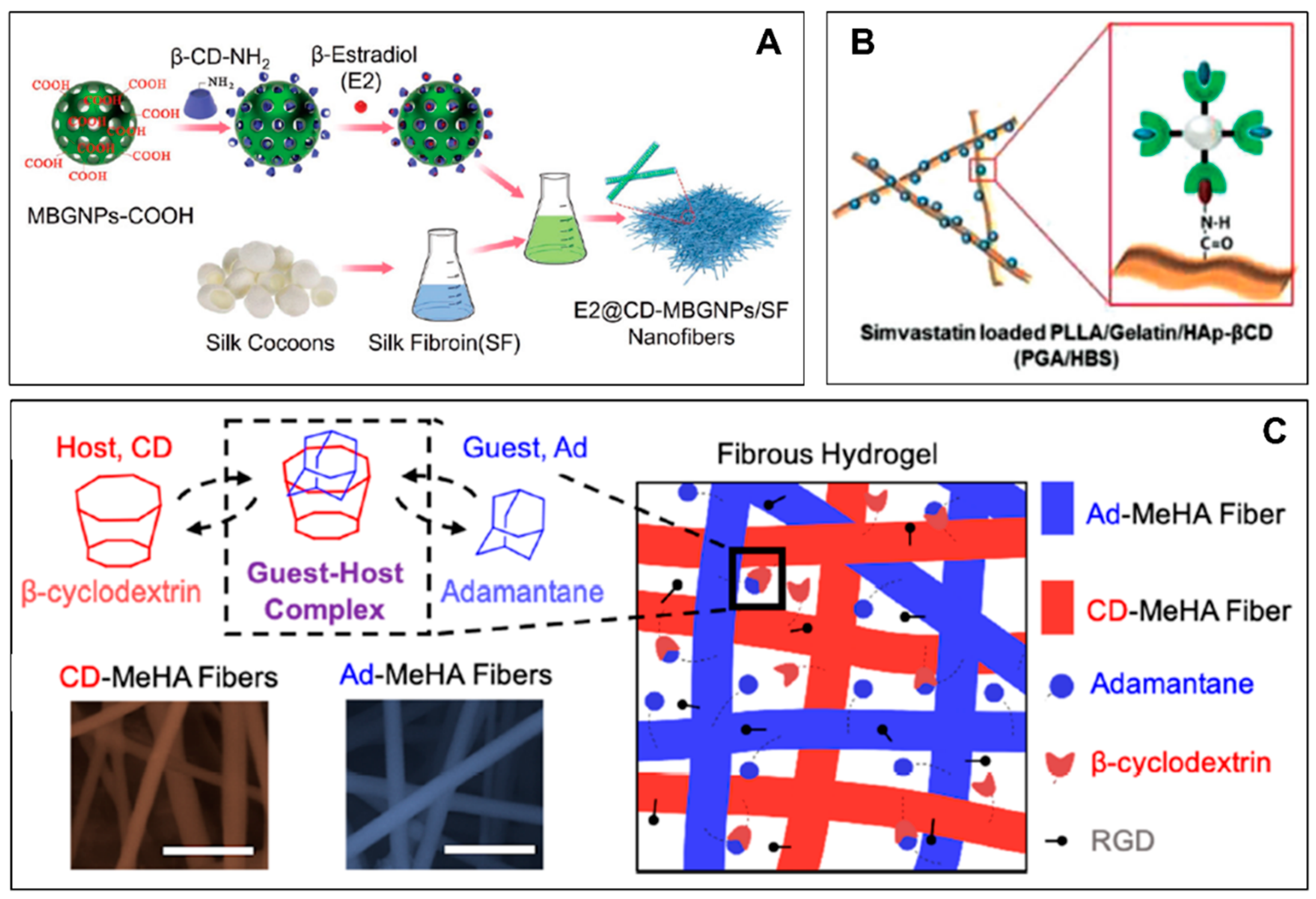
An HPβCDs-based IC with sulfisoxazole was embedded in hydroxypropyl cellulose fibers via electrospinning. The results depicted the fabrication of uniform nanofibers and the amorphous distribution of ICs in the nanocomposite. A higher amount of drugs were released from the IC-embedded nanofibers than from the sulfisoxazole-embedded nanofibers. The significant improvement to the drug’s solubility could be attributed to the incorporated ICs, which could assist in developing promising delivery systems of hydrophobic drugs [34] (Figure 2D). The ICs between the CD and adamantane-conjugated epitope were successfully entrapped in the electrospun nanofibers. The nanocomposite scaffold was investigated against peripheral nerve regeneration. The structural properties of the nanocomposite promoted cell adhesion and proliferation, while the bioactive epitopes on the nanofibers’ surfaces guided cellular differentiation. Both the chemical and physical cues were utilized for an effective neuronal differentiation process [35]. The bioactive glass nanoparticles with inherent osteogenic properties are modified with βCDs to enhance their affinity for exogenous estradiol. The estradiol-loaded βCDs functionalized glass nanoparticles were embedded onto silk fibroin-based nanofibers. The localized delivery of estradiol could circumvent the systemic adverse effects, and the resultant nanocomposite enhances the in vitro apatite formation and sustains the release of estradiol. It significantly enhanced the osteoblast proliferation, differentiation, and matrix-maturation, thus, it could be crucial in bone reconstruction [36] (Figure 3A). Naproxen has been complexed with βCDs. The pristine drug and ICs were then incorporated into PCL nanofibers. The drug release elucidates that the supramolecular nanofibers release a higher amount of naproxen than the PCL/naproxen nanofibers do due to the solubility enhancement of naproxen by the CD-ICs [37].
The electrospinning of the hydrocortisone/CDs complex has been introduced to fabricate a rapid-dissolving oral medication delivery system, without using an additional polymer matrix. Electrospinning successfully yielded homogenous nanofibrous structures, and the resultant complex improved the water solubility and distribution of the drugs in an amorphous state. The nanofibrous membrane could rapidly be dissolved in water and artificial saliva, therefore, it could be applied as a rapidly dissolving oral drug delivery system [38]. HPβCDs and tetracycline have been used to form an IC. The resultant IC was then blended with pullulan and electrospun to form nanofibers. The electrospinning yielded defect-free fibers with a high drug loading capacity in an amorphous state. The supramolecular nanofibers enhanced the water solubility and rapid drug release profile more in artificial saliva than the simple pullulan/tetracycline nanofibers did. Furthermore, ICs-functionalized fibers exhibited a strong antibacterial potential against Gram-positive and Gram-negative bacteria, and they could be an attractive material for rapid-dissolving oral drug delivery systems [39].
Eucalyptol/βCDs ICs have been embedded in gellan/polyvinyl alcohol nanofibers to eradicate antifungal biofilms. The resultant nanocomposite possesses good hydrophilic properties, facilitates rapid drug release, and inhibits the biofilms of C. albicans and C. glabrata. The introduced fibrous systems enhanced the antibiofilm activity of eucalyptol to treat fungal infections, and they can be applied as a cost-effective implant coating biomaterial [40]. The electrospinning of polymer-free (acyclovir/HP-βCD) and polymer-based materials (acyclovir/polyvinylpyrrolidone) has been carried out to form functional nanofibers. Both nanocomposites were embedded with high concentrations of acyclovir. The results show that the polymer-free nanocomposite provided a better drug loading efficiency (98%) than the polymer-based fibers did (66%), and they showed faster drug release and disintegration profiles. This could act as a promising strategy for fabricating a rapid-dissolving drug delivery system in good dosage formulation against viral infections [41].
A meloxicam/βCDs-functionalized polyvinylpyrrolidone nanofiber was fabricated using an electrospinning process. The results indicated that the resultant supramolecular nanocomposite possesses suitable mechanical properties, incorporated the meloxicam in an amorphous form, and exhibits faster disintegration. Furthermore, the nanocomposite membrane exhibits a better rapid drug release potential than the pure meloxicam powder and commercial meloxicam tablets do. Furthermore, the nanocomposite remained stable for up to 6 months, and it could rapidly disintegrate in the mouth, and it could mask the bitter taste of drugs, and therefore, it could be a good candidate for fast dissolving drug delivery systems for bitter medicines [42]. βCDs and thymol were self-assembled to form a water-soluble IC. The pristine thymol and resultant ICs were separately embedded into cellulose acetate nanofibers via electrospinning. The results demonstrated that ICs-functionalized fibrous membranes exhibited sustained thymol release and a lasting antibacterial potential against S. aureus, and they showed good cytocompatibility. The IC-embedded nanocomposite could be an attractive candidate for a wound dressing material [43].
Poly(L-lactic acid)/gelatin nanofibers were fabricated via electrospinning. The βCDs-conjugated hydroxyapatite was coated onto the surface of the nanofiber via a specific interaction between the βCDs and adamantane. Simvastatin (osteogenic drug) was then loaded into the remaining βCD, and overall, the results demonstrate that the simvastatin-loaded nanocomposite better promoted mineralization, osteogenic gene expression, and bone reconstruction [44] (Figure 3B). The engineering design brings together hyaluronic acid electrospun hydrogel nanofiber segments that are functionalized with either an adamantane-based guest or βCD-based host supramolecular moieties. The host–guest interaction creates a shear-thinning and self-healing hydrogel fiber network via the guest−host complexation [45] (Figure 3C).
The electrospun nanofibers incorporated with IC of niclosamide and HPβCDs were produced using pH-responsive polymer. The IC-embedded nanocomposite disintegrated at pH values that were greater than six, and it had the potential to be utilized for the targeted and regulated release of niclosamide to the colon [46]. Overall, the CD-based nanofibers could carry more significant amounts of drugs, and they are more crucial for rapidly dissolving oral medication delivery systems compared to conventional drug formulations. Furthermore, the structural, mechanical, and functional features of the fibrous composite could control the drug release properties (according to the desired application) and regulate the cell function. The supramolecular nanocomposite could maximize site-specific drug targeting for tissue engineering, cancer treatment, and other biomedical applications.

5. Conclusions and Future Perspectives

There are numerous factors favoring CD-based ICs and electrospun nanofibers in biomedical applications. However, the use of pure CD-IC is unsuited for biomedical applications due to their poor structural and physicochemical properties. On the other hand, since most drugs are water insoluble, the incorporation of high concentrations of drugs into the nanofibrous composite, while maintaining their functional activity, can be problematic for biomedical applications. Considering the properties of the CD-ICs and nanofibers, our review highlights that incorporating CD-ICs into the nanofiber matrix is a simple procedure to engineer drug-encapsulated fibers, since distinct properties and synergistic effects can be obtained. The supramolecular nanocomposite has been investigated as a drug delivery system. It is suited for biomedical applications because it can assemble into different forms, engineer diverse assemblies such as nanoparticles, structurally tune the drug release mechanism, and it can integrate many types of drugs. Furthermore, it can respond to physiological cues, and maximize the applicability of the nanocomposite in a range of biomedical applications. Most of these investigations are still in the proof-of-concept stage, nevertheless, the future of supramolecular nanofibers in the biomedical domain is promising. There is a need to strengthen the biodistribution, biodegradability, biosafety, and stimuli-responsive composite design for enhanced biomedical applications.

Author Contributions

Study conception and design, S.U., Z.H. and L.W.; Literature and data collection, S.M., B.L., Y.Z., M.W.G.; review and editing, I.U., R.P. and J.W. All authors have read and agreed to the published version of the manuscript.

Funding

This research received no external funding.

Institutional Review Board Statement

Not applicable.

Informed Consent Statement

Not applicable.

Acknowledgments

This study was supported by the Strategic Priority Research Program of Chinese Academy of Sciences (XDA16040700) and Science and Technology Project of Suzhou (SZS201904). Z.H. is grateful to CAS-TWAS President’s Ph.D. Fellowship Program.

Conflicts of Interest

The authors declare no conflict of interest.

References

  1. Kim, D.-H.; Lee, S.-E.; Pyo, Y.-C.; Tran, P.; Park, J.-S. Solubility enhancement and application of cyclodextrins in local drug delivery. J. Pharm. Investig. 2019, 50, 17–27. [Google Scholar] [CrossRef]
  2. Jacob, S.; Nair, A.B. Cyclodextrin complexes: Perspective from drug delivery and formulation. Drug Dev. Res. 2018, 79, 201–217. [Google Scholar] [CrossRef] [PubMed]
  3. Jansook, P.; Ogawa, N.; Loftsson, T. Cyclodextrins: Structure, physicochemical properties and pharmaceutical applications. Int. J. Pharm. 2018, 535, 272–284. [Google Scholar] [CrossRef]
  4. Liu, Z.; Ye, L.; Xi, J.; Wang, J.; Feng, Z.G. Cyclodextrin polymers: Structure, synthesis, and use as drug carriers. Prog. Polym. Sci. 2021, 118, 101408. [Google Scholar] [CrossRef]
  5. Narayanan, G.; Shen, J.; Matai, I.; Sachdev, A.; Boy, R.; Tonelli, A.E. Cyclodextrin-based nanostructures. Prog. Mater. Sci. 2021, 124, 100869. [Google Scholar] [CrossRef]
  6. Suvarna, V.; Bore, B.; Bhawar, C.; Mallya, R. Complexation of phytochemicals with cyclodextrins and their derivatives- an update. Biomed. Pharmacother. 2022, 149, 112862. [Google Scholar] [CrossRef]
  7. Paiva-Santos, A.C.; Ferreira, L.; Peixoto, D.; Silva, F.; Soares, M.J.; Zeinali, M.; Zafar, H.; Mascarenhas-Melo, F.; Raza, F.; Mazzola, P.G.; et al. Cyclodextrins as an encapsulation molecular strategy for volatile organic compounds—Pharmaceutical applications. Colloids Surf. B Biointerfaces 2022, 218, 112758. [Google Scholar] [CrossRef]
  8. Liu, L.; Guo, Q.-X. The Driving Forces in the Inclusion Complexation of Cyclodextrins. J. Incl. Phenom. Macrocycl. Chem. 2002, 42, 1–14. [Google Scholar] [CrossRef]
  9. Tang, W.; Zou, C.; Da, C.; Cao, Y.; Peng, H. A review on the recent development of cyclodextrin-based materials used in oilfield applications. Carbohydr. Polym. 2020, 240, 116321. [Google Scholar] [CrossRef] [PubMed]
  10. Topuz, F.; Uyar, T. Advances in the development of cyclodextrin-based nanogels/microgels for biomedical applications: Drug delivery and beyond. Carbohydr. Polym. 2022, 297, 120033. [Google Scholar] [CrossRef]
  11. Wankar, J.; Kotla, N.G.; Gera, S.; Rasala, S.; Pandit, A.; Rochev, Y.A. Recent Advances in Host–Guest Self-Assembled Cyclodextrin Carriers: Implications for Responsive Drug Delivery and Biomedical Engineering. Adv. Funct. Mater. 2020, 30, 1909049. [Google Scholar] [CrossRef]
  12. Harada, A.; Takashima, Y.; Nakahata, M. Supramolecular Polymeric Materials via Cyclodextrin–Guest Interactions. Accounts Chem. Res. 2014, 47, 2128–2140. [Google Scholar] [CrossRef] [PubMed]
  13. Samprasit, W.; Akkaramongkolporn, P.; Kaomongkolgit, R.; Opanasopit, P. Cyclodextrin-based oral dissolving films formulation of taste-masked meloxicam. Pharm. Dev. Technol. 2017, 23, 530–539. [Google Scholar] [CrossRef]
  14. Carneiro, S.B.; Costa Duarte, F.Í.; Heimfarth, L.; Siqueira Quintans, J.D.S.; Quintans-Júnior, L.J.; Veiga Júnior, V.F.D.; Neves de Lima, Á.A. Cyclodextrin–drug inclusion complexes: In vivo and in vitro approaches. Int. J. Mol. Sci. 2019, 20, 642. [Google Scholar] [CrossRef] [PubMed]
  15. Bhardwaj, N.; Kundu, S.C. Electrospinning: A fascinating fiber fabrication technique. Biotechnol. Adv. 2010, 28, 325–347. [Google Scholar] [CrossRef]
  16. Persano, L.; Camposeo, A.; Tekmen, C.; Pisignano, D. Industrial Upscaling of Electrospinning and Applications of Polymer Nanofibers: A Review. Macromol. Mater. Eng. 2013, 298, 504–520. [Google Scholar] [CrossRef]
  17. Hussain, Z.; Ding, P.; Zhang, L.; Zhang, Y.; Ullah, S.; Liu, Y.; Ullah, I.; Wang, Z.; Zheng, P.; Pei, R. Multifaceted tannin crosslinked bioinspired dECM decorated nanofibers modulating cell–scaffold biointerface for tympanic membrane perforation bioengineering. Biomed. Mater. 2022, 17, 034102. [Google Scholar] [CrossRef]
  18. Gao, Y.; Bach Truong, Y.; Zhu, Y.; Louis Kyratzis, I. Electrospun antibacterial nanofibers: Production, activity, and in vivo applications. J. Appl. Polym. Sci. 2014, 131, 18. [Google Scholar] [CrossRef]
  19. Hussain, Z.; Khan, M.A.; Iqbal, F.; Raffi, M.; Hafeez, F.Y. Electrospun Microbial-Encapsulated Composite-Based Plasticized Seed Coat for Rhizosphere Stabilization and Sustainable Production of Canola (Brassica napus L.). J. Agric. Food Chem. 2019, 67, 5085–5095. [Google Scholar] [CrossRef]
  20. Hussain, Z.; Ullah, S.; Yan, J.; Wang, Z.; Ullah, I.; Ahmad, Z.; Zhang, Y.; Cao, Y.; Wang, L.; Mansoorianfar, M.; et al. Electrospun tannin-rich nanofibrous solid-state membrane for wastewater environmental monitoring and remediation. Chemosphere 2022, 307, 135810. [Google Scholar] [CrossRef]
  21. Khan, M.A.; Hussain, Z.; Ali, S.; Qamar, Z.; Imran, M.; Hafeez, F.Y. Fabrication of Electrospun Probiotic Functionalized Nanocomposite Scaffolds for Infection Control and Dermal Burn Healing in a Mice Model. ACS Biomater. Sci. Eng. 2019, 5, 6109–6116. [Google Scholar] [CrossRef] [PubMed]
  22. Pilehvar-Soltanahmadi, Y.; Akbarzadeh, A.; Moazzez-Lalaklo, N.; Zarghami, N. An update on clinical applications of electrospun nanofibers for skin bioengineering. Artif. Cells Nanomed. Biotechnol. 2016, 44, 1350–1364. [Google Scholar] [CrossRef]
  23. Hussain, Z.; Ullah, I.; Liu, X.; Shen, W.; Ding, P.; Zhang, Y.; Gao, T.; Mansoorianfar, M.; Gao, T.; Pei, R. Tannin-reinforced iron substituted hydroxyapatite nanorods functionalized collagen-based composite nanofibrous coating as a cell-instructive bone-implant interface scaffold. Chem. Eng. J. 2022, 438, 135611. [Google Scholar] [CrossRef]
  24. Hussain, Z.; Ullah, I.; Wang, Z.; Ding, P.; Ullah, S.; Zhang, Y.; Zhang, Z.; Yan, J.; Luo, B.; Pei, R. Electrospun nanofibrous membrane functionalized with dual drug-cyclodextrin inclusion complexes for the potential treatment of otitis externa. Colloids Surf. A Physicochem. Eng. Asp. 2022, 651, 129742. [Google Scholar] [CrossRef]
  25. Muñoz-Shugulí, C.; Vidal, C.P.; Cantero-López, P.; Lopez-Polo, J. Encapsulation of plant extract compounds using cyclodextrin inclusion complexes, liposomes, electrospinning and their combinations for food purposes. Trends Food Sci. Technol. 2021, 108, 177–186. [Google Scholar] [CrossRef]
  26. Vidal, C.P.; de Dicastillo, C.L.; Rodríguez-Mercado, F.; Guarda, A.; Galotto, M.J.; Muñoz-Shugulí, C. Electrospinning and cyclodextrin inclusion complexes: An emerging technological combination for developing novel active food packaging materials. Crit. Rev. Food Sci. Nutr. 2021, 62, 5495–5510. [Google Scholar] [CrossRef]
  27. Wang, Y.; Chou, J.; Sun, Y.; Wen, S.; Vasilescu, S.; Zhang, H. Supramolecular-based nanofibers. Mater. Sci. Eng. C 2019, 101, 650–659. [Google Scholar] [CrossRef]
  28. Dodero, A.; Schlatter, G.; Hébraud, A.; Vicini, S.; Castellano, M. Polymer-free cyclodextrin and natural polymer-cyclodextrin electrospun nanofibers: A comprehensive review on current applications and future perspectives. Carbohydr. Polym. 2021, 264, 118042. [Google Scholar] [CrossRef]
  29. Souza, S.O.L.; Cotrim, M.A.P.; Oréfice, R.L.; Carvalho, S.G.; Dutra, J.A.P.; Careta, F.D.P.; Resende, J.A.; Villanova, J.C.O. Electrospun poly(ε-caprolactone) matrices containing silver sulfadiazine complexed with β-cyclodextrin as a new pharmaceutical dosage form to wound healing: Preliminary physicochemical and biological evaluation. J. Mater. Sci. Mater. Med. 2018, 29, 67. [Google Scholar] [CrossRef]
  30. Narayanan, G.; Ormond, B.R.; Gupta, B.S.; Tonelli, A.E. Efficient wound odor removal by b-cyclodextrin functionalized poly(e-caprolactone) nanofibers. J. Appl. Polym. 2015, 25, 132. [Google Scholar] [CrossRef]
  31. Li, C.; Liu, Z.; Liu, S.; Tiwari, S.K.; Thummavichai, K.; Ola, O.; Ma, Z.; Zhang, S.; Wang, N.; Zhu, Y. Antibacterial properties and drug release study of cellulose acetate nanofibers containing ear-like Ag-NPs and Dimethyloxallyl Glycine/beta-cyclodextrin. Appl. Surf. Sci. 2022, 590, 153132. [Google Scholar] [CrossRef]
  32. Liu, C.; Zhu, Y.; Lun, X.; Sheng, H.; Yan, A. Effects of wound dressing based on the combination of silver@curcumin nanoparticles and electrospun chitosan nanofibers on wound healing. Bioengineered 2022, 13, 4328–4339. [Google Scholar] [CrossRef]
  33. Nalbandi, B.; Amiri, S. Antibacterial activity of PVA-based nanofibers loaded with silver sulfadiazine/cyclodextrin nanocapsules. Int. J. Polym. Mater. Polym. Biomater. 2018, 68, 647–659. [Google Scholar] [CrossRef]
  34. Aytac, Z.; Sen, H.S.; Durgun, E.; Uyar, T. Sulfisoxazole/cyclodextrin inclusion complex incorporated in electrospun hydroxypropyl cellulose nanofibers as drug delivery system. Colloids Surf. B Biointerfaces 2015, 128, 331–338. [Google Scholar] [CrossRef] [PubMed]
  35. Hamsici, S.; Cinar, G.; Celebioglu, A.; Uyar, T.; Tekinay, A.B.; Guler, M.O. Bioactive peptide functionalized aligned cyclodextrin nanofibers for neurite outgrowth. J. Mater. Chem. B 2016, 5, 517–524. [Google Scholar] [CrossRef] [PubMed]
  36. Wang, D.; Steffi, C.; Wang, Z.; Kong, C.H.; Lim, P.N.; Shi, Z.; Thian, E.S.; Wang, W. Beta-cyclodextrin modified mesoporous bioactive glass nanoparticles/silk fibroin hybrid nanofibers as an implantable estradiol delivery system for the potential treatment of osteoporosis. Nanoscale 2018, 10, 18341–18353. [Google Scholar] [CrossRef] [PubMed]
  37. Canbolat, M.F.; Celebioglu, A.; Uyar, T. Drug delivery system based on cyclodextrin-naproxen inclusion complex incorporated in electrospun polycaprolactone nanofibers. Colloids Surf. B Biointerfaces 2014, 115, 15–21. [Google Scholar] [CrossRef]
  38. Celebioglu, A.; Uyar, T. Hydrocortisone/cyclodextrin complex electrospun nanofibers for a fast-dissolving oral drug delivery system. RSC Med. Chem. 2020, 11, 245–258. [Google Scholar] [CrossRef]
  39. Hsiung, E.; Celebioglu, A.; Chowdhury, R.; Kilic, M.E.; Durgun, E.; Altier, C.; Uyar, T. Antibacterial nanofibers of pullulan/tetracycline-cyclodextrin inclusion complexes for Fast-Disintegrating oral drug delivery. J. Colloid Interface Sci. 2022, 610, 321–333. [Google Scholar] [CrossRef]
  40. Mishra, P.; Gupta, P.; Srivastava, A.K.; Poluri, K.M.; Prasad, R. Eucalyptol/ β-cyclodextrin inclusion complex loaded gellan/PVA nanofibers as antifungal drug delivery system. Int. J. Pharm. 2021, 609, 121163. [Google Scholar] [CrossRef]
  41. Celebioglu, A.; Uyar, T. Electrospun formulation of acyclovir/cyclodextrin nanofibers for fast-dissolving antiviral drug delivery. Mater. Sci. Eng. C 2020, 118, 111514. [Google Scholar] [CrossRef]
  42. Samprasit, W.; Akkaramongkolporn, P.; Ngawhirunpat, T.; Rojanarata, T.; Kaomongkolgit, R.; Opanasopit, P. Fast releasing oral electrospun PVP/CD nanofiber mats of taste-masked meloxicam. Int. J. Pharm. 2015, 487, 213–222. [Google Scholar] [CrossRef] [PubMed]
  43. Chen, Y.; Mensah, A.; Wang, Q.; Li, D.; Qiu, Y.; Wei, Q. Hierarchical porous nanofibers containing thymol/beta-cyclodextrin: Physico-chemical characterization and potential biomedical applications. Mater. Sci. Eng. C 2020, 115, 111155. [Google Scholar] [CrossRef] [PubMed]
  44. Lee, J.B.; Kim, J.E.; Balikov, D.A.; Bae, M.S.; Heo, D.N.; Lee, D.; Rim, H.J.; Lee, D.W.; Sung, H.J.; Kwon, I.K. Poly(l-lactic acid)/gelatin fibrous scaffold loaded with simvastatin/beta-cyclodextrin-modified hydroxyapatite inclusion complex for bone tissue regeneration. Macromol. Biosci. 2016, 16, 1027–1038. [Google Scholar] [CrossRef] [PubMed]
  45. Miller, B.; Hansrisuk, A.; Highley, C.B.; Caliari, S.R. Guest–host supramolecular assembly of injectable hydrogel nanofibers for cell encapsulation. ACS Biomater. Sci. Eng. 2021, 7, 4164–4174. [Google Scholar] [CrossRef]
  46. Coban, O.; Aytac, Z.; Yildiz, Z.I.; Uyar, T. Colon targeted delivery of niclosamide from β-cyclodextrin inclusion complex incorporated electrospun Eudragit® L100 nanofibers. Colloids Surf. B Biointerfaces 2020, 197, 111391. [Google Scholar] [CrossRef]
Figure 1. (A) Schematic chemical structure of αCDs, βCDs, and γCDs. (B) The main physicochemical properties of αCDs, βCDs, and γCDs. (C) Schematics representation of ICs formation between CDs and guest molecules. (D) Schematic electrospinning setup. (E) Schematic representation of polymer-free CDs nanofiber. (F) Schematic representation of drug-entrapped polymer-free CDs nanofiber. (G) Schematic representation of drug-entrapped polymer-based nanofiber.
Chemproc 12 00060 g001
Figure 2. (A) The schematic diagram show releases of dual biocides from IC incorporated nanofibers to inhibit and reduce E. coli growth and inflammatory mediators. Reprinted with permission from [24]. Copyright: (2022) Elsevier. (B) The chitosan-based nanofibrous material functionalized with curcumin@βCD/AgNPs nanoparticles. Reprinted with permission from [32]. Copyright: (2022) Taylor & Francis. (C) Schematics representation of ICs formation between CDs/SSD and their incorporation into nanofibers. Reprinted with permission from [33]. Copyright: (2019) Taylor & Francis. (D) HPβCDs/SFS complex was incorporated in hydroxypropyl cellulose nanofibers via electrospinning to modulate the drug release profile. Reprinted with permission from [34]. Copyright: (2015) Elsevier.
Chemproc 12 00060 g002
Figure 3. (A) The bioactive and osteogenic glass nanoparticles (MBGNPs) are conjugated with βCDs (CD-MBGNPs) to enhance their encapsulation affinity for exogenous estradiol. Reprinted with permission from [36]. Copyright: (2018) Royal Society of Chemistry. (B) are fabricated via electrospinning. The βCDs conjugated HAp is coated onto the surface of poly(L-lactic acid)/gelatin-based nanofibers through interaction between βCDs and adamantane, and at the latter stage, simvastatin is loaded into the remaining βCD. Reprinted with permission from [44]. Copyright: (2016) Wiley-VCH. (C) The formation of a macroscale fibrous hydrogel scaffold is achieved as a consequence of interactions involving adamantane and CDs on complimentary bioactive nanofibers. Reprinted with permission from [45]. Copyright: (2021) American Chemical Society.
Chemproc 12 00060 g003
Disclaimer/Publisher’s Note: The statements, opinions and data contained in all publications are solely those of the individual author(s) and contributor(s) and not of MDPI and/or the editor(s). MDPI and/or the editor(s) disclaim responsibility for any injury to people or property resulting from any ideas, methods, instructions or products referred to in the content.