Review

Ion-Channel Antiepileptic Drugs: An Analytical Perspective on the Therapeutic Drug Monitoring (TDM) of Ezogabine, Lacosamide, and Zonisamide

1
Department for Life Quality Studies (QuVi), Alma Mater Studiorum—University of Bologna, Corso d’Augusto 237, 47921 Rimini, Italy
2
Research Group of Pharmaco-Toxicological Analysis (PTA Lab), Department of Pharmacy and Biotechnology (FaBiT), Alma Mater Studiorum—University of Bologna, Via Belmeloro 6, 40126 Bologna, Italy
*
Author to whom correspondence should be addressed.
Analytica 2021, 2(4), 171-194; https://doi.org/10.3390/analytica2040016
Submission received: 8 August 2021 / Revised: 24 November 2021 / Accepted: 27 November 2021 / Published: 30 November 2021

Abstract

:
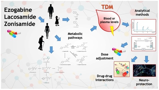
The term seizures includes a wide array of different disorders with variable etiology, which currently represent one of the most important classes of neurological illnesses. As a consequence, many different antiepileptic drugs (AEDs) are currently available, exploiting different activity mechanisms and providing different levels of performance in terms of selectivity, safety, and efficacy. AEDs are currently among the psychoactive drugs most frequently involved in therapeutic drug monitoring (TDM) practices. Thus, the plasma levels of AEDs and their metabolites are monitored and correlated to administered doses, therapeutic efficacy, side effects, and toxic effects. As for any analytical endeavour, the quality of plasma concentration data is only as good as the analytical method allows. In this review, the main techniques and methods are described, suitable for the TDM of three AEDs belonging to the class of ion channel agents: ezogabine (or retigabine), lacosamide, and zonisamide. In addition to this analytical overview, data are provided, pertaining to two of the most important use cases for the TDM of antiepileptics: drug–drug interactions and neuroprotection activity studies. This review contains 146 references.

Graphical Abstract

1. Introduction

Epilepsy, and more generally seizures, are one of the most widespread and incapacitating neurologic disorders. Recent reports have calculated a 0.63% prevalence of epilepsy over the total world population, corresponding to about 50 million people [1]. In 2016, the Global Burden of Diseases, Injuries, and Risk Factors (GBD) Study [2] estimated that epilepsy accounted for >13 million disability-adjusted life-years (DALYs) and was responsible for 0.5% of the total disease burden, as well as 5.0% of DALYs attributable to neurological disorders [3]. In terms of DALY rates for all neurological disorders, epilepsy ranked second to eighth depending on the geographic region, with highest impact in sub-Saharan Africa, Central Asia, Central and Andean Latin America, and Southeast Asia [2].
As can be expected, the rates of success of antiepileptic treatment can be considerably lower in low-middle income countries (LMIC) than in other parts of the world [4]: lower access to affordable or free public health structures, lower access to cheap medicines, and in some cases even prejudices and open discrimination toward the patients themselves can produce these effects [5], which also result in higher disease severity and lethality. It has been estimated that in LMIC up to 50% of epileptic people are undiagnosed and/or untreated [6]. In addition, more difficult perinatal conditions and higher incidence of traumatic brain injuries and central nervous system (CNS) infections in LMIC can contribute to the observed increased disease incidence and severity [7].
The first effective antiepileptic drugs (AEDs) were bromides, introduced in the mid-1850s, and then paraldehyde, in the late 1880s. Since then, many other, vastly more effective and safer drugs have been devised, belonging to many chemical classes, starting from barbiturates in the 1910s, their analogues oxazolidinediones and pyrimidinediones, and hydantoins in the 1930–1960s. Then, benzodiazepines, carboxamides, fatty acids, and their derivatives, fructose derivatives, γ-aminobutyric acid (GABA) derivatives, hydantoins, pyrrolidines, succinimides, sulphonamides, and triazines have been used against seizures [8]. Pharmacological activity mechanisms are equally varied, although for some drugs, and entire chemical classes, the precise mechanism is still uncertain [9]. The two oldest AEDs, bromide and paraldehyde, are thought to non-selectively depress brain activity; if they have any molecular target, it is still unknown. A large group of AEDs interacts directly with ion channels, acting either as sodium channel blockers (such as carbamazepine, oxcarbazepine, lamotrigine, phenytoin) or as calcium channel blockers (such as ethosuximide); ezogabine seems to act as a potassium channel opener. Many AEDs interact with GABAergic transmission; since GABA is known as the main inhibitory neurotransmitter in the brain, its activation or facilitation is intuitively connected with a possible anticonvulsant effect. Thus, GABA transaminase inhibitors (vigabatrin) and glutamic acid decarboxylase (GAD) inducers (gabapentin) are used as antiepileptic agents. On the other hand, glutamate is generally recognized as the main excitatory brain neurotransmitter, and therefore drugs acting on the glutamatergic system (topiramate) are also known as antiepileptic agents [10]. The exact mechanisms of activity of valproic acid, levetiracetam, and stiripentol are still unknown, although of course hypotheses have been proposed [11].
The need for an accurate titration of the AED levels in patient plasma has been acknowledged relatively early in the history of anticonvulsant therapy and has been often confirmed in the following years [12]. To this day, many patients receive a constant monitoring of their AED levels, especially at the beginning of the treatment, to ascertain that correct concentrations are reached without undue risks of overdose and toxic phenomena [13]. This practice is commonly named therapeutic drug monitoring (TDM) and is currently being increasingly used to optimize and personalize pharmacological therapy, in particular in the treatment of central nervous system (CNS) disorders [14,15,16,17,18]. TDM uses clinical information (administered doses, symptoms, side or toxic effects) and chemical data (plasma levels of drugs and metabolites) to find possible chemical–clinical correlations (CCC), i.e., those correlations (if they exist) that connect administered doses with plasma levels, and plasma levels with clinical outcomes.
In the case of AEDs in particular, TDM utility is evident [19,20], due to the frequent need for polytherapy and the capacity of several AEDs to induce and/or to inhibit AED-metabolizing enzymes, and the consequent high frequency of pharmacokinetic and metabolic drug–drug interactions (DDI) [21]. Thus, the need for precise, accurate, and sensitive analytical methods for AED determination in patient blood is apparent as well.
In 2004 the German Workgroup for Neuropsychopharmacology and Pharmacopsychiatry (Arbeitsgemeinschaft für Neuropsychopharmakologie und Pharmakopsychiatrie, AGNP) started publishing a series of papers reporting consensus guidelines [22,23,24] on the TDM of psychotropic drugs, including AEDs. For each drug, a “level of recommendation to use TDM”, is reported as a number in the 1–4 range: 1 corresponds to “strongly recommended”, 2 to “recommended”, 3 to “useful”, and 4 to “potentially useful”. These recommendation classes are mostly relative to routine TDM practices, but TDM can be useful also in non-routine conditions, for example in cases of insufficient response, suggested non-adherence, adverse drug reaction at therapeutic doses, potential DDI. This highlights the real benefit of TDM even for those drugs with low recommendation levels, and even when CCC are unavailable or unclear.
In this review, the most important and recent methods devised for TDM purposes are described and briefly commented upon, in order to highlight the current status of advancement in this field. This information could be useful for both analytical chemists and clinicians, providing a common base from which to start discussing the actual TDM needs of the patients and the best way to satisfy them.
We addressed in a previous review the main TDM characteristics of the following AEDs: carbamazepine, oxcarbazepine, lamotrigine, phenytoin, ethosuximide, gabapentin, vigabatrin, topiramate, levetiracetam, and valproic acid [20]. In this mini-review, information will be reported on the three other most significant AEDs that interact with ion channels; i.e., ezogabine (or retigabine, EZG), lacosamide (LCS), and zonisamide (ZNS). In addition to the most important analytical methods for TDM purposes, information will be provided on these drugs’ metabolism and DDI, which are among the most important characteristics when deciding whether TDM is appropriate and, in fact, useful. Some brief notes on possible neuroprotective effects are also included, since neuroprotection is one more field where TDM can be eminently useful.

2. Methods

Electronic searches of the following publication databases were conducted: Scopus [25] and PubMed [26]. Search results covered the time span from 2005 to June 2021, in order to retrieve the most recent and updated information available. If this information proved to be incomplete or in any way unsatisfactory, the search was extended back in time in 10-year intervals.
Search terms for the “Analytical Methods” section used the following string: drug name AND (HPLC OR LC OR MS OR mass spectrometry OR GC OR CE OR electrophoresis OR immunoassay). Within these results, papers were filtered for application to human blood/plasma/serum or other human biological tissues relevant for TDM purposes. Papers with a clear analytical purpose were chosen preferentially, as opposed to clinical papers including some notes or a simple section on the analytical techniques and methods used.

3. Ion Channel Agents

3.1. Ezogabine (Retigabine)

EZG (ethyl N-[2-amino-4-[(4-fluorophenyl)methylamino]phenyl]carbamate, Figure 1a), also known as retigabine in the USA, was used as an add-on treatment for drug-resistant partial onset seizures with or without secondary generalization in people aged 18 or older [27]. It was approved in Europe and the USA in 2011, commercialized by GlaxoSmithKline under the name Trobalt. Since 2013, it has been known to produce blue skin discoloration and eye abnormalities [28]. In 2016, the manufacturer stopped its production, probably for commercial reasons (i.e., limited clinical use) [29].
EZG exerts its activity by activating the Kv7 (KCNQ) slowly-inactivating voltage-gated potassium channels [30], which is a mechanism different from that of most other antiepileptic drugs.
In vivo in humans, EZG is mainly N-glucuronidated (Figure 1b,c) by uridine diphosphate-glucuronosyltransferases (UGT) 1A1, 1A3, 1A4, and 1A9 and, to a lesser extent, N-acetylated after decarbamoylation (Figure 1d) [31]. The N-acetyl metabolite has weak biological activity [32].

3.1.1. Therapeutic Drug Monitoring

The AGNP’s recommendation level for EZG TDM is 3, “useful” [24]. Therapeutic plasma levels are in the 450–900 ng/mL range, corresponding to a 600 mg/day dose; levels above 1800 ng/mL are considered potentially toxic [33].
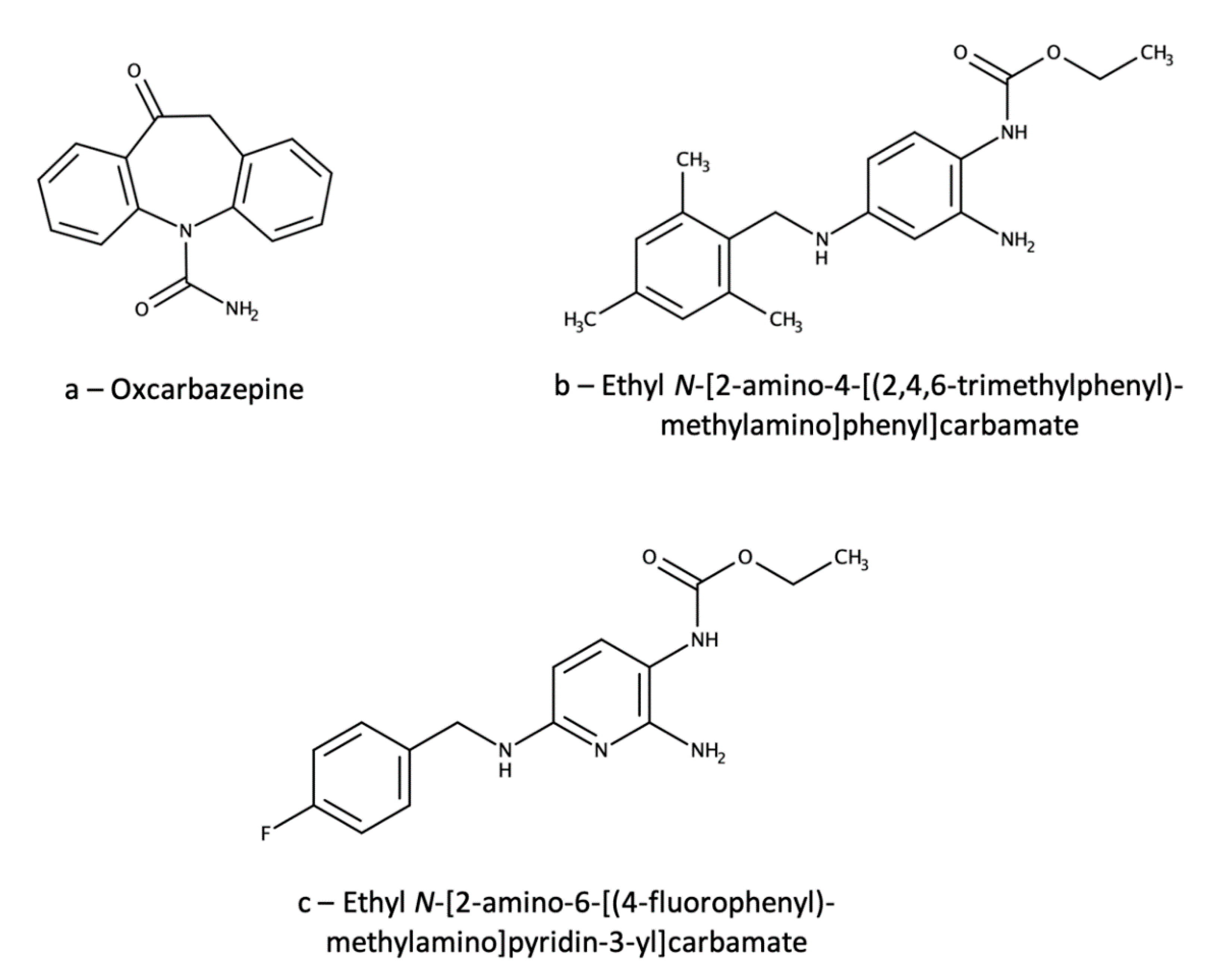
Just a few analytical methods are available for the TDM of EZG; most of them are based on liquid chromatography coupled with tandem mass spectrometry (LC-MS/MS) [34,35,36,37]. One of these methods [34] was applied to evaluate gender differences in EZG pharmacokinetics. Analytical separation was achieved on a C18 column kept at 40 °C with a mobile phase consisting of an aqueous ammonium acetate/methanol/acetonitrile mixture. Sample pretreatment was carried out by liquid–liquid extraction (LLE) of 500 µL of plasma using 5 mL of a diethyl ether/dichloromethane (70/30, v/v) mixture, followed by centrifugation, separation, drying of the organic phase and reconstitution with 100 µL of mobile phase. Detection was performed by MS/MS with triple quadrupole (QqQ) detection. Linearity was achieved in the 10–2000 ng/mL range, with precision RSD values between 1.9% and 9.3%. Extraction yields were in the 88–91% range and accuracy values in the 102–111% range. Stability assays provided recovery values always higher than 80% under all short- and log-term conditions tested. Matrix effect was almost negligible (mean = 3.8%), as was carryover (0.3–0.7% of the limit of quantitation (LOQ) peak area). Instead of a deuterated EZG analogue, oxcarbazepine (Figure 2a) was used as the internal standard (IS), which is obviously a sub-optimal choice. In fact, oxcarbazepine does not bear any close structural resemblance to EZG. Moreover, it is a very popular antiepileptic drug, and this makes its simultaneous administration with EZG quite probable. In these cases, the IS would be totally useless.
Another method included both EZG and its active N-acetyl metabolite and used automated on-line solid-phase extraction in the form of column-switching [35]. In this case, a C8 column specifically suited for basic compounds (LiChrospher RP-Select B) was used, coupled with an acetonitrile/pH 6 ammonium acetate (55/45, v/v) mobile phase. MS/MS detection was carried out by atmospheric pressure chemical ionization (APCI)—QqQ. Sample preparation consisted of a sample dilution step with 800 µL of 13% (v/v) aqueous acetonitrile added to 200 µL of plasma, followed by injection of 500 µL of the mixture into a column-switching system using a preparative C2 column. Interfering matrix components (salts, proteins) were flushed out with 10% (v/v) aqueous acetonitrile, then the analytes were eluted with the mobile phase and switched to the analytical column. In this case, the EZG analogue ethyl N-[2-amino-4-[(2,4,6-trimethylphenyl)methylamino]phenyl]carbamate (Figure 2b) was used as the IS; again, the choice is suboptimal for an LC-MS/MS method, but the structural similarity is quite high. Linearity was obtained in the 1−1000 ng/mL range for both analytes and precision RSD values were in the 1.58−7.87% range for EZG and in the 5.52−10.07% range for the metabolite. Accuracy varied between 89.6% and 104.5% for EZG and between 94.8% and 109.5% for the metabolite. Stability was not tested. The matrix effect was not explicitly assayed, but the complex pre-treatment is claimed to produce very clean extracts and matrix-independent results. Carryover was stated as negligible. Among the cited methods, this is probably the most complete, sensitive and reliable one, although the use of MS/MS detection makes it expensive and not universally applicable by common TDM laboratories. The column-switching pretreatment also makes this method rather complicated to set up and equipment-intensive.
The third method [36] deals with the determination of the N-acetyl metabolite by LC-APCI-MS/MS(QqQ) after pretreatment of 200 µL of plasma by micro-solid phase extraction (µSPE) in 96-well plates with a hydrophilic-lipophilic balance (HLB) sorbent; washing was carried out with two different ammonium acetate/acetonitrile/isopropyl alcohol mixtures, followed by elution with 1.2 mL of acetonitrile/isopropyl alcohol (40/60, v/v). To avoid the possible back-conversion of N-acetyl-EZG glucuronide to the parent N-acetyl-EZG, all materials and reagents for the dilution and µSPE procedures were refrigerated in ice; moreover, the glucuronide was eliminated from the sample by the washing step of the SPE procedure. However, the method only includes N-acetyl-EZG and has only been applied to dog plasma; hence, its usefulness for TDM is almost negligible. The IS was a 13C6-analogue of the analyte, which is a satisfactory choice. Linearity was obtained in the 1−1000 ng/mL range, with precision RSD values in the 1.2−17.3% range. Accuracy was always between 88% and 107%. The matrix effect was tested as the relative variability of signal between different dog plasma samples, and it was found to be 0.2−3.6%. Mean extraction yield was 34.8%, while stability assays provided accuracy levels in the 90−103% range; carryover was not explicitly tested.
Recently, an HPLC-UV method has also been published [38]. A C18 column and a mixture of water/acetonitrile/methanol/phosphoric acid were used, and detection was carried out at 240 nm. Sample pretreatment was by SPE and made use of EMPORE extraction disks, where a small amount of sorbent particles is trapped into a mostly inert PTFE matrix to create a mechanically stable sorbent disk. This setup allowed us to obtain much higher flow rates and throughput with lower cartridge clogging, while maintaining excellent performance. Then, 300 µL of plasma were mixed with 600 µL of ammonium acetate buffer, and 800 µL of the mixture were loaded onto a disk. Interferences were washed out with 1 mL of ammonium acetate/acetonitrile/isopropyl alcohol (95/3/2, v/v/v) mixture, then EZG was eluted with 100 µL of methanol and 150 µL of water. Linearity was found in the 25–2000 ng/mL range, with precision RSD ranging from 1.6% to 12.6% and accuracy ranging from 99.7% to 108.7%. No significant loss of the analyte was observed in stability assays, which were however carried out only at relatively high concentrations (75–1500 ng/mL). The IS was flupirtine, an analgesic agent that is neither an opioid nor a nonsteroidal anti-inflammatory drug (NSAID); since flupirtine is a pyridine analogue of EZG (ethyl N-[2-amino-6-[(4-fluorophenyl)methylamino]pyridin-3-yl]carbamate, Figure 2c), structural and chemical–physical similarity is very high. However, the use of another drug as the IS exposes the method to possible interference due to concomitant administration of flupirtine. This method is clearly much less sensitive than LC-MS/MS ones, but has the advantage of being less expensive, requiring much cheaper instrumentation. The inability to analyze the biologically active decarbamoylated metabolite is a severe drawback for a TDM method.
An UHPLC-MS/MS method was applied to exactly assess the concentrations of EZG and its main metabolite, specifically considering the marked lability of glucuronidated EZG metabolites [37]. In fact, both glucuronidated metabolites are present in plasma at much higher concentrations than the parent drug (about 25-fold) and have been demonstrated to be quite prone to degradation both in vivo and during storage at non-freezing temperature. Thus, relatively small amounts of glucuronide cleavage are likely, and could lead to substantial increases in detected EZG, especially during sample preparation and analysis; both temperature and low pH values seem to play an important role. For these reasons, column temperature was not raised above room temperature and neutral solutions and mobile phases were used throughout the procedure; moreover, an LLE step with ethyl acetate was introduced as early as possible in the workflow, since the glucuronides are not extracted by this solvent, thus limiting the chances of undue analyte concentration increase. Sample preparation consisted in an LLE procedure carried out in a 96-well plate by mixing plasma and 1 mL of diethyl ether; the organic phase was then dried up under a nitrogen stream and redissolved with 100 µL of acetonitrile/pH 7.9 ammonium bicarbonate (50/50, v/v). Separation was achieved on a C8 column with high resistance to dewetting by using a mixture of aqueous ammonium formate and acetonitrile/methanol, under gradient conditions (from 85% to 5% of aqueous phase); QqQ detection was employed. Linearity was obtained in the 5–2500 ng/mL range for both analytes, with accuracy ranging from 93.2% to 103.6%. While mean extraction yields were quite low (>37% for EZG, >47% for the metabolite), they were acceptable, since both the precision (RSD < 15%) and matrix effect (RSD < 15%) were within specification. Relatively extensive analyte stability assays were carried out, and they showed that the analytes were stable under most usual storage conditions (i.e., at 4 or 37 °C for a few hours, at −80 °C for more than two years). The ISs were D4-analogues of the analytes.
As one can see, analytical methods for the TDM of EZG are few and far between, and some of them have limited scope or are outright unsuitable for TDM without extensive validation or modification. Although EZG currently has very limited clinical application, this situation makes its TDM more complicated and expensive. More studies in this field would be welcome.

3.1.2. Ezogabine Interactions

Since EZG is not metabolized through the cytochrome P450 (CYP) system and does not induce or inhibit these isozymes at clinically relevant concentrations, it has a low potential for pharmacokinetic interactions with other drugs via CYP.

Clinically Relevant Data

Although the acetyl EZG metabolite has the capacity of inhibiting the P-glycoprotein (P-gp) transporter, a clinical study has found just a small increase in the digoxin plasma area under the concentration-time curve, which should not have any clinical significance [39]. EZG did not have any clinically relevant impact on exposure of hormones in a combined oral contraceptive agent containing norethindrone and ethinyl oestradiol, and the hormones did not alter EZG pharmacokinetic parameters [40]. In healthy humans, EZG did not significantly alter the pharmacokinetics of phenobarbital [41] and phenobarbitone [42].
Regarding other antiepileptic drugs (lamotrigine, valproate, topiramate, carbamazepine, phenytoin), small increases in EZG plasma levels were observed with carbamazepine, phenytoin, and lamotrigine; conversely, EZG increased lamotrigine concentrations by about 20% [43]. None of those interactions is likely to be clinically relevant [44]. Other reports have confirmed these findings: Ferron et al. [45] have observed that EZG pharmacokinetics are not altered by valproate or topiramate, while phenytoin and carbamazepine increased EZG clearance; and that EZG does not alter the PK of valproate, topiramate, phenytoin, or carbamazepine.

Data with Low or Uncertain Clinical Relevance, Case Reports

In humans, ethanol increases exposure to EZG, but this interaction is probably not clinically significant [46].

3.1.3. Neuroprotection

Chemotherapy-induced peripheral neuropathy is a major dose-limiting side effect of many commonly used chemotherapeutic agents, including cisplatin. One mechanism underlying this neuronal damage is via drug-induced membrane depolarization. Accordingly, one potential approach for preventing chemotherapy-induced neuropathy is the maintenance of normal membrane potential during exposure to the neurotoxic drug. In a study, it was found that cisplatin caused membrane depolarization and peripheral axon loss in mice sensory nerves, and both actions were partially prevented by EZG pretreatment [47]. In the rat model, EZG reduced the stimulation of glutamate release and prevented the neuronal damage caused by 4-aminopyridine; however, it did not block its epileptic activity with equal effectiveness [48]. In mice, chronic high-fat-diet-induced neuroinflammation can be effectively treated by retigabine [49]. In the same animal, EZG attenuated focal cerebral ischemic injury, probably through reducing oxidative stress and mitochondria-mediated apoptosis via inhibition of protein phosphorylation by p38 and c-Jun N-terminal kinases (JNKs) [50]. In rats, EZG has been shown to protect the blood–brain barrier integrity by regulating tight junctions between cerebral vascular endothelial cells in cerebral ischemia-reperfusion [51]. Finally, EZG demonstrated effectiveness against specific forms of neuronal damage, such as salicylate ototoxicity [52] and spinal nerve ligation-induced neuropathic pain [53].

3.2. Lacosamide

LCS ((2R)-2-acetamido-N-benzyl-3-methoxypropanamide, Figure 3a) is used under the tradename Vimpat for the adjunctive treatment of partial-onset seizures and for neuropathic pain. The therapeutic activity of LCS has been attributed to the inactivation of sodium channels, and in particular to selective, fast binding to the slow inactivated state of Na+ channels [54], although slow binding to the fast inactivated state has been alternatively proposed recently [55]. It has also been proposed that LCS targets GABAA receptors as well [56]. LCS is a chiral compound, but it was traditionally only available as a racemate.
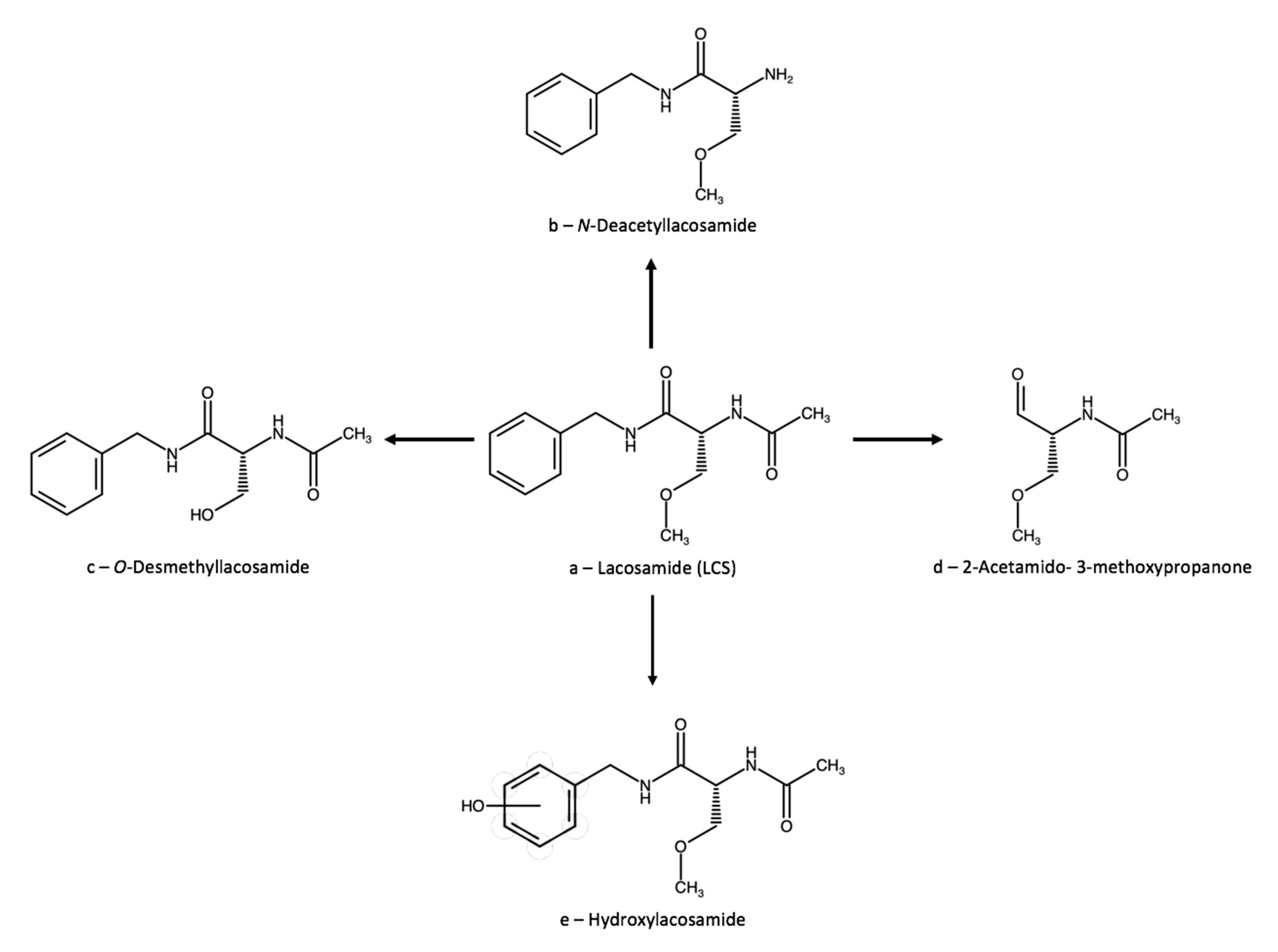
Since the end of the 2000s, the resolved (R)-LCS enantiomer has also been available for the treatment of partial onset seizures [57]. The (S)-enantiomer is inactive as an antiepileptic but could be useful in neuropathic pain management [58]. Hepatic LCS metabolism has recently been clarified, and involves three main pathways: deacetylation, amide hydrolysis, and O-demethylation (Figure 3b–d). The O-demethyl metabolite is inactive and its formation seems to be catalysed by CYP2C19 [59]. Aryl hydroxylation (Figure 3e) also seems to happen in different metabolic tissues, possibly renal ones [60]. Neither age nor sex seem to significantly affect the pharmacokinetic profile of LCS in adults [61].

3.2.1. Therapeutic Drug Monitoring

Single-Drug Methods

A clear relationship has been found between daily dose and plasma concentration of LCS; however, there does not appear to be any clear relationship between concentration and efficacy, and for this reason some authors think that the TDM of LCS is scarcely useful [59]. Nonetheless, the usefulness of TDM has recently been proposed in patients taking enzyme-inducer antiepileptics, in patients with decreased renal function or on dialysis, and older adults, due to the high probability of altered pharmacokinetics in these populations [62]. A paper has reported the use of LC-MS/MS to compare the serum and cerebrospinal fluid levels of LCS. A good correlation was found, thus confirming that serum is a significant and suitable matrix for the TDM of LCS [63]. The AGNP proposes a TDM recommendation level of 3, “useful”, for LCS, with therapeutic plasma levels in the 1–10 µg/mL and toxic levels above 20 µg/mL [24]. Yamamoto et al. have evaluated that the effective LCS concentration range in non-drug-fasting patients (1–3 h after administration) is presumably 20–40 µg/mL [64]. The analytical method in this case was based on HPLC-MS/MS after a plasma protein precipitation (PPP) procedure with formic acid carried out on 50 mL of serum, but unfortunately more details are missing.
A few HPLC-UV methods for LCS analysis in human plasma have been published. One method uses a C18 column and a mobile phase consisting of pH 2 phosphate buffer and methanol; detection is carried out at 225 nm [65]. Sample pretreatment is performed by a simple LLE from 500 µL of plasma with 4 mL of a diethyl ether/dichloromethane (70/30, v/v) mixture. The organic phase was evaporated to dryness at 40 °C under a nitrogen stream, then reconstituted with 200 μL of water/methanol (20:80, v/v). Linearity was achieved in the 0.2–20 µg/mL range and extraction yields ranged from 94% to 95%. Precision RSD was 4.0–7.8% and accuracy ranged from 92.4% to 111.7%. A few short-term stability assays were also carried out, with 94–96% accuracy. The IS was ranolazine (Figure 4a), an anti-angina agent with little structural resemblance to LCS.
Another HPLC-UV method uses a C18 column and a pH 9 ammonium formate buffer/acetonitrile mixture as the mobile phase under gradient mode, with detection at 210 nm [66]. Sample pretreatment consists of PPP with 300 µL of methanol added to 100 µL of plasma. The linearity range was 0.5–12.5 µg/mL, with precision RSD in the 2–18% range. Accuracy ranged from 97.6% to 115.6%. The method was cross-validated with an LC-MS/MS method, but no reference was provided for this latter method. Twenty-two antiepileptic drug solutions and six different blank plasma samples were tested for method selectivity, with satisfactory results. The choice of Ac-Phe-NHMe ((2S)-2-acetamido-N-methyl-3-phenylpropanamide, Figure 4b) as the IS was quite apt, since it is a modified amino acid (phenylalanine) with relatively close structural resemblance to LCS.
UHPLC-MS/MS(QqQ) has also been applied for LCS determination in plasma [67]. Sample pretreatment included diluting 150 mL of plasma with 50 µL of methanol/water (50/50, v/v) and PPP with 1 mL of acetonitrile/methanol/0.1% formic acid (40/40/20, v/v/v). After centrifugation, 5 µL of the supernatant were injected into a C18 column, with a mobile phase consisting of aqueous formic acid/methanol/acetonitrile under gradient conditions. Linearity was found in the 20–16,000 ng/mL range, with precision RSD values always lower than 4.7%. Extraction yields were higher than 97%, matrix effect was lower than 4%, and carryover was almost negligible (0.85%). Stability was tested under a wide set of conditions for up to 65 days and minimum recovery was 95.8%. As expected from a UHPLC method, the run time was very short: 2.2 min. The IS was an LCS-13C,D3 analogue.
Brandt et al. [68] studied the relationships between LCS daily dose and plasma and saliva levels and concluded that saliva has great clinical potential for TDM of the drug. Unfortunately, details of the analytical method are unavailable. On the other hand, another paper by Cawello et al. [69] had previously reported important information on the saliva (as well as plasma and urine) concentrations of LCS and O-demethyl-LCS. This in-formation was obtained by an LC-electrospray ionization (ESI)-MS method using a C8 column as the stationary phase. Saliva sample pretreatment was carried out by solid phase extraction (SPE) on C18 cartridges; no details of the procedure are reported. A different LC-ESI-MS method was used for plasma and urine analysis on a C8 column specifically designed for basic analyte separation (LiChrospher 60 RP-Select B). Next, 100-µL plasma samples were pretreated by PPP with 30 µL of trichloroacetic acid, centrifuged, and the supernatant diluted with 500 µL of water. Then, 10-µL urine samples were simply diluted with 5 mL of water (dilute-and-shoot, DAS). Linearity ranges for LCS were 0.1–20 µg/mL in plasma, 5–500 µg/mL in urine; for O-demethyl-LCS, 0.02–4 µg/mL in plasma, 1–100 µg/mL in urine; for both analytes, 0.02–12 µg/mL in saliva. The ISs were LCS-D7 and O-demethyl-LCS-D3. Further details on method performance are missing, but the methods are claimed to be validated according to the 2001 FDA guidelines for industry [70]. This is probably the most useful method available for LCS TDM, since it includes both its main metabolite and three different biological matrices.
A simple HPLC-UV method was also used to evaluate the possible correlations between serum and saliva LCS concentrations, using a C18 column and a methanol/formic acid/water mixture with detection at 215 nm to determine LCS in either matrix [71]; this method had already been used for LCS determination in plasma [72]. Next, 100-µL aliquots of biological sample were treated by PPP with 10 µL of 60% (m/m) perchloric acid for all matrices (serum, saliva, and serum ultrafiltrate for the determination of the free LCS fraction); after centrifugation, the supernatant was directly injected. Good linearity was found in the 2.5–62.6 µg/mL range, with precision RSD values in the 0.6–4.0% range. Extraction yields ranged from 99% to 106%. Selectivity assays reported no interference from any other co-administered AED, and stability studies proved satisfactory analyte stability for up to 8 days under the tested conditions. The paper’s results seem also to point out to a good correlation between serum and saliva LCS concentrations.
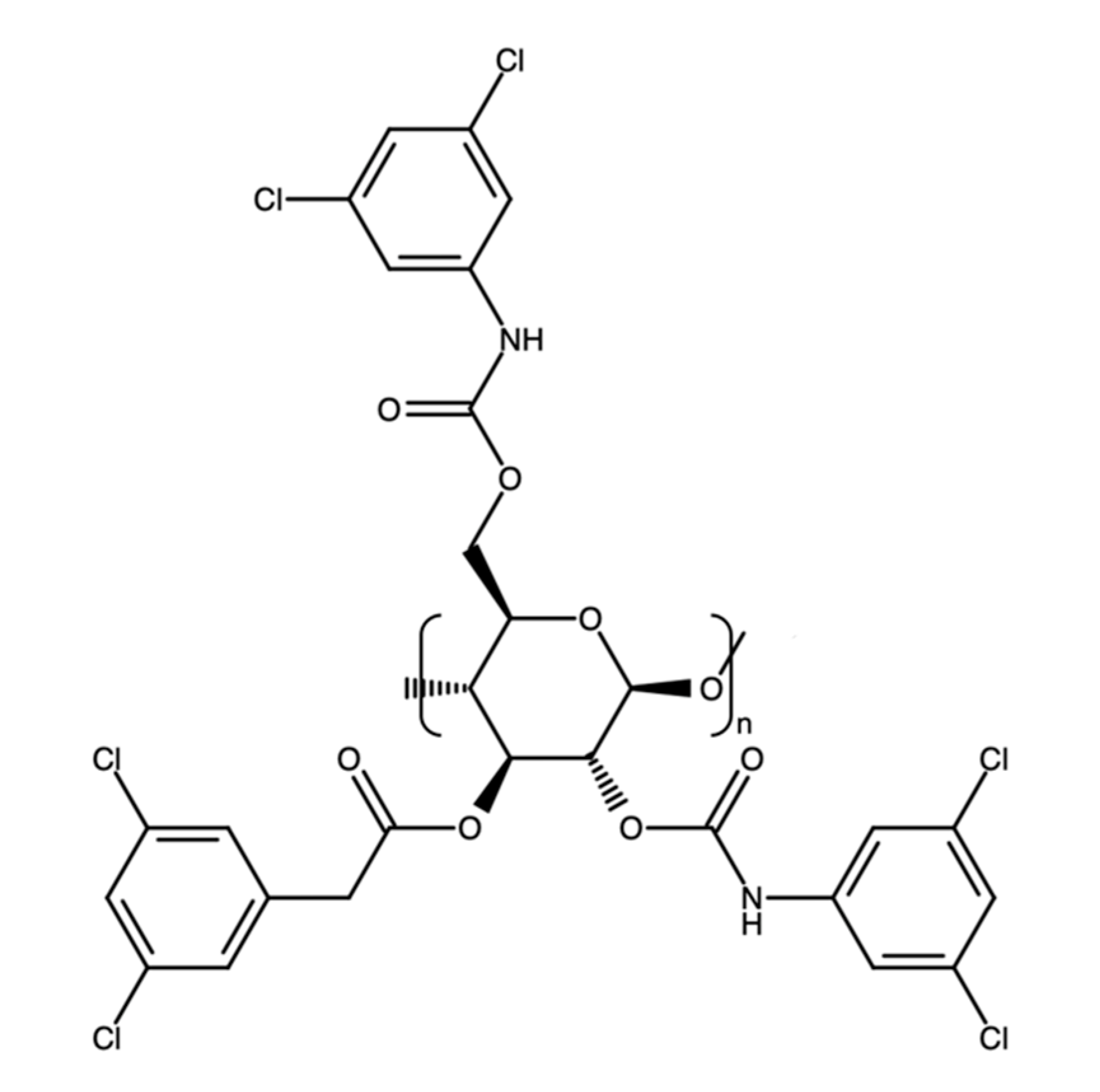
Very recently, a chiral LC-MS/MS method has been developed for the determination of (R)-LCS in the presence of the (S)- enantiomer [73]. Complete enantioresolution is obtained using a Daicel-IC3 chiral column, a polysaccharide-based column where the selector is immobilized on a silica matrix. In this case, the selector was cellulose tris(3,5-dichlorophenylcarbamate), Figure 5. LCS extraction was carried out on 100 µL of plasma by LLE with 1 mL of methyl t-butyl ether. The samples were then centrifuged, flash frozen in an alcohol bath, and the organic phase was evaporated to dryness (nitrogen stream at 40 °C) and redissolved in 500 µL of acetonitrile. Linearity was established in the 0.1–15 µg/mL range. Selectivity was satisfactory on normal, hemolyzed, and lipemic plasma samples. Carryover was always lower than 3.4%, while the matrix effect was lower than 5.8%; lipemic and haemolysed samples had higher matrix effect values, up to 12%. The least satisfactory parameter was probably extraction yield, which ranged from 46.8% to 62.1%; precision RSD values were up to 14.7%. As usual for methods involving MS detection, the IS was a deuterated (R)-LCS analogue, namely (R)-LCS-D6.
A gas chromatographic (GC)-MS method has been reported for LCS analysis in whole blood [74], although this kind of method is mostly useful for forensic analyses, not for TDM. After LLE with ethyl acetate at pH 12, the blood sample was injected without derivatization into the GC system, which featured a (5%-phenyl)-methylpolysiloxane column and helium as the carried gas under programmed temperature (120–300 °C). The MS detector was a single quadrupole coupled to an electron impact (EI) ionization chamber.

Multi-Drug Methods

A few multi-drug methods for LCS with possible TDM interest have been published. An LC-MS/MS method was used for the monitoring LCS and 16 other drugs (not all of them AEDs) in human breast milk. Protein precipitation followed by LC separation and detection by hybrid QqQ-ion trap (QTRAP) [75]. An HPLC-UV method was used for the application of TDM to saliva samples for LCS and three other AEDs (carbamazepine, licarbazepine, and levetiracetam). However, LCS saliva concentrations did not correlate well with plasmatic ones [76]. A simple HPLC—photodiode array detection (PDA) method has also been published for the simultaneous determination of LCS, ZNS, and levetiracetam in plasma [77] after LLE with ethyl acetate.
A resume of the main validation and performance parameters of these latter methods for LCS analysis is found in Table 1. As one can see, performances were often quite similar to those of dedicated, single-drug methods. Notably, none of these methods included any LCS metabolite (as is often the case in multi-drug methods). As can be surmised form the many analytical methods presented here, LCS has been extensively studied from an analytical point of view in the last few years. However, methods including one or more metabolites are still few, as is lacking complete knowledge about their actual biological activity. This should be a primary research objective in the future. In the future, enantioselective methods for LCS TDM will probably be the norm to obtain more.

3.2.2. Lacosamide Interactions

The potential of LCS for interactions, both with other AEDs and with different classes of drugs, seems to be relatively low [78].

Clinically Relevant Data

In a retrospective study on 157 drug-resistant patients treated with LCS, levetiracetam resulted to be the compound most frequently associated with LCS in the responder subgroup. This allows us to hypothesize that LCS and levetiracetam can interact synergistically at the pharmacodynamic level [56]. Phase I clinical studies have found no significant interactions of LCS with omeprazole [79] or digoxin [80]. Two separate TDM studies on 128 [81] and 75 [82] patients found that enzyme-inducing AEDs (carbamazepine and phenytoin in the first study, carbamazepine, phenytoin, and phenobarbital in the second one) reduce LCS serum concentrations by 30–40%; these results were later confirmed by another TDM study, which also found even more pronounced effects in children [83].

Data with Low or Uncertain Clinical Relevance, Case Reports

A small post-marketing assessment has found that concomitant use of LCS and other voltage-gated sodium channel-blocking AEDs can cause neurotoxicity (diplopia, dizziness, drowsiness), probably due to pharmacodynamic interactions [84]. A case report found reduced plasma levels of valproate and levetiracetam during LCS polypharmacy [85]. A case of LCS overdose with concomitant levetiracetam and cyclobenzaprine intake produced ventricular dysrhythmias, which was attributed to elevated sodium channel blockade caused by LCS and cyclobenzaprine [86]. In an open-label crossover study, no interaction between LCS and warfarin was observed [87]. Clinical trials have found no significant interactions between LCS and combined contraceptives [88], valproate [89], or carbamazepine [90]. Since LCS treatment has been described as a possible cause of QT-interval prolongation, its simultaneous administration with other drug with similar effects on heart conduction could theoretically cause life-threatening arrhythmias [91]. In this regard, a case report has found that an LCS overdose combined with therapeutic concentrations of sodium channel blocking agents (cyclobenzaprine) may have caused cardiac conduction delays and cardiac arrest [86].

3.2.3. Neuroprotection

In rats, LCS has been observed to reduce oxidative stress, inflammation, and apoptosis caused by lipopolysaccharide in rat brain [92]; it also decreases the production of reactive oxygen species by increasing antioxidant enzyme expression, inhibiting lipid peroxidation and attenuating glial cell activation in experimental spinal cord injury [93]. A similar effect has been observed in gerbils, where LCS protected animals from transient cerebral ischemia by increasing catalase and glutathione peroxidase expression [94]. In mice, the drug seems to protect striatal and hippocampal neurones from energy metabolism failure due to ischemia [95].

3.3. Zonisamide

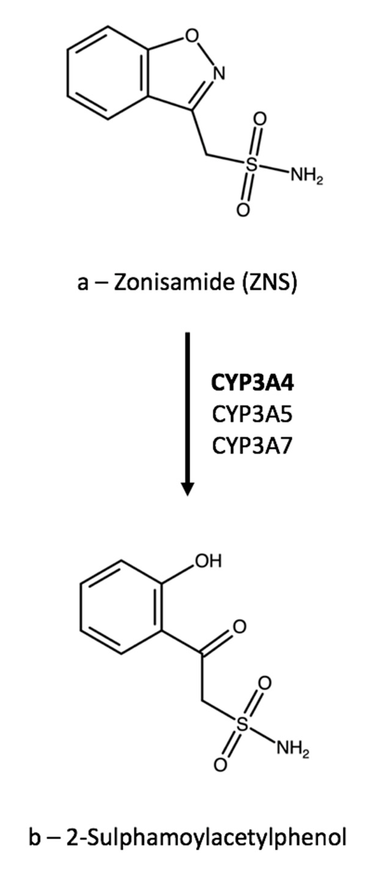
Zonisamide (1,2-benzoxazol-3-ylmethanesulfonamide, ZNS, Figure 6a) is an anticonvulsant agent used primarily for adjunctive treatment of partial seizures. However, it can also be used for a variety of other indications either on- or off-label, including partial and generalized seizures and absence [96], bipolar depression as a mood stabilizer [97], Parkinson’s disease [98], tardive dyskinesia during antipsychotic therapy [99], and migraine [100]. The mechanism of ZNS antiepileptic activity is still debated, but it is currently reputed to be the blockade of sodium and T-type calcium channels [101]. Moreover, the modulation of GABAergic and glutamatergic neurotransmission could have a role in seizure inhibition [102]. ZNS is mainly metabolized to 2-sulphamoylacetylphenol (cleavage of the benzisoxazole ring, Figure 6b) by CYP3A4, with minor contributions from CYP3A7 and CYP3A5 [103].

3.3.1. Therapeutic Drug Monitoring

Single-Drug Methods

According to an early report [104], the ZNS dose-serum level correlation is linear up to doses of 10 to 15 mg/kg per day, and the therapeutic range is 10–40 μg/mL. However, the relationship between serum ZNS levels, clinical response, and adverse effects appears weak. The AGNP reports a similar therapeutic range and an immediately adjacent toxic range, starting from 40 µg/mL. Due to this close proximity between therapeutic and toxic levels, the TDM recommendation level for ZNS is 2, “recommended” [24].
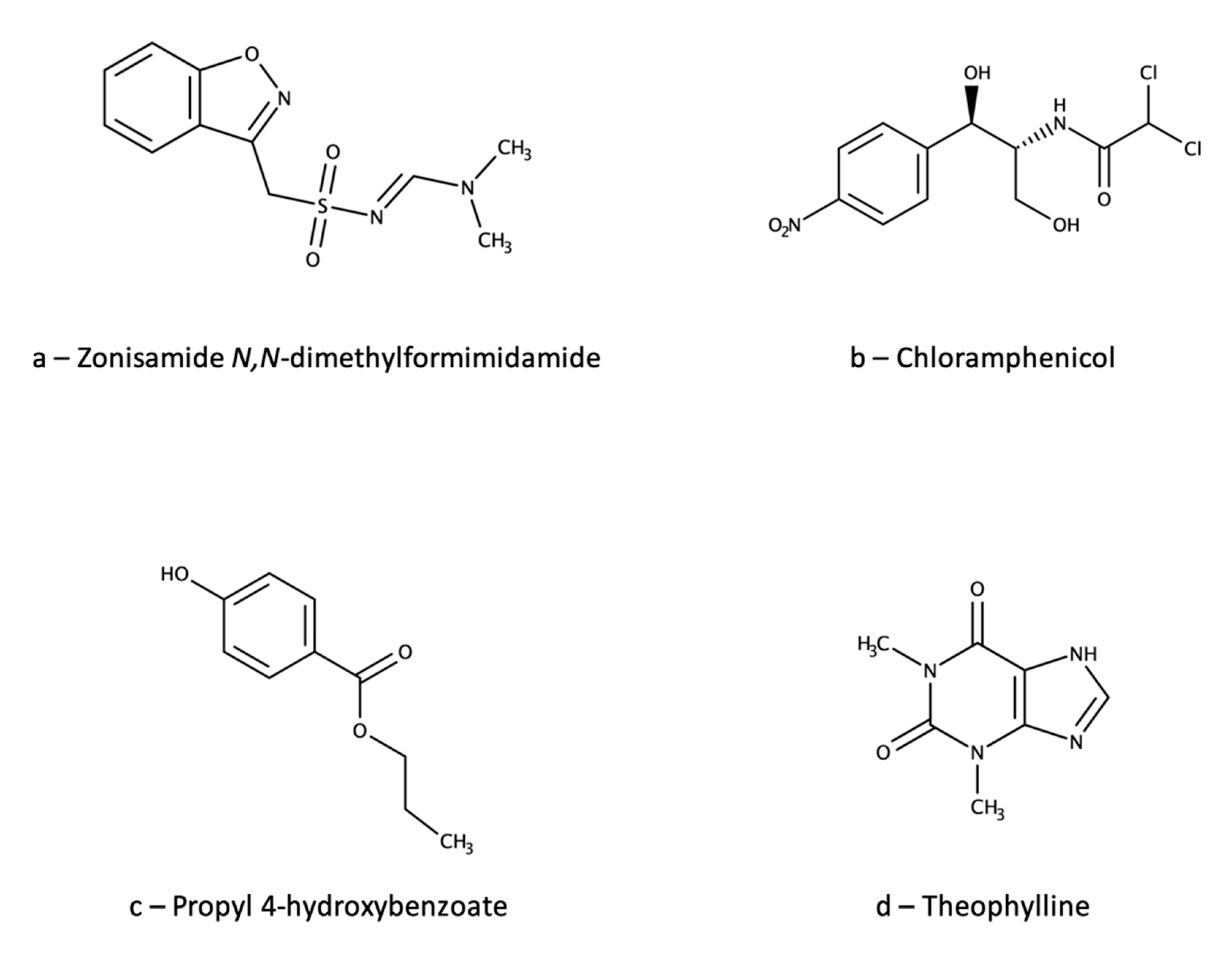
Recently, an HPLC-UV method has been used to verify the reliability of a routine TDM kit based on latex particle-enhanced turbidimetric immunoassay (LTIA) for ZNS [105]. The HPLC method used a C18 column and a mobile phase consisting of water, isopropyl alcohol, acetonitrile, and acetic acid. Column temperature was set at 40 °C and detection wavelength at 238 nm. Plasma sample (100 µL) pretreatment was based on a simple PPP with 200 µL of methanol, followed by centrifugation and supernatant injection. The IS was ZNS N,N-dimethylformimidamide (Figure 7a), which is reasonably similar to the analyte; however, the introduction of a relatively big functional group could impair the IS performance and make the method less than ideally reliable. The method was validated for ZNS quantification in the 1–75 µg/mL range, with accuracy in the 93.7–103.7% range and precision RSD values up to 10.3%. The corresponding validation parameters for the LTIA method were: linearity 5–50 µg/mL; accuracy 84.2–106.7; precision RSD ≤ 5.2%. The paper reports strong correlation between the results of the two methods, thus ruling out possible interferences from ZNS metabolites with the LTIA method. However, since the HPLC-UV method did not separate nor analyse any metabolites, and was not validated for selectivity toward them, similar interferences from metabolites in both methods could theoretically not be ruled out.
ZNS was also determined in plasma using an HPLC-UV method [106] with a C18 column kept at 35 °C and a mobile phase consisting in a water/acetonitrile (65/35) mixture. Detection wavelength was 240 nm. Sample preparation was carried out on a semimicro scale, with 100 µL of plasma being subjected to PPP with 400 µL of ice-cold acetonitrile and then to microextraction by packed sorbent (MEPS) on C18 sorbent. MEPS is a miniaturized version of SPE, where small amounts (a few milligrams) of sorbent are contained in a barrel insert and needle (BIN) assembly and all typical SPE steps (activation, conditioning, loading, washing, and elution) are functionally carried out by drawing into, and discharging from, a custom syringe through the BIN. In this case, no washing step was used and elution was carried out with 60 µL of acetonitrile, followed by dilution with 90 µL of water and injection. Experimental design was used to optimize the most critical parameters of the PPP+MEPS sample preparation procedure. Linearity was obtained in the 0.2–80 µg/mL range, with precision RSD values ≤ 12.5% and accuracy values between 87.7% and 111.5%. Absolute recovery ranged between 63% and 65% and the analyte was found to be stable under most tested conditions. The IS was chloramphenicol (Figure 7b), which is both structurally dissimilar from ZNS and also a commercially available antibacterial drug, so this choice appears to be relatively unsound.
A micellar electrokinetic chromatographic (MEKC) method is also available for the TDM of ZNS [107]. This method uses an extended-path capillary with a 150-µm internal diameter at the optical window (as compared to 50 µm for the rest of the capillary) to increase sensitivity, the limit of detection (LOD) thus obtained when detecting at 210 nm was 3 µg/mL. The running buffer was a pH 8 phosphate buffer containing 20 mM of sodium dodecyl sulphate (SDS). Sample pretreatment (200 µL of serum) was carried out by LLE with 1 mL of ethyl acetate containing propyl 4-hydroxybenzoate (Figure 7c) as the IS; this choice appears less than optimal due to structural dissimilarity. After centrifugation, 500 µL of supernatant was evaporated to dryness (nitrogen stream), redissolved in 50 µL of 5% (v/v) aqueous methanol and injected. Linearity was obtained in the 5–40 µg/mL range, with precision RSD values between 1.3% and 16.4% for peak areas and between 0.3% and 3.7% for migration times. Mean extraction yield was 73.3%. The MEKC method results were compared to those of an HPLC-UV method, with good agreement between them, although the serum levels found with MEKC tended to be lower than those found by HPLC, since linear correlation graph had a slope of 0.87.
A peculiar technique for TDM, namely high-performance thin-layer chromatography (HPTLC), has been also used for ZNS analysis in human plasma [108]. Silica gel-coated aluminium plates were used, and the mobile phase was an ethyl acetate/methanol/toluene mixture; densitometric detection was carried out at 254 nm. Aliquots of 150 µL of serum were pretreated by PPP with 450 µL of acetonitrile, then centrifuged and the supernatant evaporated to dryness (nitrogen stream), redissolved in 60 µL of methanol and injected. Linearity was obtained in the 5–80 µg/mL range, with precision RSD values ranging from 2.1% to 4.4% and accuracy from 97.5% to 115.2%. Robustness was also evaluated, with satisfactory results. Theophylline (Figure 7d) was used as the IS; although not optimal, this choice appears to be at least acceptable.
Overall, many analytical methods for the TDM of ZNS have been published. They provide a wide choice to laboratories for the best suited methods in relation to their instrumental availability and specific patients’ needs or clinicians’ requests. However, none of the methods reported above included the determination of the main ZNS metabolite. This is probably dictated by the fact that the metabolite does not appear to contribute to the therapeutic effect; however, its determination could provide interesting information on the metabolic status of the patient and their compliance with the therapy.

Multi-Drug Methods

Several analytical methods with possible TDM application include ZNS together with other AEDs. Among them, and especially in recent years, methods using microsampling techniques are becoming commonplace. These techniques can provide several advantages over traditional venepuncture sampling, including reduced invasiveness, increased analyte stability, and minimal storage and shipping requirements [109,110,111,112]. For example, LC-MS/MS was used to analyze 14 drugs using volumetric absorptive microsampling (VAMS) [113], a technique based on a plastic device with a porous polymeric tip that is able to absorb fixed biological matrix volumes [114,115,116]. Another microsampling technique exploiting dried sample spot devices (DSSD) was used for plasma sampling. In this case, the DSSD is a simple filter paper sheet where the desired plasma volume is deposited (and absorbed) as a series of spots, which are then dried and properly extracted [117]. Analysis is then carried out by LC-MS/MS. Another miniaturized matrix, dried plasma spots (DPS), was used to analyze six AEDs, including ZNS, by HPLC-UV [118].
LC-MS/MS [119,120,121,122], HPLC-PDA [77], and HPLC-UV [123,124,125,126] were also used for the analysis of “traditional” plasma or serum samples. HPTLC with UV densitometry was used to analyse ZNS, lamotrigine, and levetiracetam in human plasma [127]. Finally, an HPLC-spectrofluorometric detection (FL) method was used to analyze ZNS, topiramate, and sulpiride in plasma after their derivatization with 4-chloro-7-nitrobenzofurazan (NBD-Cl) [128]. The validation and performance parameters of these multi-drug methods are summarized in Table 2. As already noted for ZNS single-drug methods, none of the multi-drug methods included any ZNS metabolite.

3.3.2. Zonisamide Interactions

Clinically Relevant Data

Carbamazepine, phenytoin, and phenobarbital all increase ZNS clearance by 24–29%, an interaction that may necessitate a dosage increase [129]. Otherwise, ZNS is essentially devoid of clinically significant interactions with other AEDs (such as valproate [130] and lamotrigine [131]), oral contraceptives, and most other classes of therapeutic agents [132]. Conversely, ZNS does not appear to significantly alter the pharmacokinetics of phenytoin [133] and carbamazepine [134], nor those of oral contraceptives [135]. ZNS is being studied as a possible agent to counter the weight gain observed during atypical antipsychotic treatment, and in particular during olanzapine therapy. However, it is still unknown whether this is the effect of a DDI, or the net result of two different, independent activities. Due to the broad spectrum of receptor and channel systems targeted by both atypical antipsychotics and ZNS, it is difficult to exclude an interaction [136]. Possible points of confluence could be dopamine release and modulation of dopamine D2 receptors, or hypothalamic feeding circuits [137].

Data with Low or Uncertain Clinical Relevance, Case Reports

ZNS is a competitive inhibitor of human MAO B, but not of MAO A [138]. As such, it has the potential to interact pharmacodynamically with some Alzheimer’s and Parkinson’s disease medications.

3.3.3. Neuroprotection

In experimental preclinical mouse models of Parkinson’s disease, ZNS reduces neuroinflammation through the downregulation of microglial voltage-gated sodium channels and the decrease of pro-inflammatory cytokine levels [139]. It also reduces nigrostriatal dopaminergic neurodegeneration in a mouse genetic model of Parkinson’s disease [140], possibly by increasing GSH levels by enhancing the astroglial cystine transport system and/or astroglial proliferation [141].
Moreover, the very antiepileptic effect of ZNS could have neuroprotective consequences: The drug has been found to reduce seizure-induced free radical production and neuroinflammation in animals [142]. In rat brain slices, ZNS significantly reduces long-term hippocampal potentiation caused by ischemia, and this has been demonstrated to happen through the nitric oxide/cyclic guanosine monophosphate/protein kinase G (NO/cGMP/PKG) pathway. These findings support the possible use of ZNS as a neuroprotective agent in brain ischemia [143].

4. Multi-Drug Methods

A couple of papers report multi-drug methods, which include all three drugs considered in this review: EZG, LCS, and ZNS. Of these, one is mainly focused on forensic samples (blood, plasma, serum) and is based on LC-MS/MS (QqQ) on a C18 column [144]. This method allows the determination of 22 different AEDs (including a few metabolites). Sample preparation is carried out on 100 µL of biological matrix by PPP with 400 µL of methanol. After centrifugation, 200 µL of supernatant were diluted with 1.3 mL of water and injected (5 µL). Performance and validation parameters of this method for the three analytes of interest in plasma are reported in Table 3. As with many methods of this kind, suboptimal IS choice is one of its main weak points.
The second paper is focused on determining the serum protein binding ratio of 25 AEDs and 2 metabolites [145]. More than one analytical LC-MS/MS(QqQ) method was used, and many experimental details seem to be missing; however, they include the use of a C18 column and sample (24 µL) preparation by PPP with 500 mL of acetonitrile. As usual for multi-drug methods, neither one includes any EZG, LCS, or ZNS metabolite.

5. Conclusions

In this review, an overview was presented on the most important analytical methods potentially useful in the TDM of EZG, LCS, and ZNS. All of this information has been collected and updated to 2021 in a single article for the first time here, also including additional notes on drug–drug interactions and neuroprotection studies, which represent two of the most frequent use and interest cases for the TDM of AEDs and for related analytical methods.
Reversed phase LC is absolutely the most frequently used technique, due to its versatility, precision, accuracy, and selectivity. LC is most often coupled with MS, which in turn is optimal in terms of sensitivity and selectivity. Its use is also supported by the lack of strong UV absorption at high wavelengths (>250 nm) in the considered drugs, rendering UV and FL more difficult to use for them than for other drug classes. Nonetheless, the relatively high typical plasma concentrations of these drugs (in the tens of µg/mL range) can make them amenable to spectrometric detection means, especially at low UV wavelengths and/or following suitable derivatization. GC and CE are much less frequent, in part due to the relative scarcity of the corresponding equipment in typical clinical analysis laboratories. Due in part to the low sensitivity required, HPTLC with densitometric detection is also a viable option. Future developments will reasonably see an increase in the use of LC-MS/MS methods, as instrumentation prices will decrease making them more commonplace, and suitably trained personnel will be more easily available. As highlighted above, just a few methods are currently completely suitable for the TDM of the considered drugs, with many having more or less severe drawbacks, usually regarding IS choice or lack of one or more validation parameter. Probably the most glaring defect of most TDM methods for EZG, LCS, and ZNS is that their metabolites are almost never determined. This is a big problem especially for EZG, whose main metabolite is biologically active and whose glucuronidated metabolites are labile and present in plasma at very high concentrations. However, it is our strong opinion that if possible, at least one or two metabolites should always be included in TDM methods. In fact, metabolite concentrations always provide much needed information regarding the metabolic setup of the patient and their compliance, thus greatly helping clinicians in taking critical therapeutic decisions, such as whether to change the drug dose regimen, to switch to another drug or to request other tests and verifications.
So, it is clear that there is still place for new scientific research simultaneously addressing most or all of these weak spots. Reducing sample preparation time, complication, and variability is another field of real interest, and in the next few years one expects to see a rapid increase in automation and in “dilute-and-shoot” approaches. In this regard, microsampling (through DPS, VAMS, and other techniques) could be a real boon and is already increasingly being employed to reduce invasiveness, costs, and refrigeration/storage/shipping requirements [146], although its use is by no means widespread. Similarly, alternative matrices with lower or no invasiveness and better manageability are currently under scrutiny, with saliva as the most attractive candidate. Nonetheless, studies on the clinical significance of saliva levels (and their possible correlation with plasma/serum levels), however promising, are still underway.

Author Contributions

Conceptualization, R.M., M.P. and L.M. (Laura Mercolini); methodology, R.M., M.P. and L.M. (Laura Mercolini); investigation, R.M., L.M. (Lorenzo Marincich), M.P. and L.M. (Laura Mercolini); resources, R.M. and L.M. (Laura Mercolini); data curation, R.M., L.M. (Lorenzo Marincich), M.P. and L.M. (Laura Mercolini); writing—original draft preparation, R.M., L.M. (Lorenzo Marincich), M.P. and L.M. (Laura Mercolini); writing—review and editing, R.M., L.M., M.P. and L.M. (Laura Mercolini); visualization, R.M., L.M. (Lorenzo Marincich), M.P. and L.M. (Laura Mercolini); supervision, R.M.; project administration, R.M.; funding acquisition, R.M. and L.M. (Laura Mercolini). All authors have read and agreed to the published version of the manuscript.

Funding

This research was funded by University of Bologna with RFO (Ricerca Fondamentale Orientata) funds.

Institutional Review Board Statement

Not applicable.

Informed Consent Statement

Not applicable.

Data Availability Statement

No new data were created or analyzed in this study. Data sharing is not applicable to this article.

Conflicts of Interest

The authors declare no conflict of interest.

References

  1. World Health Organization. Epilepsy. Available online: http://www.who.int/mediacentre/factsheets/fs999/en/ (accessed on 2 August 2021).
  2. GBD 2016, Neurology Collaborators. Global, regional, and national burden of neurological disorders, 1990–2016: A systematic analysis for the Global Burden of Disease Study 2016. Lancet Neurol. 2019, 18, 357–375. [Google Scholar] [CrossRef] [Green Version]
  3. Beghi, E. The Epidemiology of Epilepsy. Neuroepidemiol. 2020, 54, 185–191. [Google Scholar] [CrossRef] [PubMed]
  4. Jost, J.; Moyano, L.M.; Auditeau, E.; Boumediene, F.; Ratsimbazafy, V.; Preux, P.-M. Interventional programs to improve therapeutic management of people with epilepsy in low– and middle–income countries. Epilepsy Behav. 2018, 80, 337–345. [Google Scholar] [CrossRef]
  5. Meyer, A.-C.; Dua, T.; Ma, J.; Saxena, S.; Birbeck, G. Global disparities in the epilepsy treatment gap: A systematic review. Bull. World Health Organ. 2010, 88, 260–266. [Google Scholar] [CrossRef] [PubMed]
  6. Katchanov, J.; Birbeck, G.L. Epilepsy care guidelines for low– and middle– income countries: From WHO mental health GAP to national programs. BMC Med. 2012, 10, 107–113. [Google Scholar] [CrossRef] [Green Version]
  7. Fiest, K.M.; Sauro, K.M.; Wiebe, S.; Patten, S.B.; Kwon, C.S.; Dykeman, J. Prevalence and incidence of epilepsy: A systematic review and meta–analysis of international studies. Neurology 2017, 88, 296–303. [Google Scholar] [CrossRef]
  8. Aboul–Enein, M.N.; El–Azzouny, A.A.; Saleh, O.A.; Maklad, Y.A. On chemical structures with potent antiepileptic/anticonvulsant profile. Mini Rev. Med. Chem. 2012, 12, 671–700. [Google Scholar] [CrossRef] [PubMed]
  9. Goldenberg, M.M. Overview of Drugs Used For Epilepsy and Seizures: Etiology; Diagnosis; and Treatment. Pharm. Ther. 2010, 35, 392–415. [Google Scholar]
  10. Łukawski, K.; Gryta, P.; Łuszczki, J.; Czuczwar, S.J. Exploring the latest avenues for antiepileptic drug discovery and development. Expert Opin. Drug Dis. 2016, 11, 369–382. [Google Scholar] [CrossRef]
  11. Lasoñ, W.; Chlebicka, M.; Rejdak, K. Research advances in basic mechanisms of seizures and antiepileptic drug action. Pharmacol. Rep. 2013, 65, 787–801. [Google Scholar] [CrossRef]
  12. Jacob, S.; Nair, A.B. An Updated Overview on Therapeutic Drug Monitoring of Recent Antiepileptic Drugs. Drugs R. D. 2016, 16, 303–316. [Google Scholar] [CrossRef] [Green Version]
  13. Patsalos, P.N.; Berry, D.J.; Bourgeois, B.F.D.; Cloyd, J.C.; Glauser, T.A.; Johannessen, S.I.; Leppik, I.E.; Tomson, T.; Perucca, E. Antiepileptic drugs—Best practice guidelines for therapeutic drug monitoring: A position paper by the subcommission on therapeutic drug monitoring, ILAE Commission on Therapeutic Strategies. Epilepsia 2008, 49, 1239–1276. [Google Scholar] [CrossRef]
  14. Preskorn, S.H. Outliers on the dose–response curve: How to minimize this problem using therapeutic drug monitoring, an underutilized tool in psychiatry. J. Psychiatr. Pract. 2010, 16, 177–182. [Google Scholar] [CrossRef]
  15. Mandrioli, R.; Mercolini, L.; Saracino, M.A.; Raggi, M.A. Selective serotonin reuptake inhibitors (SSRIs): Therapeutic drug monitoring and pharmacological interactions. Curr. Med. Chem. 2012, 19, 1846–1863. [Google Scholar] [CrossRef]
  16. Mandrioli, R.; Protti, M.; Mercolini, L. Novel atypical antipsychotics: Metabolism and Therapeutic Drug Monitoring (TDM). Curr. Drug Metab. 2015, 16, 141–151. [Google Scholar] [CrossRef]
  17. Raggi, M.A.; Mandrioli, R.; Sabbioni, C.; Pucci, V. Atypical antipsychotics: Pharmacokinetics, therapeutic drug monitoring and pharmacological interactions. Curr. Med. Chem. 2004, 11, 279–296. [Google Scholar] [CrossRef]
  18. Pichini, S.; Papaseit, E.; Joya, X.; Vall, O.; Farré, M.; Garcia-Algar, O.; De Latorre, R. Pharmacokinetics and therapeutic drug monitoring of psychotropic drugs in pediatrics. Ther. Drug Monit. 2009, 31, 283–318. [Google Scholar] [CrossRef]
  19. Clarke, W.; McMillin, G. Application of TDM, pharmacogenomics and biomarkers for neurological disease pharmacotherapy: Focus on antiepileptic drugs. Pers. Med. 2006, 3, 139–149. [Google Scholar] [CrossRef]
  20. Mandrioli, R.; Mercolini, L. Metabolism of drugs used in the therapy of seizures: An analytical point of view. Part 1. Curr.Drug Metab. 2017, 18, 735–756. [Google Scholar] [CrossRef]
  21. Johannessen, S.I.; Tomson, T. Pharmacokinetic variability of newer antiepileptic drugs: When is monitoring needed? Clin. Pharmacokin. 2006, 45, 1061–1075. [Google Scholar] [CrossRef]
  22. Baumann, P.; Hiemke, C.; Ulrich, S.; Eckermann, G.; Gaertner, I.; Gerlach, M.; Kuss, H.J.; Laux, G.; Müller–Oerlinghausen, B.; Rao, M.L.; et al. Arbeitsgemeinschaft für neuropsychopharmakologie und pharmakopsychiatrie. The AGNP–TDM expert group consensus guidelines: Therapeutic drug monitoring in psychiatry. Pharmacopsychiatry 2004, 37, 243–265. [Google Scholar] [CrossRef] [PubMed]
  23. Hiemke, C.; Baumann, P.; Bergemann, N.; Conca, A.; Dietmaier, O.; Egberts, K.; Fric, M.; Gerlach, M.; Greiner, C.; Gründer, G.; et al. AGNP consensus guidelines for therapeutic drug monitoring in psychiatry: Update 2011. Pharmacopsychiatry 2011, 44, 195–235. [Google Scholar] [CrossRef] [PubMed] [Green Version]
  24. Hiemke, C.; Bergemann, N.; Clement, H.W.; Conca, A.; Deckert, J.; Domschke, K.; Eckermann, G.; Egberts, K.; Gerlach, M.; Greiner, C.; et al. Consensus guidelines for therapeutic drug monitoring in neuropsychopharmacology: Update 2017. Pharmacopsychiatry 2018, 51, 9–62. [Google Scholar] [PubMed] [Green Version]
  25. Scopus Document Search. Available online: https://www.scopus.com/home.uri (accessed on 2 August 2021).
  26. PubMed. Available online: https://www.ncbi.nlm.nih.gov/pubmed (accessed on 2 August 2021).
  27. Baftiu, A.; Johannessen Landmark, C.; Nikaj, V.; Neslein, I.-L.; Johannessen, S.I.; Perucca, E. Availability of antiepileptic drugs across Europe. Epilepsia 2015, 56, e191–e197. [Google Scholar] [CrossRef] [Green Version]
  28. Clark, S.; Antell, A.; Kaufman, K. New antiepileptic medication linked to blue discoloration of the skin and eyes. Ther. Adv. Drug Saf. 2015, 6, 15–19. [Google Scholar] [CrossRef] [Green Version]
  29. Advance notification of Trobalt® discontinuation. Available online: https://assets.publishing.service.gov.uk/media/57fe4b6640f0b6713800000c/Trobalt_letter.pdf (accessed on 2 August 2021).
  30. Orhan, G.; Wuttke, T.V.; Nies, A.T.; Schwab, M.; Lerche, H. Retigabine/Ezogabine, a KCNQ/KV7 channel opener: Pharmacological and clinical data. Exp. Opin. Pharmacother. 2012, 13, 1807–1816. [Google Scholar] [CrossRef]
  31. Hempel, R.; Schupke, H.; McNeilly, P.J.; Heinecke, K.; Kronbach, C.; Grunwald, C.; Zimmermann, G.; Griesinger, C.; Engel, J.; Kronbach, T. Metabolism of retigabine (D–23129), a novel anticonvulsant. Drug Metab. Dispos. 1999, 27, 613–622. [Google Scholar]
  32. McNeilly, P.J.; Torchin, C.D.; Anderson, L.W.; Kapetanovic, I.M.; Kupferberg, H.J.; Strong, J.M. In vitro glucuronidation of D–23129, a new anticonvulsant, by human liver microsomes and liver slices. Xenobiotica 1997, 27, 431–441. [Google Scholar] [CrossRef]
  33. Patsalos, P.N.; Berry, D.J. Therapeutic drug monitoring of antiepileptic drugs by use of saliva. Ther. Drug Monit. 2013, 35, 4–29. [Google Scholar] [CrossRef]
  34. Elkady, E.F.; Aboelwafa, A.A.; Fouad, M.A. Study of gender–related pharmacokinetics of ezogabine in Egyptian volunteers by a validated LC–MS/MS bioanalytical method. J. Adv. Res. 2020, 22, 99–104. [Google Scholar] [CrossRef]
  35. Knebel, N.G.; Grieb, S.; Leisenheimer, S.; Locher, M. Determination of retigabine and its acetyl metabolite in biological matrices by on–line solid–phase extraction (column switching) liquid chromatography with tandem mass spectrometry. J. Chromatogr. B 2000, 748, 97–111. [Google Scholar] [CrossRef]
  36. Bu, W.; Nguyen, M.; Xu, C.; Lin, C.-C.; Yeh, L.-T.; Borges, V. Determination of N-acetyl retigabine in dog plasma by LC/MS/MS following off-line μElution 96-well solid phase extraction. J. Chromatogr. B 2007, 852, 465–472. [Google Scholar] [CrossRef]
  37. Licea Perez, H.; Boram, S.L.; Evans, C.A. Development and validation of a quantitative method for determination of retigabine and its N-acetyl metabolite; overcoming challenges associated with circulating labile N-glucuronide metabolites. Anal. Meth. 2015, 7, 723–735. [Google Scholar] [CrossRef]
  38. Franco, V.; Baruffi, K.; Gatti, G.; Marchiselli, R.; Fattore, C.; Canevini, M.P.; Crema, F.; Perucca, E. A simple and rapid HPLC–UV method for the determination of retigabine in human plasma. Biomed. Chromatogr. 2018, 32, e4168. [Google Scholar] [CrossRef]
  39. Tompson, D.J.; Crean, C.S.; Buraglio, M.; Arumugham, T. Lack of effect of ezogabine/retigabine on the pharmacokinetics of digoxin in healthy individuals: Results from a drug–drug interaction study. Clin. Pharmacol. 2014, 6, 149–159. [Google Scholar] [CrossRef] [Green Version]
  40. Crean, C.S.; Tompson, D.J.; Buraglio, M. The effect of ezogabine on the pharmacokinetics of an oral contraceptive agent. Int. J. Clin. Pharmacol. Ther. 2013, 51, 847–853. [Google Scholar] [CrossRef]
  41. Ferron, G.M.; Patat, A.; Parks, V.; Rolan, P.; Troy, S. Lack of pharmacokinetic interaction between retigabine and phenobarbital at steady–state in healthy subjects. Clin. Pharmacol. Ther. 2001, 69, P48. [Google Scholar]
  42. Ferron, G.M.; Patat, A.; Parks, V.; Rolan, P.; Troy, S.M. Lack of pharmacokinetic interaction between retigabine and phenobarbitone at steady–state in healthy subjects. Br. J. Clin. Pharmacol. 2003, 56, 39–45. [Google Scholar] [CrossRef] [Green Version]
  43. Luszczki, J.J.; Wu, J.Z.; Raszewski, G.; Czuczwar, S.J. Isobolographic characterization of interactions of retigabine with carbamazepine, lamotrigine, and valproate in the mouse maximal electroshock–induced seizure model. N.-S. Arch. Pharmacol. 2009, 379, 163–179. [Google Scholar] [CrossRef]
  44. Tompson, D.J.; Crean, C.S. The interaction potential of retigabine (ezogabine) with other antiepileptic drugs. Curr. Clin. Pharmacol. 2014, 9, 148–156. [Google Scholar] [CrossRef]
  45. Ferron, G.M.; Sachdeo, R.; Partiot, A.; Fritz, T.; Althouse, S.; Troy, S. Pharmacokinetic interaction between valproic acid, topiramate, phenytoin or carbamazepine and retigabine in epileptic patients. Clin. Pharmacol. Ther. 2001, 69, P18. [Google Scholar]
  46. Crean, C.S.; Tompson, D.J. The Effects of Ethanol on the Pharmacokinetics, Pharmacodynamics, Safety, and Tolerability of Ezogabine (Retigabine). Clin. Ther. 2013, 35, 87–93. [Google Scholar] [CrossRef]
  47. Nodera, H.; Spieker, A.; Sung, M.; Rutkove, S. Neuroprotective effects of Kv7 channel agonist, retigabine, for cisplatin–induced peripheral neuropathy. Neurosci. Lett. 2011, 505, 223–227. [Google Scholar] [CrossRef]
  48. Mora, G.; Tapia, R. Effects of retigabine on the neurodegeneration and extracellular glutamate changes induced by 4–aminopyridine in rat hippocampus in vivo. Neurochem. Res. 2005, 30, 1557–1565. [Google Scholar] [CrossRef]
  49. Feng, M.; Crowley, N.A.; Patel, A.; Guo, Y.; Bugni, S.E.; Luscher, B. Reversal of a Treatment–Resistant, Depression–Related Brain State with the Kv7 Channel Opener Retigabine. Neuroscience 2019, 406, 109–125. [Google Scholar] [CrossRef]
  50. Li, G.-B.; Liu, J.-Y.; Feng, X.-M.; Zhang, B.-L.; Zhang, R.-S. Retigabine attenuates focal cerebral ischemic injury through inhibiting mitochondria–dependent apoptotic pathway. Eur. Rev. Med. Pharmacol. Sci. 2018, 22, 5018–5023. [Google Scholar]
  51. Zhao, Y.-J.; Nai, Y.; Li, S.-Y.; Zheng, Y.-H. Retigabine protects the blood–brain barrier by regulating tight junctions between cerebral vascular endothelial cells in cerebral ischemia-reperfusion rats. Eur. Rev. Med. Pharmacol. Sci. 2018, 22, 8509–8518. [Google Scholar]
  52. Sheppard, A.M.; Chen, G.-D.; Salvi, R. Potassium ion channel openers, Maxipost and Retigabine, protect against peripheral salicylate ototoxicity in rats. Hear. Res. 2015, 327, 1–8. [Google Scholar] [CrossRef] [Green Version]
  53. Pottabathini, R.; Kumar, A.; Bhatnagar, A.; Garg, S. Possible Involvement of Nitric Oxide Modulatory Mechanism in the Protective Effect of Retigabine Against Spinal Nerve Ligation-Induced Neuropathic Pain. Cell. Mol. Neurobiol. 2014, 35, 137–146. [Google Scholar] [CrossRef] [PubMed]
  54. Peng, Y.-S.; Wu, H.-T.; Lai, Y.-C.; Chen, J.-L.; Yang, Y.-C.; Kuo, C.-C. Inhibition of neuronal Na+ currents by lacosamide: Differential binding affinity and kinetics to different inactivated states. Neuropharmacology 2020, 179, 108266. [Google Scholar] [CrossRef] [PubMed]
  55. Jo, S.; Bean, B.P. Lacosamide inhibition of Nav1.7 voltage-gated sodium channels: Slow binding to fast-inactivated states. Mol. Pharmacol. 2017, 91, 277–286. [Google Scholar] [CrossRef] [PubMed] [Green Version]
  56. Limatola, C.; Aronica, E.; Palma, E.; Giallonardo, A.T. A novel action of lacosamide on GABAA currents sets the ground for a synergic interaction with levetiracetam in treatment of epilepsy. Neurobiol. Dis. 2018, 115, 59–68. [Google Scholar]
  57. Perucca, E.; Yasothan, U.; Clincke, G.; Kirkpatrick, P. Lacosamide. Nature Rev. Drug Disc. 2008, 7, 973–974. [Google Scholar] [CrossRef] [PubMed]
  58. Moutal, A.; Chew, L.A.; Yang, X.; Wang, Y.; Yeon, S.K.; Telemi, E.; Meroueh, S.; Park, K.D.; Shrinivasan, R.; Gilbraith, K.B.; et al. (S)-lacosamide inhibition of CRMP2 phosphorylation reduces postoperative and neuropathic pain behaviors through distinct classes of sensory neurons identified by constellation pharmacology. Pain 2016, 157, 1448–1463. [Google Scholar] [CrossRef] [Green Version]
  59. Bentué-Ferrer, D.; Tribut, O.; Verdier, M.-C. Therapeutic drug monitoring of lacosamide Suivi thérapeutique pharmacologique du lacosamide. Therapie 2012, 67, 151–155. [Google Scholar] [CrossRef]
  60. Gawlik, M.; Skibiaski, R.; Komsta, L. Characterization of Lacosamide Metabolites by UHPLC-ESI-HRMS Method. Acta Chromatogr. 2020, 32, 107–116. [Google Scholar] [CrossRef]
  61. Schaefer, C.; Cawello, W.; Waitzinger, J.; Elshoff, J.-P. Effect of Age and Sex on Lacosamide Pharmacokinetics in Healthy Adult Subjects and Adults with Focal Epilepsy. Clin. Drug Investig. 2015, 35, 255–265. [Google Scholar] [CrossRef]
  62. Schultz, L.; Mahmoud, S.H. Is Therapeutic Drug Monitoring of Lacosamide Needed in Patients with Seizures and Epilepsy? Eur. J. Drug Metab. Pharmacokin. 2020, 45, 315–349. [Google Scholar] [CrossRef]
  63. May, T.W.; Brandt, C.; Helmer, R.; Bien, C.G.; Cawello, W. Comparison of lacosamide concentrations in cerebrospinal fluid and serum in patients with epilepsy. Epilepsia 2015, 56, 1134–1140. [Google Scholar] [CrossRef]
  64. Yamamoto, Y.; Terada, K.; Araki, Y.; Fukushima, Y.; Imai, K.; Kagawa, Y.; Takahashi, Y. Therapeutic Monitoring of Lacosamide in Japanese Patients With Epilepsy: Clinical Response, Tolerability, and Optimal Therapeutic Range. Ther. Drug Monit. 2020, 42, 754–759. [Google Scholar] [CrossRef]
  65. Sindhu Devi, M.; Peda Varma, D.; Tulja Rani, G.; Srinivas, N. Liquid chromatographic assay of lacosamide in human plasma using liquidliquid extraction. Int. J. Phar. Pharm. Sci. 2014, 6, 530–533. [Google Scholar]
  66. Kestelyn, C.; Lastelle, M.; Higuet, N.; Dell’Aiera, S.; Staelens, L.; Boulanger, P.; Boekens, H.; Smith, S. A simple HPLC–UV method for the determination of lacosamide in human plasma. Bioanalysis 2011, 3, 2515–2522. [Google Scholar] [CrossRef]
  67. Bharwad, K.D.; Shah, P.A.; Shrivastav, P.S.; Sharma, V.S. Selective quantification of lacosamide in human plasma using UPLC–MS/MS: Application to pharmacokinetic study in healthy subjects with different doses. Biomed. Chromatogr. 2020, 34, e4928. [Google Scholar] [CrossRef]
  68. Brandt, C.; Bien, C.G.; Helmer, R.; May, T.W. Assessment of the correlations of lacosamide concentrations in saliva and serum in patients with epilepsy. Epilepsia 2018, 59, e34–e39. [Google Scholar] [CrossRef] [Green Version]
  69. Cawello, W.; Bökens, H.; Nickel, B.; Andreas, J.O.; Halabi, A. Tolerability, pharmacokinetics, and bioequivalence of the tablet and syrup formulations of lacosamide in plasma, saliva, and urine: Saliva as a surrogate of pharmacokinetics in the central compartment. Epilepsia 2013, 54, 81–84. [Google Scholar] [CrossRef] [Green Version]
  70. FDA Guidance for Industry: Bioanalytical Method Validation (2001). Available online: https://www.fda.gov/files/drugs/published/Bioanalytical-Method-Validation-Guidance-for-Industry.pdf (accessed on 10 November 2021).
  71. Greenaway, C.; Ratnaraj, N.; Sander, J.W.; Patsalos, P.N. Saliva and serum lacosamide concentrations in patients with epilepsy. Epilepsia 2011, 52, 258–263. [Google Scholar] [CrossRef]
  72. Greenaway, C.; Ratnaraj, N.; Sander, J.W.; Patsalos, P.N. A high–performance liquid chromatography assay to monitor the new antiepileptic drug lacosamide in patients with epilepsy. Ther. Drug Monit. 2010, 32, 448–452. [Google Scholar] [CrossRef]
  73. Jalakam, S.P.; Tambe, V.S.; Deodhar, M.N.; Prakya, V.; Waghmode, J.; Pawar, P. Direct Chiral HPLC–MS/MS Method for Determination of R–Lacosamide in Human Plasma. Pharm. Chem. J. 2020, 54, 90–97. [Google Scholar] [CrossRef]
  74. Mouskeftara, T.; Alexandridou, A.; Krokos, A.; Gika, H.; Mastrogianni, O.; Orfanidis, A.; Raikos, N. A Simple Method for the Determination of Lacosamide in Blood by GC-MS. J. Forensic Sci. 2020, 65, 288–294. [Google Scholar] [CrossRef] [Green Version]
  75. Monfort, A.; Jutras, M.; Martin, B.; Boucoiran, I.; Ferreira, E.; Leclair, G. Simultaneous quantification of 19 analytes in breast milk by liquid chromatography–tandem mass spectrometry (LC-MS/MS). J. Pharm. Biomed. Anal. 2021, 204, 114236. [Google Scholar] [CrossRef]
  76. Carona, A.; Bicker, J.; Silva, R.; Silva, A.; Santana, I.; Sales, F.; Falcão, A.; Fortuna, A. HPLC method for the determination of antiepileptic drugs in human saliva and its application in therapeutic drug monitoring. J. Pharm. Biomed. Anal. 2021, 197, 113961. [Google Scholar] [CrossRef]
  77. Gonçalves, J.; Alves, G.; Bicker, J.; Falcão, A.; Fortuna, A. Development and full validation of an innovative HPLC–diode array detection technique to simultaneously quantify lacosamide, levetiracetam and zonisamide in human plasma. Bioanalysis 2018, 10, 541–557. [Google Scholar] [CrossRef]
  78. Cawello, W.; Stockis, A.; Andreas, J.-O.; Dimova, S. Advances in epilepsy treatment: Lacosamide pharmacokinetic profile. Ann. N.Y. Acad. Sci. 2014, 1329, 18–32. [Google Scholar] [CrossRef]
  79. Cawello, W.; Mueller-Voessing, C.; Fichtner, A. Pharmacokinetics of lacosamide and omeprazole coadministration in healthy volunteers: Results from a phase I, randomized, crossover trial. Clin. Drug Investig. 2014, 34, 317–325. [Google Scholar] [CrossRef]
  80. Cawello, W.; Mueller-Voessing, C.; Andreas, J.-O. Effect of lacosamide on the steady-state pharmacokinetics of digoxin: Results from a phase I, multiple-dose, double-blind, randomised, placebo-controlled, crossover trial. Clin. Drug Investig. 2014, 34, 327–334. [Google Scholar] [CrossRef]
  81. Markoula, S.; Teotonio, R.; Ratnaraj, N.; Duncan, J.S.; Sander, J.W.; Patsalos, P.N. Lacosamide serum concentrations in adult patients with epilepsy: The influence of gender, age, dose, and concomitant antiepileptic drugs. Ther. Drug Monit. 2014, 36, 494–498. [Google Scholar] [CrossRef] [PubMed]
  82. Contin, M.; Albani, F.; Riva, R.; Candela, C.; Mohamed, S.; Baruzzi, A. Lacosamide therapeutic monitoring in patients with epilepsy: Effect of concomitant antiepileptic drugs. Ther. Drug Monit. 2013, 35, 849–852. [Google Scholar] [CrossRef]
  83. May, T.W.; Helmer, R.; Bien, C.G.; Brandt, C. Influence of dose and antiepileptic comedication on lacosamide serum concentrations in patients with epilepsy of different ages. Ther. Drug Monit. 2018, 40, 620–627. [Google Scholar] [CrossRef] [PubMed]
  84. Novy, J.; Patsalos, P.N.; Sander, J.W.; Sisodiya, S.M. Lacosamide neurotoxicity associated with concomitant use of sodium channel–blocking antiepileptic drugs: A pharmacodynamic interaction? Epilepsy Behav. 2011, 20, 20–23. [Google Scholar] [CrossRef]
  85. Tountopoulou, M.; Weschke, B.; Kaindl, A.M. Lacosamide Lowers Valproate and Levetiracetam Levels. Neuropediatrics 2017, 48, 188–189. [Google Scholar] [CrossRef]
  86. Chua-Tuan, J.L.; Cao, D.; Iwanicki, J.L.; Hoyte, C.O. Cardiac sodium channel blockade after an intentional ingestion of lacosamide, cyclobenzaprine, and levetiracetam: Case report. Clin. Toxicol. 2015, 53, 565–568. [Google Scholar] [CrossRef]
  87. Stockis, A.; Van Lier, J.J.; Cawello, W.; Kumke, T.; Eckhardt, K. Lack of effect of lacosamide on the pharmacokinetic and pharmacodynamic profiles of warfarin. Epilepsia 2013, 54, 1161–1166. [Google Scholar] [CrossRef] [Green Version]
  88. Cawello, W.; Rosenkranz, B.; Schmid, B.; Wierich, W. Pharmacodynamic and pharmacokinetic evaluation of coadministration of lacosamide and an oral contraceptive (levonorgestrel plus ethinylestradiol) in healthy female volunteers. Epilepsia 2013, 54, 530–536. [Google Scholar] [CrossRef]
  89. Cawello, W.; Bonn, R. No pharmacokinetic interaction between lacosamide and valproic acid in healthy volunteers. J. Clin. Pharmacol. 2012, 52, 1739–1748. [Google Scholar] [CrossRef]
  90. Cawello, W.; Nickel, B.; Eggert-Formella, A. No pharmacokinetic interaction between lacosamide and carbamazepine in healthy volunteers. J. Clin. Pharmacol. 2010, 50, 459–471. [Google Scholar] [CrossRef]
  91. Lacosamide: New Drug. Refractory partial epilepsy: Optimise existing combinations. Prescrire Int. 2009, 18, 196. [Google Scholar]
  92. Savran, M.; Ozmen, O.; Erzurumlu, Y.; Savas, H.B.; Asci, S.; Kaynak, M. The Impact of Prophylactic Lacosamide on LPS–Induced Neuroinflammation in Aged Rats. Inflammation 2019, 42, 1913–1924. [Google Scholar] [CrossRef]
  93. Demiroz, S.; Ur, K.; Ulucan, A.; Bengu, A.S.; Demiralin Ur, F.; Gergin, O.O.; Erdem, S. Neuroprotective effects of lacosamide in experimental traumatic spinal cord injury in rats. Turk. Neurosurg. 2019, 29, 718–723. [Google Scholar] [CrossRef] [PubMed]
  94. Choi, H.Y.; Park, J.H.; Chen, B.H.; Shin, B.N.; Lee, Y.L.; Kim, I.H.; Cho, J.-H.; Lee, T.-K.; Lee, J.-C.; Won, M.-H.; et al. Increases of Catalase and Glutathione Peroxidase Expressions by Lacosamide Pretreatment Contributes to Neuroprotection Against Experimentally Induced Transient Cerebral Ischemia. Neurochem. Res. 2016, 41, 2380–2390. [Google Scholar] [CrossRef] [PubMed]
  95. Mazzocchetti, P.; Tantucci, M.; Bastioli, G.; Calabrese, V.; Di Filippo, M.; Tozzi, A.; Calabresi, P.; Costa, C. Lacosamide protects striatal and hippocampal neurons from in vitro ischemia without altering physiological synaptic plasticity. Neuropharmacology 2018, 135, 424–430. [Google Scholar] [CrossRef] [PubMed]
  96. Excegran Tablets 100 mg & Excegran Powder 20%. Available online: https://web.archive.org/web/20070928063802/http://www.e-search.ne.jp/~jpr/PDF/DAINIP03.PDF (accessed on 2 August 2021).
  97. Zonegran. Available online: http://www.bellaireneurology.com/seizure/epil_trt_zonegran.html (accessed on 2 August 2021).
  98. Grover, N.D.; Limaye, R.P.; Gokhale, D.V.; Patil, T.R. Zonisamide: A review of the clinical and experimental evidence for its use in Parkinson’s disease. Indian J. Pharmacol. 2013, 45, 547–555. [Google Scholar] [CrossRef]
  99. Iwata, Y.; Irie, S.; Uchida, H.; Suzuki, T.; Watanabe, K.; Iwashita, S.; Mimura, M. Effects of zonisamide on tardive dyskinesia: A preliminary open–label trial. J. Neurol. Sci. 2012, 315, 137–140. [Google Scholar] [CrossRef]
  100. Zonisamide. Martindale: The Complete Drug Reference; Brayfield, A., Ed.; MedicinesComplete: London, UK, 2016. [Google Scholar]
  101. Leppik, I.E. Zonisamide: Chemistry, mechanism of action, and pharmacokinetics. Seizure 2004, 13 (Suppl. 1), S5–S10. [Google Scholar] [CrossRef] [Green Version]
  102. Ueda, Y.; Doi, T.; Tokumaru, J.; Willmore, L.J. Effect of zonisamide on molecular regulation of glutamate and GABA transporter proteins during epileptogenesis in rats with hippocampal seizures. Brain Res. Mol. 2003, 116, 1–6. [Google Scholar] [CrossRef]
  103. Stiff, D.D.; Robicheau, J.T.; Zemaitis, M.A. Reductive metabolism of the anticonvulsant agent zonisamide, a 1,2–benzisoxazole derivative. Xenobiotica 1992, 22, 1–11. [Google Scholar] [CrossRef]
  104. Mimaki, T. Clinical pharmacology and therapeutic drug monitoring of zonisamide. Ther. Drug Monit. 1998, 20, 593–597. [Google Scholar] [CrossRef]
  105. Eto, D.; Tanaka, R.; Suzuki, Y.; Sato, Y.; Itoh, H. Comparison of performance characteristics between high–performance liquid chromatography and latex agglutination turbidimetric immunoassay for therapeutic drug monitoring of zonisamide. J. Clin. Lab. Anal. 2019, 33, e22940. [Google Scholar] [CrossRef] [Green Version]
  106. Lourenço, D.; Sarraguça, M.; Alves, G.; Coutinho, P.; Araujo, A.R.T.S.; Rodrigues, M. A novel HPLC method for the determination of zonisamide in human plasma using microextraction by packed sorbent optimised by experimental design. Anal. Meth. 2017, 9, 5910–5919. [Google Scholar] [CrossRef]
  107. Makino, K.; Goto, Y.; Sueyasu, M.; Futagami, K.; Kataoka, Y.; Oishi, R. Micellar electrokinetic capillary chromatography for therapeutic drug monitoring of zonisamide. J. Chromatogr. B 1997, 695, 417–425. [Google Scholar] [CrossRef]
  108. Munshi, R.P.; Gawde, N. Development and Validation of a HPTLC Method to Determine Serum Zonisamide levels for Therapeutic Drug Monitoring in Clinical Settings. Indian J. Pharm. Sci. 2019, 81, 385–389. [Google Scholar] [CrossRef]
  109. Protti, M.; Sberna, P.M.; Sberna, A.E.; Ferrante, R.; Mandrioli, R.; Mercolini, L. Enhanced urinary stability of peptide hormones and growth factors by dried urine microsampling. J. Pharm. Biomed. Anal. 2021, 204, 114234. [Google Scholar] [CrossRef]
  110. Mercolini, L.; Mandrioli, R.; Protti, M. Quantitative microsampling for bioanalytical applications related to the SARS–CoV–2 pandemic: Usefulness, benefits and pitfalls. J. Pharm. Biomed. Anal. 2020, 191, 113597. [Google Scholar]
  111. Protti, M.; Marasca, C.; Cirrincione, M.; Mandrioli, R.; Cavalli, A.; Mercolini, L. Assessment of capillary volumetric blood microsampling for the analysis of central nervous system drugs and metabolites. Analyst 2020, 145, 5744–5753. [Google Scholar] [CrossRef]
  112. Marasca, C.; Protti, M.; Mandrioli, R.; Atti, A.R.; Armirotti, A.; Cavalli, A.; De Ronchi, D.; Mercolini, L. Whole blood and oral fluid microsampling for the monitoring of patients under treatment with antidepressant drugs. J. Pharm. Biomed. Anal. 2020, 118, 113384. [Google Scholar] [CrossRef]
  113. Vincze, I.; Rudge, J.; Vásárhelyi, B.; Karvaly, G.B. Analysis of 14 drugs in dried blood microsamples in a single workflow using whole blood and serum calibrators. Bioanalysis 2020, 12, 1243–1261. [Google Scholar] [CrossRef]
  114. Mandrioli, R.; Mercolini, L.; Protti, M. Blood and plasma volumetric absorptive microsampling (VAMS) coupled to LC–MS/MS for the forensic assessment of cocaine consumption. Molecules 2020, 25, 1046. [Google Scholar] [CrossRef] [Green Version]
  115. Mercolini, L.; Mandrioli, R.; Protti, M. Tutorial: Volumetric absorptive microsampling (VAMS). Anal. Chim. Acta 2019, 1046, 32–47. [Google Scholar]
  116. Protti, M.; Sberna, P.M.; Sardella, R.; Vovk, T.; Mercolini, L.; Mandrioli, R. VAMS and StAGE as innovative tools for the enantioselective determination of clenbuterol in urine by LC–MS/MS. J. Pharm. Biomed. Anal. 2021, 195, 113873. [Google Scholar] [CrossRef] [PubMed]
  117. D’Urso, A.; Cangemi, G.; Barco, S.; Striano, P.; D’avolio, A.; De Grazia, U. LC-MS/MS-Based Quantification of 9 Antiepileptic Drugs From a Dried Sample Spot Device. Ther. Drug Monit. 2019, 41, 331–339. [Google Scholar] [CrossRef]
  118. Baldelli, S.; Cattaneo, D.; Giodini, L.; Baietto, L.; Di Perri, G.; D’Avolio, A.; Clementi, E. Development and validation of a HPLC-UV method for the quantification of antiepileptic drugs in dried plasma spots. Clin. Chem. Lab. Med. 2015, 53, 435–444. [Google Scholar] [CrossRef] [PubMed]
  119. Palte, M.J.; Basu, S.S.; Dahlin, J.L.; Gencheva, R.; Mason, D.; Jarolim, P.; Petrides, A.K. Development and validation of an ultra–performance liquid chromatography—tandem mass spectrometry method for the concurrent measurement of gabapentin, lamotrigine, levetiracetam, monohydroxy derivative of oxcarbazepine, and zonisamide concentrations in serum in a clinical setting. Ther. Drug Monit. 2018, 40, 469–476. [Google Scholar]
  120. Park, D.; Choi, H.; Jang, M.; Chang, H.; Woo, S.; Yang, W. Simultaneous determination of 18 psychoactive agents and 6 metabolites in plasma using LC-MS/MS and application to actual plasma samples from conscription candidates. Forensic Sci. Int. 2018, 288, 283–290. [Google Scholar] [CrossRef]
  121. Carlow, D.C.; Shi, H.; Schofield, R.C. Simultaneous quantitation of lamotrigine, levetiracetam, 10–hydroxycarbazepine, topiramate, and zonisamide in serum using HPLC-MS/MS. In Clinical Applications of Mass Spectrometry in Drug Analysis (Methods in Molecular Biology series); Garg, U., Ed.; Humana Press: New York, NY, USA, 2016; Volume 1383, pp. 29–37. [Google Scholar]
  122. Kim, K.-B.; Seo, K.-A.; Kim, S.-E.; Bae, S.K.; Kim, D.-H.; Shin, J.-G. Simple and accurate quantitative analysis of ten antiepileptic drugs in human plasma by liquid chromatography/tandem mass spectrometry. J. Pharm. Biomed. Anal. 2011, 56, 771–777. [Google Scholar] [CrossRef]
  123. Behbahani, M.; Najafi, F.; Bagheri, S.; Bojdi, M.K.; Salarian, M.; Bagheri, A. Application of surfactant assisted dispersive liquid-liquid microextraction as an efficient sample treatment technique for preconcentration and trace detection of zonisamide and carbamazepine in urine and plasma samples. J. Chromatogr. A 2013, 1308, 25–31. [Google Scholar] [CrossRef]
  124. Heideloff, C.; Bunch, D.R.; Wang, S. A novel HPLC method for quantification of 10 antiepileptic drugs or metabolites in serum/plasma using a monolithic column. Ther. Drug Monit. 2010, 32, 102–106. [Google Scholar] [CrossRef]
  125. Contin, M.; Mohamed, S.; Candela, C.; Albani, F.; Riva, R.; Baruzzi, A. Simultaneous HPLC–UV analysis of rufinamide, zonisamide, lamotrigine, oxcarbazepine monohydroxy derivative and felbamate in deproteinized plasma of patients with epilepsy. J. Chromatogr. B 2010, 878, 461–465. [Google Scholar] [CrossRef]
  126. Greiner–Sosanko, E.; Giannoutsos, S.; Lower, D.R.; Virji, M.A.; Krasowski, M.D. Drug monitoring: Simultaneous analysis of lamotrigine, oxcarbazepine, 10-hydroxycarbazepine, and zonisamide by HPLC–UV and a rapid GC method using a nitrogen–phosphorus detector for levetiracetam. J. Chromatogr. Sci. 2007, 45, 616–622. [Google Scholar] [CrossRef]
  127. Antonilli, L.; Brusadin, V.; Filipponi, F.; Guglielmi, R.; Nencini, P. Development and validation of an analytical method based on high performance thin layer chromatography for the simultaneous determination of lamotrigine, zonisamide and levetiracetam in human plasma. J. Pharm. Biomed. Anal. 2011, 56, 763–770. [Google Scholar] [CrossRef]
  128. Ibrahim, F.A.; El–Yazbi, A.F.; Wagih, M.M.; Barary, M.A. Chromatographic determination of zonisamide, topiramate and sulpiride in plasma by a fluorescent ‘turn-on’ chemosensor. Bioanalysis 2017, 9, 1049–1064. [Google Scholar] [CrossRef]
  129. Okada, Y.; Seo, T.; Ishitsu, T.; Wanibuchi, A.; Hashimoto, N.; Higa, Y.; Nakagawa, K. Population estimation regarding the effects of cytochrome P450 2C19 and 3A5 polymorphisms on zonisamide clearance. Ther. Drug Monit. 2008, 30, 540–543. [Google Scholar] [CrossRef]
  130. Ragueneau-Majlessi, I.; Levy, R.H.; Brodie, M.; Smith, D.; Shah, J.; Grundy, J.S. Lack of pharmacokinetic interactions between steady-state zonisamide and valproic acid in patients with epilepsy. Clin. Pharmacokin. 2005, 44, 517–523. [Google Scholar] [CrossRef] [PubMed]
  131. Levy, R.H.; Ragueneau-Majlessi, I.; Brodie, M.J.; Smith, D.F.; Shah, J.; Pan, W.-J. Lack of clinically significant pharmacokinetic interactions between zonisamide and lamotrigine at steady state in patients with epilepsy. Ther. Drug Monit. 2005, 27, 193–198. [Google Scholar] [CrossRef] [PubMed]
  132. Sills, G.J.; Brodie, M.J. Pharmacokinetics and drug interactions with zonisamide. Epilepsia 2007, 48, 435–441. [Google Scholar] [CrossRef] [PubMed]
  133. Levy, R.H.; Ragueneau-Majlessi, I.; Garnett, W.R.; Schmerler, M.; Rosenfeld, W.; Shah, J.; Pan, W.-J. Lack of a clinically significant effect of zonisamide on phenytoin steady-state pharmacokinetics in patients with epilepsy. J. Clin. Pharmacol. 2004, 44, 1230–1234. [Google Scholar] [CrossRef]
  134. Ragueneau-Majlessi, I.; Levy, R.H.; Bergen, D.; Garnett, W.; Rosenfeld, W.; Mather, G.; Shah, J.; Grundy, J.S. Carbamazepine pharmacokinetics are not affected by zonisamide: In vitro mechanistic study and in vivo clinical study in epileptic patients. Epilepsy Res. 2004, 62, 1–11. [Google Scholar] [CrossRef]
  135. Griffith, S.G.; Dai, Y. Effect of zonisamide on the pharmacokinetics and pharmacodynamics of a combination ethinyl estradiol–norethindrone oral contraceptive in healthy women. Clin. Ther. 2004, 26, 2056–2065. [Google Scholar] [CrossRef]
  136. Stefanidis, A.; Watt, M.J.; Cowley, M.A.; Oldfield, B.J. Prevention of the adverse effects of olanzapine on lipid metabolism with the antiepileptic zonisamide. Neuropharmacology 2017, 123, 55–66. [Google Scholar] [CrossRef]
  137. Wallingford, N.M.; Sinnayah, P.; Bymaster, F.P.; Gadde, K.M.; Krishnan, R.K.; McKinney, A.A.; Landbloom, R.P.; Tollefson, G.D.; Cowley, M.A. Zonisamide prevents olanzapine–associated hyperphagia, weight gain, and elevated blood glucose in rats. Neuropsychopharmacology 2008, 33, 2922–2933. [Google Scholar] [CrossRef]
  138. Binda, C.; Aldeco, M.; Mattevi, A.; Edmondson, D.E. Interactions of monoamine oxidases with the antiepileptic drug zonisamide: Specificity of inhibition and structure of the human monoamine oxidase B complex. J. Med. Chem. 2011, 54, 909–912. [Google Scholar] [CrossRef] [Green Version]
  139. Hossain, M.M.; Weig, B.; Reuhl, K.; Gearing, M.; Wu, L.J.; Richardson, J.R. The anti–parkinsonian drug zonisamide reduces neuroinflammation: Role of microglial Nav 1.6. Exp. Neurol. 2018, 308, 111–119. [Google Scholar] [CrossRef]
  140. Sano, H.; Murata, M.; Nambu, A. Zonisamide reduces nigrostriatal dopaminergic neurodegeneration in a mouse genetic model of Parkinson’s disease. J. Neurochem. 2015, 134, 371–381. [Google Scholar] [CrossRef]
  141. Asanuma, M.; Miyazaki, I.; Diaz-Corrales, F.J.; Kimoto, N.; Kikkawa, Y.; Takeshima, M.; Miyoshi, K.; Murata, M. Neuroprotective effects of zonisamide target astrocyte. Ann. Neurol. 2010, 67, 239–249. [Google Scholar] [CrossRef]
  142. Kumar, B.; Medhi, B.; Modi, M.; Saikia, B.; Attri, S.V.; Patial, A. A mechanistic approach to explore the neuroprotective potential of zonisamide in seizures. Inflammopharmacology 2018, 26, 1125–1131. [Google Scholar] [CrossRef]
  143. Costa, C.; Tozzi, A.; Siliquini, S.; Galletti, F.; Cardaioli, G.; Tantucci, M.; Pisani, F.; Calabresi, P. A critical role of NO/cGMP/PKG dependent pathway in hippocampal post-ischemic LTP: Modulation by zonisamide. Neurobiol. Dis. 2011, 44, 185–191. [Google Scholar] [CrossRef]
  144. Deeb, S.; McKeown, D.A.; Torrance, H.J.; Wylie, F.M.; Logan, B.K.; Scott, K.S. Simultaneous analysis of 22 antiepileptic drugs in postmortem blood, serum and plasma using LC-MS/MS with a focus on their role in forensic cases. J. Anal. Toxicol. 2014, 38, 485–494. [Google Scholar] [CrossRef] [Green Version]
  145. Patsalos, P.N.; Zugman, M.; Lake, C.; James, A.; Ratnaraj, N.; Sander, J.W. Serum protein binding of 25 antiepileptic drugs in a routine clinical setting: A comparison of free non-protein-bound concentrations. Epilepsia 2017, 58, 1234–1243. [Google Scholar] [CrossRef] [Green Version]
  146. Mercolini, L.; Saracino, M.A.; Protti, M. Current advances in biosampling for therapeutic drug monitoring of psychiatric CNS drugs. Bioanalysis 2015, 7, 1925–1942. [Google Scholar] [CrossRef]
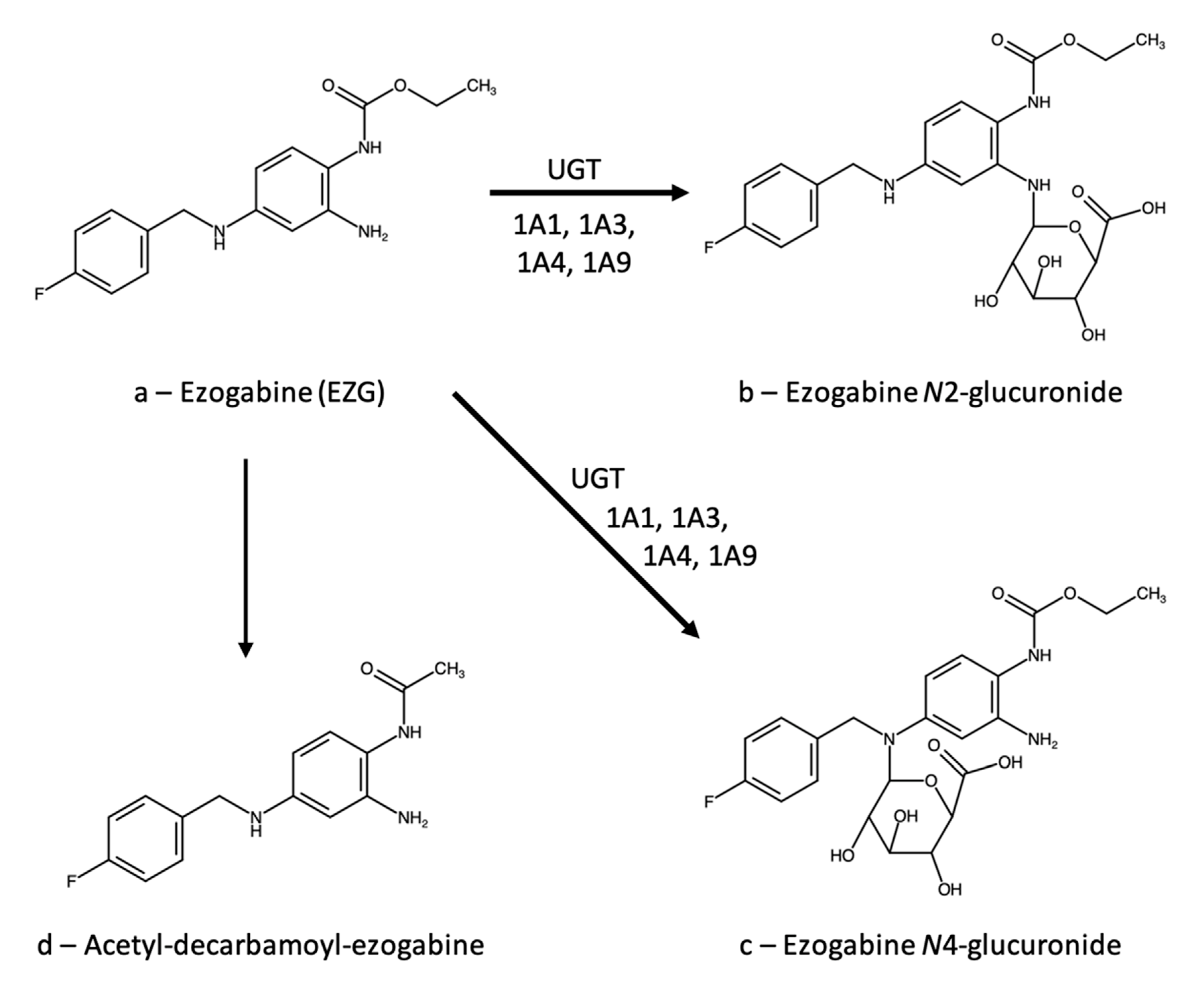
Figure 1. Metabolic pathways of ezogabine.
Analytica 02 00016 g001
Figure 2. Chemical structures of the internal standards (ISs) used in references (a) [34], (b) [35], and (c) [36].
Analytica 02 00016 g002
Figure 3. Metabolic pathways of lacosamide.
Analytica 02 00016 g003
Figure 4. Chemical structures of the internal standards (ISs) used in references (a) [65] and (b) [66].
Analytica 02 00016 g004
Figure 5. Chemical structure of cellulose tris(3,5-dichlorophenylcarbamate).
Analytica 02 00016 g005
Figure 6. Metabolic pathways of zonisamide.
Analytica 02 00016 g006
Figure 7. Chemical structures of the internal standards (ISs) used in references (a) [105], (b) [106], (c) [107], and (d) [108].
Analytica 02 00016 g007
Table 1. Performance and validation parameters of LCS analytical methods: GC-MS and multi-drug methods.
Ref.TechniqueLinearity Range, µg/mLExtraction Yield, %Precision, RSD %Accuracy, %Matrix Effect, %Carry-Over 1ISNotes
[74]GC-MS1–7091–1144.6–10.1---MoclobemideStability
assessed; forensic
[75]LC-MS/MS0.001–0.5-2.4-18.795-115<10.45.6LCS-13CStability
assessed;
breast milk
[76]HPLC-PDA1–3078–904.2–13.0100–102n.a.10AntipyrineStability
assessed; saliva
[77]HPLC-PDA0.5–3073–992.3–8.888–108n.a.<5AntipyrineStability assessed
n.a. = not applicable. 1 Expressed as % of the LOQ area.
Table 2. Performance and validation parameters of multi-drug methods for ZNS analysis.
Ref.TechniqueLinearity Range, µg/mLExtraction Yield, %Precision, RSD%Accuracy, %Matrix Effect, %Carry-Over 1ISNotes
[113]LC-MS/MS0.4–39.8<1191.1–13.891–1161–5-ZNS-D4,15NStability
assessed; VAMS
[118]HPLC-UV2.4–6084.5<15.085–115n.a.≅0LinezolidStability
assessed; DPS
[119]UHPLC-MS/MS0.1–100101
(mean)
2.8–11.1≤1184Yes 2ZNS-13C6Stability
assessed
[120]LC-MS/MS0.025–1104
(mean)
6.5–7.6
(mean)
89–91
(mean)
≤3.9-Topiramate-D12
[121]LC-MS/MS0.8–80-<7 3---Clonazepam-D4
[122]LC-MS/MS1.6–6096–1072.3–9.988–112≤12≅0Valproate-D6Stability
assessed
[77]HPLC-PDA0.5–3073–992.3–8.888–108n.a.<5AntipyrineStability
assessed
[123]HPLC-UV0.5–300≤97.56.2–6.995–96n.a.-None
[124]HPLC-UV1–9989–1090.3–7.699 (mean)n.a.≅05-Ethyl-5-p- tolylbarbituric acid
[125]HPLC-UV2–80-1.5–9–7100–108n.a.-Citalopram
[126]HPLC-UV1–40-0.7–8.2101–104n.a.-Chloramphenicol
[127]HPTLC-UV3.7–20-1.5–6.2 3-n.a.-None
[128]HPLC-FL0.1–3-0.9–1.399–103--Sulpiride or
topiramate
Stability
assessed
n.a. = not applicable. 1 Expressed as % of the LOQ area. 2 Carryover not quantified; significant for ZNS concentrations ≤ 0.3 µg/mL. 3 Reported range refers to all analytes (ZNS, lamotrigine, levetiracetam).
Table 3. Performance and validation parameters of method [144] for EZG, LCS and ZNS in plasma.
AnalyteLinearity Range, µg/mLExtraction Yield, %Precision, RSD%Accuracy, %Matrix Effect, %Carry-Over 1ISNotes
Ezogabine0.05–1093–1020.8–8.2102–1076–12≅0Carbamazepine diolStability
assessed
Lacosamide0.5–5099–1041.3–4.5102–1079–10≅0Gabapentin-D10Stability
assessed
Zonisamide1–5098–1043.1–8.794–1006–8≅0TolbutamideStability
assessed
1 Expressed as % of the LOQ area.
Publisher’s Note: MDPI stays neutral with regard to jurisdictional claims in published maps and institutional affiliations.