Review

Oxytocin: From Biomarker to Therapy for Postmenopausal Osteoporosis

1
FCS-UBI, Faculty of Health Sciences, University of Beira Interior, 6200-506 Covilhã, Portugal
2
Rheumatology Department, Unidade Local de Saúde da Guarda, 6300-749 Guarda, Portugal
3
RISE-Health, Department of Medical Sciences, Faculty of Health Sciences, University of Beira Interior, Av. Infante D. Henrique, 6200-506 Covilhã, Portugal
*
Author to whom correspondence should be addressed.
Women 2025, 5(3), 27; https://doi.org/10.3390/women5030027 (registering DOI)
Submission received: 30 June 2025 / Revised: 21 July 2025 / Accepted: 28 July 2025 / Published: 1 August 2025

Abstract

Postmenopausal osteoporosis is estrogen-dependent and results in an imbalance between bone formation and resorption. The approved therapy is intended to reduce the risk and consequences of fractures, but still has a number of contraindications and associated adverse effects. Recently, oxytocin has been shown to have an anabolic effect on bone tissue, increasing the production of osteoblasts and inhibiting the activity of osteoclasts. Thus, this study aimed to examine the potential of oxytocin as a biomarker and therapeutic agent for postmenopausal osteoporosis. A PubMed search yielded 16 articles upon analysis of the inclusion and exclusion criteria. The results showed that, compared to women in the same age group without bone loss, those diagnosed with osteoporosis exhibited lower blood oxytocin levels, possibly related to a greater tendency towards fractures. The administration of oxytocin could be a promising strategy to enhance bone quality and, consequently, to reduce the incidence of fragility fractures; however, no human studies have been conducted regarding its use as a possible treatment. Thus, it is essential to increase the number of clinical trials in women with ovarian dysfunction and bone loss, in which oxytocin could become a viable therapeutic alternative.

1. Introduction

Osteoporosis (OP) is a systemic and metabolic disease characterized by a reduction in bone mass and progressive deterioration of the microarchitecture of skeletal tissue [1]. As a consequence of this process, there is an increase in bone fragility, along with a heightened risk of fractures. Based on data from 2021, it is projected that approximately 18.3% of the global population is affected by this most prevalent metabolic skeletal disorder [2], with women being more likely to be affected than men (23.1% vs. 11.7%, based on data from the global population). It is estimated that 50% of postmenopausal women will experience an osteoporotic fracture over the course of their lifetime [3].
The principal consequence of osteoporotic pathology is fragility fractures. Vertebral, hip, distal forearm and proximal humerus fractures are the most commonly observed in these patients [1,4]. Fragility fractures represent the primary cause of morbidity in the population and are associated with a fourfold increased risk of mortality within three to six months of the primary event, with the exception of forearm fractures [4,5]. Hip fractures are the most prevalent and have the highest incidence of post-event complications, primarily in postmenopausal women [1].
The pathophysiology of OP in menopause is associated with an imbalance between bone formation and resorption, which is related to changes in hormone levels. Oxytocin (OT) is a hormone known for its functional roles in lactation and parturition, since it stimulates the contraction of smooth muscles [2,6,7]. However, recently, a potential role in bone metabolism regulation has also been identified for oxytocin [2,7]. The presence of OT receptors in osteoblasts, osteoclasts, osteocytes, chondrocytes and adipocytes and the consequent responsiveness elucidates the role of OT in bone metabolism [6]. Specifically, there is a positive feedback between OT, estrogens and osteoblasts, which culminates in the increase of OT and bone formation [6]. Therefore, to counteract the reduction in bone mass, it is imperative to persist in the active pursuit of a suitable therapeutic option for postmenopausal OP. Novel pharmacological formulations have demonstrated encouraging efficacy while exhibiting a comparatively reduced incidence of adverse effects, and OT appears to have great potential. Thus, this study aims to examine the potential of OT as a biomarker and therapeutic agent for postmenopausal OP.

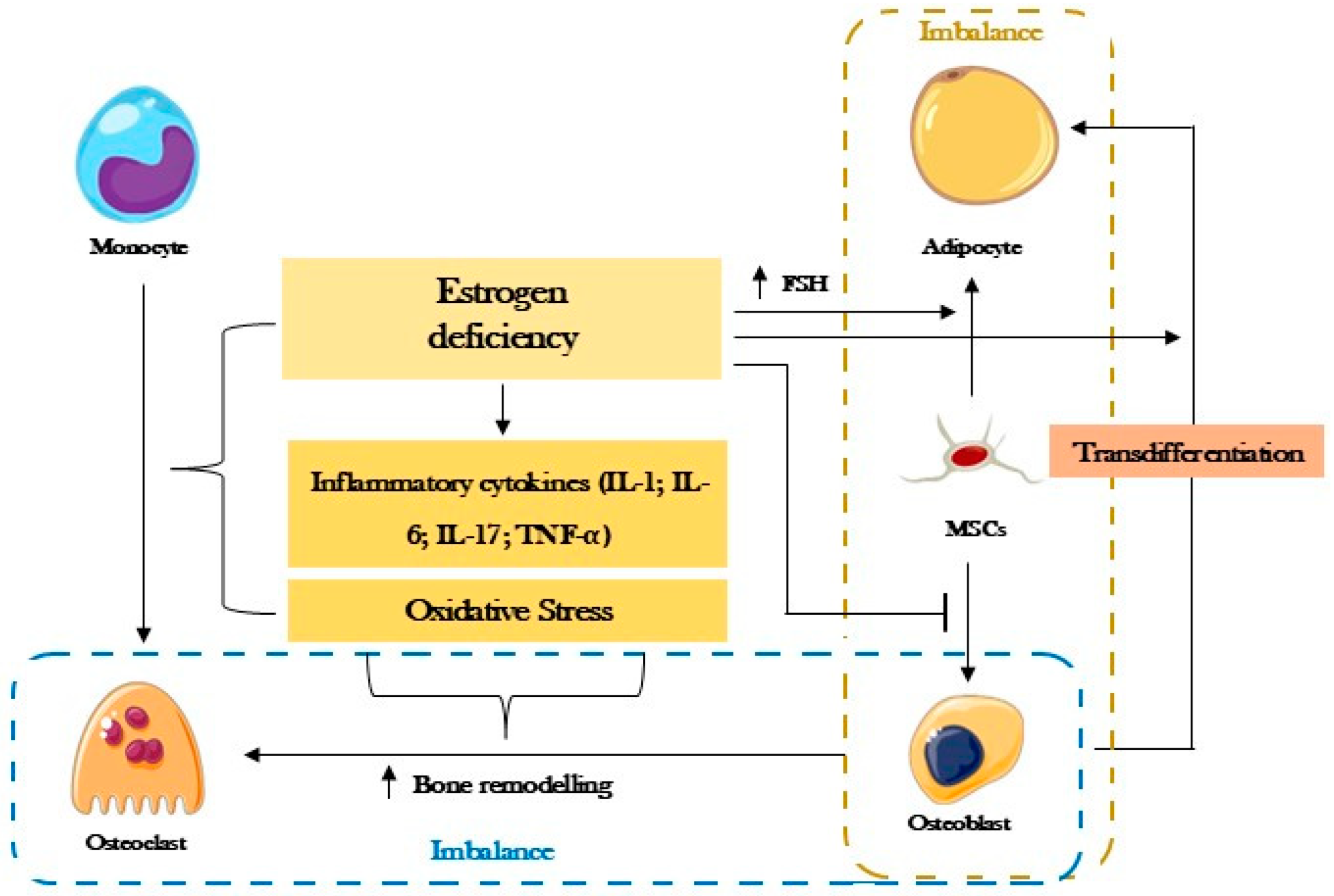
2. Postmenopausal Osteoporosis

The etiology of postmenopausal OP is sex hormone-dependent. During the menopause and postmenopause, there is a progressive decrease in estrogen concentration, which results in a change in bone metabolism [8].
Estrogens exert their effects on the bone system through a number of different mechanisms (Figure 1). RANK-L (receptor activator of nuclear factor kappa-B ligand) exerts its effects on osteoclastogenesis through its connection to RANK (receptor activator of nuclear factor kappa-B). This mechanism results in an increase in the differentiation from monocytes and macrophages, as well as the activation and survival of osteoclasts. This process is inhibited by the action of Osteoprotegerin (OPG) until the age at which ovarian function declines. OPG, produced by bone marrow cells and B lymphocytes, competitively binds to RANK and thereby inhibits osteoclastogenesis [8]. A reduction in estrogen causes an increase in RANK-L expression in mesenchymal cells (MSCs), osteocytes and bone lining cells, accompanied by a decline in OPG production. Consequently, bone resorption is enhanced at the expense of inhibiting osteoclast apoptosis, which in turn increases the cell death of osteocytes, which are fundamental for regulating bone remodeling, and osteoblasts [8].
Moreover, a deficiency in estrogen has been demonstrated to result in a pro-inflammatory state and oxidative stress [8]. In such circumstances, the production of inflammatory cytokines by T lymphocytes is increased, which results in the overstimulation of RANK-L and, consequently, a greater proliferation and differentiation of osteoclasts. Furthermore, high levels of tumor necrosis factor-α (TNF-α) prevent osteoblastic differentiation. A reduction in estrogen levels results in a decline in the activity of antioxidant enzymes, leading to an accumulation of reactive oxygen species and, consequently, an exacerbated pro-inflammatory state. This, in turn, promotes increased osteoclast activity and a reduction in osteoblast durability. It has been demonstrated that the degree of systemic oxidative stress is inversely correlated with bone mineral density (BMD) in the femoral neck, hip, and lumbar vertebrae [8].
Both osteoblasts and medullary adipocytes differentiate from MSCs. The lack of estrogen and increased levels of follicle-stimulating hormone (FSH), characteristic of the menopause, contribute to the differentiation of MSCs in favor of adipocytes. It may be reasonably inferred that with advancing age, there will be a progressive increase in the number of adipocytes within the bone marrow, accompanied by a corresponding decrease in the number of osteoblasts. Additionally, the pro-inflammatory state and oxidative stress, which have already been documented, contribute to bone marrow adipogenesis [8].

3. Oxytocin

Oxytocin (OT) is a neuropeptide synthesized in the supraventricular and paraventricular nuclei of the hypothalamus and released through the neurohypophysis following stimulation [6,7]. In terms of its functional role, oxytocin is primarily associated with the processes of lactation and parturition, given that it stimulates the contraction of smooth muscles [2,6,7]. Recently, OT, also known as the “love hormone”, has been identified as a potential regulator of bone metabolism [2,7].
In addition to central synthesis, which is responsible for regulating social interaction and reproduction, OT has peripheral synthesis. The uterus, ovaries, testicles, placenta, kidney, muscle tissue, pancreas, adipose tissue, thymus, heart and vascular endothelium, adrenal gland and bone tissue cells are all capable of producing and responding to OT [2,6,7]. It is the OT synthesized in these organs and tissues that plays a role in bone homeostasis [6].
The extensive spectrum of effects observed is attributable to the association between OT and its receptor. The responsiveness of osteoblasts, osteoclasts, osteocytes, chondrocytes and adipocytes to OT elucidates the role of OT in bone metabolism. In addition to these cells, MSCs are also subject to the hormonal effect [6]. When exposed to peripheral OT, these cells demonstrate enhanced proliferation, differentiation, and activity, with the capacity to differentiate into osteoblasts rather than adipocytes. Osteoblasts are capable of synthesizing OT under the control of estrogen and have a high expression of receptors for this hormone [2,6]. The synthesis of OT acts in a self-paracrine manner, resulting in the mineralizing phenotype of osteoblasts and an increase in genes that contribute to bone formation [5,6,8]. It is now acknowledged that OT, estrogens and osteoblasts constitute a positive feedback mechanism, whereby estrogens stimulate osteoblasts to produce OT, which subsequently triggers the release of further OT [6].
In addition to its effects on bone-forming cells, OT also exerts influence over osteoclasts. OT has been demonstrated to enhance the expression of RANK-L in osteoblasts while concurrently reducing OPG levels [2,7]. This results in an increase in the number of osteoclasts, accompanied by a 40% decrease in their resorptive activity. This process is the result of an increase in the concentration of intracellular calcium ([Ca2+]i) and, consequently, nitric oxide [2,6,7]. This mechanism also occurs in osteoblasts, where the release of Ca2+ is more instantaneous and the return to baseline is equally rapid. In contrast, the increase and decline in osteoclasts is slower and more progressive [2]. This discrepancy elucidates the enhancement in osteoblastic functionality and the diminution in osteoclastic functionality.
Additionally, OT has been observed to target chondrocytes. The hormonal effect facilitates the formation of cartilage in the subchondral bone and inhibits the immune and inflammatory response, thereby preserving cartilage and maintaining the quality of the bone microarchitecture. As they share the same precursor as osteoblasts, adipocytes are quantitatively reduced as a consequence of the privilege that OT confers upon osteoblastic differentiation [6].
It can thus be concluded that the pathophysiology of OP associated with the menopause is a consequence of an imbalance between osteoblasts and osteoclasts, that is, an imbalance between bone formation and resorption. Furthermore, bone marrow adiposity is a significant contributing factor to the development of the disease [8]. However, OT and its analogues, including Carbetocin (CB), have been demonstrated to possess anabolic properties with regard to bone tissue, thereby counteracting the effects associated with estrogen deprivation [7]. This function is attributable to the OT role in the genesis of osteoblasts and its dual role in osteoclasts [6]. The selection of a suitable therapeutic option for postmenopausal OP should be based on an assessment of the patient’s risk of fracture. As in all therapies, the decision-making process regarding the most appropriate course of treatment should take into account the benefits and risks inherent to each option, with due consideration given to the patient’s characteristics and preferences. However, the adverse effects associated with these therapeutic options remain a significant concern, potentially undermining adherence to treatment. One hypothesis that has recently emerged with potential for further investigation is the use of OT. This has been shown to have positive effects at the bone level and a reduced list of adverse effects and, therefore, should be considered as a potential option.

4. Oxytocin in Osteoporosis

4.1. Oxytocin as a Biomarker

Following an examination of the role of OT in bone metabolism, it is necessary to investigate a potential correlation between serum levels of this hormone released by the neurohypophysis and the diagnosis of OP. The retrieved human studies are analyzed below, and the main results are presented in Table 1.
In a study conducted between May 2006 and January 2008, Breuil et al. examined the serum concentration of OT and other hormones in a total of 36 postmenopausal women, comprising 16 healthy controls and 20 women diagnosed with severe OP who had not undergone any treatment. The researchers found that OP was negatively associated with OT, since women diagnosed with bone decline exhibited OT values of approximately 50 pg/mL, in comparison to the 110 pg/mL observed in the group of healthy women. Moreover, OT was related to BMD and decreased OT levels were predictive of OP. Even after adjusting for age and both estradiol and leptin levels, which are involved in the pathophysiology of osteoporosis and the regulation of oxytocin secretion. Thus, low OT levels appear to be related to the pathophysiology of severe OP and consequently to bone remodeling [9]. In a six-year prospective study conducted later by the same research group, serum samples from 1094 German, British and French women, aged between 55 and 79, were analyzed for the connection between OT and bone status. After quantifying OT, leptin and estradiol hormonal levels and analyzing different bone parameters, their findings demonstrated that women with normal BMD values (T-score > −1) exhibited higher OT levels compared to those with osteopenia (T-score between −1 and −2.5) and OP. However, among women with osteopenia and OP, the difference was only significant in those with a deficit in bone density in the lumbar spine and in those with a deficit affecting both the lumbar spine and the hip. In addition, women with a history of fractures had lower levels of OT than those without such a history, but no significant association was found. Overall, high OT levels are associated with greater bone density, especially in the hip and in women with low estradiol or high leptin levels [10].
In a study published in 2021, Du et al. examined the outcomes of a cohort of 478 postmenopausal women of Chinese origin to identify biomarkers associated with the risk of osteoporosis and sarcopenia. Upon measuring hormonal levels and muscle strength and bone/muscle mass, OT was, once more, demonstrated to have a positive correlation with BMD. The OT values were found to be higher in women in the no osteoporosis/no sarcopenia group (612 pg/mL) when compared to the other three groups: osteoporosis/no sarcopenia group (425 pg/mL), osteoporosis/sarcopenia group (398 pg/mL) and no osteoporosis/sarcopenia group (500 pg/mL). The findings of this study indicate that menopausal women with OP and a personal history of fracture are more likely to exhibit reduced levels of OT. This study suggests that OT is a potential risk marker for osteoporosis in elderly women and increased OT levels could confer protection against fracture risk [11]. In agreement with this study, Yu et al. gathered a total of 149 Chinese women (74 premenopausal and 75 postmenopausal) to analyze the relationship between plasma concentrations of OT and the risk of OP. The parameters analyzed showed that serum OT, estradiol and BMD were significantly higher in the premenopausal women, specifically, markedly elevated OT concentrations (777 pg/mL) were observed in the premenopausal group in comparison to the postmenopausal one (364 pg/mL). These findings suggest that OT may have a protective role against the loss of bone mass in women [12].
In accordance with the preceding findings, a trial conducted in France by Elabd et al. demonstrated that circulating OT levels were 55% lower in postmenopausal women developing osteoporosis compared to healthy women (50.2 ± 8.8 pg/mL and 110.6 ± 19.8 pg/mL, respectively). A total of 36 women were included in the study, comprising 20 individuals with osteoporotic conditions and 16 controls. The findings indicated that, irrespective of age, body mass index (BMI), and weight, OT was inversely associated with the development of OP. This highlights the role of this hormone in the pathophysiology of bone imbalance that is typical of women in the postmenopausal period [13].
Overall, these results revealed a correlation between OT blood levels in women and BMD. Specific correlations have been identified between OT levels and BMD at the lumbar vertebrae [9], hip [11,12] and femoral neck [9,11,12]. In accordance with the aforementioned findings, Breuil et al. demonstrated that in women with markedly low estrogen levels, a condition indicative of menopause, OT plays a pivotal role in hip BMD [10]. Furthermore, it is evident that OT plays a crucial role in restoring bone metabolism in women [11].

4.2. Oxytocin as a Treatment

The findings presented in the previous section provide a potential avenue for further investigation into the therapeutic potential of OT in postmenopausal OP. Although there are no human studies regarding the use of OT as a therapeutic approach, experimental investigations using human cell cultures and in vivo studies have been conducted on this subject. These are described below and summarized in Table 2.

4.2.1. In Vitro Studies: Human and Animal Cells

In a further reference to the French study published in 2008 by Elabd et al., human multipotent adipose-derived stem cells (hMADS) from the pubic region of five-year-old donors and human bone marrow stromal cells (hBMSC) were employed to investigate the potential regulation of the OT receptor pathway on the osteoblast/adipocyte balance. The published results demonstrated that, in both cell populations, OT and CB stimulate osteoblastic differentiation while simultaneously inhibiting differentiation into adipocytes. The results are presented as being dose-dependent, with 30 nM of OT and 300 nM of CB. Moreover, the qualitative analysis conducted by the investigators revealed that female rats treated with OT following ovarian removal surgery exhibited a higher trabecular bone volume fraction (BV/TV), a greater number of trabeculae per unit length (Tb.N), and a lower trabecular spacing (Tb.Sp). These properties rendered the bone tissue less susceptible to deformation and fracture. In ovariectomized rats subjected to OT therapy, the femur exhibited a 22–25% increase in stiffness compared to rats not subjected to the same conditions. Furthermore, enhancements were observed in the cortical bone, notably in its strength [13]. The results demonstrated that OT has the capacity to reverse the osteoblast/adipocyte imbalance, thereby facilitating the recuperation of bone loss and microarchitecture.
Two studies from the same research group [14,15] investigated the effects of OT on rat bone marrow mesenchymal cells (BMMSCs). However, in contrast with the previous study, the results obtained suggested that OT had limited efficacy in promoting recovery from osteoporosis [14]. Nevertheless, its capacity to stimulate the genetic expression of the mineralizing phenotype in MSCs was acknowledged. This fact was evidenced by the elevated levels of osteocalcin and OPG observed in the exposed cells [15].

4.2.2. In Vivo Studies

In a different perspective, Tamma et al., using mice deficient in OT and its receptors, confirmed that when injected intraperitoneally, OT was able to increase the number of osteoclasts and BMD. High levels of OP were detected in these animals, highlighting the essential role of peripheral OT in maintaining bone mass. Furthermore, when osteoblasts in culture were deprived of OT for 21 days, there was a decrease in mineralization that was restored after 21 days of exposure to the same hormone. Under the same conditions, there was a decrease in cell proliferation. Quantitatively, the concentration of osteocalcin and osteopontin increased with exposure to OT, suggesting an active bone matrix production by the osteoblasts [16].
With the purpose of studying OT effect in the prevention and treatment of postmenopausal OP, Beranger et al. used female rats as a study subject. The animals, divided into groups of 6 to 12, were subjected to bilateral ovariectomies or simulated surgeries—the control group. Subsequently, two different therapeutic strategies, with variants, were followed: (1) OT or placebo was administered 2 weeks after surgery—the placebo was administered daily for 8 weeks, while administration of OT followed three different protocols: OT 1 mg/kg administered daily for 8 weeks; OT 1 mg/kg administered in the first 4 weeks followed by an equal injection time with placebo or OT 0.1 mg/kg; OT 1 mg/kg administered twice a week for 8 weeks. (2) Treatment with OT or placebo, of 8 weeks duration, started 8 weeks after surgery. The results presented indicate that, when compared to the control, OT for 8 weeks, in the groups of rats treated after 2 weeks, was able to normalize the femoral trabecular parameters—thickness (Tb.Th); Tb.Sp and Tb.N. Similarly, positive results were documented in experiments carried out on rats that only started treatment after 8 weeks. A total quantitative normalization of osteoblasts and partial normalization of osteoclasts was observed in the treated rats, via an increase in the RANK-L/OPG ratio. It was thus concluded that OT is able to normalize bone deficit and microarchitecture in animal models mimicking menopause. Moreover, considering that after treatment, rats with osteoporosis had a scientifically relevant increase in P1NP (Procollagen Type I N-terminal Propeptide), without altering other markers of bone activity, OT could be an alternative to current treatment with human parathyroid hormone or Romosozumab (the only anabolic treatments for OP) [17].
In a 2014 Chinese publication, OT was identified as a highly promising pharmaceutical agent for the prevention of postmenopausal osteoporosis. This conclusion was achieved following a study in which an OP rabbit model was used to analyze the effect of early OT treatment on bone and bone fat masses. In total, 60 female rabbits were randomly assigned to one of three groups: (1) control group, (2) OP group, and (3) OP with OT treatment group. In order to differentiate the treatment groups from the control group, the rabbits underwent ovariectomy. Moreover, the treatment group received OT at a dose of 1 mg/kg for 8 weeks, commencing 2 weeks post-surgery, and the other groups received 15 mL of saline solution. With regard to BMD, the results showed no significant differences between the control group and the group treated with OT; however, the OP group showed a 58.6% decline in BMD. Accordingly, the bone parameters regarding trabecular bone (BV/TV; Tb.N and Tb.Th) exhibited an increase of 14–33% in the OT treatment group when the OP group was taken as a reference. Conversely, there was a reduction of approximately 20% in Tb.Sp and Structural Model Index (SMI), indicating a lower trabecular space and a change in the trabecular structure to a more plate-like shape, respectively. Furthermore, the study demonstrated the impact of OT in medullary adiposity, which was significantly associated with BMD. There was a reduction in bone deterioration and bone marrow adiposity accumulation when OT was administered early, with results that suggest OT as a possible treatment for OP [18]. In 2019, the same research group used the same rabbit OP model to investigate the impact of OT on skeletal health. The 65 rabbits were randomly assigned to one of three groups described in the previous study by Qiu et al., but this time the OT treatment group received OT at 1 mg/kg for 10 days. The findings suggest that OT may retard bone deterioration, although no significant differences in BMD were observed up to 12 weeks into the study. At the 10th week post-surgery, notable discrepancies were observed in the parameters pertaining to microarchitecture when comparing the OT treatment group to the placebo group. Specifically, there was a 37% increase in Tb.N and Tb.Th, an 18% decrease in SMI, a 49% increase in BV/TV, and a 59% decrease in Tb.Sp in the OT treatment group. Therefore, Qiu et al. concluded that OT, when administered in a timely manner, is capable of preventing the loss of cancellous bone resulting from ovariectomy, thereby restoring the quality of the microarchitecture. Furthermore, BV/TV and Tb.Sp were identified as highly sensitive to OT, suggesting their potential as markers for assessing therapeutic efficacy [19].
In a different perspective, to study the osseointegration of implants, Wang et al. employed a methodology that involved 20 female rats that had undergone bilateral ovariectomy, followed by the subsequent placement of femoral implants. One group was administered subcutaneous OT at a dose of 1 mg/kg/day, while a placebo was administered to the remaining group. Following a 12-week experimental period, the femurs were extracted for observation. The bones from the OT treatment group demonstrated optimal bone–implant articulation, exhibiting a relative increase of 0.62 times. The continuous and substantial formation of bone around the implant resulted in a 2.25 increase in fixation, as assessed by the expulsion force, in comparison to the control group. Furthermore, the rats that received a placebo during the trial period exhibited thin, diffuse, and disconnected trabeculae, accompanied by a notable decline in the bone layer, opposite to the OT group. In light of the aforementioned data, it can be inferred that OT exerts a beneficial influence on the osseointegration of titanium implants in ovariectomized rats, resulting in increased bone volume and enhanced trabecular microarchitecture [20].
In 2020, Moghazy and co-workers randomly assigned 60 female rats to one of three groups: (1) a control group that underwent a simulated surgery, (2) a second group that underwent an ovariectomy, and (3) a third group that underwent both the ovariectomy and the OT treatment. The treatment consisted of intraperitoneal administration of 0.1 mg/kg/day of OT for eight weeks. The authors observed a significant reduction in BMD in the second group, which underwent physical castration with no treatment, compared to the control group. The serum concentrations of bone alkaline phosphatase (bone ALP), osteocalcin, TRAP and RANK-L were found to be elevated in the second group, while osteocytes and osteoblasts were decreased. The observed differences between the control and the treatment groups were not statistically significant, indicating that OT might be an effective intervention for mitigating bone loss caused by ovariectomy. The group treated with OT exhibited an improvement in Tb.Th compared to the untreated group. These findings demonstrate that bone loss associated with gonadal decline is contingent upon serum OT levels. It is, therefore, plausible that OT may serve as a preventative measure for OP in postmenopausal women [7].
Three different studies from the same Brazilian research group aimed to analyze the role of OT on bone metabolism in female rats with irregular estrous cycles. In the first study, from 2020, there was a potential preventive action of OT on osteoporosis. Specifically, the animals were administered two injections of sodium chloride or OT (134 μg/kg) at 12 h intervals and the data collected after 35 days. The results demonstrated that OT facilitates bone remodeling by increasing bone formation and decreasing Tartrate-resistant acid phosphatase (TRAP), which is a marker of osteoclastic activity. As reported by Fernandes et al., OT showed an anabolic activity on bone tissue since in animals exposed to OT, there was an increase in transcription factors essential for osteogenesis and osteoblastic differentiation. OT facilitated improvements in the trabecular and cortical regions, enhancing rigidity and augmenting the maximum load tolerated, thereby reducing the probability of fracture. Therefore, it was shown that OT leads to the reduction of osteopenia and thus represents a promising anabolic strategy for the prevention of primary OP [21]. The second study was conducted to examine the effects of OT (134 μg/kg) on the musculoskeletal system of the female rats. The rats were divided into four groups: (1) a control group, (2) an OT group, (3) an Atosiban (AT) group (an OT receptor antagonist), and (4) an AT and OT group (with OT administered five minutes apart). Following the administration of four doses over a two-month period, the results indicate that the rats of the OT group showed more osteoblasts and osteocytes on the surface of the bone matrix, presented a thicker cortical bone (while in the AT group it was thinner), and had low concentrations of TRAP and high concentrations of osteocalcin. Moreover, the OT group also showed better results in the polar moment of inertia and higher permissible load and stiffness. On the other hand, the AT group experienced a worsening of the bone deficit and higher cortical porosity. Both groups exposed to OT had higher BMD. From these results, the authors concluded that both endogenous and exogenous OT contribute to the improvement of the histomorphometry of the femoral neck, with a reduction in cortical porosity, an increase in BMD, a higher polar moment of inertia and greater bone strength. Therefore, OT is crucial in preventing age-related bone transformations, with a beneficial impact on functionality, as evidenced by the longer strides recorded in rats exposed exclusively to OT [22]. In the third study, the authors administered the same OT dose (134 μg/kg) as in the previous analyses to study the effect of OT and strength training on rats’ bone quality. OT was administered bimonthly for a period of four months. The combination of these therapeutic approaches yielded synergistic effects, including an increased maximum load, enhanced BMD and stiffness, and a reduction in porosity. In isolation, OT had a marked effect on bone trabeculae, increasing Tb.Th and Tb.N. The polar moment of inertia demonstrated enhanced values when strength training was combined with OT, although this outcome was markedly dependent on OT. The findings of this study indicate that, while OT is beneficial, the results are more readily achieved when bone decline is addressed through a combined OT + strength training therapy. The combination preserves the mechanical and structural aspects, as well as stimulating the genetic factors that help prevent the bone changes associated with ageing and, consequently, osteoporosis [23].

5. Discussion

Given the decline in bone mass associated with menopause, it is essential to continue searching for effective treatments for postmenopausal osteoporosis. Emerging pharmacological approaches with reduced side effects have been suggested as promising candidates, including OT; however, the literature, as a consequence of the few scientific studies carried out, is sparse. This review, therefore, explored the potential of OT in the context of postmenopausal osteoporosis. The studies presented in this manuscript followed two different perspectives of oxytocin function and its potential as a biomarker for postmenopausal osteoporosis, analyzing observational studies in humans, and as a possible therapeutic agent, with most of the literature from pre-clinical animal studies.

5.1. Human Cross-Sectional Studies

The human studies analyzed in this review have consistently shown that lower circulating OT levels are associated with reduced BMD, which can increase the risk of fractures in postmenopausal women. Women with OP tend to have significantly lower OT levels than healthy women, and this difference is particularly evident when there is bone loss in the spine or hip. In addition, lower OT levels have also been associated with a history of fractures and the coexistence of sarcopenia. Comparisons between premenopausal and postmenopausal women further support the protective role of OT against bone loss, since there is a decrease in hormone levels after the menopause.
Thus, when examined as a biomarker, clinical studies in humans consistently reveal a substantial reduction (approximately 50%) in circulating OT levels among women suffering from bone loss compared to unaffected women. Furthermore, a correlation was identified between OT and the likelihood of bone fracture [10,11]. These findings suggest that OT may serve as a diagnostic criterion, therapeutic choice, and prognostic factor when bone mass decreases in postmenopausal women. However, there is a significant discrepancy in values between European and Asian populations, which makes it challenging to propose a coherent cut-off point based on the current and limited human data.
Nevertheless, the results from these studies suggest that OT may contribute to bone health regulation and could serve as a valuable biomarker and potential therapeutic target for postmenopausal OP.

5.2. Animal Model Studies

Regarding the animal studies, which analyzed OT as a treatment, most have shown that OT plays a crucial role in bone health. Mutant mice and rats lacking OT or its receptor developed OP, while treatment with OT restored bone density and improved trabecular structure. In ovariectomized models, OT prevented bone loss, improved bone architecture, and strengthened bones mechanically. It also promotes osteoblast activity, reduces bone resorption and improves bone healing around implants. When combined with strength training, there was a synergistic effect, improving the mechanical load tolerance and BMD. Collectively, these studies consistently demonstrate that OT supports bone formation, preserves microarchitecture, and increases bone strength, suggesting its potential as a preventative and therapeutic agent in models of osteoporosis.
Most of the current therapies proposed for OP primarily inhibit trabecular bone loss. A review of the available data suggests that OT may represent a significant advance in the treatment of postmenopausal OP, given its capacity to influence the cortical component of the bone as well as the trabecular bone, potentially offering more comprehensive skeletal protection [12,13]. Preclinical studies predominantly conducted in animal models have demonstrated that when OT is administered intraperitoneally, which simulates the peripheral action of the hormone, it improves bone tissue architecture, which is crucial for the prevention of fragility fractures. The majority of these animal studies simulate or induce bone decline by inducing estrogen deficiency through ovariectomy; however, none of them measure circulating OT levels after the procedure or after the treatment was administered. Although the most frequently used dose in the studies analyzed was 1 mg/kg, variations in dosing regimens and species differences preclude direct extrapolation to humans. As pointed out in this manuscript, studies in humans are scarce; thus, this paucity of knowledge renders it challenging to ascertain the minimum serum concentrations that must be attained to substantiate the efficacy of the therapy.

5.3. Therapeutic Approach

It has been demonstrated that OT has the capacity to positively impact general health. Unlike current pharmacologic options for OP, OT has anti-inflammatory and antioxidant properties, enhances immune function, and modifies body composition, cognitive functions, and mood [2]. These effects are particularly relevant in the postmenopausal context, a period associated with elevated risks of cardiovascular disease, diabetes, cognitive decline, sleep disorders and depression [2]. As a consequence of the ageing process, there is a redistribution of subcutaneous to visceral adipose tissue, which is deleterious to health [2]. Thus, OT has been presented as an alternative to these setbacks by reducing fat mass, improving lipid metabolism, and contributing to cardiovascular regulation [2]. Similarly, OT exerts an effect at the cognitive and psychiatric levels. While the evidence is inconclusive, there is a suggestion that OT may also have a role in breast cancer, although this is a controversial area of research [2,24].
It is essential to consider that only peripheral OT exerts an effect on bone tissue [2] and that the oral form is not a viable option due to its rapid degradation in the digestive system [24]. In consideration of the available alternatives, the intranasal route appears to offer the most promising benefit, as it increases both central and peripheral OT and is, therefore, capable of a broader therapeutic action [2]. However, a major pharmacokinetic limitation of native OT is its short plasma half-life (approximately five minutes), which presents a significant barrier to its clinical use [7,25,26]. Therefore, CB has emerged as a promising avenue for further investigation. It is an analogue of OT with the capacity to remain in the body for a longer period of time, due to its longer half-life and heat stability. These properties mean that in cases of uterine atony, the main current indication for the use of OT, CB can be administered as a single dose, in contrast to OT, which is given as an infusion to ensure constant concentrations in the body [25]. Despite this, OT and CB have different pharmacological properties, meaning that their effects on bone and systemic physiology may not be identical [26]. A further obstacle inherent to this option is the lower supply of CB on the pharmaceutical market, which consequently results in a higher price [27]. This naturally reduces the opportunities to study the molecule and makes the conclusions drawn about it still superficial and in need of confirmation.
It is crucial to consider the potential adverse effects associated with any therapeutic intervention. A review of data collected in 2021 from women who had been exposed to OT and CB following postpartum hemorrhage identified tachycardia and hypotension as the most commonly encountered adverse effects. Additionally, episodes of nausea, vomiting, headaches, fever, chills, abdominal pain, diarrhea, bradycardia, and hypertension were observed in these women. Interestingly, while CB was more likely to induce side effects, these were generally less severe than those associated with OT. Moreover, the potential for drug interactions with glucose, sodium chloride, dinoprostone and misoprostol, prostaglandin analogues used as labor inducers, must be considered when evaluating OT or CB for broader clinical use [25].

5.4. Final Considerations and Future Perspectives
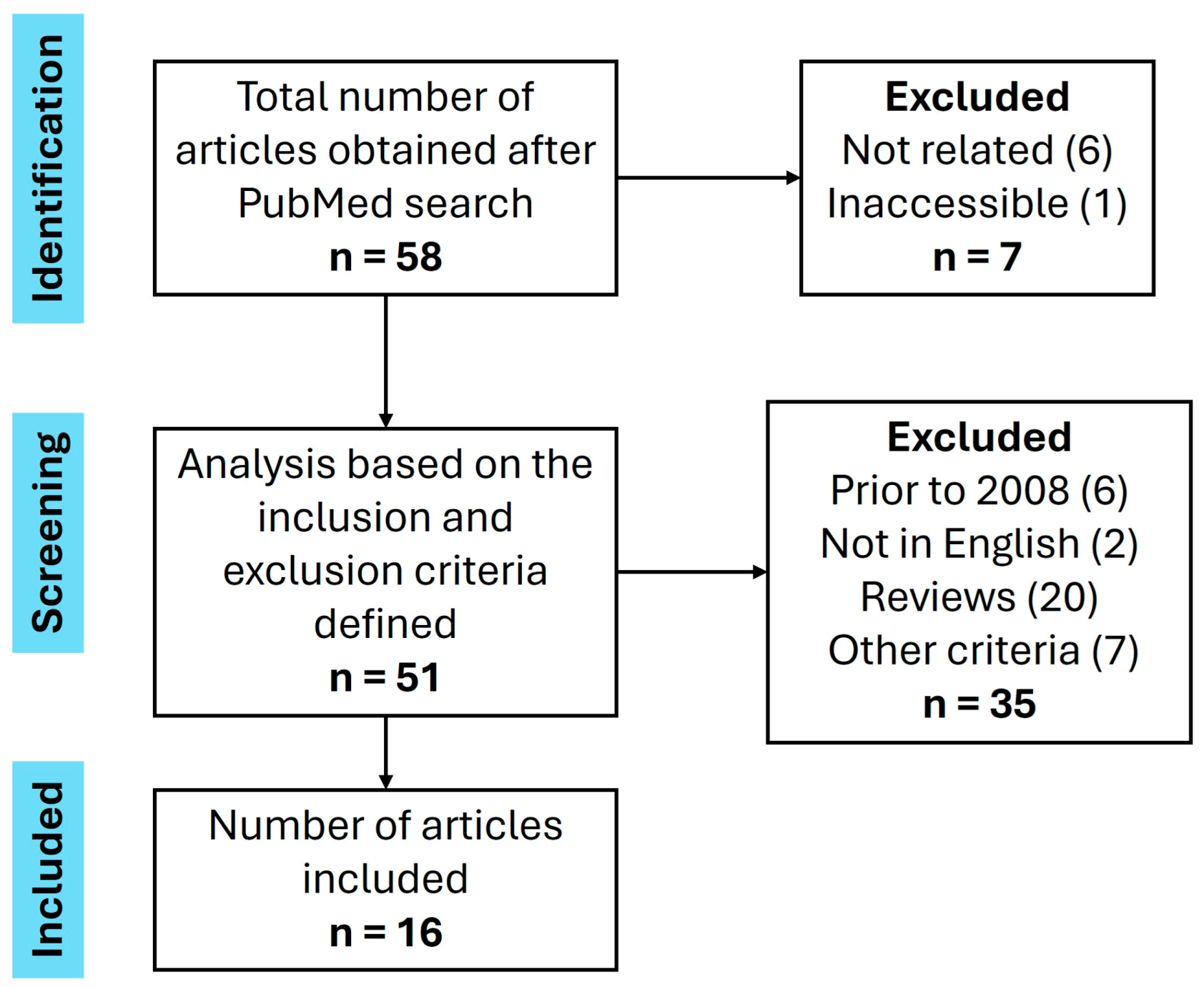
OT seems to have great potential for intervening in OP; however, the existing literature on this subject remains scarce, especially in humans. Furthermore, the existing barriers must be overcome so that one day, either as an original molecule or through an analog, OT can be among the therapeutic options available to women with this highly life-limiting and potentially fatal condition. Future clinical trials that aim to establish the precise and safe route of administration and dosage to be used in these situations are recommended, as well as to identify the criteria that qualify women to benefit from this specific therapy. As the number of studies of exposure to OT increases, it is probable that any adverse effects will become more apparent. This will inform the safety of the therapy, which has never been studied in women of the age group addressed in this study. The current treatment guidelines utilize Ca2+ and vitamin D as an adjuvant; thus, it is imperative to develop studies to assess the efficacy of OT in monotherapy as well as in combination with other substances. According to the literature, if OT therapy were approved for postmenopausal women, there would be no need to resort to hormone replacement therapy with estrogens. This contrasts with osteoporosis in men, where existing data indicates that men need to replenish testosterone levels in order to benefit from improvements in bone condition [28]. Consequently, OT could be a viable treatment option for OP in both sexes in the future, albeit with a distinct administration approach.
For all these reasons, it is highly recommended that more studies should be carried out on this subject so that, if it proves viable, OT could become a therapeutic option in the near future for this disease, which affects nearly 20% of the world’s population.

6. Methods

The objective of this study is to provide data on the validity and usefulness of OT as a diagnostic criterion for postmenopausal OP (Section 4.1) and as a therapeutic molecule in the management of postmenopausal OP (Section 4.2).
In order to meet the aforementioned objectives, the PubMed database was used. The search was based on combining the concepts “osteoporosis” and “oxytocin” using the Boolean operator “AND”.
The selection of relevant articles for presentation in this dissertation was made in accordance with the following inclusion criteria:
-
Articles published from 2008 inclusive to June 2024;
-
Articles written in English;
-
Original articles;
-
Articles with postmenopausal women;
-
Articles with an animal population in a life cycle corresponding to the human postmenopause period;
-
Articles in which Oxytocin is studied as an option for diagnosing postmenopausal Osteoporosis;
-
Articles in which Oxytocin is presented as an option for the treatment of postmenopausal Osteoporosis.
The following exclusion criteria were taken into account in the selection:
-
Articles published before 2008 and after June 2024;
-
Articles written in a language other than English;
-
Non-original articles;
-
Articles with an exclusively male population;
-
Articles with animal populations at a stage in their life cycle that is not equivalent to the human postmenopause period;
-
Articles in which Oxytocin is presented as useful for situations other than the diagnosis and/or treatment of postmenopausal Osteoporosis.
Following the application of the aforementioned inclusion and exclusion criteria, 16 articles were selected for detailed analysis, as seen in Figure 2. The results of this analysis are presented below.

7. Conclusions

The evidence from the studies analyzed indicates that oxytocin is a molecule with the potential to eventually be used as a biomarker for osteoporosis in postmenopausal women. Furthermore, although there are still few studies, the existing results seem to show that oxytocin has the potential to become a suitable therapy for women with this condition and also has the power to intervene in other medical conditions. However, despite these positive results, the full potential of oxytocin and its analogues in the treatment of postmenopausal osteoporosis has yet to be elucidated. More research is, therefore, needed to determine the ideal ways to harness the systemic potential of this hormone.

Author Contributions

Conceptualization, T.F. and E.C.; methodology, T.F. and E.C.; validation, J.F.F. and E.C.; investigation, T.F. and E.C.; writing—original draft preparation, T.F.; writing—review and editing, J.F.F., M.M. and E.C.; visualization, T.F., J.F.F., M.M. and E.C.; supervision, J.F.F. and E.C.; project administration, E.C.; funding acquisition, E.C. All authors have read and agreed to the published version of the manuscript.

Funding

The article was developed within the scope of the CICS-UBI projects DOI 10.54499/UIDB/00709/2020 (https://doi.org/10.54499/UIDB/00709/2020) and DOI 10.54499/UIDP/00709/2020 (https://doi.org/10.54499/UIDP/00709/2020), financed by national funds through the Portuguese Foundation for Science and Technology (FCT)/Ministry of Science, Technology, and Higher Education (MCTES).

Institutional Review Board Statement

Not applicable.

Informed Consent Statement

Not applicable.

Data Availability Statement

Not applicable.

Conflicts of Interest

The authors declare no conflicts of interest.

Abbreviations

The following abbreviations are used in this manuscript:
ALPAlkaline phosphatase
BMDBone mineral density
BMIBody mass index
BMMSCsBone marrow mesenchymal cells
BV/TVTrabecular bone volume fraction to total volume ratio
CBCarbetocin
FSHFollicle-stimulating hormone
hBMSCHuman mesenchymal stromal cells
hMADSAdipose tissue-derived multipotent cells
ILInterleukin
MSCsMesenchymal cells
OPOsteoporosis
OPGOsteoprotegerin
OTOxytocin
P1NPProcollagen Type I N-terminal Propeptide
RANKReceptor activator of nuclear factor kappa-B
RANK-LReceptor activator of nuclear factor kappa-B ligand
SMIStructural model index
Tb.NTrabeculae per unit length
Tb.SpTrabecular spacing
Tb.ThTrabecular thickness
TNF-αTumor necrosis factor-α
TRAPTartrate-resistant acid phosphatase

References

  1. Kanis, J.A.; Cooper, C.; Rizzoli, R.; Reginster, J.Y.; on behalf of the Scientific Advisory Board of the European Society for Clinical and Economic Aspects of Osteoporosis (ESCEO) and the Committees of Scientific Advisors and National Societies of the International Osteoporosis Foundation (IOF). European guidance for the diagnosis and management of osteoporosis in postmenopausal women. Osteoporos. Int. 2019, 30, 3–44. [Google Scholar] [CrossRef] [PubMed]
  2. Breuil, V.; Trojani, M.C.; Ez-Zoubir, A. Oxytocin and Bone: Review and Perspectives. Int. J. Mol. Sci. 2021, 22, 8551. [Google Scholar] [CrossRef] [PubMed]
  3. Eastell, R.; Rosen, C.J.; Black, D.M.; Cheung, A.M.; Murad, M.H.; Shoback, D. Pharmacological Management of Osteoporosis in Postmenopausal Women: An Endocrine Society* Clinical Practice Guideline. J. Clin. Endocrinol. Metab. 2019, 104, 1595–1622. [Google Scholar] [CrossRef]
  4. Gregson, C.L.; Armstrong, D.J.; Bowden, J.; Cooper, C.; Edwards, J.; Gittoes, N.J.L.; Harvey, N.; Kanis, J.; Leyland, S.; Low, R.; et al. UK clinical guideline for the prevention and treatment of osteoporosis. Arch. Osteoporos. 2022, 17, 58. [Google Scholar] [CrossRef] [PubMed]
  5. Salari, N.; Ghasemi, H.; Mohammadi, L.; Behzadi, M.H.; Rabieenia, E.; Shohaimi, S.; Mohammadi, M. The global prevalence of osteoporosis in the world: A comprehensive systematic review and meta-analysis. J. Orthop. Surg. Res. 2021, 16, 609. [Google Scholar] [CrossRef]
  6. Feixiang, L.; Yanchen, F.; Xiang, L.; Yunke, Z.; Jinxin, M.; Jianru, W.; Zixuan, L. The mechanism of oxytocin and its receptors in regulating cells in bone metabolism. Front. Pharmacol. 2023, 14, 1171732. [Google Scholar] [CrossRef]
  7. Moghazy, H.; Abdeen Mahmoud, A.; Elbadre, H.; Abdel Aziz, H.O. Protective Effect of Oxytocin Against Bone Loss in a Female Rat Model of Osteoporosis. Rep. Biochem. Mol. Biol. 2020, 9, 147–155. [Google Scholar] [CrossRef]
  8. Li, J.; Chen, X.; Lu, L.; Yu, X. The relationship between bone marrow adipose tissue and bone metabolism in postmenopausal osteoporosis. Cytokine Growth Factor Rev. 2020, 52, 88–98. [Google Scholar] [CrossRef]
  9. Breuil, V.; Amri, E.Z.; Panaia-Ferrari, P.; Testa, J.; Elabd, C.; Albert-Sabonnadiere, C.; Roux, C.H.; Ailhaud, G.; Dani, C.; Carle, G.F.; et al. Oxytocin and bone remodelling: Relationships with neuropituitary hormones, bone status and body composition. Jt. Bone Spine 2011, 78, 611–615. [Google Scholar] [CrossRef]
  10. Breuil, V.; Panaia-Ferrari, P.; Fontas, E.; Roux, C.; Kolta, S.; Eastell, R.; Ben Yahia, H.; Faure, S.; Gossiel, F.; Benhamou, C.L.; et al. Oxytocin, a new determinant of bone mineral density in post-menopausal women: Analysis of the OPUS cohort. J. Clin. Endocrinol. Metab. 2014, 99, E634–E641. [Google Scholar] [CrossRef]
  11. Du, Y.; Xu, C.; Shi, H.; Jiang, X.; Tang, W.; Wu, X.; Chen, M.; Li, H.; Zhang, X.; Cheng, Q. Serum concentrations of oxytocin, DHEA and follistatin are associated with osteoporosis or sarcopenia in community-dwelling postmenopausal women. BMC Geriatr. 2021, 21, 542. [Google Scholar] [CrossRef] [PubMed]
  12. Yu, W.J.; Shi, H.L.; Wu, X.Q.; Du, Y.P.; Li, H.L.; Tang, W.J.; Chen, M.M.; Zhang, X.M.; Shen, L.; Cheng, Q. Association between Serum Oxytocin, Bone Mineral Density and Body Composition in Chinese Adult Females. Medicina 2022, 58, 1625. [Google Scholar] [CrossRef] [PubMed]
  13. Elabd, C.; Basillais, A.; Beaupied, H.; Breuil, V.; Wagner, N.; Scheideler, M.; Zaragosi, L.E.; Massiera, F.; Lemichez, E.; Trajanoski, Z.; et al. Oxytocin controls differentiation of human mesenchymal stem cells and reverses osteoporosis. Stem Cells 2008, 26, 2399–2407. [Google Scholar] [CrossRef]
  14. Fallahnezhad, S.; Piryaei, A.; Darbandi, H.; Amini, A.; Ghoreishi, S.K.; Jalalifirouzkouhi, R.; Bayat, M. Effect of low-level laser therapy and oxytocin on osteoporotic bone marrow-derived mesenchymal stem cells. J. Cell. Biochem. 2018, 119, 983–997. [Google Scholar] [CrossRef] [PubMed]
  15. Fallahnezhad, S.; Jajarmi, V.; Shahnavaz, S.; Amini, A.; Ghoreishi, S.K.; Kazemi, M.; Chien, S.; Bayat, M. Improvement in viability and mineralization of osteoporotic bone marrow mesenchymal stem cell through combined application of photobiomodulation therapy and oxytocin. Lasers Med. Sci. 2020, 35, 557–566. [Google Scholar] [CrossRef]
  16. Tamma, R.; Colaianni, G.; Zhu, L.L.; DiBenedetto, A.; Greco, G.; Montemurro, G.; Patano, N.; Strippoli, M.; Vergari, R.; Mancini, L.; et al. Oxytocin is an anabolic bone hormone. Proc. Natl. Acad. Sci. USA 2009, 106, 7149–7154. [Google Scholar] [CrossRef]
  17. Beranger, G.E.; Pisani, D.F.; Castel, J.; Djedaini, M.; Battaglia, S.; Amiaud, J.; Boukhechba, F.; Ailhaud, G.; Michiels, J.F.; Heymann, D.; et al. Oxytocin reverses ovariectomy-induced osteopenia and body fat gain. Endocrinology 2014, 155, 1340–1352. [Google Scholar] [CrossRef]
  18. Qiu, Y.; Yao, J.; Wu, X.; Zhou, B.; Shao, H.; Hua, T.; Xiong, Z.; Tang, G. Longitudinal assessment of oxytocin efficacy on bone and bone marrow fat masses in a rabbit osteoporosis model through 3.0-T magnetic resonance spectroscopy and micro-CT. Osteoporos. Int. 2015, 26, 1081–1092. [Google Scholar] [CrossRef]
  19. Qiu, Y.; Tang, C.; Serrano-Sosa, M.; Hu, J.; Zhu, J.; Tang, G.; Huang, C.; Huang, M. Bone microarchitectural parameters can detect oxytocin induced changes prior to bone density on mitigating bone deterioration in rabbit osteoporosis model using micro-CT. BMC Musculoskelet. Disord. 2019, 20, 560. [Google Scholar] [CrossRef]
  20. Wang, M.; Lan, L.; Li, T.; Li, J.; Li, Y. The effect of oxytocin on osseointegration of titanium implant in ovariectomized rats. Connect. Tissue Res. 2016, 57, 220–225. [Google Scholar] [CrossRef]
  21. Fernandes, F.; Stringhetta-Garcia, C.T.; Peres-Ueno, M.J.; Fernandes, F.; Nicola, A.C.; Castoldi, R.C.; Ozaki, G.; Louzada, M.J.Q.; Chaves-Neto, A.H.; Ervolino, E.; et al. Oxytocin and bone quality in the femoral neck of rats in periestropause. Sci. Rep. 2020, 10, 7937. [Google Scholar] [CrossRef]
  22. Santos, L.F.G.; Fernandes-Breitenbach, F.; Silva, R.A.S.; Santos, D.R.; Peres-Ueno, M.J.; Ervolino, E.; Chaves-Neto, A.H.; Dornelles, R.C.M. The action of oxytocin on the bone of senescent female rats. Life Sci. 2022, 297, 120484. [Google Scholar] [CrossRef]
  23. Fernandes-Breitenbach, F.; Peres-Ueno, M.J.; Santos, L.F.G.; Brito, V.G.B.; Castoldi, R.C.; Louzada, M.J.Q.; Chaves-Neto, A.H.; Oliveira, S.H.P.; Dornelles, R.C.M. Analysis of the femoral neck from rats in the periestropause treated with oxytocin and submitted to strength training. Bone 2022, 162, 116452. [Google Scholar] [CrossRef]
  24. Elabd, S.; Sabry, I. Two Birds with One Stone: Possible Dual-Role of Oxytocin in the Treatment of Diabetes and Osteoporosis. Front. Endocrinol. 2015, 6, 121. [Google Scholar] [CrossRef] [PubMed]
  25. Stampfli, D.; Dommrich, R.; Orbach-Zinger, S.; Burden, A.M.; Heesen, M. An explorative analysis of pharmacovigilance data of oxytocin and its analogue carbetocin, with a focus on haemodynamic adverse effects. Int. J. Clin. Pharm. 2023, 45, 922–928. [Google Scholar] [CrossRef] [PubMed]
  26. Passoni, I.; Leonzino, M.; Gigliucci, V.; Chini, B.; Busnelli, M. Carbetocin is a Functional Selective Gq Agonist That Does Not Promote Oxytocin Receptor Recycling After Inducing beta-Arrestin-Independent Internalisation. J. Neuroendocrinol. 2016, 28. [Google Scholar] [CrossRef]
  27. Hodgins, S. New Evidence on Carbetocin: Another Arrow in Our Quiver. Glob. Health Sci. Pract. 2018, 6, 405–407. [Google Scholar] [CrossRef]
  28. Beranger, G.E.; Djedaini, M.; Battaglia, S.; Roux, C.H.; Scheideler, M.; Heymann, D.; Amri, E.Z.; Pisani, D.F. Oxytocin reverses osteoporosis in a sex-dependent manner. Front. Endocrinol. 2015, 6, 81. [Google Scholar] [CrossRef] [PubMed]
Figure 1. Pathophysiology of postmenopausal osteoporosis. (IL: interleukin; FSH: Follicle stimulating hormone; TNF-α: Tumor necrosis factor α; MSCs: Mesenchymal cells).
Women 05 00027 g001
Figure 2. Flow diagram of the literature review process.
Women 05 00027 g002
Table 1. Characteristics of the studies included in the review related to the activity of oxytocin as a biomarker of osteoporosis.
Year PublishedAuthorCountryStudy DesignPopulationOxytocin ValuesMain Conclusions
Women Without OPWomen with OP
2011
[9]
Breuil et al.FranceCross-sectionalPostmenopausal women110
pg/mL
50
pg/mL
Women with OP have lower serum OT than healthy women.
2014
[10]
Breuil et al.France; Germany; EnglandCross-sectionalPostmenopausal women--Low OT levels are associated with a decline in bone mass and fractures.
2021
[11]
Du et al.ChinaCross-sectionalPostmenopausal women612
pg/mL
425
pg/mL
Women with OP have lower OT values.
History of fractures associated with lower OT levels.
2022
[12]
Yu et al.ChinaCross-sectionalPostmenopausal women777
pg/mL
364
pg/mL
OT protects against loss of bone mass in women.
2008
[13]
Elabd et al.FrancePre-clinicalPostmenopausal women110.6 ± 19.8 pg/mL50.2 ± 8.8 pg/mLOT is inversely correlated with the development of OP
Table 2. Characteristics of the studies included in the review related to the role played by oxytocin in the treatment of osteoporosis.
Year PublishedAuthorsCountrySpecies/CellsDose of OT AdministeredMain Conclusions
2008
[13]
Elabd et al.FrancehMADS
hBMSC
C57Bl/6J mice
30 nM

1 mg/kg
OT and CB stimulate osteoblastic differentiation.
OT makes the bone less prone to deformation and fracture and improves strength at the cortical level.
2017
[14]
Fallahnezhad et al.IranRat BMMSCs-OT alone has limited effects on bone recovery after severe bone loss.
2019
[15]
Fallahnezhad et al.IranRat BMMSCs-OT induces a mineralizing phenotype and increases the concentration of osteocalcin and OPG.
2009
[16]
Tamma et al.Italy
USA
C57BL/6 and 129 SvEv mice mutant or wild-type for OT or Oxtr -OT deprivation induces OP.
OT induces a pattern of osteoblastic mineralization.
2014
[17]
Beranger et al.FranceC57Bl/6J mice0.1 mg/kg and 1 mg/kgOT can normalize femoral trabecular parameters.
2014
[18]
Qiu et al.ChinaNew Zealand white rabbits1 mg/kgOT prevents a decrease in BMD and maintains the quality of trabecular bone.
OT reduces medullary adiposity.
2019
[19]
Qiu et al.ChinaNew Zealand white rabbits1 mg/kgOT slows down bone deterioration and restores the quality of bone microarchitecture.
BV/TV and Tb.Sp are sensitive to follow-up therapy with OT.
2016
[20]
Wang et al.ChinaSprague Dawley rats1 mg/kgOT improves the osseointegration of femoral implants.
OT maintains the structural quality of trabecular bone.
2020
[7]
Moghazy et al.EgyptSprague-Dawley rats0.1 mg/kgBone loss related to gonadal decline is dependent on serum OT levels, with OT having the capacity to recover the structural deficit.
2020
[21]
Fernandes et al.BrazilRattus norvegicus albinus134 μg/kgOT favors bone tissue formation.
The likelihood of fracture is lower due to the improvement in trabecular and cortical bone.
2022
[22]
Santos et al.BrazilWistar rats134 μg/kgOT improves the histomorphometry of the femoral neck.
The group exposed to AT showed high bone deterioration.
2022
[23]
Fernandes-Breitenbach et al.BrazilRattus norvegicus albinus134 μg/kgOT and strength training are synergistic for gains in bone properties.
OP: Osteoporosis; OT: Oxytocin; BMD: Bone Mineral Density; OPG: Osteoprotegerin; BV/TV: Ratio of trabecular bone volume to total bone volume; Tb.Sp: Trabecular spacing; AT: Atosiban.
Disclaimer/Publisher’s Note: The statements, opinions and data contained in all publications are solely those of the individual author(s) and contributor(s) and not of MDPI and/or the editor(s). MDPI and/or the editor(s) disclaim responsibility for any injury to people or property resulting from any ideas, methods, instructions or products referred to in the content.

